Oxygen regulation of macrophage migration inhibitory factor in human placenta
Upload
khangminh22Category
view
2download
0
295
Personal non-commercial use only. EBX copyright © 2021. All rights reserved DOI:10.21608/ebwhj.2019.17220.1021
Original Article
Maternal and Fetal Outcome of Placenta Previa Patients Attending Ain-Shams University Maternity Hospital : Prospective Study
Ahmed Hamdy Awad2, Dina Yahia Mansour1, Sherif Mohamed Habib1
Department of Obstetrics and Gynecology, Faculty of Medicine, 1Ain-Shams University, 2Egyptian Ministry of Health, One Day Surgeries Hospital, Cairo, Egypt
ABSTRACTAim: This study aimed to analyze maternal and neonatal outcome measures of placenta previa patients presented to Ain Shams university maternity hospital.Materials and Methods: A prospective study was conducted in Ain shams Maternity Hospital, in the period between September 2018 and February 2019. Study included 85 patients. Outcome measures: Maternal outcome measures included: Estimated blood loss, Amount of blood transfusion, Mode of delivery, Complications, pre and postoperative hemoglobin level, Postoperative fever, Postoperative wound infection, ICU admission. Neonatal outcome measures included: Neonatal weight, APGAR score, Prematurity, NICU admission, IUFD.Results: The Mean age (31.82 yrs), gravidity (4.06), parity (2.71), previous CS deliveries (2.52). Other RF: E and C (7.1%), hystrotomy (2.4%), twin pregnancy (2.4%), ICSI (1.2%) and IUD (1.2%). Mean blood loss (1576.47 ml). Mean used packed RBCs units (3.48 units), mean used FFP (3.12 units). Mean hospital stay (9.39 days). Uneventful CS delivery (54.1%), CS hysterectomy (42.4%) and CS delivery plus bilateral tubal ligation (3.5%). Complications(8.2%) including: UB injury (4.7%), intestinal injury(1.2%), ureteric injury (1.2%), aspiration (1.2%), post-partum haemorrhage(3.5%), fever (8.2%), wound infection (5.9%), ICU admission(12.9%). Mean pre-operative Hb level (11.01) and mean post-operative Hb levels (8.97). Mean GA on admission very preterm (11.8%), preterm (62.4%), term (25.9%). GA at TOP: very preterm (10.6%), preterm (45.9%) and term (43.5%). Mean birth weight (2599.18g), LBW (30.6%). APGAR scores at 1 minute; normal(42.4%), moderately depressed (40%), severely depressed(17.6%). At 5 minutes; normal(85.9%) moderately depressed(10.6%), severely depressed(3.5%). NICU admission(22.4%). IUFD (5.9%).Conclusion: Placenta previa has higher incidence rate with increasing maternal age, gravidity, parity and number of previous caesarean section deliveries. Placenta previa patients at risk of numerous maternal and neonatal morbidities, maternal morbidities include life threatening haemorrhage, caesarean hysterectomy, blood component transfusion, prolonged hospital stay and ICU admission. Neonatal morbidities include premature delivery, low birth weight, intrauterine foetal death and NICU admission.
Key Words: Maternal, neonatal, outcome measures, placenta accreta spectrum, PAS, placenta previa
Received: 23 September 2019, Accepted: 27 September 2019
Corresponding Author: Ahmed Hamdy Awad, Department of Obstetrics and Gynecology, Egyptian Ministry of Health, One Day Surgeries Hospital, Cairo, Egypt, Tel.: +201002988208, E-mail: [email protected]
ISSN: 2090-7265, November 2021, Vol.11, No. 4
INTRODUCTION
Placenta previa (PP) is characterized by the abnormal placenta overlying the endocervical os, and it is known as one of the most feared adverse maternal and fetal-neonatal complications in obstetrics[1]. All placentas overlying the os (to any degree) are termed previas and those near to but not overlying the os are termed low-lying[2].
It appears to be an association between endometrial damage and uterine scarring and subsequent placenta previa[3]. Risk factors for placenta praevia include those that increase the likelihood of uterine scar tissue (including higher parity, prior caesarean delivery or prior abortion) or multiple gestations[4]. It occurs in 2.8/1000 singleton
pregnancies and 3.9/1000 twin pregnancies[5]. For pregnant women with placenta previa, the odds of placenta accreta are 11% for one prior cesarean section (CS) and 67% for a fifth or more repeated CS[6]. Other risk factor associated with placenta previa is smoking during pregnancy[7].
Placenta praevia can result in life-threatening maternal complications such as haemorrhage and shock and in adverse infant outcomes such as prematurity, stillbirth and neonatal death[8]. It represents a significant clinical problem, because the patient may need to be admitted to hospital for observation, she may need blood transfusion, and she is at risk for premature delivery.The incidence of hysterectomy after Caesarean section (CS) for placenta previa is 5.3% (relative risk compared with those undergoing CS without

296
OUTCOME OF P.P IN AIN SHAMS HOSPITAL
in the anterior group, irrespective of placenta previa[19].
PATIENTS AND METHODS
This prospective study was conducted in Ain-Shams Maternity Hospital, Cairo, Egypt in the period between September 2018 and February 2019 after approval of the hospital.
The total number of deliveries during the study period was 4680. The number of CS deliveries was 2894 and vaginal deliveries 1786. Of these deliveries 87 women were fitted to the inclusion criteria, diagnosed with placenta previa either anterior, posterior or centralis and admitted in Ain Shams University maternity hospital. 2 of them requested discharge against medical advice and 85 were included in the study.
The inclusion criteria of the study were any patient diagnosed as placenta previa and admitted to hospital either accidentally discovered or presented with to emergency by vaginal bleeding. Placenta previa is diagnosed and confirmed through documented ultrasound scan. Results were reported as means ± standard deviation and p < 0.05 was considered significant.
Complete history was taken including age, gravidity, parity, no. of previous CS deliveries and other possible risk factors for PP. other collected data included day of admission, serial hemoglobin levels, mode of delivery, estimated blood loss, amount of blood transfusion, ICU admission, intraoperative complications, GA on admission and at termination, neonatal APGAR scores at 1 min and 5 min, neonatal sex , weight, NICU admission and occurrence of IUFD.
STATISTICAL ANALYSIS
Recorded data were analyzed using the statistical package for social sciences, version 20.0 (SPSS Inc., Chicago, Illinois, USA). Quantitative data were expressed as mean± standard deviation (SD). Qualitative data were expressed as frequency and percentage.
The following tests were done; independent-samples t-test of significance was used when comparing between two means ; one sample t-test, Mann Whitney U test for two-group comparisons in non-parametric data. Chi-square (x2) test of significance was used in order to compare proportions between qualitative parameters.
The confidence interval was set to 95% and the margin of error accepted was set to 5%. So, the p-value was considered significant as the following; probability (P-value); P-value <0.05 was considered significant. P-value <0.001 was considered as highly significant. P-value >0.05 was considered insignificant.
placenta previa is 33)[9]. Perinatal mortality rates are three to four times higher than in normal pregnancies[5].
Transvaginal sonography (TVS) for the diagnosis of placenta previa has become the gold standard[10]. The accurate localization of the placental edge in relation to the discrete point of the internal os by TVS makes the use of the terms marginal, partial, and low-lying outmoded. What the clinician really wants to know to guide treatment is the likelihood of antepartum hemorrhage and need for cesarean section delivery, based on the exact distance from the cervix. There is now a growing literature on this relationship[11-13]. A placental edge lying 2 cm away from the internal os on TVS has become generally accepted as the threshold for the performance of cesarean section delivery for previa at term. Transvaginal scans improve the accuracy of placental localization and are safe, so the suspected diagnosis of placenta previa at 20 weeks of gestation by abdominal scan should be confirmed by transvaginal scan. Women with a placenta that is situated 11-20 mm away can be offered a trial of labor[14].
A new classification could describe the distance on TVS that is performed within 28 days of term in the following way; 20 mm away from the internal os; cesarean section delivery for previa not indicated; 11-20 mm; lower likelihood of bleeding and need for cesarean section delivery; 0-10 mm; higher likelihood of bleeding and need for cesarean section delivery and overlap of the internal os by any distance: cesarean section delivery indicated[15].
Three variants of abnormally invasive placentation are recognized: placenta accreta, in which placental villi invade the surface of the myometrium; placenta increta, in which placental villi extend into the myometrium; and placenta percreta, where the villi penetrate through the myometrium to the uterine serosa and may invade adjacent organs, such as the bladder. Placenta accreta, increta, or percreta is associated with major pregnancy complications, including life-threatening maternal hemorrhage, large-volume blood transfusion, and peripartum[16-17].
Placental locations can be categorized into anterior, posterior or lateral according to ultrasound findings. Anterior and posterior low lying placentas have marked difference in migration rates; anterior low lying placenta migrates faster than posterior low lying placenta. Other differences include incidences of cesarean section were relatively high without significant difference (p>0.05) between the anterior and posterior low lying placentas, incidences of premature delivery and vaginal spotting during pregnancy were significantly higher in the posterior group as well.
These results suggest a better prognosis of anterior placenta previa compared with posterior placenta previa[18].
The incidence of placenta accreta was significantly higher
297
Awad et al.
RESULTS
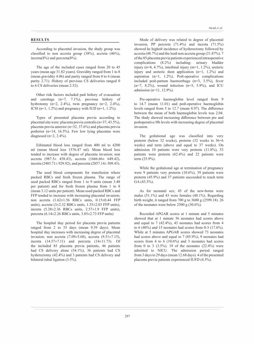
According to placental invasion, the study group was classified to non accreta group (38%), accreta (46%), increta(8%) and percreta(8%).
The age of the included cases ranged from 20 to 45 years (mean age 31.82 years). Gravidity ranged from 1 to 8 (mean gravidity 4.06) and parity ranged from 0 to 6 (mean parity 2.71). History of previous CS deliveries ranged 0 to 6 CS deliveries (mean 2.52).
Other risk factors included past history of evacuation and curettage (n=7, 7.1%), previous history of hystrotomy (n=2, 2.4%), twin pregnancy (n=2, 2.4%), ICSI (n=1, 1.2%) and pregnancy with IUD (n=1, 1.2%).
Types of presented placenta previa according to placental site were: placenta previa centralis (n=37, 43.5%), placenta previa anterior (n=32, 37.6%) and placenta previa posterior (n=14, 16.5%). Few low lying placentas were diagnosed (n=2, 2.4%).
Estimated blood loss ranged from 400 ml to 4200 ml (mean blood loss 1576.47 ml). Mean blood loss tended to increase with degree of placenta invasion; non accreta (987.5± 458.43), accreta (1666.66± 649.42), increta (2485.71± 929.92), and percreta (2857.14± 509.43).
The used blood components for transfusion where packed RBCs and fresh frozen plasma. The range of used packed RBCs ranged from 1 to 9 units (mean 3.48 per patient) and for fresh frozen plasma from 1 to 8 (mean 3.12 units per patient). Mean used packed RBCs and FFP tended to increase with increasing placental invasion; non accreta (1.62±1.56 RBCs units, 0.15±0.44 FFP units), accreta (3±2.32 RBCs units, 1.35±2.03 FFP units), increta (5.28±2.36 RBCs units, 2.57±1.9 FFP units), percreta (6.14±2.26 RBCs units, 3.85±2.73 FFP units)
The hospital stay period for placenta previa patients ranged from 2 to 35 days (mean 9.39 days). Mean hospital stay increases with increasing degree of placental invasion; non accreta (7.09±5.68), accreta (9.51±7.15), increta (14.57±7.11) and percreta (14±11.73). Of the included 85 placenta previa patients, 46 patients had CS delivery alone (54.1%), 36 patients had CS hysterectomy (42.4%) and 3 patients had CS delivery and bilateral tubal ligation (3.5%).
Mode of delivery was related to degree of placental invasion, PP percreta (71.4%) and increta (71.5%) showed he highest incidence of hysterectomy followed by accreta (48.7%) and the least non accreta group (21.87%). 7 of the 85 placenta previa patients experienced intraoperative complications (8.2%) including: urinary bladder injury (n=4, 4.7%), intestinal injury (m=1, 1.2%), ureteric injury and ureteric stent application (n=1, 1.2%) and aspiration (n=1, 1.2%). Post-operative complications included post-partum haemorrhage (n=3, 3.5%), fever (n=7, 8.2%), wound infection (n=5, 5.9%), and ICU admission (n=11, 12.9%).
Pre-operative haemoglobin level ranged from 9 to 14.7 (mean 11.01) and post-operative haemoglobin levels ranged from 5 to 12.7 (mean 8.97). The difference between the mean of both haemoglobin levels was 2.04. The study showed increasing difference between pre and postoperative Hb levels with increasing degree of placental invasion.
The gestational age was classified into very preterm (below 32 weeks), preterm (32 weeks to 36+6 weeks) and term (above and equal to 37 weeks). On admission 10 patients were very preterm (11.8%), 53 patients were preterm (62.4%) and 22 patients were term (25.9%).
While the gestational age at termination of pregnancy were 9 patients very preterm (10.6%), 39 patients were preterm (45.9%) and 37 patients succeeded to reach term GA (43.5%).
As for neonatal sex; 45 of the new-borns were males (51.1%) and 43 were females (48.1%). Regarding birth weight, it ranged from 700 g to 3600 g (2599.18). 26 of the neonates were below 2500 g (30.6%).
Recorded APGAR scores at 1 minute and 5 minutes showed that at 1 minute 36 neonates had scores above and equal to 7 (42.4%), 43 neonates had scores from 4 to 6 (40%) and 15 neonates had scores from 0-3 (17.6%). While at 5 minutes APGAR scores showed 73 neonates had scores above and equal to 7 (85.9%), 9 neonates had scores from 4 to 6 (10.6%) and 3 neonates had scores from 0 to 3 (3.5%). 19 of the neonates (22.4%) were admitted to NICU. The admission period ranged from 3 days to 29 days (mean 12.68 days). 4 of the presented placenta previa patients experienced IUFD (4.5%).
298
OUTCOME OF P.P IN AIN SHAMS HOSPITAL
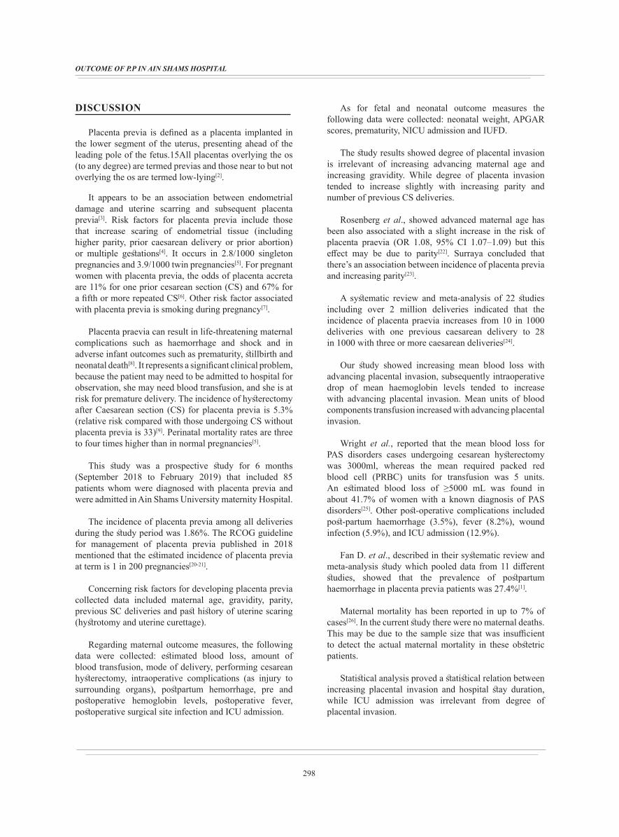
DISCUSSION
Placenta previa is defined as a placenta implanted in the lower segment of the uterus, presenting ahead of the leading pole of the fetus.15All placentas overlying the os (to any degree) are termed previas and those near to but not overlying the os are termed low-lying[2].
It appears to be an association between endometrial damage and uterine scarring and subsequent placenta previa[3]. Risk factors for placenta previa include those that increase scaring of endometrial tissue (including higher parity, prior caesarean delivery or prior abortion) or multiple gestations[4]. It occurs in 2.8/1000 singleton pregnancies and 3.9/1000 twin pregnancies[5]. For pregnant women with placenta previa, the odds of placenta accreta are 11% for one prior cesarean section (CS) and 67% for a fifth or more repeated CS[6]. Other risk factor associated with placenta previa is smoking during pregnancy[7].
Placenta praevia can result in life-threatening maternal complications such as haemorrhage and shock and in adverse infant outcomes such as prematurity, stillbirth and neonatal death[8]. It represents a significant clinical problem, because the patient may need to be admitted to hospital for observation, she may need blood transfusion, and she is at risk for premature delivery. The incidence of hysterectomy after Caesarean section (CS) for placenta previa is 5.3% (relative risk compared with those undergoing CS without placenta previa is 33)[9]. Perinatal mortality rates are three to four times higher than in normal pregnancies[5].
This study was a prospective study for 6 months (September 2018 to February 2019) that included 85 patients whom were diagnosed with placenta previa and were admitted in Ain Shams University maternity Hospital.
The incidence of placenta previa among all deliveries during the study period was 1.86%. The RCOG guideline for management of placenta previa published in 2018 mentioned that the estimated incidence of placenta previa at term is 1 in 200 pregnancies[20-21].
Concerning risk factors for developing placenta previa collected data included maternal age, gravidity, parity, previous SC deliveries and past history of uterine scaring (hystrotomy and uterine curettage).
Regarding maternal outcome measures, the following data were collected: estimated blood loss, amount of blood transfusion, mode of delivery, performing cesarean hysterectomy, intraoperative complications (as injury to surrounding organs), postpartum hemorrhage, pre and postoperative hemoglobin levels, postoperative fever, postoperative surgical site infection and ICU admission.
As for fetal and neonatal outcome measures the following data were collected: neonatal weight, APGAR scores, prematurity, NICU admission and IUFD.
The study results showed degree of placental invasion is irrelevant of increasing advancing maternal age and increasing gravidity. While degree of placenta invasion tended to increase slightly with increasing parity and number of previous CS deliveries.
Rosenberg et al., showed advanced maternal age has been also associated with a slight increase in the risk of placenta praevia (OR 1.08, 95% CI 1.07–1.09) but this effect may be due to parity[22]. Surraya concluded that there’s an association between incidence of placenta previa and increasing parity[23].
A systematic review and meta-analysis of 22 studies including over 2 million deliveries indicated that the incidence of placenta praevia increases from 10 in 1000 deliveries with one previous caesarean delivery to 28 in 1000 with three or more caesarean deliveries[24].
Our study showed increasing mean blood loss with advancing placental invasion, subsequently intraoperative drop of mean haemoglobin levels tended to increase with advancing placental invasion. Mean units of blood components transfusion increased with advancing placental invasion.
Wright et al., reported that the mean blood loss for PAS disorders cases undergoing cesarean hysterectomy was 3000ml, whereas the mean required packed red blood cell (PRBC) units for transfusion was 5 units. An estimated blood loss of ≥5000 mL was found in about 41.7% of women with a known diagnosis of PAS disorders[25]. Other post-operative complications included post-partum haemorrhage (3.5%), fever (8.2%), wound infection (5.9%), and ICU admission (12.9%).
Fan D. et al., described in their systematic review and meta-analysis study which pooled data from 11 different studies, showed that the prevalence of postpartum haemorrhage in placenta previa patients was 27.4%[1].
Maternal mortality has been reported in up to 7% of cases[26]. In the current study there were no maternal deaths. This may be due to the sample size that was insufficient to detect the actual maternal mortality in these obstetric patients.
Statistical analysis proved a statistical relation between increasing placental invasion and hospital stay duration, while ICU admission was irrelevant from degree of placental invasion.

299
Awad et al.
No statistical correlation between each; maternal age, gravidity, parity, no. of previous cs deliveries and preoperative hemoglobin levels and estimated intraoperative blood loss.
Our study also showed statistical relation between placenta previa and prematurity either at time of presentation and on termination of pregnancy. Similarly Bahar et al., found that hemorrhage in women with placenta previa was associated with premature delivery (P<0.001)[8].
The study showed insignificant relation between placenta previa and neonatal sex (P=0.831). Similarly Anath et al., found no relation between neonatal sex and placenta previa[5].
Regarding birth weight, our study showed high statistically significant relation between placenta previa and low birth weight. Anath et al., described that low birth weight in placenta previa patients is mainly due to prematurity and to a lesser extent due to growth restriction[5].
As for the recorded neonatal APGAR scores, our study showed statistically significant relation between placenta previa and low APGAR scores at 1 minute and 5 minutes. At 1 min, 17.6% were below 3, 40% were below 7 and 42.4% more than 7. At 5 min, 3.5% were below 3, 10.6a5 below 7, 85.9% were below 7. Regarding NICU admission, 22.4% of the newborn were admitted to NICU, mean duration (12.68 ± 7.08).
A large case-control study done by Lal et al., found that neonatal morbidities in women with placenta previa include an increased risk of lower 5-minute APGAR scores, NICU admission, anemia, respiratory distress syndrome, mechanical ventilation and intraventricular hemorrhage[27].
In addition to the previous neonatal outcome measures, our study recorded 4 cases of IUFD (4.5%). Salah R. et al., described in their prospective study that took place in Sohag university maternity hospital over a 6 months period that the incidence of IUFD among placenta previa patients was 13.2%[28].
This prospective study had some limitations. The sample size was insufficient to identify the magnitude of some risk factors for placenta previa as ICSI, multiple pregnancy and smoking. In addition, it failed to show the significant relationship between placenta previa and maternal mortality, wound infection and IUFD.
CONCOLUSION
In conclusion, placenta previa has higher incidence rate with increasing maternal age, gravidity, parity and number of previous caesarean section deliveries. Placenta previa patients at risk of numerous maternal and neonatal morbidities, maternal morbidities include life threatening haemorrhage, caesarean hysterectomy, blood component
transfusion, prolonged hospital stay and ICU admission. Neonatal morbidities include premature delivery, low birth weight, intrauterine foetal death and NICU admission.
CONFLICT OF INTEREST
There are no conflicts of interests.
REFERENCES
1. Fan D, Xia Q, Liu L, Wu S, Tian G, WangW, et al. (2017): The Incidence of Postpartum Hemorrhage in Pregnant Women with Placenta Previa: A Systematic Review and Meta-Analysis. PLoS ONE 12(1): e0170194.
2. Reddy U, Abuhamad A, Levine D, Saade G. (2014): Fetal Imaging Workshop Invited P. Fetal imaging: executive summary of a joint Eunice Kennedy Shriver National Institute of Child Health and Human Development, Society for Maternal-Fetal Medicine, American Institute of Ultrasound in Medicine, American College of Obstetricians and Gynecologists, American College of Radiology, Society for Pediatric Radiology, and Society of Radiologists in Ultrasound Fetal Imaging Workshop. Journal of ultrasound in medicine: official journal of the American Institute of Ultrasound in Medicine; 33(5):745–757.
3. Mastrolia S, Baumfeld Y, Loverro G, Yohai D, Hershkovitz R, Weintraub A. (2016): Placenta previa associated with severe bleeding leading to hospitalization and delivery: a retrospective population-based cohort study. Journal of Maternal-Fetal and Neonatal Medicine; 29:3467-3471.
4. Gurol-Urganci I, Cromwell D, Edozien L. (2011): Risk of placenta previa in second birth after first birth cesarean section: a population-based study and meta-analysis. BMC Pregnancy Childbirth; 11, 95.
5. Ananth C, Smulian J, Vintzileos A. (2003): The effect of placenta previa on neonatal mortality: a population based study in United States 1989 through 1997. American Journal of Obstetrics and Gynecology; 188:1299-1304.
6. Silver R, Landon M, Rouse D. (2006): Maternal morbidity associated with multiple repeat cesarean deliveries. Obstetrics & Gynecology;107:1226–1232
7. Fatemeh SH, Masoumi SZ, Jenabi E. (2017): The association between maternal smoking and placenta abruption: a meta-analysis. Journal of Maternal-Fetal and Neonatal Medicine.
300
OUTCOME OF P.P IN AIN SHAMS HOSPITAL
8. Bahar A, Abusham A, Eskandar M, Sobande A & Alsunaidi M. (2009): Risk factors and pregnancy outcome in different types of placenta previa. Journal of Obstetrics & Gynecology Canada; 31, 126–131.
9. Crane J, Van den Hof M, Dodds L, Armson B, Liston R. (2000): Maternal complications with placenta previa. American Journal of Perinatology; 17:101–105.
10. Merz E. (2007): Ultrasound in obstetrics and gynecology, 2nd ed. New York, NY: Thieme.
11. Sallout B, Oppenheimer L. (2002): The classification of placenta previa based on os-placental edge distance at transvaginal sonography. Am J Obstet Gynecol;187(6):S94.
12. Bhide A, Prefumo F, Moore J, Hollis B, Thilaganathan B. (2003): Placental edge to internal os distance in the late third trimester and mode of delivery in placenta previa. BJOG; 110:860-864.
13. Predanic M, Perni S, Baergen R, Jean-Pierre C, Chasen S, Chervenak F. (2005): A sonographic assessment of different patterns of placenta previa “migration” in the third trimester of pregnancy. J Ultrasound Med; 24:773–780.
14. Vergani P, Ornaghi S, Pozzi I. (2009): Placenta previa: distance to internal os and mode of delivery. American Journal of Obstetrics and Gynecology; 201:266-268.
15. Oppenheimer L, Farine D. (2009): A new classification of placenta previa: measuring progress in obstetrics. American journal of Obstetrics and Gynecology; 201:227–229.
16. Oyelese Y, Smulian J. (2006): Placenta Previa, Placenta Accreta, and Vasa Previa. Obstetrics & Gynecology. 107 (4): 927–941.
17. Bauer S, Bonanno C. (2009): Abnormal placentation. Seminars in Perinatology; 33:88–96.
18. Jeong Y, Young H, Min H, Jung H. (2008): Difference in Migration of Placenta According to the Location and Type of Placenta Previa. Wiley InterScience, J Clin Ultrasound 36:79–84.
19. Atsuko S, Akihito N, Ikuno K, Masako H, Toshiyuki T. (2013): Type and Location of Placenta Previa Affect Preterm Delivery Risk Related to Antepartum Hemorrhage. Int J Med Sci; 10(12):1683-1688.
20. Silver R. (2015): Abnormal placentation: Placenta previa, vasa previa and placenta accreta. Obstet Gynecol; 126:654–668.
21. Vahanian S, Lavery J, Ananth C, Vintzileos A. (2015): Placental implantation abnormalities and risk of preterm delivery: a systematic review and metaanalysis. Am J Obstet Gynecol; 213:S78–90.
22. Rosenberg T, Pariente G, Sergienko R, Wiznitzer A, Sheiner E. (2011): Critical analysis of risk factors and outcome of placenta previa. Arch Gynecol Obstet; 284:47–51.
23. Surraya Halimi (2011): Association of placenta previa with multiparity and previous cesarean section. JPMI; 02:139-142.
24. Marshall N, Fu R, Guise J. (2011): Impact of multiple cesarean deliveries on maternal morbidity: a systematic review. Am J Obstet Gynecol; 205:262.e1–8.
25. Wright J, Pri-Paz S, Herzog T, Shah M, Bonanno C, Lewin S. (2011): Predictors of massive blood loss in women with placenta accreta. Am J Obstet Gynecol; 205:38. e1-6.
26. Judson E, Polyakov A, Lawrence A.(2008): Intra-abdominal haemorrhage at 17 weeks’ gestation caused by placenta percreta: a case report. Royal Austr N Zeal Col Obstet Gynecol; 48:218–224.
27. Lal A, Hibbard J. (2015): Placenta previa: an outcome-based cohort study in a contemporary obstetric population. Arch Gynecol Obstet; 292:299–305.
28. Salah Roshdy, Abdusaeed Aitallah, Hazem M, Mohamed Alkhatim (2015): Major Placenta Previa: Rate, Maternal and Neonatal Outcomes Experience at a Tertiary Maternity Hospital, Sohag, Egypt: A Prospective Study. Journal of Clinical and Diagnostic Research; Vol-9(11): QC17-QC19.
Copyright © 2022 FDOKUMEN