Antenatal Fetal Assessment - KSUMSC
10
Antenatal Fetal Assessment Objectives: Describe how to test for each of the following: -Fetal well-being -Fetal growth -Fetal movement -Amniotic fluid -Fetal lung maturity Done by: Lina Aljurf ★ Resources used: Slides, Kaplan Explanation Important ★ Editing file
-
Upload
khangminh22 -
Category
Documents
-
view
0 -
download
0
Transcript of Antenatal Fetal Assessment - KSUMSC

Antenatal Fetal
Assessment
Objectives:Describe how to test for each of the following:
-Fetal well-being-Fetal growth
-Fetal movement-Amniotic fluid
-Fetal lung maturity
Done by: Lina Aljurf
★ Resources used:Slides, KaplanExplanationImportant
★ Editing file
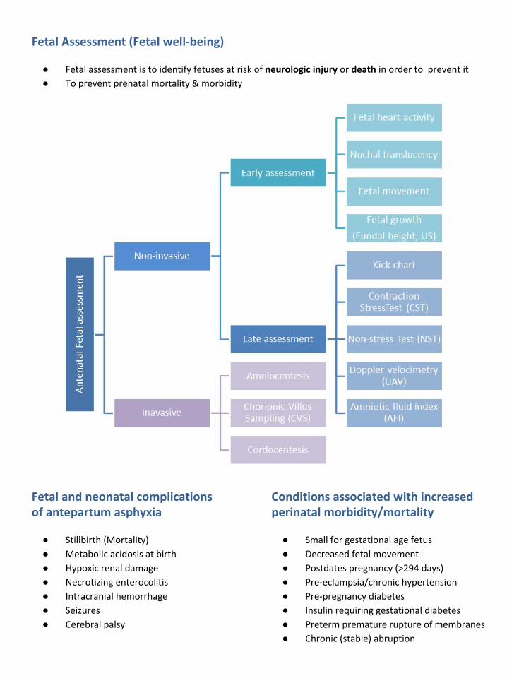
Fetal Assessment (Fetal well-being)
● Fetal assessment is to identify fetuses at risk of neurologic injury or death in order to prevent it
● To prevent prenatal mortality & morbidity
Fetal and neonatal complications of antepartum asphyxia
● Stillbirth (Mortality)
● Metabolic acidosis at birth
● Hypoxic renal damage
● Necrotizing enterocolitis
● Intracranial hemorrhage
● Seizures
● Cerebral palsy
Conditions associated with increased perinatal morbidity/mortality
● Small for gestational age fetus
● Decreased fetal movement
● Postdates pregnancy (>294 days)
● Pre-eclampsia/chronic hypertension
● Pre-pregnancy diabetes
● Insulin requiring gestational diabetes
● Preterm premature rupture of membranes
● Chronic (stable) abruption

Rational
Fetal oxygenation challenged:
● Blood flow directed to brain, heart and adrenal glands
● Blood flow away from the kidneys → decrease fetal urine production → decrease amniotic fluid
(AF) volume.
● CNS hypoxia → Fetal movement decrease
● Chemoreceptors → vagally-mediated reflex → Fetal heart rate abnormality late deceleration.
When to start fetal assessment antenatally?
Fetal assessment is done once or twice weekly
Risk assessed individually:
Case Week
Uncomplicated DM 32 weeks onward
Complicated DM 24 weeks onward
Post date pregnancy 40 weeks
Decreased fetal movement Start immediately
Early pregnancy assessment
Fetal heart activity
● fetal auscultation (special
stethoscope or Doppler)
~12 weeks
● fetal heart activity seen by US
(can be seen from 6 weeks):
Nuchal translucency
● Ultrasound to measure the thickness of the fluid
buildup at the back of the developing baby's neck.
● measurement for early screening for chromosomal
abnormality such as down syndrome, trisomy 18
● Between 11-13+ weeks

Fetal movement
● Fetal movements are usually first perceptible to mother ~17w-20w (quickening)
● 50% of isolated limb movements are perceived
● 80% of trunk and limb movements
Fetal growth
● By fundal height measurement in the clinic
● By ultrasound:
● Biometry:
○ Biparietal diameter (BPD)
○ Abdominal Circumference (AC)
○ Femur Length (FL)
○ Head Circumference (HC)
● Amniotic fluid
Growth Chart
Biparietal diameter (BPD) (AC) Femur Length (FL)
Amniotic Fluid Index (AFI) (Kaplan)
The 4-quadrant amniotic fluid index test assesses in centimeters the deepest single vertical amniotic fluid
pocket in each of the 4 quadrants of the uterus. The sum of the pockets is known as the amniotic fluid
index, or AFI. Interpretation is as follows:
<5 cm → oligohydramnios
5-8 cm → borderline
9-25 cm → normal
> 25 cm → polyhydramnios
Late pregnancy assessment
Fetal movement counting (kick chart)
● It should be started ~28w in normal
pregnancy &~24w in high risk pregnancy
● It can reduce avoidable stillbirth
CARDIFF TECHNIQUE
● 10 movements in 12 hours
● If abnormal patient should get further
assessment
SADOVSKY TECHNIQUE
● 4 movement/hour, if not felt, another hour
● If not, patient needs more assessment

Non Stress Test (NST) (Kaplan + Lecture)
This test assesses the frequency of fetal movements using and external fetal heart rate (FHR) monitoring
device to detect the presence or absence of accelerations. These are abrupt increases in FHR above the
baseline lasting <2 min and are unrelated to contractions. The criteria vary by gestational age:
● <32 weeks, the increase should be >10 beats/min lasting >10 s
● >32 weeks, the increase should be >15 beats/min lasting >15 s
They are mediated by the sympathetic nervous system and always occur in response to fetal
movements.
Interpretation: accelerations are always reassuring. (check the pic at the bottom of the page)
● Reactive: At least two accelerations from baseline of 15 bpm for at least 15 sec within 20 minutes
● Non reactive: No acceleration after 20 minutes- proceed for another 20 minutes
○ If non reactive in 40 minutes---proceed for contraction stress test or biophysical profile
○ The positive predictive value of NST to predict fetal acidosis at birth is 55%
● Main advantage over CST: no need for contraction
● False +ve and false -ve is higher than CST
● The base line 120-160 beats/minute
Reactive NST
Criteria: >2 accelerations in 20 min:↑ FHR >15 beats/min and lasting >15 seconds
Assessment: reassuring of fetal well-being
Follow-up: repeat weekly/biweekly
Nonreactive NST
Criteria: no FHR accelerations or did not meet criteria
Assessment: sleeping, immature, or sedated fetus; acidotic, compromised fetus?
Follow-up: Vibroacoustic Stimulation (VAS)
If still NR: do contraction stress test (CST) or biophysical profile (BPP)
Cardiotocography (CTG) interpretation
Normal Baseline FHR 110–160 bpm
Moderate bradycardia 100–109 bpm
Moderate tachycardia 161–180 bpm
Abnormal bradycardia < 100 bpm
Abnormal tachycardia > 180 bpm
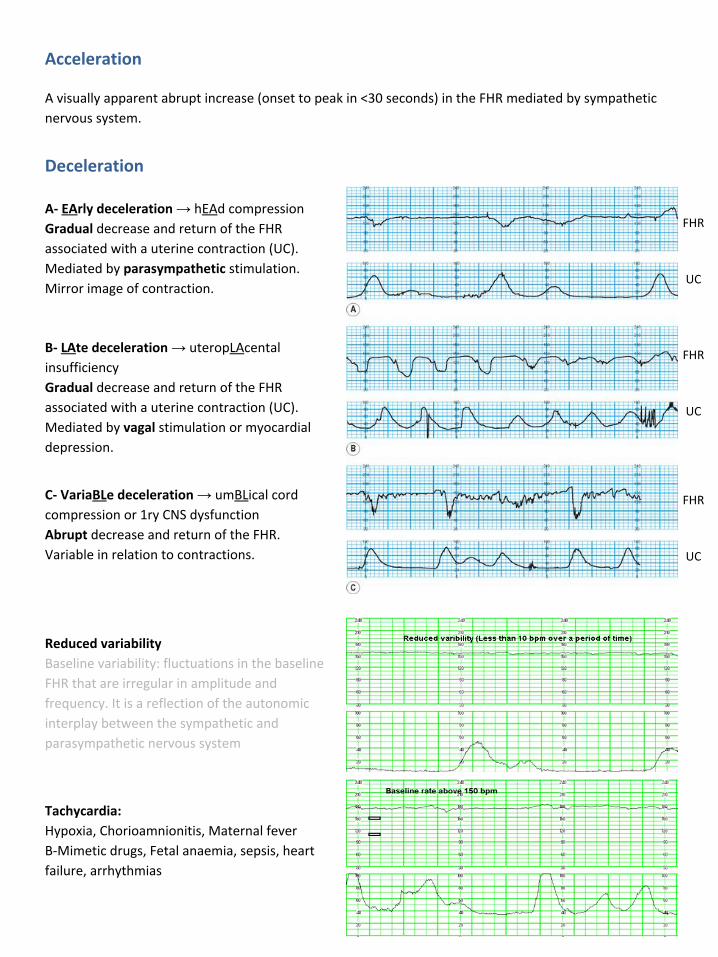
Acceleration
A visually apparent abrupt increase (onset to peak in <30 seconds) in the FHR mediated by sympathetic
nervous system.
Deceleration
A- EArly deceleration → hEAd compression
Gradual decrease and return of the FHR
associated with a uterine contraction (UC).
Mediated by parasympathetic stimulation.
Mirror image of contraction.
FHR
UC
FHR
UC
FHR
UC
B- LAte deceleration → uteropLAcental
insufficiency
Gradual decrease and return of the FHR
associated with a uterine contraction (UC).
Mediated by vagal stimulation or myocardial
depression.
C- VariaBLe deceleration → umBLical cord
compression or 1ry CNS dysfunction
Abrupt decrease and return of the FHR.
Variable in relation to contractions.
Reduced variability
Baseline variability: fluctuations in the baseline
FHR that are irregular in amplitude and
frequency. It is a reflection of the autonomic
interplay between the sympathetic and
parasympathetic nervous system
Tachycardia:
Hypoxia, Chorioamnionitis, Maternal fever
B-Mimetic drugs, Fetal anaemia, sepsis, heart
failure, arrhythmias
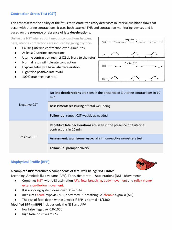
Contraction Stress Test (CST)
This test assesses the ability of the fetus to tolerate transitory decreases in intervillous blood flow that
occur with uterine contractions. It uses both external FHR and contraction monitoring devices and is
based on the presence or absence of late decelerations.
Unlike the NST where spontaneous contractions happen,
here, uterine contractions are induced by giving oxytocin
● Causing uterine contraction over 20minutes
● At least 2 uterine contractions
● Uterine contraction restrict O2 delivery to the fetus
● Normal fetus will tolerate contraction
● Hypoxic fetus will have late deceleration
● High false positive rate ~50%
● 100% true negative rate
Negative CST
No late decelerations are seen in the presence of 3 uterine contractions in 10 min
Assessment: reassuring of fetal well-being
Follow-up: repeat CST weekly as needed
Positive CST
Repetitive late decelerations are seen in the presence of 3 uterine contractions in 10 min
Assessment: worrisome, especially if nonreactive non-stress test
Follow-up: prompt delivery
Biophysical Profile (BPP)
A complete BPP measures 5 components of fetal well-being: “BAT HAM”
Breathing, Amniotic fluid volume (AFV), Tone, Heart rate + Accelerations (NST), Movements
● Combines NST with USS estimation AFV, fetal breathing, body movement and reflex /tone/
extension-flexion movement.
● It is a scoring system done over 30 minute
● measures acute hypoxia (NST, body mov. & breathing) & chronic hypoxia (AFI)
● The risk of fetal death within 1 week if BPP is normal~ 1/1300
Modified BPP (mBPP) includes only the NST and AFV
● low false negative 0.8/1000
● high false positives ~60%

Biophysical profile cont.
● Score of 8 or 10 → highly reassuring of fetal well-being
○ Management: repeat test weekly or as indicated
● Score of 4 or 6 → worrisome
○ Management:
○ if the fetus is >36 weeks → delivery
○ if the fetus is <36 weeks → repeat biophysical profile in 12-24 hours
● Score of 0 or 2 → highly predictive of fetal hypoxia with low probability of false positive
○ Management: prompt delivery regardless of gestational age
Doppler velocimetry/Umbilical Artery doppler
This test measures the ratio of systolic and diastolic blood flow in the umbilical artery. The umbilical
circulation normally has low resistance, so significant diastolic blood flow is expected.
It is predictive of poor perinatal outcome only in IUGR fetuses. Nonreassuring findings, which may
indicate need for delivery, are absent diastolic flow and reversed diastolic flow.
● Measurement of blood flow
velocities in maternal & fetal
vessels
● Reflect feto-placental circulation
● Doppler indices from UA, Uterine
A & MCA
● Doppler studies is mostly valuable
IUGR
● In IUGR absent or reversed EDF
(end diastolic flow) associated
with fetal hypoxia

Invasive fetal assessment (Check kaplan chapter 3, pg 31-34)
Amniocentesis
● Obtaining a sample of amniotic fluid during pregnancy.
● Usually done after 15w (can be done after 11w)
● Indications
○ genetic (karyotype)
○ bilirubin level (RH-isoimmunisation)
○ fetal lung maturity (L/S)
○ therapeutic in polyhydramnios
● Risks: ROM ~1%, abortion 0.5%, infection 1/1000
Chorionic Villus Sampling (CVS)
● Usually done after 10w
● It is the procedure of choice for first trimester prenatal diagnosis of genetic disorders
● Complication: fetal loss (0.7 percent within 14 days of a TA CVS procedure and 1.3 percent
within 30 days), Procedure-induced limb defects
● Second trimester amniocentesis is associated with the lowest risk of pregnancy loss; chorionic
villus samplings safer than early (i.e, before 15 weeks) amniocentesis.
Cordocentesis
● Indications:
○ rapid karyotyping
○ diagnosis of inherited disorders
○ fetal HB assessment
○ fetal plt level
○ fetal blood transfusion
● Complication: bleeding, bradycardia, infection….
Amniocentesis
Chorionic Villus Sampling (CVS)
Cordocentesis
Fetal Lung Maturity (FLM)
● A test for fetal lung maturity is performed before
semi-elective but medically indicated births <39 weeks
● Tests for fetal lung maturity are generally not performed
before 32 weeks of gestation
● RDS develops as a consequence of surfactant deficiency and
immature lung development.
● L/S ratio is the most commonly used (ratio should be 2:1)

Fetal Lung Maturity cont.
FLM testing may have value in the following clinical situations:
● Premature rupture of membranes (≥32 weeks) – if FLM test is mature, delivery is likely safer than
“wait and see” approach
● Assessment of need for NICU – possible only if early delivery has medical mandate and time
allows for FLM testing
● Other selected late preterm and early preterm pregnancy issues where FLM may guide
management of at-risk pregnancy
Comparison of FLM Laboratory test options:
All tests require amniocentesis for obtaining amniotic fluid
Lamellar body count (LBC) Phosphatidylglycerol (PG) Lecithin-sphingomyelin ratio
(L/S)
•Initial FLM of choice
•Rapid, sensitive
•New data indicates that one
can estimate risk of respiratory
distress syndrome (RDS) as a
function of gestational age and
LBC
•Not useful unless gestational
age ≥35 weeks
•Limited availability
•Sensitive
•Main role is in adjudication of
immature LBC or PG
•Last test of choice
•Labor intensive, imprecise
•Limited availability
•Results take >24 hrs unless
performed at a local laboratory
..وبسIf you like this work, please make dua’ for me
If you don’t, also make dua’ for me :o)
Indications for antepartum fetal surveillance: