chapter 2 - KSUMSC
33
contrast to a unipotent cell, which differentiates into a single cell type, a hematopoietic stem cell is multipotent, or pluripo- tent, able to differentiate in various ways and thereby generate erythrocytes, granulocytes, monocytes, mast cells, lympho- cytes, and megakaryocytes. These stem cells are few, normally fewer than one HSC per 5 10 4 cells in the bone marrow. The study of hematopoietic stem cells is difficult both be- cause of their scarcity and because they are hard to grow in vitro. As a result, little is known about how their proliferation and differentiation are regulated. By virtue of their capacity for self-renewal, hematopoietic stem cells are maintained at stable levels throughout adult life; however, when there is an increased demand for hematopoiesis, HSCs display an enor- mous proliferative capacity. This can be demonstrated in mice whose hematopoietic systems have been completely de- stroyed by a lethal dose of x-rays (950 rads; one rad repre- sents the absorption by an irradiated target of an amount of radiation corresponding to 100 ergs/gram of target). Such ir- radiated mice will die within 10 days unless they are infused with normal bone-marrow cells from a syngeneic (genetically identical) mouse. Although a normal mouse has 3 10 8 bone-marrow cells, infusion of only 10 4 –10 5 bone-marrow cells (i.e., 0.01%–0.1% of the normal amount) from a donor is sufficient to completely restore the hematopoietic system, chapter 2 ■ Hematopoiesis ■ Cells of the Immune System ■ Organs of the Immune System ■ Systemic Function of the Immune System ■ Lymphoid Cells and Organs—Evolutionary Comparisons Cells and Organs of the Immune System T organs and tissues that are found throughout the body. These organs can be classified functionally into two main groups. The primary lymphoid organs provide appropriate microenvironments for the development and maturation of lymphocytes. The secondary lymphoid organs trap antigen from defined tissues or vascular spaces and are sites where mature lymphocytes can interact effectively with that antigen. Blood vessels and lymphatic systems connect these organs, uniting them into a functional whole. Carried within the blood and lymph and populating the lymphoid organs are various white blood cells, or leuko- cytes, that participate in the immune response. Of these cells, only the lymphocytes possess the attributes of diversity, specificity, memory, and self/nonself recognition, the hall- marks of an adaptive immune response. All the other cells play accessory roles in adaptive immunity, serving to activate lymphocytes, to increase the effectiveness of antigen clear- ance by phagocytosis, or to secrete various immune-effector molecules. Some leukocytes, especially T lymphocytes, se- crete various protein molecules called cytokines. These mol- ecules act as immunoregulatory hormones and play important roles in the regulation of immune responses. This chapter describes the formation of blood cells, the properties of the various immune-system cells, and the functions of the lymphoid organs. Hematopoiesis All blood cells arise from a type of cell called the hematopoi- etic stem cell (HSC). Stem cells are cells that can differentiate into other cell types; they are self-renewing—they maintain their population level by cell division. In humans, hematopoiesis, the formation and development of red and white blood cells, begins in the embryonic yolk sac during the first weeks of development. Here, yolk-sac stem cells differen- tiate into primitive erythroid cells that contain embryonic hemoglobin. In the third month of gestation, hematopoietic stem cells migrate from the yolk sac to the fetal liver and then to the spleen; these two organs have major roles in hematopoiesis from the third to the seventh months of gesta- tion. After that, the differentiation of HSCs in the bone mar- row becomes the major factor in hematopoiesis, and by birth there is little or no hematopoiesis in the liver and spleen. It is remarkable that every functionally specialized, ma- ture blood cell is derived from the same type of stem cell. In Macrophage Interacting with Bacteria 8536d_ch02_024-056 9/6/02 9:00 PM Page 24 mac85 Mac 85:365_smm:Goldsby et al. / Immunology 5e:
-
Upload
khangminh22 -
Category
Documents
-
view
0 -
download
0
Transcript of chapter 2 - KSUMSC

contrast to a unipotent cell, which differentiates into a singlecell type, a hematopoietic stem cell is multipotent, or pluripo-tent, able to differentiate in various ways and thereby generateerythrocytes, granulocytes, monocytes, mast cells, lympho-cytes, and megakaryocytes. These stem cells are few, normallyfewer than one HSC per 5 � 104 cells in the bone marrow.
The study of hematopoietic stem cells is difficult both be-cause of their scarcity and because they are hard to grow invitro. As a result, little is known about how their proliferationand differentiation are regulated. By virtue of their capacityfor self-renewal, hematopoietic stem cells are maintained atstable levels throughout adult life; however, when there is anincreased demand for hematopoiesis, HSCs display an enor-mous proliferative capacity. This can be demonstrated inmice whose hematopoietic systems have been completely de-stroyed by a lethal dose of x-rays (950 rads; one rad repre-sents the absorption by an irradiated target of an amount ofradiation corresponding to 100 ergs/gram of target). Such ir-radiated mice will die within 10 days unless they are infusedwith normal bone-marrow cells from a syngeneic (geneticallyidentical) mouse. Although a normal mouse has 3 � 108
bone-marrow cells, infusion of only 104–105 bone-marrowcells (i.e., 0.01%–0.1% of the normal amount) from a donoris sufficient to completely restore the hematopoietic system,
chapter 2
� Hematopoiesis
� Cells of the Immune System
� Organs of the Immune System
� Systemic Function of the Immune System
� Lymphoid Cells and Organs—EvolutionaryComparisons
Cells and Organs of theImmune System
T
organs and tissues that are found throughout thebody. These organs can be classified functionally
into two main groups. The primary lymphoid organs provideappropriate microenvironments for the development andmaturation of lymphocytes. The secondary lymphoid organstrap antigen from defined tissues or vascular spaces and aresites where mature lymphocytes can interact effectively withthat antigen. Blood vessels and lymphatic systems connectthese organs, uniting them into a functional whole.
Carried within the blood and lymph and populating thelymphoid organs are various white blood cells, or leuko-cytes, that participate in the immune response. Of thesecells, only the lymphocytes possess the attributes of diversity,specificity, memory, and self/nonself recognition, the hall-marks of an adaptive immune response. All the other cellsplay accessory roles in adaptive immunity, serving to activatelymphocytes, to increase the effectiveness of antigen clear-ance by phagocytosis, or to secrete various immune-effectormolecules. Some leukocytes, especially T lymphocytes, se-crete various protein molecules called cytokines. These mol-ecules act as immunoregulatory hormones and playimportant roles in the regulation of immune responses. Thischapter describes the formation of blood cells, the propertiesof the various immune-system cells, and the functions of thelymphoid organs.
HematopoiesisAll blood cells arise from a type of cell called the hematopoi-etic stem cell (HSC). Stem cells are cells that can differentiateinto other cell types; they are self-renewing—they maintaintheir population level by cell division. In humans,hematopoiesis, the formation and development of red andwhite blood cells, begins in the embryonic yolk sac during thefirst weeks of development. Here, yolk-sac stem cells differen-tiate into primitive erythroid cells that contain embryonichemoglobin. In the third month of gestation, hematopoieticstem cells migrate from the yolk sac to the fetal liver and thento the spleen; these two organs have major roles inhematopoiesis from the third to the seventh months of gesta-tion. After that, the differentiation of HSCs in the bone mar-row becomes the major factor in hematopoiesis, and by birththere is little or no hematopoiesis in the liver and spleen.
It is remarkable that every functionally specialized, ma-ture blood cell is derived from the same type of stem cell. In
Macrophage Interacting with Bacteria
8536d_ch02_024-056 9/6/02 9:00 PM Page 24 mac85 Mac 85:365_smm:Goldsby et al. / Immunology 5e:

Cells and Organs of the Immune System C H A P T E R 2 25
which demonstrates the enormous proliferative and differ-entiative capacity of the stem cells.
Early in hematopoiesis, a multipotent stem cell differenti-ates along one of two pathways, giving rise to either a com-mon lymphoid progenitor cell or a common myeloid
progenitor cell (Figure 2-1). The types and amounts ofgrowth factors in the microenvironment of a particular stemcell or progenitor cell control its differentiation. During thedevelopment of the lymphoid and myeloid lineages, stem cells differentiate into progenitor cells, which have lost the
TH helper cell
TC cytotoxic T cell
Natural killer(NK) cell
Myeloidprogenitor
Lymphoidprogenitor
Hematopoieticstem cell
Self -renewing
B cell
Dendritic cell
T -cellprogenitor
B-cellprogenitor
Eosinophil
Monocyte
Neutrophil
Basophil
Platelets
Erythrocyte
Erythroid progenitor
Megakaryocyte
Eosinophilprogenitor
Granulocyte-monocyte progenitor
Basophil progenitor
Macrophage
Dendritic cell
V I S U A L I Z I N G C O N C E P T S
FIGURE 2-1 Hematopoiesis. Self-renewing hematopoieticstem cells give rise to lymphoid and myeloid progenitors. All lym-phoid cells descend from lymphoid progenitor cells and all cells
of the myeloid lineage arise from myeloid progenitors. Note thatsome dendritic cells come from lymphoid progenitors, othersfrom myeloid precursors.
8536d_ch02_024-056 8/5/02 4:02 PM Page 25 mac79 Mac 79:45_BW:Goldsby et al. / Immunology 5e:

26 P A R T I Introduction
capacity for self-renewal and are committed to a particular celllineage. Common lymphoid progenitor cells give rise to B, T,and NK (natural killer) cells and some dendritic cells. Myeloidstem cells generate progenitors of red blood cells (erythro-cytes), many of the various white blood cells (neutrophils,eosinophils, basophils, monocytes, mast cells, dendritic cells),and platelets. Progenitor commitment depends on the acquisi-tion of responsiveness to particular growth factors and cy-tokines. When the appropriate factors and cytokines arepresent, progenitor cells proliferate and differentiate into thecorresponding cell type, either a mature erythrocyte, a partic-ular type of leukocyte, or a platelet-generating cell (themegakaryocyte). Red and white blood cells pass into bone-marrow channels, from which they enter the circulation.
In bone marrow, hematopoietic cells grow and mature ona meshwork of stromal cells, which are nonhematopoieticcells that support the growth and differentiation of hema-topoietic cells. Stromal cells include fat cells, endothelial cells,fibroblasts, and macrophages. Stromal cells influence the dif-ferentiation of hematopoietic stem cells by providing ahematopoietic-inducing microenvironment (HIM) con-sisting of a cellular matrix and factors that promote growthand differentiation. Many of these hematopoietic growth factors are soluble agents that arrive at their target cells bydiffusion, others are membrane-bound molecules on thesurface of stromal cells that require cell-to-cell contact be-tween the responding cells and the stromal cells. During in-fection, hematopoiesis is stimulated by the production ofhematopoietic growth factors by activated macrophages andT cells.
Hematopoiesis Can Be Studied In VitroCell-culture systems that can support the growth and differ-entiation of lymphoid and myeloid stem cells have made it
possible to identify many hematopoietic growth factors. Inthese in vitro systems, bone-marrow stromal cells are cul-tured to form a layer of cells that adhere to a petri dish;freshly isolated bone-marrow hematopoietic cells placed onthis layer will grow, divide, and produce large visible colonies(Figure 2-2). If the cells have been cultured in semisolid agar,their progeny will be immobilized and can be analyzed forcell types. Colonies that contain stem cells can be replated toproduce mixed colonies that contain different cell types, in-cluding progenitor cells of different cell lineages. In contrast,progenitor cells, while capable of division, cannot be replatedand produce lineage-restricted colonies.
Various growth factors are required for the survival, pro-liferation, differentiation, and maturation of hematopoieticcells in culture. These growth factors, the hematopoietic cytokines, are identified by their ability to stimulate the for-mation of hematopoietic cell colonies in bone-marrow cultures. Among the cytokines detected in this way was afamily of acidic glycoproteins, the colony-stimulating fac-tors (CSFs), named for their ability to induce the formationof distinct hematopoietic cell lines. Another importanthematopoietic cytokine detected by this method was the gly-coprotein erythropoietin (EPO). Produced by the kidney,this cytokine induces the terminal development of erythro-cytes and regulates the production of red blood cells. Fur-ther studies showed that the ability of a given cytokine tosignal growth and differentiation is dependent upon thepresence of a receptor for that cytokine on the surface of thetarget cell—commitment of a progenitor cell to a particulardifferentiation pathway is associated with the expression ofmembrane receptors that are specific for particular cy-tokines. Many cytokines and their receptors have since beenshown to play essential roles in hematopoiesis. This topic isexplored much more fully in the chapter on cytokines(Chapter 11).
FIGURE 2-2 (a) Experimental scheme for culturing hematopoieticcells. Adherent bone-marrow stromal cells form a matrix on whichthe hematopoietic cells proliferate. Single cells can be transferredto semisolid agar for colony growth and the colonies analyzed fordifferentiated cell types. (b) Scanning electron micrograph of cells
Add fresh bone-marrow cells
Culture insemisolid agar
Adherent layer ofstromal cells
Visible colonies ofbone-marrow cells
(a) (b)
in long-term culture of human bone marrow. [Photograph from M. J. Cline and D. W. Golde, 1979, Nature 277:180; reprinted by permission; © 1979 Macmillan Magazines Ltd., micrograph cour-tesy of S. Quan.]
8536d_ch02_024-056 8/5/02 4:02 PM Page 26 mac79 Mac 79:45_BW:Goldsby et al. / Immunology 5e:

Cells and Organs of the Immune System C H A P T E R 2 27
Hematopoiesis Is Regulated at the Genetic LevelThe development of pluripotent hematopoietic stem cellsinto different cell types requires the expression of differentsets of lineage-determining and lineage-specific genes at ap-propriate times and in the correct order. The proteins speci-fied by these genes are critical components of regulatorynetworks that direct the differentiation of the stem cell andits descendants. Much of what we know about the depen-dence of hematopoiesis on a particular gene comes fromstudies of mice in which a gene has been inactivated or“knocked out” by targeted disruption, which blocks the pro-duction of the protein that it encodes (see Targeted Disrup-tion of Genes, in Chapter 23). If mice fail to produce red cellsor particular white blood cells when a gene is knocked out,we conclude that the protein specified by the gene is neces-sary for development of those cells. Knockout technology isone of the most powerful tools available for determining theroles of particular genes in a broad range of processes and ithas made important contributions to the identification ofmany genes that regulate hematopoiesis.
Although much remains to be done, targeted disruptionand other approaches have identified a number of transcrip-tion factors (Table 2-1) that play important roles inhematopoiesis. Some of these transcription factors affectmany different hematopoietic lineages, and others affect onlya single lineage, such as the developmental pathway that leadsto lymphocytes. One transcription factor that affects multi-ple lineages is GATA-2, a member of a family of transcriptionfactors that recognize the tetranucleotide sequence GATA, anucleotide motif in target genes. A functional GATA-2 gene,which specifies this transcription factor, is essential for thedevelopment of the lymphoid, erythroid, and myeloid lin-eages. As might be expected, animals in which this gene isdisrupted die during embryonic development. In contrast toGATA-2, another transcription factor, Ikaros, is requiredonly for the development of cells of the lymphoid lineage. Al-though Ikaros knockout mice do not produce significant
numbers of B, T, and NK cells, their production of erythro-cytes, granulocytes, and other cells of the myeloid lineage isunimpaired. Ikaros knockout mice survive embryonic devel-opment, but they are severely compromised immunologi-cally and die of infections at an early age.
Hematopoietic Homeostasis Involves Many FactorsHematopoiesis is a continuous process that generally main-tains a steady state in which the production of mature bloodcells equals their loss (principally from aging). The averageerythrocyte has a life span of 120 days before it is phagocytosedand digested by macrophages in the spleen. The various whiteblood cells have life spans ranging from a few days, for neu-trophils, to as long as 20–30 years for some T lymphocytes. Tomaintain steady-state levels, the average human being mustproduce an estimated 3.7 � 1011 white blood cells per day.
Hematopoiesis is regulated by complex mechanisms thataffect all of the individual cell types. These regulatory mech-anisms ensure steady-state levels of the various blood cells,yet they have enough built-in flexibility so that production ofblood cells can rapidly increase tenfold to twentyfold in re-sponse to hemorrhage or infection. Steady-state regulation ofhematopoiesis is accomplished in various ways, which in-clude:
� Control of the levels and types of cytokines produced bybone-marrow stromal cells
� The production of cytokines with hematopoietic activityby other cell types, such as activated T cells andmacrophages
� The regulation of the expression of receptors forhematopoietically active cytokines in stem cells andprogenitor cells
� The removal of some cells by the controlled induction ofcell death
A failure in one or a combination of these regulatory mecha-nisms can have serious consequences. For example, abnormal-ities in the expression of hematopoietic cytokines or theirreceptors could lead to unregulated cellular proliferation andmay contribute to the development of some leukemias. Ulti-mately, the number of cells in any hematopoietic lineage is setby a balance between the number of cells removed by cell deathand the number that arise from division and differentiation.Any one or a combination of regulatory factors can affect ratesof cell reproduction and differentiation. These factors can alsodetermine whether a hematopoietic cell is induced to die.
Programmed Cell Death Is an EssentialHomeostatic MechanismProgrammed cell death, an induced and ordered process inwhich the cell actively participates in bringing about its owndemise, is a critical factor in the homeostatic regulation of
TABLE 2-1 Some transcription factors essentialfor hematopoietic lineages
Factor Dependent lineage
GATA-1 Erythroid
GATA-2 Erythroid, myeloid, lymphoid
PU.1 Erythroid (maturational stages), myeloid (laterstages), lymphoid
BM11 Myeloid, lymphoid
Ikaros Lymphoid
Oct-2 B lymphoid (differentiation of B cells into plasmacells)
8536d_ch02_024-056 8/5/02 4:02 PM Page 27 mac79 Mac 79:45_BW:Goldsby et al. / Immunology 5e:

28 P A R T I Introduction
many types of cell populations, including those of thehematopoietic system.
Cells undergoing programmed cell death often exhibitdistinctive morphologic changes, collectively referred to as apoptosis (Figures 2-3, 2-4). These changes include a pronounced decrease in cell volume, modification of the cy-toskeleton that results in membrane blebbing, a condensa-tion of the chromatin, and degradation of the DNA intosmaller fragments. Following these morphologic changes, anapoptotic cell sheds tiny membrane-bounded apoptotic bod-ies containing intact organelles. Macrophages quickly phago-cytose apoptotic bodies and cells in the advanced stages ofapoptosis. This ensures that their intracellular contents, in-cluding proteolytic and other lytic enzymes, cationic pro-teins, and oxidizing molecules are not released into thesurrounding tissue. In this way, apoptosis does not induce alocal inflammatory response. Apoptosis differs markedlyfrom necrosis, the changes associated with cell death arisingfrom injury. In necrosis the injured cell swells and bursts, re-
leasing its contents and possibly triggering a damaging in-flammatory response.
Each of the leukocytes produced by hematopoiesis has acharacteristic life span and then dies by programmed celldeath. In the adult human, for example, there are about5 � 1010 neutrophils in the circulation. These cells have alife span of only a few days before programmed cell deathis initiated. This death, along with constant neutrophilproduction, maintains a stable number of these cells. Ifprogrammed cell death fails to occur, a leukemic state maydevelop. Programmed cell death also plays a role in main-taining proper numbers of hematopoietic progenitor cells.For example, when colony-stimulating factors are re-moved, progenitor cells undergo apoptosis. Beyondhematopoiesis, apoptosis is important in such immuno-logical processes as tolerance and the killing of target cellsby cytotoxic T cells or natural killer cells. Details of themechanisms underlying apoptosis are emerging; Chapter13 describes them in detail.
NECROSIS APOPTOSIS
Chromatin clumpingSwollen organellesFlocculent mitochondria
Mild convolutionChromatin compaction and segregationCondensation of cytoplasm
Nuclear fragmentationBlebbingApoptotic bodies
Phagocytosis
Phagocytic cell
Apoptotic body
Disintegration
Release of intracellularcontents
Inflammation
FIGURE 2-3 Comparison of morphologic changes that occur inapoptosis and necrosis. Apoptosis, which results in the programmedcell death of hematopoietic cells, does not induce a local inflamma-
tory response. In contrast, necrosis, the process that leads to deathof injured cells, results in release of the cells’ contents, which may in-duce a local inflammatory response.
Go to www.whfreeman.com/immunology AnimationCell Death
8536d_ch02_024-056 9/6/02 9:00 PM Page 28 mac85 Mac 85:365_smm:Goldsby et al. / Immunology 5e:

Cells and Organs of the Immune System C H A P T E R 2 29
The expression of several genes accompanies apoptosisin leukocytes and other cell types (Table 2-2). Some of theproteins specified by these genes induce apoptosis, othersare critical during apoptosis, and still others inhibit apop-tosis. For example, apoptosis can be induced in thymocytesby radiation, but only if the protein p53 is present; manycell deaths are induced by signals from Fas, a molecule pre-sent on the surface of many cells; and proteases known ascaspases take part in a cascade of reactions that lead toapoptosis. On the other hand, members of the bcl-2 (B-celllymphoma 2) family of genes, bcl-2 and bcl-XL encode pro-tein products that inhibit apoptosis. Interestingly, the firstmember of this gene family, bcl-2, was found in studies thatwere concerned not with cell death but with the uncon-trolled proliferation of B cells in a type of cancer known asB-lymphoma. In this case, the bcl-2 gene was at the break-point of a chromosomal translocation in a human B-celllymphoma. The translocation moved the bcl-2 gene intothe immunoglobulin heavy-chain locus, resulting in tran-
scriptional activation of the bcl-2 gene and overproductionof the encoded Bcl-2 protein by the lymphoma cells. Theresulting high levels of Bcl-2 are thought to help transformlymphoid cells into cancerous lymphoma cells by inhibit-ing the signals that would normally induce apoptotic celldeath.
Bcl-2 levels have been found to play an important role inregulating the normal life span of various hematopoietic celllineages, including lymphocytes. A normal adult has about 5 L of blood with about 2000 lymphocytes/mm3 for a total ofabout 1010 lymphocytes. During acute infection, the lym-phocyte count increases 4- to 15-fold, giving a total lympho-cyte count of 40–50 � 109. Because the immune systemcannot sustain such a massive increase in cell numbers for anextended period, the system needs a means to eliminate un-needed activated lymphocytes once the antigenic threat haspassed. Activated lymphocytes have been found to expresslower levels of Bcl-2 and therefore are more susceptible to theinduction of apoptotic death than are naive lymphocytes or
(a)
(c)
(b)
(d)
FIGURE 2-4 Apoptosis. Light micrographs of (a) normal thymo-cytes (developing T cells in the thymus) and (b) apoptotic thymo-cytes. Scanning electron micrographs of (c) normal and (d)
apoptotic thymocytes. [From B. A. Osborne and S. Smith, 1997, Jour-nal of NIH Research 9:35; courtesy B. A. Osborne, University of Mass-achusetts at Amherst.]
8536d_ch02_024-056 8/5/02 4:02 PM Page 29 mac79 Mac 79:45_BW:Goldsby et al. / Immunology 5e:

30 P A R T I Introduction
memory cells. However, if the lymphocytes continue to beactivated by antigen, then the signals received during activa-tion block the apoptotic signal. As antigen levels subside, sodoes activation of the block and the lymphocytes begin to dieby apoptosis (Figure 2-5).
Hematopoietic Stem Cells Can Be EnrichedI. L. Weissman and colleagues developed a novel way of en-riching the concentration of mouse hematopoietic stem cells,which normally constitute less than 0.05% of all bone-marrow cells in mice. Their approach relied on the use of an-tibodies specific for molecules known as differentiationantigens, which are expressed only by particular cell types.They exposed bone-marrow samples to antibodies that hadbeen labeled with a fluorescent compound and were specificfor the differentiation antigens expressed on the surface ofmature red and white blood cells (Figure 2-6). The labeled cellswere then removed by flow cytometry with a fluorescence-activated cell sorter (see Chapter 6).After each sorting,the remain-ing cells were assayed to determine the number needed forrestoration of hematopoiesis in a lethally x-irradiated mouse.As the pluripotent stem cells were becoming relatively morenumerous in the remaining population, fewer and fewer cells were needed to restore hematopoiesis in this system.Because stem cells do not express differentiation antigens
TABLE 2-2 Genes that regulate apoptosis
Gene Function Role in apoptosis
bcl-2 Prevents apoptosis Inhibits
bax Opposes bcl-2 Promotes
bcl-XL (bcl-Long) Prevents apoptosis Inhibits
bcl-XS (bcl-Short) Opposes bcl-XL Promotes
caspase (several Protease Promotesdifferent ones)
fas Induces apoptosis Initiates
FIGURE 2-5 Regulation of activated B-cell numbers by apoptosis.Activation of B cells induces increased expression of cytokine recep-tors and decreased expression of Bcl-2. Because Bcl-2 prevents apop-tosis, its reduced level in activated B cells is an important factor in
TH cellB cell
Antigen
Cytokinereceptor
↓ Bcl-2↑ Cytokine receptors
Cessation of, or inappropriate,activating signals
Continued activating signals(e.g., cytokines, TH cells, antigen)
B memory cellPlasma cell
Activated B cell
Apoptotic cell
Cytokines
making activated B cells more susceptible to programmed cell deaththan either naive or memory B cells. A reduction in activating signalsquickly leads to destruction of excess activated B cells by apoptosis.Similar processes occur in T cells.
8536d_ch02_024-056 8/5/02 4:02 PM Page 30 mac79 Mac 79:45_BW:Goldsby et al. / Immunology 5e:

Cells and Organs of the Immune System C H A P T E R 2 31
known to be on developing and mature hematopoietic cells, by removing those hematopoietic cells that expressknown differentiation antigens, these investigators were ableto obtain a 50- to 200-fold enrichment of pluripotent stemcells. To further enrich the pluripotent stem cells, the re-maining cells were incubated with various antibodies raisedagainst cells likely to be in the early stages of hematopoiesis.One of these antibodies recognized a differentiation antigencalled stem-cell antigen 1 (Sca-1). Treatment with this anti-body aided capture of undifferentiated stem cells and yieldeda preparation so enriched in pluripotent stem cells that analiquot containing only 30–100 cells routinely restoredhematopoiesis in a lethally x-irradiated mouse, whereas
more than 104 nonenriched bone-marrow cells were neededfor restoration. Using a variation of this approach, H.Nakauchi and his colleagues have devised procedures that al-low them to show that, in 1 out of 5 lethally irradiated mice,a single hematopoietic cell can give rise to both myeloid andlymphoid lineages (Table 2-3).
It has been found that CD34, a marker found on about 1%of hematopoietic cells, while not actually unique to stemcells, is found on a small population of cells that containsstem cells. By exploiting the association of this marker withstem cell populations, it has become possible to routinely en-rich preparations of human stem cells. The administration ofhuman-cell populations suitably enriched for CD34� cells
Restore hematopoiesis,mouse lives
E
Eo
LP
L
BE
NDifferentiatedcells
M
N
P
SP
React withFl-antibodiesagainst Sca-1
Lethally irradiatedmouse (950 rads)
Restore hematopoiesis,mouse lives
2 × 10⁵
unenriched cells
1 × 10³
partly enriched cells
30–100fully enriched cells
(a)
E
Eo
L
P
L
B
E
NM
N
P
P
S
React withFl-antibodiesto differentiationantigens
SP
P
Stemcell
Progenitorcells
P
Restore hematopoiesis,mouse lives
Surv
ival
rat
e, %
100
101 102 103 104 105
Number of cells injected into lethally irradiated mouse
Fullyenrichedcells
Partlyenrichedcells
Unenrichedcells
(b)
FIGURE 2-6 Enrichment of the pluripotent stem cells from bonemarrow. (a) Differentiated hematopoietic cells (white) are removedby treatment with fluorescently labeled antibodies (Fl-antibodies)specific for membrane molecules expressed on differentiated lin-eages but absent from the undifferentiated stem cells (S) and prog-enitor cells (P). Treatment of the resulting partly enriched preparationwith antibody specific for Sca-1, an early differentiation antigen, re-moved most of the progenitor cells. M = monocyte; B = basophil; N = neutrophil; Eo = eosinophil; L = lymphocyte; E = erythrocyte. (b) Enrichment of stem-cell preparations is measured by their abilityto restore hematopoiesis in lethally irradiated mice. Only animals inwhich hematopoiesis occurs survive. Progressive enrichment ofstem cells is indicated by the decrease in the number of injected cellsneeded to restore hematopoiesis. A total enrichment of about 1000-fold is possible by this procedure.
8536d_ch02_024-056 8/5/02 4:02 PM Page 31 mac79 Mac 79:45_BW:Goldsby et al. / Immunology 5e:

32 P A R T I Introduction
(the “�” indicates that the factor is present on the cell mem-brane) can reconstitute a patient’s entire hematopoietic sys-tem (see Clinical Focus).
A major tool in studies to identify and characterize thehuman hematopoietic stem cell is the use of SCID (severecombined immunodeficiency) mice as in vivo assay systemsfor the presence and function of HSCs. SCID mice do nothave B and T lymphocytes and are unable to mount adaptiveimmune responses such as those that act in the normal rejec-tion of foreign cells, tissues, and organs. Consequently, theseanimals do not reject transplanted human cell populationscontaining HSCs or tissues such as thymus and bone mar-row. It is necessary to use immunodeficient mice as surrogateor alternative hosts in human stem-cell research becausethere is no human equivalent of the irradiated mouse. SCIDmice implanted with fragments of human thymus and bonemarrow support the differentiation of human hematopoieticstem cells into mature hematopoietic cells. Different subpop-ulations of CD34� human bone-marrow cells are injectedinto these SCID-human mice, and the development of vari-ous lineages of human cells in the bone-marrow fragment issubsequently assessed. In the absence of human growth fac-tors, only low numbers of granulocyte-macrophage progeni-tors develop. However, when appropriate cytokines such aserythropoietin and others are administered along withCD34� cells, progenitor and mature cells of the myeloid,lymphoid, and erythroid lineages develop. This system hasenabled the study of subpopulations of CD34� cells and theeffect of human growth factors on the differentiation of var-ious hematopoietic lineages.
Cells of the Immune SystemLymphocytes are the central cells of the immune system, re-sponsible for adaptive immunity and the immunologic at-tributes of diversity, specificity, memory, and self/nonselfrecognition. The other types of white blood cells play impor-
tant roles, engulfing and destroying microorganisms, pre-senting antigens, and secreting cytokines.
Lymphoid CellsLymphocytes constitute 20%–40% of the body’s white bloodcells and 99% of the cells in the lymph (Table 2-4). There areapproximately 1011 (range depending on body size and age:~1010–1012) lymphocytes in the human body. These lym-phocytes continually circulate in the blood and lymph andare capable of migrating into the tissue spaces and lymphoidorgans, thereby integrating the immune system to a high degree.
The lymphocytes can be broadly subdivided into threepopulations—B cells, T cells, and natural killer cells—on thebasis of function and cell-membrane components. Naturalkiller cells (NK cells) are large, granular lymphocytes that donot express the set of surface markers typical of B or T cells.Resting B and T lymphocytes are small, motile, nonphago-cytic cells, which cannot be distinguished morphologically. Band T lymphocytes that have not interacted with antigen—referred to as naive, or unprimed—are resting cells in the G0
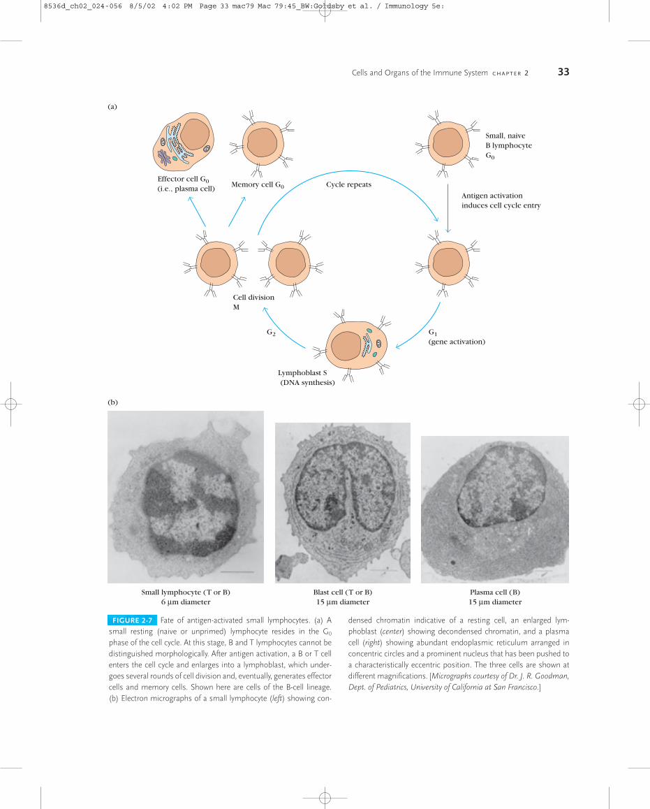
phase of the cell cycle. Known as small lymphocytes, thesecells are only about 6 �m in diameter; their cytoplasm formsa barely discernible rim around the nucleus. Small lympho-cytes have densely packed chromatin, few mitochondria, anda poorly developed endoplasmic reticulum and Golgi appa-ratus. The naive lymphocyte is generally thought to have ashort life span. Interaction of small lymphocytes with anti-gen, in the presence of certain cytokines discussed later, in-duces these cells to enter the cell cycle by progressing from G0
into G1 and subsequently into S, G2, and M (Figure 2-7a). Asthey progress through the cell cycle, lymphocytes enlargeinto 15 �m-diameter blast cells, called lymphoblasts; thesecells have a higher cytoplasm:nucleus ratio and more or-ganellar complexity than small lymphocytes (Figure 2-7b).
Lymphoblasts proliferate and eventually differentiate intoeffector cells or into memory cells. Effector cells function invarious ways to eliminate antigen. These cells have short life
TABLE 2-3 Reconstitution of hematopoeisisby HSCs
Number of Number of miceenriched HSCs reconstituted (%)
1 9 of 41 (21.9%)
2 5 of 21 (23.8%)
5 9 of 17 (52.9%)
10 10 of 11 (90.9%)
20 4 of 4 (100%)
SOURCE: Adapted from M. Osawa, et al. 1996. Science 273:242.
TABLE 2-4 Normal adult blood-cell counts
Cell type Cells/mm3 %
Red blood cells 5.0 � 106
Platelets 2.5 � 105
Leukocytes 7.3 � 103
Neutrophil 50–70
Lymphocyte 20–40
Monocyte 1–6
Eosinophil 1–3
Basophil �1
Go to www.whfreeman.com/immunology AnimationCells and Organs of the Immune System
8536d_ch02_024-056 9/6/02 9:00 PM Page 32 mac85 Mac 85:365_smm:Goldsby et al. / Immunology 5e:
Cells and Organs of the Immune System C H A P T E R 2 33
Lymphoblast S (DNA synthesis)
Effector cell G0(i.e., plasma cell) Memory cell G0
Small, naiveB lymphocyteG0
Antigen activationinduces cell cycle entry
Cycle repeats
Cell divisionM
G1(gene activation)
(a)
(b)
Small lymphocyte (T or B)6 µm diameter
Blast cell (T or B)15 µm diameter
Plasma cell (B)15 µm diameter
G2
FIGURE 2-7 Fate of antigen-activated small lymphocytes. (a) Asmall resting (naive or unprimed) lymphocyte resides in the G0
phase of the cell cycle. At this stage, B and T lymphocytes cannot bedistinguished morphologically. After antigen activation, a B or T cellenters the cell cycle and enlarges into a lymphoblast, which under-goes several rounds of cell division and, eventually, generates effectorcells and memory cells. Shown here are cells of the B-cell lineage. (b) Electron micrographs of a small lymphocyte (left) showing con-
densed chromatin indicative of a resting cell, an enlarged lym-phoblast (center) showing decondensed chromatin, and a plasmacell (right) showing abundant endoplasmic reticulum arranged inconcentric circles and a prominent nucleus that has been pushed toa characteristically eccentric position. The three cells are shown atdifferent magnifications. [Micrographs courtesy of Dr. J. R. Goodman,Dept. of Pediatrics, University of California at San Francisco.]
8536d_ch02_024-056 8/5/02 4:02 PM Page 33 mac79 Mac 79:45_BW:Goldsby et al. / Immunology 5e:

lines in the laboratory. Strikingly, these EScells can be induced to generate many dif-ferent types of cells. Mouse ES cells havebeen shown to give rise to muscle cells,nerve cells, liver cells, pancreatic cells, and,of course, hematopoietic cells.
Recent advances have made it possibleto grow lines of human pluripotent cells.This is a development of considerable im-portance to the understanding of humandevelopment, and it has great therapeuticpotential. In vitro studies of the factors thatdetermine or influence the development ofhuman pluripotent stem cells along one de-velopmental path as opposed to anotherwill provide considerable insight into thefactors that affect the differentiation of cellsinto specialized types. There is also great in-terest in exploring the use of pluripotent
stem cells to generate cells and tissues thatcould be used to replace diseased or dam-aged ones. Success in this endeavor wouldbe a major advance because transplanta-tion medicine now depends totally upon do-nated organs and tissues. Unfortunately,the need far exceeds the number of dona-tions and is increasing. Success in derivingpractical quantities of cells, tissues, and or-gans from pluripotent stem cells would pro-vide skin replacement for burn patients,heart muscle cells for those with chronicheart disease, pancreatic islet cells for pa-tients with diabetes, and neurons for use inParkinson’s disease or Alzheimer’s disease.
The transplantation of hematopoieticstem cells (HSCs) is an important ther-apy for patients whose hematopoieticsystems must be replaced. It has threemajor applications:
1. Providing a functional immunesystem to individuals with agenetically determinedimmunodeficiency, such as severe
Stem-cell transplanta-tion holds great promise for the regener-ation of diseased, damaged, or defectivetissue. Hematopoietic stem cells are al-ready used to restore hematopoieticcells, and their use is described in theclinic below. However, rapid advances instem-cell research have raised the possi-bility that other stem-cell types, too, maysoon be routinely employed for replace-ment of other cells and tissues. Twoproperties of stem cells underlie theirutility and promise. They have the capac-ity to give rise to more differentiatedcells, and they are self-renewing, becauseeach division of a stem cell creates atleast one stem cell. If stem cells are clas-sified according to their descent and de-velopmental potential, four levels ofstem cells can be recognized: totipotent,pluripotent, multipotent, and unipotent.
Totipotent cells can give rise to an en-tire organism. A fertilized egg, the zygote,is a totipotent cell. In humans the initial di-visions of the zygote and its descendantsproduce cells that are also totipotent. Infact, identical twins, each with its own pla-centa, develop when totipotent cells sepa-rate and develop into genetically identicalfetuses. Pluripotent stem cells arise fromtotipotent cells and can give rise to mostbut not all of the cell types necessary for fe-tal development. For example, humanpluripotent stem cells can give rise to all ofthe cells of the body but cannot generate aplacenta. Further differentiation of pluripo-tent stem cells leads to the formation ofmultipotent and unipotent stem cells.Multipotent stem cells can give rise to onlya limited number of cell types, and unipo-tent cells to a single cell type. Pluripotentcells, called embryonic stem cells, or sim-ply ES cells, can be isolated from early em-bryos, and for many years it has beenpossible to grow mouse ES cells as cell
C L I N I C A L F O C U S
Stem Cells—Clinical Uses and Potential
Bone marrow Nerve cells Heart muscle cells
Human pluripotent stem cells
Pancreatic islet cells
Human pluripotent stem cells can differentiate into a variety of different cell types,some of which are shown here. [Adapted from Stem Cells: A Primer, NIH web sitehttp://www.nih.gov/news/stemcell/primer.htm. Micrographs (left to right):Biophoto Associates/Science Source/Photo Researchers; Biophoto Associates/PhotoResearchers; AFIP/Science Source/Photo Researchers; Astrid & Hanns-FriederMichler/Science Photo Library/Photo Researchers.]
34 P A R T I Introduction
8536d_ch02_024-056 8/5/02 4:02 PM Page 34 mac79 Mac 79:45_BW:Goldsby et al. / Immunology 5e:

Cells and Organs of the Immune System C H A P T E R 2 35
sible for individuals to store their ownhematopoietic cells for transplantation tothemselves at a later time. Currently, thisprocedure is used to allow cancer patientsto donate cells before undergoing chemo-therapy and radiation treatments and thento reconstitute their hematopoietic systemfrom their own stem cells. Hematopoieticstem cells are found in cell populations thatdisplay distinctive surface antigens. One ofthese antigens is CD34, which is present ononly a small percentage (~1%) of the cellsin adult bone marrow. An antibody specificfor CD34 is used to select cells displayingthis antigen, producing a population en-riched in CD34� stem cells. Various ver-sions of this selection procedure have beenused to enrich populations of stem cellsfrom a variety of sources.
Transplantation of stem cell popula-tions may be autologous (the recipient isalso the donor), syngeneic (the donor isgenetically identical, i.e., an identical twinof the recipient), or allogeneic (the donorand recipient are not genetically identical).In any transplantation procedure, geneticdifferences between donor and recipientcan lead to immune-based rejection reac-tions. Aside from host rejection of trans-planted tissue (host versus graft),lymphocytes in the graft can attack the re-cipient’s tissues, thereby causing graft-versus-host disease (GVHD), a life-threatening affliction. In order to suppressrejection reactions, powerful immunosup-pressive drugs must be used. Unfortu-nately, these drugs have serious sideeffects, and immunosuppression in-creases the patient’s risk of infection andfurther growth of tumors. Consequently,HSC transplantation has fewest complica-tions when there is genetic identity be-tween donor and recipient.
At one time, bone-marrow transplanta-tion was the only way to restore thehematopoietic system. However, the essen-tial element of bone-marrow transplanta-tion is really stem-cell transplantation.Fortunately, significant numbers of stemcells can be obtained from other tissues,such as peripheral blood and umbilical-cordblood (“cord blood”). These alternativesources of HSCs are attractive because the
donor does not have to undergo anesthesiaand the subsequent highly invasive proce-dure that extracts bone marrow. Many in thetransplantation community believe that pe-ripheral blood will replace marrow as themajor source of hematopoietic stem cellsfor many applications. To obtain HSC-en-riched preparations from peripheral blood,agents are used to induce increased num-bers of circulating HSCs, and then the HSC-containing fraction is separated from theplasma and red blood cells in a processcalled leukopheresis. If necessary, furtherpurification can be done to remove T cellsand to enrich the CD34� population.
Umbilical cord blood already contains asignificant number of hematopoietic stemcells. Furthermore, it is obtained from pla-cental tissue (the “afterbirth”) which is nor-mally discarded. Consequently, umbilicalcord blood has become an attractivesource of cells for HSC transplantation. Al-though HSCs from cord blood fail to en-graft somewhat more often than do cellsfrom peripheral blood, grafts of cord bloodcells produce GVHD less frequently thando marrow grafts, probably because cordblood has fewer mature T cells.
Beyond its current applications in can-cer treatment, many researchers feel thatautologous stem-cell transplantation willbe useful for gene therapy, the introductionof a normal gene to correct a disordercaused by a defective gene. Rapid ad-vances in genetic engineering may soonmake gene therapy a realistic treatment forgenetic disorders of blood cells, andhematopoietic stem cells are attractive ve-hicles for such an approach. The therapywould entail removing a sample ofhematopoietic stem cells from a patient,inserting a functional gene to compensatefor the defective one, and then reinjectingthe engineered stem cells into the donor.The advantage of using stem cells in genetherapy is that they are self renewing. Con-sequently, at least in theory, patients wouldhave to receive only a single injection of en-gineered stem cells. In contrast, gene ther-apy with engineered mature lymphocytesor other blood cells would require periodicinjections because these cells are not ca-pable of self renewal.
combined immunodeficiency(SCID).
2. Replacing a defective hematopoieticsystem with a functional one to curesome patients who have a life-threatening nonmalignant geneticdisorder in hematopoiesis, such assickle-cell anemia or thalassemia.
3. Restoring the hematopoietic systemof cancer patients after treatmentwith doses of chemotherapeuticagents and radiation so high thatthey destroy the system. Thesehigh-dose regimens can be muchmore effective at killing tumor cellsthan are therapies that use moreconventional doses of cytotoxicagents. Stem-cell transplantationmakes it possible to recover fromsuch drastic treatment. Also, certaincancers, such as some cases ofacute myeloid leukemia, can becured only by destroying the sourceof the leukemia cells, the patient’sown hematopoietic system.
Restoration of the hematopoietic sys-tem by transplanting stem cells is facili-tated by several important technicalconsiderations. First, HSCs have extraordi-nary powers of regeneration. Experimentsin mice indicate that only a few—perhaps,on occasion, a single HSC—can com-pletely restore the erythroid population andthe immune system. In humans it is neces-sary to administer as little as 10% of adonor’s total volume of bone marrow toprovide enough HSCs to completely re-store the hematopoietic system. Once in-jected into a vein, HSCs enter thecirculation and find their own way to thebone marrow, where they begin the processof engraftment. There is no need for a sur-geon to directly inject the cells into bones.In addition, HSCs can be preserved byfreezing. This means that hematopoieticcells can be “banked.” After collection, thecells are treated with a cryopreservative,frozen, and then stored for later use. Whenneeded, the frozen preparation is thawedand infused into the patient, where it re-constitutes the hematopoietic system. Thiscell-freezing technology even makes it pos-
8536d_ch02_024-056 8/7/02 8:25 AM Page 35 mac79 Mac 79:45_BW:Goldsby et al. / Immunology 5e:

36 P A R T I Introduction
spans, generally ranging from a few days to a few weeks.Plasma cells—the antibody-secreting effector cells of the B-cell lineage—have a characteristic cytoplasm that containsabundant endoplasmic reticulum (to support their high rateof protein synthesis) arranged in concentric layers and alsomany Golgi vesicles (see Figure 2-7). The effector cells of theT-cell lineage include the cytokine-secreting T helper cell(TH cell) and the T cytotoxic lymphocyte (TC cell). Some ofthe progeny of B and T lymphoblasts differentiate into mem-ory cells. The persistence of this population of cells is respon-sible for life-long immunity to many pathogens. Memorycells look like small lymphocytes but can be distinguishedfrom naive cells by the presence or absence of certain cell-membrane molecules.
Different lineages or maturational stages of lymphocytescan be distinguished by their expression of membrane mole-cules recognized by particular monoclonal antibodies (anti-bodies that are specific for a single epitope of an antigen; seeChapter 4 for a description of monoclonal antibodies). All ofthe monoclonal antibodies that react with a particular mem-brane molecule are grouped together as a cluster of dif-ferentiation (CD). Each new monoclonal antibody that recognizes a leukocyte membrane molecule is analyzed forwhether it falls within a recognized CD designation; if it does
not, it is given a new CD designation reflecting a new mem-brane molecule. Although the CD nomenclature was origi-nally developed for the membrane molecules of humanleukocytes, the homologous membrane molecules of otherspecies, such as mice, are commonly referred to by the sameCD designations. Table 2-5 lists some common CD mole-cules (often referred to as CD markers) found on humanlymphocytes. However, this is only a partial listing of themore than 200 CD markers that have been described. A com-plete list and description of known CD markers is in the ap-pendix at the end of this book.
The general characteristics and functions of B and T lym-phocytes were described in Chapter 1 and are reviewedbriefly in the next sections. These central cells of the immunesystem will be examined in more detail in later chapters.
B LYMPHOCYTES
The B lymphocyte derived its letter designation from its siteof maturation, in the bursa of Fabricius in birds; the nameturned out to be apt, for bone marrow is its major site of mat-uration in a number of mammalian species, including hu-mans and mice. Mature B cells are definitively distinguishedfrom other lymphocytes by their synthesis and display ofmembrane-bound immunoglobulin (antibody) molecules,
TABLE 2-5 Common CD markers used to distinguish functional lymphocyte subpopulations
T CELL
CD designation* Function B cell TH TC NK cell
CD2 Adhesion molecule; signal transduction � � � �
CD3 Signal-transduction element of T-cell � � � �receptor
CD4 Adhesion molecule that binds to class II � � � �MHC molecules; signal transduction (usually) (usually)
CD5 Unknown � � � �(subset)
CD8 Adhesion molecule that binds to class I � � � �MHC molecules; signal transduction (usually) (usually) (variable)
CD16 (Fc�RIII) Low-affinity receptor for Fc region of IgG � � � �
CD21 (CR2) Receptor for complement (C3d) and � � � �Epstein-Barr virus
CD28 Receptor for co-stimulatory B7 molecule � � � �on antigen-presenting cells
CD32 (Fc�RII) Receptor for Fc region of IgG � � � �
CD35 (CR1) Receptor for complement (C3b) � � � �
CD40 Signal transduction � � � �
CD45 Signal transduction � � � �
CD56 Adhesion molecule � � � �
*Synonyms are shown in parentheses.
8536d_ch02_024-056 8/5/02 4:02 PM Page 36 mac79 Mac 79:45_BW:Goldsby et al. / Immunology 5e:
Cells and Organs of the Immune System C H A P T E R 2 37
which serve as receptors for antigen. Each of the approxi-mately 1.5 � 105 molecules of antibody on the membrane ofa single B cell has an identical binding site for antigen.Among the other molecules expressed on the membrane ofmature B cells are the following:
� B220 (a form of CD45) is frequently used as a markerfor B cells and their precursors. However, unlikeantibody, it is not expressed uniquely by B-lineage cells.
� Class II MHC molecules permit the B cell to function asan antigen-presenting cell (APC).
� CR1 (CD35) and CR2 (CD21) are receptors for certaincomplement products.
� Fc�RII (CD32) is a receptor for IgG, a type of antibody.
� B7-1 (CD80) and B7-2 (CD86) are molecules thatinteract with CD28 and CTLA-4, important regulatorymolecules on the surface of different types of T cells,including TH cells.
� CD40 is a molecule that interacts with CD40 ligand onthe surface of helper T cells. In most cases thisinteraction is critical for the survival of antigen-stimulated B cells and for their development intoantibody-secreting plasma cells or memory B cells.
Interaction between antigen and the membrane-bound anti-body on a mature naive B cell, as well as interactions with Tcells and macrophages, selectively induces the activation anddifferentiation of B-cell clones of corresponding specificity.In this process, the B cell divides repeatedly and differentiatesover a 4- to 5-day period, generating a population of plasmacells and memory cells. Plasma cells, which have lower levelsof membrane-bound antibody than B cells, synthesize andsecrete antibody. All clonal progeny from a given B cell se-crete antibody molecules with the same antigen-bindingspecificity. Plasma cells are terminally differentiated cells,and many die in 1 or 2 weeks.
T LYMPHOCYTES
T lymphocytes derive their name from their site of matura-tion in the thymus. Like B lymphocytes, these cells havemembrane receptors for antigen. Although the antigen-binding T-cell receptor is structurally distinct from im-munoglobulin, it does share some common structuralfeatures with the immunoglobulin molecule, most notably inthe structure of its antigen-binding site. Unlike the mem-brane-bound antibody on B cells, though, the T-cell receptor(TCR) does not recognize free antigen. Instead the TCR rec-ognizes only antigen that is bound to particular classes ofself-molecules. Most T cells recognize antigen only when it isbound to a self-molecule encoded by genes within the majorhistocompatibility complex (MHC). Thus, as explained inChapter 1, a fundamental difference between the humoraland cell-mediated branches of the immune system is that theB cell is capable of binding soluble antigen, whereas the T cell
is restricted to binding antigen displayed on self-cells. To berecognized by most T cells, this antigen must be displayed to-gether with MHC molecules on the surface of antigen-pre-senting cells or on virus-infected cells, cancer cells, andgrafts. The T-cell system has developed to eliminate these al-tered self-cells, which pose a threat to the normal functioningof the body.
Like B cells, T cells express distinctive membrane mole-cules. All T-cell subpopulations express the T-cell receptor, acomplex of polypeptides that includes CD3; and most can bedistinguished by the presence of one or the other of twomembrane molecules, CD4 and CD8. In addition, most ma-ture T cells express the following membrane molecules:
� CD28, a receptor for the co-stimulatory B7 family ofmolecules present on B cells and other antigen-presenting cells
� CD45, a signal-transduction molecule
T cells that express the membrane glycoprotein moleculeCD4 are restricted to recognizing antigen bound to class IIMHC molecules, whereas T cells expressing CD8, a dimericmembrane glycoprotein, are restricted to recognition of anti-gen bound to class I MHC molecules. Thus the expression ofCD4 versus CD8 corresponds to the MHC restriction of theT cell. In general, expression of CD4 and of CD8 also definestwo major functional subpopulations of T lymphocytes.CD4� T cells generally function as T helper (TH) cells and areclass-II restricted; CD8� T cells generally function as T cyto-toxic (TC) cells and are class-I restricted. Thus the ratio of TH
to TC cells in a sample can be approximated by assaying thenumber of CD4� and CD8� T cells. This ratio is approxi-mately 2:1 in normal human peripheral blood, but it may besignificantly altered by immunodeficiency diseases, autoim-mune diseases, and other disorders.
The classification of CD4� class II–restricted cells as TH
cells and CD8� class I–restricted cells as TC cells is not ab-solute. Some CD4� cells can act as killer cells. Also, some TC
cells have been shown to secrete a variety of cytokines and ex-ert an effect on other cells comparable to that exerted by TH
cells. The distinction between TH and TC cells, then, is not al-ways clear; there can be ambiguous functional activities.However, because these ambiguities are the exception andnot the rule, the generalization of T helper (TH) cells as beingCD4� and class-II restricted and of T cytotoxic cells (TC) asbeing CD8� and class-I restricted is assumed throughoutthis text, unless otherwise specified.
TH cells are activated by recognition of an antigen–class IIMHC complex on an antigen-presenting cell. After activa-tion, the TH cell begins to divide and gives rise to a clone ofeffector cells, each specific for the same antigen–class IIMHC complex. These TH cells secrete various cytokines,which play a central role in the activation of B cells, T cells,and other cells that participate in the immune response.Changes in the pattern of cytokines produced by TH cells canchange the type of immune response that develops among
8536d_ch02_024-056 8/5/02 4:02 PM Page 37 mac79 Mac 79:45_BW:Goldsby et al. / Immunology 5e:

38 P A R T I Introduction
other leukocytes. The TH1 response produces a cytokineprofile that supports inflammation and activates mainly cer-tain T cells and macrophages, whereas the TH2 response ac-tivates mainly B cells and immune responses that dependupon antibodies. TC cells are activated when they interactwith an antigen–class I MHC complex on the surface of analtered self-cell (e.g., a virus-infected cell or a tumor cell) inthe presence of appropriate cytokines. This activation, whichresults in proliferation, causes the TC cell to differentiate intoan effector cell called a cytotoxic T lymphocyte (CTL). Incontrast to TH cells, most CTLs secrete few cytokines. In-stead, CTLs acquire the ability to recognize and eliminate al-tered self-cells.
Another subpopulation of T lymphocytes—called T sup-pressor (TS) cells—has been postulated. It is clear that someT cells help to suppress the humoral and the cell-mediatedbranches of the immune system, but the actual isolation andcloning of normal TS cells is a matter of controversy and dis-pute among immunologists. For this reason, it is uncertainwhether TS cells do indeed constitute a separate functionalsubpopulation of T cells. Some immunologists believe thatthe suppression mediated by T cells observed in some sys-tems is simply the consequence of activities of TH or TC sub-populations whose end results are suppressive.
NATURAL KILLER CELLS
The natural killer cell was first described in 1976, when it wasshown that the body contains a small population of large,granular lymphocytes that display cytotoxic activity against awide range of tumor cells in the absence of any previous im-munization with the tumor. NK cells were subsequentlyshown to play an important role in host defense both againsttumor cells and against cells infected with some, though notall, viruses. These cells, which constitute 5%–10% of lym-phocytes in human peripheral blood, do not express themembrane molecules and receptors that distinguish T- andB-cell lineages. Although NK cells do not have T-cell recep-tors or immunoglobulin incorporated in their plasma mem-branes, they can recognize potential target cells in twodifferent ways. In some cases, an NK cell employs NK cell re-ceptors to distinguish abnormalities, notably a reduction inthe display of class I MHC molecules and the unusual profileof surface antigens displayed by some tumor cells and cellsinfected by some viruses. Another way in which NK cells rec-ognize potential target cells depends upon the fact that sometumor cells and cells infected by certain viruses display anti-gens against which the immune system has made an anti-body response, so that antitumor or antiviral antibodies arebound to their surfaces. Because NK cells express CD16, amembrane receptor for the carboxyl-terminal end of the IgGmolecule, called the Fc region, they can attach to these anti-bodies and subsequently destroy the targeted cells. This is anexample of a process known as antibody-dependent cell-mediated cytotoxicity (ADCC). The exact mechanism ofNK-cell cytotoxicity, the focus of much current experimentalstudy, is described further in Chapter 14.
Several observations suggest that NK cells play an impor-tant role in host defense against tumors. For example, in hu-mans the Chediak-Higashi syndrome—an autosomalrecessive disorder—is associated with impairment in neu-trophils, macrophages, and NK cells and an increased inci-dence of lymphomas. Likewise, mice with an autosomalmutation called beige lack NK cells; these mutants are moresusceptible than normal mice to tumor growth following in-jection with live tumor cells.
There has been growing recognition of a cell type, theNK1-T cell, that has some of the characteristics of both Tcells and NK cells. Like T cells, NK1-T cells have T cell recep-tors (TCRs). Unlike most T cells, the TCRs of NK1-T cells in-teract with MHC-like molecules called CD1 rather than withclass I or class II MHC molecules. Like NK cells, they havevariable levels of CD16 and other receptors typical of NKcells, and they can kill cells. A population of triggered NK1-Tcells can rapidly secrete large amounts of the cytokinesneeded to support antibody production by B cells as well asinflammation and the development and expansion of cyto-toxic T cells. Some immunologists view this cell type as a kind of rapid response system that has evolved to pro-vide early help while conventional TH responses are still developing.
Mononuclear PhagocytesThe mononuclear phagocytic system consists of monocytescirculating in the blood and macrophages in the tissues (Figure 2-8). During hematopoiesis in the bone marrow,granulocyte-monocyte progenitor cells differentiate intopromonocytes, which leave the bone marrow and enter the blood, where they further differentiate into maturemonocytes. Monocytes circulate in the bloodstream forabout 8 h, during which they enlarge; they then migrate intothe tissues and differentiate into specific tissue macrophagesor, as discussed later, into dendritic cells.
Differentiation of a monocyte into a tissue macrophageinvolves a number of changes: The cell enlarges five- to ten-fold; its intracellular organelles increase in both number andcomplexity; and it acquires increased phagocytic ability, pro-duces higher levels of hydrolytic enzymes, and begins to se-crete a variety of soluble factors. Macrophages are dispersedthroughout the body. Some take up residence in particulartissues, becoming fixed macrophages, whereas others remainmotile and are called free, or wandering, macrophages. Freemacrophages travel by amoeboid movement throughout the tissues. Macrophage-like cells serve different functions indifferent tissues and are named according to their tissue location:
� Alveolar macrophages in the lung
� Histiocytes in connective tissues
� Kupffer cells in the liver
� Mesangial cells in the kidney
8536d_ch02_024-056 8/5/02 4:02 PM Page 38 mac79 Mac 79:45_BW:Goldsby et al. / Immunology 5e:

Cells and Organs of the Immune System C H A P T E R 2 39
� Microglial cells in the brain
� Osteoclasts in bone
Although normally in a resting state, macrophages are acti-vated by a variety of stimuli in the course of an immune re-sponse. Phagocytosis of particulate antigens serves as aninitial activating stimulus. However, macrophage activity canbe further enhanced by cytokines secreted by activated TH
cells, by mediators of the inflammatory response, and bycomponents of bacterial cell walls. One of the most potentactivators of macrophages is interferon gamma (IFN-�) se-creted by activated TH cells.
Activated macrophages are more effective than restingones in eliminating potential pathogens, because they exhibitgreater phagocytic activity, an increased ability to kill in-gested microbes, increased secretion of inflammatory medi-ators, and an increased ability to activate T cells. In addition,
activated macrophages, but not resting ones, secrete variouscytotoxic proteins that help them eliminate a broad range ofpathogens, including virus-infected cells, tumor cells, and in-tracellular bacteria. Activated macrophages also expresshigher levels of class II MHC molecules, allowing them tofunction more effectively as antigen-presenting cells. Thus,macrophages and TH cells facilitate each other’s activationduring the immune response.
PHAGOCYTOSIS
Macrophages are capable of ingesting and digesting exoge-nous antigens, such as whole microorganisms and insolubleparticles, and endogenous matter, such as injured or deadhost cells, cellular debris, and activated clotting factors. In thefirst step in phagocytosis, macrophages are attracted by andmove toward a variety of substances generated in an immuneresponse; this process is called chemotaxis. The next step inphagocytosis is adherence of the antigen to the macrophagecell membrane. Complex antigens, such as whole bacterialcells or viral particles, tend to adhere well and are readilyphagocytosed; isolated proteins and encapsulated bacteriatend to adhere poorly and are less readily phagocytosed. Ad-herence induces membrane protrusions, called pseudopo-dia, to extend around the attached material (Figure 2-9a).Fusion of the pseudopodia encloses the material within amembrane-bounded structure called a phagosome, whichthen enters the endocytic processing pathway (Figure 2-9b).In this pathway, a phagosome moves toward the cell interior,where it fuses with a lysosome to form a phagolysosome.Lysosomes contain lysozyme and a variety of other hy-drolytic enzymes that digest the ingested material. The di-gested contents of the phagolysosome are then eliminated ina process called exocytosis (see Figure 2-9b).
The macrophage membrane has receptors for certainclasses of antibody. If an antigen (e.g., a bacterium) is coatedwith the appropriate antibody, the complex of antigen andantibody binds to antibody receptors on the macrophagemembrane more readily than antigen alone and phagocyto-sis is enhanced. In one study, for example, the rate of phago-cytosis of an antigen was 4000-fold higher in the presence ofspecific antibody to the antigen than in its absence. Thus, an-tibody functions as an opsonin, a molecule that binds toboth antigen and macrophage and enhances phagocytosis.The process by which particulate antigens are rendered moresusceptible to phagocytosis is called opsonization.
ANTIMICROBIAL AND CYTOTOXIC ACTIVITIES
A number of antimicrobial and cytotoxic substances pro-duced by activated macrophages can destroy phagocytosedmicroorganisms (Table 2-6). Many of the mediators of cyto-toxicity listed in Table 2-6 are reactive forms of oxygen.
OXYGEN-DEPENDENT KILLING MECHANISMS Activatedphagocytes produce a number of reactive oxygen intermedi-ates (ROIs) and reactive nitrogen intermediates that have
(a) Monocyte
Lysosome
Nucleus
Phagosome
(b) Macrophage
Phagosome
PhagosomePhagolysosome
Lysosome
Pseudopodia
FIGURE 2-8 Typical morphology of a monocyte and amacrophage. Macrophages are five- to tenfold larger than monocytesand contain more organelles, especially lysosomes.
8536d_ch02_024-056 8/5/02 4:02 PM Page 39 mac79 Mac 79:45_BW:Goldsby et al. / Immunology 5e:

40 P A R T I Introduction
oxide and chloride ions. Hypochlorite, the active agent ofhousehold bleach, is toxic to ingested microbes. Whenmacrophages are activated with bacterial cell-wall compo-nents such as lipopolysaccharide (LPS) or, in the case of my-cobacteria, muramyl dipeptide (MDP), together with aT-cell–derived cytokine (IFN-�), they begin to express highlevels of nitric oxide synthetase (NOS), an enzyme that oxi-dizes L-arginine to yield L-citrulline and nitric oxide (NO), agas:
L-arginine � O2 �NADPH →NO � L-citrulline � NADP
Nitric oxide has potent antimicrobial activity; it also cancombine with the superoxide anion to yield even more po-tent antimicrobial substances. Recent evidence suggests thatmuch of the antimicrobial activity of macrophages againstbacteria, fungi, parasitic worms, and protozoa is due to nitricoxide and substances derived from it.
OXYGEN-INDEPENDENT KILLING MECHANISMS Acti-vated macrophages also synthesize lysozyme and various hy-drolytic enzymes whose degradative activities do not requireoxygen. In addition, activated macrophages produce a groupof antimicrobial and cytotoxic peptides, commonly knownas defensins. These molecules are cysteine-rich cationic pep-tides containing 29–35 amino-acid residues. Each peptide,which contains six invariant cysteines, forms a circular mole-cule that is stabilized by intramolecular disulfide bonds.These circularized defensin peptides have been shown toform ion-permeable channels in bacterial cell membranes.Defensins can kill a variety of bacteria, including Staphylo-coccus aureus, Streptococcus pneumoniae, Escherichia coli,
potent antimicrobial activity. During phagocytosis, a meta-bolic process known as the respiratory burst occurs in acti-vated macrophages. This process results in the activation of amembrane-bound oxidase that catalyzes the reduction ofoxygen to superoxide anion, a reactive oxygen intermediatethat is extremely toxic to ingested microorganisms. The su-peroxide anion also generates other powerful oxidizingagents, including hydroxyl radicals and hydrogen peroxide.As the lysosome fuses with the phagosome, the activity ofmyeloperoxidase produces hypochlorite from hydrogen per-
FIGURE 2-9 Macrophages can ingest and degrade particulateantigens, including bacteria. (a) Scanning electron micrograph of amacrophage. Note the long pseudopodia extending toward and mak-ing contact with bacterial cells, an early step in phagocytosis. (b)Phagocytosis and processing of exogenous antigen by macrophages.
Pseudopodia
Lysosome
Phagolysosome
Class IIMHC
Bacteria
Phagosome
Exocytosed degraded material
Antigenic peptide/class II MHC
(b)(a)
Most of the products resulting from digestion of ingested materialare exocytosed, but some peptide products may interact with class IIMHC molecules, forming complexes that move to the cell surface,where they are presented to TH cells. [Photograph by L. Nilsson, ©Boehringer Ingelheim International GmbH.]
TABLE 2-6Mediators of antimicrobial andcytotoxic activity of macrophages and neutrophils
Oxygen-dependent killing Oxygen-independent killing
Reactive oxygen intermediates Defensins
O•2
� (superoxide anion) Tumor necrosis factor �
OH• (hydroxyl radicals) (macrophage only)
H2O2 (hydrogen peroxide) Lysozyme
ClO�(hypochlorite anion) Hydrolytic enzymes
Reactive nitrogen intermediates
NO (nitric oxide)
NO2 (nitrogen dioxide)
HNO2 (nitrous acid)
Others
NH2CL (monochloramine)
8536d_ch02_024-056 8/5/02 4:02 PM Page 40 mac79 Mac 79:45_BW:Goldsby et al. / Immunology 5e:

Cells and Organs of the Immune System C H A P T E R 2 41
Pseudomonas aeruginosa, and Haemophilus influenzae. Acti-vated macrophages also secrete tumor necrosis factor �(TNF-�), a cytokine that has a variety of effects and is cyto-toxic for some tumor cells.
ANTIGEN PROCESSING AND PRESENTATION
Although most of the antigen ingested by macrophages is de-graded and eliminated, experiments with radiolabeled anti-gens have demonstrated the presence of antigen peptides onthe macrophage membrane. As depicted in Figure 2-9b,phagocytosed antigen is digested within the endocytic pro-cessing pathway into peptides that associate with class IIMHC molecules; these peptide–class II MHC complexesthen move to the macrophage membrane. Activation ofmacrophages induces increased expression of both class IIMHC molecules and the co-stimulatory B7 family of mem-brane molecules, thereby rendering the macrophages moreeffective in activating TH cells. This processing and presenta-tion of antigen, examined in detail in Chapter 7, are critical toTH-cell activation, a central event in the development of bothhumoral and cell-mediated immune responses.
SECRETION OF FACTORS
A number of important proteins central to development ofimmune responses are secreted by activated macrophages(Table 2-7). These include a collection of cytokines, such asinterleukin 1 (IL-1), TNF-� and interleukin 6 (IL-6), thatpromote inflammatory responses. Typically, each of theseagents has a variety of effects. For example, IL-1 activateslymphocytes; and IL-1, IL-6, and TNF-� promote fever by af-fecting the thermoregulatory center in the hypothalamus.
Activated macrophages secrete a variety of factors in-volved in the development of an inflammatory response. Thecomplement proteins are a group of proteins that assist ineliminating foreign pathogens and in promoting the ensuinginflammatory reaction. The major site of synthesis of com-plement proteins is the liver, although these proteins are alsoproduced in macrophages. The hydrolytic enzymes con-tained within the lysosomes of macrophages also can be se-creted when the cells are activated. The buildup of theseenzymes within the tissues contributes to the inflammatoryresponse and can, in some cases, contribute to extensive tis-sue damage. Activated macrophages also secrete soluble fac-tors, such as TNF-�, that can kill a variety of cells. Thesecretion of these cytotoxic factors has been shown to con-tribute to tumor destruction by macrophages. Finally, asmentioned earlier, activated macrophages secrete a numberof cytokines that stimulate inducible hematopoiesis.
Granulocytic CellsThe granulocytes are classified as neutrophils, eosinophils,or basophils on the basis of cellular morphology and cyto-plasmic staining characteristics (Figure 2-10). The neu-trophil has a multilobed nucleus and a granulated cytoplasmthat stains with both acid and basic dyes; it is often called apolymorphonuclear leukocyte (PMN) for its multilobed nu-cleus. The eosinophil has a bilobed nucleus and a granulatedcytoplasm that stains with the acid dye eosin red (hence itsname). The basophil has a lobed nucleus and heavily granu-lated cytoplasm that stains with the basic dye methylene blue.Both neutrophils and eosinophils are phagocytic, whereasbasophils are not. Neutrophils, which constitute 50%–70%of the circulating white blood cells, are much more numer-ous than eosinophils (1%–3%) or basophils (�1%).
NEUTROPHILS
Neutrophils are produced by hematopoiesis in the bone mar-row. They are released into the peripheral blood and circulatefor 7–10 h before migrating into the tissues, where they havea life span of only a few days. In response to many types of in-fections, the bone marrow releases more than the usual num-ber of neutrophils and these cells generally are the first toarrive at a site of inflammation. The resulting transient in-crease in the number of circulating neutrophils, called leuko-cytosis, is used medically as an indication of infection.
Movement of circulating neutrophils into tissues, calledextravasation, takes several steps: the cell first adheres to the vascular endothelium, then penetrates the gap betweenadjacent endothelial cells lining the vessel wall, and finallypenetrates the vascular basement membrane, moving outinto the tissue spaces. (This process is described in detail inChapter 15.) A number of substances generated in an inflam-matory reaction serve as chemotactic factors that promoteaccumulation of neutrophils at an inflammatory site. Amongthese chemotactic factors are some of the complement
TABLE 2-7 Some factors secreted by activatedmacrophages
Factor Function
Interleukin 1 (IL-1) Promotes inflammatory responsesand fever
Interleukin 6 (IL-6) Promote innate immunity andTNF-� elimination of pathogens
Complement proteins Promote inflammatory responseand elimination of pathogens
Hydrolytic enzymes Promote inflammatory response
Interferon alpha Activates cellular genes, resulting(IFN-�) in the production of proteins that
confer an antiviral state on the cell
Tumor necrosis factor Kills tumor cells(TNF-�)
GM-CSF
G-CSF Promote inducible hematopoiesis
M-CSF
8536d_ch02_024-056 8/5/02 4:02 PM Page 41 mac79 Mac 79:45_BW:Goldsby et al. / Immunology 5e:

42 P A R T I Introduction
components, components of the blood-clotting system,and sev-eral cytokines secreted by activated TH cells and macrophages.
Like macrophages, neutrophils are active phagocytic cells.Phagocytosis by neutrophils is similar to that described formacrophages, except that the lytic enzymes and bactericidalsubstances in neutrophils are contained within primary andsecondary granules (see Figure 2-10a). The larger, denser pri-mary granules are a type of lysosome containing peroxidase,lysozyme, and various hydrolytic enzymes. The smaller sec-ondary granules contain collagenase, lactoferrin, and lyso-zyme. Both primary and secondary granules fuse withphagosomes, whose contents are then digested and elimi-nated much as they are in macrophages.
Neutrophils also employ both oxygen-dependent andoxygen-independent pathways to generate antimicrobialsubstances. Neutrophils are in fact much more likely thanmacrophages to kill ingested microorganisms. Neutrophilsexhibit a larger respiratory burst than macrophages and con-sequently are able to generate more reactive oxygen interme-diates and reactive nitrogen intermediates (see Table 2-6). Inaddition, neutrophils express higher levels of defensins thanmacrophages do.
EOSINOPHILS
Eosinophils, like neutrophils, are motile phagocytic cells thatcan migrate from the blood into the tissue spaces. Theirphagocytic role is significantly less important than that ofneutrophils, and it is thought that they play a role in the de-fense against parasitic organisms (see Chapter 17). The se-creted contents of eosinophilic granules may damage theparasite membrane.
BASOPHILS
Basophils are nonphagocytic granulocytes that function byreleasing pharmacologically active substances from their cy-toplasmic granules. These substances play a major role in cer-tain allergic responses.
MAST CELLS
Mast-cell precursors, which are formed in the bone marrowby hematopoiesis, are released into the blood as undifferenti-ated cells; they do not differentiate until they leave the bloodand enter the tissues. Mast cells can be found in a wide vari-ety of tissues, including the skin, connective tissues of variousorgans, and mucosal epithelial tissue of the respiratory, geni-tourinary, and digestive tracts. Like circulating basophils,these cells have large numbers of cytoplasmic granules thatcontain histamine and other pharmacologically active sub-stances. Mast cells, together with blood basophils, play an im-portant role in the development of allergies.
DENDRITIC CELLS
The dendritic cell (DC) acquired its name because it is cov-ered with long membrane extensions that resemble the den-drites of nerve cells. Dendritic cells can be difficult to isolatebecause the conventional procedures for cell isolation tend todamage their long extensions. The development of isolationtechniques that employ enzymes and gentler dispersion hasfacilitated isolation of these cells for study in vitro. There aremany types of dendritic cells, although most mature den-dritic cells have the same major function, the presentation ofantigen to TH cells. Four types of dendritic cells are known:Langerhans cells, interstitial dendritic cells, myeloid cells,and lymphoid dendritic cells. Each arises from hematopoi-etic stem cells via different pathways and in different loca-tions. Figure 2-11 shows that they descend through both themyeloid and lymphoid lineages. Despite their differences,
Phagosome
Multilobednucleus Primary
azurophilicgranule
Secondarygranule
Glycogen
(a) Neutrophil
(b) Eosinophil
Crystalloidgranule
(c) Basophil
Glycogen
Granule
FIGURE 2-10 Drawings showing typical morphology of granulo-cytes. Note differences in the shape of the nucleus and in the num-ber and shape of cytoplasmic granules.
8536d_ch02_024-056 8/5/02 4:02 PM Page 42 mac79 Mac 79:45_BW:Goldsby et al. / Immunology 5e:

Cells and Organs of the Immune System C H A P T E R 2 43
they all constitutively express high levels of both class IIMHC molecules and members of the co-stimulatory B7 fam-ily. For this reason, they are more potent antigen-presentingcells than macrophages and B cells, both of which need to beactivated before they can function as antigen-presenting cells(APCs). Immature or precursor forms of each of these typesof dendritic cells acquire antigen by phagocytosis or endocy-tosis; the antigen is processed, and mature dendritic cells pre-sent it to TH cells. Following microbial invasion or duringinflammation, mature and immature forms of Langerhanscells and interstitial dendritic cells migrate into draininglymph nodes, where they make the critical presentation ofantigen to TH cells that is required for the initiation of re-sponses by those key cells.
Another type of dendritic cell, the follicular dendritic cell(Figure 2-12), does not arise in bone marrow and has a dif-ferent function from the antigen-presenting dendritic cellsdescribed above. Follicular dendritic cells do not express classII MHC molecules and therefore do not function as antigen-presenting cells for TH-cell activation. These dendritic cellswere named for their exclusive location in organized struc-tures of the lymph node called lymph follicles, which are richin B cells. Although they do not express class II molecules,follicular dendritic cells express high levels of membrane re-ceptors for antibody, which allows the binding of antigen-an-
tibody complexes. The interaction of B cells with this boundantigen can have important effects on B cell responses.
Organs of the Immune SystemA number of morphologically and functionally diverse or-gans and tissues have various functions in the developmentof immune responses. These can be distinguished by func-tion as the primary and secondary lymphoid organs (Fig-ure 2-13). The thymus and bone marrow are the primary (orcentral) lymphoid organs, where maturation of lymphocytestakes place. The lymph nodes, spleen, and various mucosal-associated lymphoid tissues (MALT) such as gut-associatedlymphoid tissue (GALT) are the secondary (or peripheral)lymphoid organs, which trap antigen and provide sites formature lymphocytes to interact with that antigen. In addi-tion, tertiary lymphoid tissues, which normally containfewer lymphoid cells than secondary lymphoid organs, canimport lymphoid cells during an inflammatory response.Most prominent of these are cutaneous-associated lymphoidtissues. Once mature lymphocytes have been generated in theprimary lymphoid organs, they circulate in the blood andlymphatic system, a network of vessels that collect fluid thathas escaped into the tissues from capillaries of the circulatorysystem and ultimately return it to the blood.
Primary Lymphoid OrgansImmature lymphocytes generated in hematopoiesis matureand become committed to a particular antigenic specificitywithin the primary lymphoid organs. Only after a lympho-
FIGURE 2-12 Scanning electron micrograph of follicular dendriticcells showing long, beaded dendrites. The beads are coated with anti-gen-antibody complexes. The dendrites emanate from the cell body.[From A. K. Szakal et al., 1985, J. Immunol. 134:1353; © 1996 byAmerican Association of Immunologists, reprinted with permission.]
FIGURE 2-11 Dendritic cells arise from both the myeloid and lym-phoid lineages. The myeloid pathway that gives rise to the mono-cyte/macrophage cell type also gives rise to dendritic cells. Somedendritic cells also arise from the lymphoid lineage. These consider-ations do not apply to follicular dendritic cells, which are not derivedfrom bone marrow.
Common myeloidprogenitor
Common lymphoidprogenitor
Hematopoieticstem cell
Langerhanscell
Interstitialdendritic cell
Myeloiddendritic cell
Lymphoiddendritic cell
Monocyte
Go to www.whfreeman.com/immunology AnimationCells and Organs of the Immune System
8536d_ch02_024-056 9/6/02 9:00 PM Page 43 mac85 Mac 85:365_smm:Goldsby et al. / Immunology 5e:

44 P A R T I Introduction
cyte has matured within a primary lymphoid organ is the cellimmunocompetent (capable of mounting an immune re-sponse). T cells arise in the thymus, and in manymammals—humans and mice for example—B cells origi-nate in bone marrow.
THYMUS
The thymus is the site of T-cell development and maturation.It is a flat, bilobed organ situated above the heart. Each lobeis surrounded by a capsule and is divided into lobules, whichare separated from each other by strands of connective tissuecalled trabeculae. Each lobule is organized into two compart-ments: the outer compartment, or cortex, is densely packedwith immature T cells, called thymocytes, whereas the innercompartment, or medulla, is sparsely populated with thymo-cytes.
Both the cortex and medulla of the thymus are criss-crossed by a three-dimensional stromal-cell network com-posed of epithelial cells, dendritic cells, and macrophages,which make up the framework of the organ and contribute tothe growth and maturation of thymocytes. Many of thesestromal cells interact physically with the developing thymo-cytes (Figure 2-14). Some thymic epithelial cells in the outercortex, called nurse cells, have long membrane extensionsthat surround as many as 50 thymocytes, forming large mul-ticellular complexes. Other cortical epithelial cells have longinterconnecting cytoplasmic extensions that form a networkand have been shown to interact with numerous thymocytesas they traverse the cortex.
The function of the thymus is to generate and select arepertoire of T cells that will protect the body from infection.As thymocytes develop, an enormous diversity of T-cell re-ceptors is generated by a random process (see Chapter 9) thatproduces some T cells with receptors capable of recognizingantigen-MHC complexes. However, most of the T-cell recep-tors produced by this random process are incapable of recog-nizing antigen-MHC complexes and a small portion reactwith combinations of self antigen-MHC complexes. Usingmechanisms that are discussed in Chapter 10, the thymus in-duces the death of those T cells that cannot recognize anti-gen-MHC complexes and those that react with self-antigen–MHC and pose a danger of causing autoimmune disease.More than 95% of all thymocytes die by apoptosis in the thy-mus without ever reaching maturity.
THE THYMUS AND IMMUNE FUNCTION The role of thethymus in immune function can be studied in mice by exam-ining the effects of neonatal thymectomy, a procedure inwhich the thymus is surgically removed from newborn mice.These thymectomized mice show a dramatic decrease in cir-culating lymphocytes of the T-cell lineage and an absence ofcell-mediated immunity. Other evidence of the importanceof the thymus comes from studies of a congenital birth defectin humans (DiGeorge’s syndrome) and in certain mice(nude mice) in which the thymus fails to develop. In bothcases, there is an absence of circulating T cells and of cell-me-diated immunity and an increase in infectious disease.
Aging is accompanied by a decline in thymic function.This decline may play some role in the decline in immunefunction during aging in humans and mice. The thymusreaches its maximal size at puberty and then atrophies, witha significant decrease in both cortical and medullary cells and
Adenoids
Tonsil
Thoracic duct
Left subclavian vein
Lymphnodes
Spleen
Peyer'spatches
Small intestine
Bone marrow
Appendix
Largeintestine
Thymus
Rightlymphaticduct
Tissue lymphatics
FIGURE 2-13 The human lymphoid system. The primary organs(bone marrow and thymus) are shown in red; secondary organs andtissues, in blue. These structurally and functionally diverse lymphoidorgans and tissues are interconnected by the blood vessels (notshown) and lymphatic vessels (purple) through which lymphocytescirculate. Only one bone is shown, but all major bones contain mar-row and thus are part of the lymphoid system. [Adapted from H.Lodish et al., 1995, Molecular Cell Biology, 3rd ed., Scientific AmericanBooks.]
8536d_ch02_024-056 8/5/02 4:02 PM Page 44 mac79 Mac 79:45_BW:Goldsby et al. / Immunology 5e:

Cells and Organs of the Immune System C H A P T E R 2 45
an increase in the total fat content of the organ. Whereas theaverage weight of the thymus is 70 g in infants, its age-depen-dent involution leaves an organ with an average weight ofonly 3 g in the elderly (Figure 2-15).
A number of experiments have been designed to look atthe effect of age on the immune function of the thymus. Inone experiment, the thymus from a 1-day-old or 33-month-old mouse was grafted into thymectomized adults. (For mostlaboratory mice, 33 months is very old.) Mice receiving thenewborn thymus graft showed a significantly larger improve-ment in immune function than mice receiving the 33-month-old thymus.
BONE MARROW
In humans and mice, bone marrow is the site of B-cell originand development. Arising from lymphoid progenitors, im-mature B cells proliferate and differentiate within the bonemarrow, and stromal cells within the bone marrow interactdirectly with the B cells and secrete various cytokines that arerequired for development. Like thymic selection during T-cell maturation, a selection process within the bone marroweliminates B cells with self-reactive antibody receptors. Thisprocess is explained in detail in Chapter 11. Bone marrow is not the site of B-cell development in all species. In birds,a lymphoid organ called the bursa of Fabricius, a lymphoid
tissue associated with the gut, is the primary site of B-cellmaturation. In mammals such as primates and rodents, thereis no bursa and no single counterpart to it as a primary lym-phoid organ. In cattle and sheep, the primary lymphoid tis-sue hosting the maturation, proliferation, and diversificationof B cells early in gestation is the fetal spleen. Later in gesta-tion, this function is assumed by a patch of tissue embedded
Dividing thymocyte
Trabecula
CapsuleDead cell
Cortical epithelial cell
Interdigitating dendritic cell
ThymocyteNurse cell
Med
ulla
Co
rtex
Medullary epithelial cellHassall’s corpusclesMacrophage
Blood vessel
FIGURE 2-14 Diagrammatic cross section of a portion of the thy-mus, showing several lobules separated by connective tissue strands(trabeculae). The densely populated outer cortex is thought to con-tain many immature thymocytes (blue), which undergo rapid prolif-eration coupled with an enormous rate of cell death. Also present inthe outer cortex are thymic nurse cells (gray), which are specializedepithelial cells with long membrane extensions that surround asmany as 50 thymocytes. The medulla is sparsely populated and isthought to contain thymocytes that are more mature. During their
stay within the thymus, thymocytes interact with various stromalcells, including cortical epithelial cells (light red), medullary epithelialcells (tan), interdigitating dendritic cells (purple), and macrophages(yellow). These cells produce thymic hormones and express high lev-els of class I and class II MHC molecules. Hassalls corpuscles,found in the medulla, contain concentric layers of degenerating ep-ithelial cells. [Adapted, with permission, from W. van Ewijk, 1991,Annu. Rev. Immunol. 9:591, © 1991 by Annual Reviews.]
To
tal t
hym
us
wei
ght
(g)
50
40
30
20
10
010Birth 20 30 40 6050
Age (in years)
FIGURE 2-15 Changes in the thymus with age. The thymus de-creases in size and cellularity after puberty.
8536d_ch02_024-056 8/5/02 4:02 PM Page 45 mac79 Mac 79:45_BW:Goldsby et al. / Immunology 5e:

46 P A R T I Introduction
in the wall of the intestine called the ileal Peyer’s patch, whichcontains a large number (1010) B cells. The rabbit, too, usesgut-associated tissues such as the appendix as primary lym-phoid tissue for important steps in the proliferation and di-versification of B cells.
Lymphatic SystemAs blood circulates under pressure, its fluid component(plasma) seeps through the thin wall of the capillaries intothe surrounding tissue. Much of this fluid, called interstitialfluid, returns to the blood through the capillary membranes.The remainder of the interstitial fluid, now called lymph,flows from the spaces in connective tissue into a network oftiny open lymphatic capillaries and then into a series of pro-
gressively larger collecting vessels called lymphatic vessels(Figure 2-16).
The largest lymphatic vessel, the thoracic duct, emptiesinto the left subclavian vein near the heart (see Figure 2-13).In this way, the lymphatic system captures fluid lost from theblood and returns it to the blood, thus ensuring steady-statelevels of fluid within the circulatory system. The heart doesnot pump the lymph through the lymphatic system; insteadthe flow of lymph is achieved as the lymph vessels aresqueezed by movements of the body’s muscles. A series ofone-way valves along the lymphatic vessels ensures thatlymph flows only in one direction.
When a foreign antigen gains entrance to the tissues, it is picked up by the lymphatic system (which drains all the tissues of the body) and is carried to various organized lymphoid tissues such as lymph nodes, which trap the foreign antigen. As lymph passes from the tissues to lym-phatic vessels, it becomes progressively enriched in lympho-cytes. Thus, the lymphatic system also serves as a means of transporting lymphocytes and antigen from the connec-tive tissues to organized lymphoid tissues where the lympho-cytes may interact with the trapped antigen and undergoactivation.
Secondary Lymphoid OrgansVarious types of organized lymphoid tissues are locatedalong the vessels of the lymphatic system. Some lymphoidtissue in the lung and lamina propria of the intestinal wallconsists of diffuse collections of lymphocytes and macro-phages. Other lymphoid tissue is organized into structurescalled lymphoid follicles, which consist of aggregates of lym-phoid and nonlymphoid cells surrounded by a network ofdraining lymphatic capillaries. Until it is activated by anti-gen, a lymphoid follicle—called a primary follicle—com-prises a network of follicular dendritic cells and small restingB cells. After an antigenic challenge, a primary follicle be-comes a larger secondary follicle—a ring of concentricallypacked B lymphocytes surrounding a center (the germinalcenter) in which one finds a focus of proliferating B lympho-cytes and an area that contains nondividing B cells, and somehelper T cells interspersed with macrophages and folliculardendritic cells (Figure 2-17).
Most antigen-activated B cells divide and differentiateinto antibody-producing plasma cells in lymphoid follicles,but only a few B cells in the antigen-activated population findtheir way into germinal centers. Those that do undergo oneor more rounds of cell division, during which the genes thatencode their antibodies mutate at an unusually high rate.Following the period of division and mutation, there is a rig-orous selection process in which more than 90% of these Bcells die by apoptosis. In general, those B cells producing an-tibodies that bind antigen more strongly have a much betterchance of surviving than do their weaker companions. Thesmall number of B cells that survive the germinal center’s rig-orous selection differentiate into plasma cells or memory
FIGURE 2-16 Lymphatic vessels. Small lymphatic capillaries open-ing into the tissue spaces pick up interstitial tissue fluid and carry itinto progressively larger lymphatic vessels, which carry the fluid, nowcalled lymph, into regional lymph nodes. As lymph leaves the nodes,it is carried through larger efferent lymphatic vessels, which eventu-ally drain into the circulatory system at the thoracic duct or rightlymph duct (see Figure 2-13).
Lymphaticcapillaries
Lymphaticvessels
Lymphoidfollicle
Afferentlymphaticvessel
Lymphnode
Secondaryfollicle
Germinalcenter
Efferentlymphaticvessel
Tissue space
8536d_ch02_024-056 8/5/02 4:02 PM Page 46 mac79 Mac 79:45_BW:Goldsby et al. / Immunology 5e:

Cells and Organs of the Immune System C H A P T E R 2 47
cells and emerge. The process of B-cell proliferation, muta-tion, and selection in germinal centers is described more fullyin Chapter 11.
Lymph nodes and the spleen are the most highly orga-nized of the secondary lymphoid organs; they comprise notonly lymphoid follicles, but additional distinct regions of T-cell and B-cell activity, and they are surrounded by a fibrouscapsule. Less-organized lymphoid tissue, collectively calledmucosal-associated lymphoid tissue (MALT), is found invarious body sites. MALT includes Peyer’s patches (in thesmall intestine), the tonsils, and the appendix, as well as nu-merous lymphoid follicles within the lamina propria of theintestines and in the mucous membranes lining the upperairways, bronchi, and genital tract.
LYMPH NODES
Lymph nodes are the sites where immune responses aremounted to antigens in lymph. They are encapsulated bean-shaped structures containing a reticular network packedwith lymphocytes, macrophages, and dendritic cells. Clus-tered at junctions of the lymphatic vessels, lymph nodes are
the first organized lymphoid structure to encounter antigensthat enter the tissue spaces. As lymph percolates through anode, any particulate antigen that is brought in with thelymph will be trapped by the cellular network of phagocyticcells and dendritic cells (follicular and interdigitating). Theoverall architecture of a lymph node supports an ideal mi-croenvironment for lymphocytes to effectively encounterand respond to trapped antigens.
Morphologically, a lymph node can be divided into threeroughly concentric regions: the cortex, the paracortex, andthe medulla, each of which supports a distinct microenviron-ment (Figure 2-18). The outermost layer, the cortex, containslymphocytes (mostly B cells), macro-phages, and folliculardendritic cells arranged in primary follicles. After antigenicchallenge, the primary follicles enlarge into secondary folli-cles, each containing a germinal center. In children with B-celldeficiencies, the cortex lacks primary follicles and germinalcenters. Beneath the cortex is the paracortex, which is popu-lated largely by T lymphocytes and also contains interdigitat-ing dendritic cells thought to have migrated from tissues tothe node. These interdigitating dendritic cells express highlevels of class II MHC molecules, which are necessary for pre-senting antigen to TH cells. Lymph nodes taken from neona-tally thymectomized mice have unusually few cells in theparacortical region; the paracortex is therefore sometimes re-ferred to as a thymus-dependent area in contrast to the cor-tex, which is a thymus-independent area. The innermostlayer of a lymph node, the medulla, is more sparsely popu-lated with lymphoid-lineage cells; of those present, many areplasma cells actively secreting antibody molecules.
As antigen is carried into a regional node by the lymph, itis trapped, processed, and presented together with class IIMHC molecules by interdigitating dendritic cells in the para-cortex, resulting in the activation of TH cells. The initial acti-vation of B cells is also thought to take place within theT-cell-rich paracortex. Once activated, TH and B cells formsmall foci consisting largely of proliferating B cells at theedges of the paracortex. Some B cells within the foci differen-tiate into plasma cells secreting IgM and IgG. These focireach maximum size within 4–6 days of antigen challenge.Within 4–7 days of antigen challenge, a few B cells and TH
cells migrate to the primary follicles of the cortex. It is notknown what causes this migration. Within a primary follicle,cellular interactions between follicular dendritic cells, B cells,and TH cells take place, leading to development of a sec-ondary follicle with a central germinal center. Some of theplasma cells generated in the germinal center move to themedullary areas of the lymph node, and many migrate tobone marrow.
Afferent lymphatic vessels pierce the capsule of a lymphnode at numerous sites and empty lymph into the subcapsu-lar sinus (see Figure 2-18b). Lymph coming from the tissuespercolates slowly inward through the cortex, paracortex, andmedulla, allowing phagocytic cells and dendritic cells to trapany bacteria or particulate material (e.g., antigen-antibodycomplexes) carried by the lymph. After infection or the
FIGURE 2-17 A secondary lymphoid follicle consisting of a largegerminal center (gc) surrounded by a dense mantle (m) of small lym-phocytes. [From W. Bloom and D. W. Fawcett, 1975, Textbook of His-tology, 10th ed., © 1975 by W. B. Saunders Co.]
gc
m
8536d_ch02_024-056 8/5/02 4:02 PM Page 47 mac79 Mac 79:45_BW:Goldsby et al. / Immunology 5e:
48 P A R T I Introduction
introduction of other antigens into the body, the lymph leav-ing a node through its single efferent lymphatic vessel is en-riched with antibodies newly secreted by medullary plasmacells and also has a fiftyfold higher concentration of lympho-cytes than the afferent lymph.
The increase in lymphocytes in lymph leaving a node isdue in part to lymphocyte proliferation within the node inresponse to antigen. Most of the increase, however, repre-sents blood-borne lymphocytes that migrate into the nodeby passing between specialized endothelial cells that line the
postcapillary venules of the node. Estimates are that 25% ofthe lymphocytes leaving a lymph node have migrated acrossthis endothelial layer and entered the node from the blood.Because antigenic stimulation within a node can increase thismigration tenfold, the concentration of lymphocytes in anode that is actively responding can increase greatly, and thenode swells visibly. Factors released in lymph nodes duringantigen stimulation are thought to facilitate this increasedmigration.
FIGURE 2-18 Structure of a lymph node. (a) The three layers of alymph node support distinct microenvironments. (b) The left sidedepicts the arrangement of reticulum and lymphocytes within thevarious regions of a lymph node. Macrophages and dendritic cells,which trap antigen, are present in the cortex and paracortex. TH cellsare concentrated in the paracortex; B cells are located primarily in thecortex, within follicles and germinal centers. The medulla is popu-
Cortex
Paracortex
Medulla
Efferent lymphaticvesselLymphatic artery
Germinalcenters
Capsule
Primarylymphoidfollicle
(a)
(b)
Cross section post-capillary venule
B lymphocytes
B lymphocytes
Afferentlymphaticvessels
Germinalcenters
Postcapillaryvenule
Capsule
Lymphatic vein
lated largely by antibody-producing plasma cells. Lymphocytes circu-lating in the lymph are carried into the node by afferent lymphaticvessels; they either enter the reticular matrix of the node or passthrough it and leave by the efferent lymphatic vessel. The right sideof (b) depicts the lymphatic artery and vein and the postcapillaryvenules. Lymphocytes in the circulation can pass into the node fromthe postcapillary venules by a process called extravasation (inset).
8536d_ch02_024-056 8/5/02 4:02 PM Page 48 mac79 Mac 79:45_BW:Goldsby et al. / Immunology 5e:

Cells and Organs of the Immune System C H A P T E R 2 49
SPLEEN
The spleen plays a major role in mounting immune re-sponses to antigens in the blood stream. It is a large, ovoidsecondary lymphoid organ situated high in the left abdomi-nal cavity. While lymph nodes are specialized for trappingantigen from local tissues, the spleen specializes in filteringblood and trapping blood-borne antigens; thus, it can re-spond to systemic infections. Unlike the lymph nodes, thespleen is not supplied by lymphatic vessels. Instead, blood-borne antigens and lymphocytes are carried into the spleenthrough the splenic artery. Experiments with radioactivelylabeled lymphocytes show that more recirculating lympho-cytes pass daily through the spleen than through all thelymph nodes combined.
The spleen is surrounded by a capsule that extends a num-ber of projections (trabeculae) into the interior to form acompartmentalized structure. The compartments are of twotypes, the red pulp and white pulp, which are separated by adiffuse marginal zone (Figure 2-19). The splenic red pulpconsists of a network of sinusoids populated by macrophagesand numerous red blood cells (erythrocytes) and few lym-phocytes; it is the site where old and defective red blood cellsare destroyed and removed. Many of the macrophages withinthe red pulp contain engulfed red blood cells or iron pigmentsfrom degraded hemoglobin. The splenic white pulp sur-rounds the branches of the splenic artery, forming a periarte-riolar lymphoid sheath (PALS) populated mainly by Tlymphocytes. Primary lymphoid follicles are attached to the
FIGURE 2-19 Structure of the spleen. (a) The spleen, which isabout 5 inches long in adults, is the largest secondary lymphoid or-gan. It is specialized for trapping blood-borne antigens. (b) Diagram-matic cross section of the spleen. The splenic artery pierces thecapsule and divides into progressively smaller arterioles, ending invascular sinusoids that drain back into the splenic vein. The erythro-
Capsule
Trabecula
Vascularsinusoid
Red pulp
Vein Artery
Germinal center
White pulp
Renal surface
Splenic artery
Splenic vein
Gastric surface
Hilum
(a)
(b)
Primaryfollicle
Marginal zone
Periarteriolar lymphoidsheath (PALS)
cyte-filled red pulp surrounds the sinusoids. The white pulp forms asleeve, the periarteriolar lymphoid sheath (PALS), around the arteri-oles; this sheath contains numerous T cells. Closely associated withthe PALS is the marginal zone, an area rich in B cells that containslymphoid follicles that can develop into secondary follicles contain-ing germinal centers.
8536d_ch02_024-056 8/7/02 8:25 AM Page 49 mac79 Mac 79:45_BW:Goldsby et al. / Immunology 5e:

50 P A R T I Introduction
PALS. These follicles are rich in B cells and some of them con-tain germinal centers. The marginal zone, located peripheralto the PALS, is populated by lymphocytes and macrophages.
Blood-borne antigens and lymphocytes enter the spleenthrough the splenic artery, which empties into the marginalzone. In the marginal zone, antigen is trapped by interdigi-tating dendritic cells, which carry it to the PALS. Lympho-cytes in the blood also enter sinuses in the marginal zone andmigrate to the PALS.
The initial activation of B and T cells takes place in the T-cell-rich PALS. Here interdigitating dendritic cells captureantigen and present it combined with class II MHC mole-cules to TH cells. Once activated, these TH cells can then acti-vate B cells. The activated B cells, together with some TH cells,then migrate to primary follicles in the marginal zone. Uponantigenic challenge, these primary follicles develop into char-acteristic secondary follicles containing germinal centers(like those in the lymph nodes), where rapidly dividing Bcells (centroblasts) and plasma cells are surrounded by denseclusters of concentrically arranged lymphocytes.
The effects of splenectomy on the immune response de-pends on the age at which the spleen is removed. In children,splenectomy often leads to an increased incidence of bacterialsepsis caused primarily by Streptococcus pneumoniae, Neisse-ria meningitidis, and Haemophilus influenzae. Splenectomy inadults has less adverse effects, although it leads to some in-crease in blood-borne bacterial infections (bacteremia).
MUCOSAL-ASSOCIATED LYMPHOID TISSUE
The mucous membranes lining the digestive, respiratory, andurogenital systems have a combined surface area of about
400 m2 (nearly the size of a basketball court) and are the ma-jor sites of entry for most pathogens. These vulnerable mem-brane surfaces are defended by a group of organizedlymphoid tissues mentioned earlier and known collectivelyas mucosal-associated lymphoid tissue (MALT). Struc-turally, these tissues range from loose, barely organized clus-ters of lymphoid cells in the lamina propria of intestinal villito well-organized structures such as the familiar tonsils andappendix, as well as Peyer’s patches, which are found withinthe submucosal layer of the intestinal lining. The functionalimportance of MALT in the body’s defense is attested to by itslarge population of antibody-producing plasma cells, whosenumber far exceeds that of plasma cells in the spleen, lymphnodes, and bone marrow combined.
The tonsils are found in three locations: lingual at thebase of the tongue; palatine at the sides of the back of themouth; and pharyngeal (adenoids) in the roof of the na-sopharynx (Figure 2-20). All three tonsil groups are nodularstructures consisting of a meshwork of reticular cells andfibers interspersed with lymphocytes, macrophages, granulo-cytes, and mast cells. The B cells are organized into folliclesand germinal centers; the latter are surrounded by regionsshowing T-cell activity. The tonsils defend against antigensentering through the nasal and oral epithelial routes.
The best studied of the mucous membranes is the one thatlines the gastrointestinal tract. This tissue, like that of the res-piratory and urogenital tracts, has the capacity to endocytoseantigen from the lumen. Immune reactions are initiatedagainst pathogens and antibody can be generated and ex-ported to the lumen to combat the invading organisms. Asshown in Figures 2-21 and 2-22, lymphoid cells are found invarious regions within this tissue. The outer mucosal epithe-
FIGURE 2-20 Three types of tonsils. (a) The position and internalfeatures of the palatine and lingual tonsils; (b) a view of the positionof the nasopharyngeal tonsils (adenoids). [Part b adapted from
(a)
Palatine tonsil
Lingual tonsils
Lingual tonsils
Tongue
Lymphoid tissue
Crypt
Cross section of palatine tonsil
Cross section of tongue at lingual tonsil
Papilla with taste buds
Pharyngealtonsil (adenoid)
(b)
J. Klein, 1982, Immunology, The Science of Self-Nonself Discrimina-tion, © 1982 by John Wiley and Sons, Inc.]
8536d_ch02_024-056 8/5/02 4:02 PM Page 50 mac79 Mac 79:45_BW:Goldsby et al. / Immunology 5e:

Cells and Organs of the Immune System C H A P T E R 2 51
lial layer contains so-called intraepithelial lymphocytes(IELs). Many of these lymphocytes are T cells that expressunusual receptors (�T-cell receptors, or � TCRs), whichexhibit limited diversity for antigen. Although this popula-tion of T cells is well situated to encounter antigens that en-ter through the intestinal mucous epithelium, their actual
function remains largely unknown The lamina propria,which lies under the epithelial layer, contains large numbersof B cells, plasma cells, activated TH cells, and macrophagesin loose clusters. Histologic sections have revealed more than15,000 lymphoid follicles within the intestinal lamina pro-pria of a healthy child. The submucosal layer beneath thelamina propria contains Peyer’s patches, nodules of 30–40lymphoid follicles. Like lymphoid follicles in other sites,those that compose Peyer’s patches can develop into sec-ondary follicles with germinal centers.
The epithelial cells of mucous membranes play an impor-tant role in promoting the immune response by deliveringsmall samples of foreign antigen from the lumina of the res-piratory, digestive, and urogenital tracts to the underlyingmucosal-associated lymphoid tissue. This antigen transportis carried out by specialized M cells. The structure of the Mcell is striking: these are flattened epithelial cells lacking themicrovilli that characterize the rest of the mucous epithe-lium. In addition, M cells have a deep invagination, orpocket, in the basolateral plasma membrane; this pocket isfilled with a cluster of B cells, T cells, and macrophages (Fig-ure 2-22a). Luminal antigens are endocytosed into vesiclesthat are transported from the luminal membrane to the un-derlying pocket membrane. The vesicles then fuse with thepocket membrane, delivering the potentially response-acti-vating antigens to the clusters of lymphocytes containedwithin the pocket.
M cells are located in so-called inductive sites—small re-gions of a mucous membrane that lie over organized lym-phoid follicles (Figure 2-22b). Antigens transported acrossthe mucous membrane by M cells can activate B cells within
FIGURE 2-21 Cross-sectional diagram of the mucous membranelining the intestine showing a nodule of lymphoid follicles that con-stitutes a Peyer’s patch in the submucosa. The intestinal lamina pro-pria contains loose clusters of lymphoid cells and diffuse follicles.
FIGURE 2-22 Structure of M cells and production of IgA at induc-tive sites. (a) M cells, located in mucous membranes, endocytoseantigen from the lumen of the digestive, respiratory, and urogenitaltracts. The antigen is transported across the cell and released into thelarge basolateral pocket. (b) Antigen transported across the epitheliallayer by M cells at an inductive site activates B cells in the underlying
M cell
VilliFollicle
Primary follicle
Peyer’s patchGerminal center
Muscle layer
SubmucosaLamina propria
Inductive site
Intestinal lumen
Organizedlymphoidfollicle
M cell
Mucosalepithelium
Laminapropria
Antigen
Plasmacell
Lumen
IgA
Intraepitheliallymphocyte
IgA
(b)
B cells
TH cell
M cell Antigen
Macrophage
(a)
lymphoid follicles. The activated B cells differentiate into IgA-pro-ducing plasma cells, which migrate along the submucosa. The outermucosal epithelial layer contains intraepithelial lymphocytes, ofwhich many are CD8� T cells that express � TCRs with limited re-ceptor diversity for antigen.
8536d_ch02_024-056 8/5/02 4:02 PM Page 51 mac79 Mac 79:45_BW:Goldsby et al. / Immunology 5e:

52 P A R T I Introduction
these lymphoid follicles. The activated B cells differentiateinto plasma cells, which leave the follicles and secrete the IgAclass of antibodies. These antibodies then are transportedacross the epithelial cells and released as secretory IgA intothe lumen, where they can interact with antigens.
As described in Chapter 1, mucous membranes are an ef-fective barrier to the entrance of most pathogens, whichthereby contributes to nonspecific immunity. One reason forthis is that the mucosal epithelial cells are cemented to oneanother by tight junctions that make it difficult for patho-gens to penetrate. Interestingly, some enteric pathogens, in-cluding both bacteria and viruses, have exploited the M cellas an entry route through the mucous-membrane barrier. Insome cases, the pathogen is internalized by the M cell andtransported into the pocket. In other cases, the pathogenbinds to the M cell and disrupts the cell, thus allowing entryof the pathogen. Among the pathogens that use M cells inthese ways are several invasive Salmonella species, Vibriocholerae, and the polio virus.
Cutaneous-Associated Lymphoid TissueThe skin is an important anatomic barrier to the external en-vironment, and its large surface area makes this tissue impor-tant in nonspecific (innate) defenses. The epidermal (outer)layer of the skin is composed largely of specialized epithelialcells called keratinocytes. These cells secrete a number of cy-tokines that may function to induce a local inflammatory re-action. In addition, keratinocytes can be induced to expressclass II MHC molecules and may function as antigen-present-ing cells. Scattered among the epithelial-cell matrix of the epi-dermis are Langerhans cells, a type of dendritic cell, whichinternalize antigen by phagocytosis or endocytosis. TheLangerhans cells then migrate from the epidermis to regionallymph nodes, where they differentiate into interdigitatingdendritic cells. These cells express high levels of class II MHCmolecules and function as potent activators of naive TH cells.
The epidermis also contains so-called intraepidermal lym-phocytes. These are similar to the intraepithelial lymphocytesof MALT in that most of them are CD8� T cells, many ofwhich express � T-cell receptors, which have limited diver-sity for antigen. These intraepidermal T cells are well situatedto encounter antigens that enter through the skin and someimmunologists believe that they may play a role in combat-ing antigens that enter through the skin. The underlying der-mal layer of the skin contains scattered CD4� and CD8� Tcells and macrophages. Most of these dermal T cells were ei-ther previously activated cells or are memory cells.
Systemic Function of the Immune SystemThe many different cells, organs, and tissues of the immunesystem are dispersed throughout the body, yet the variouscomponents communicate and collaborate to produce an ef-
fective response to an infection. An infection that begins inone area of the body initiates processes that eventually in-volve cells, organs, and tissues distant from the site ofpathogen invasion. Consider what happens when the skin is broken, allowing bacteria to enter the body and initiate infection.
The tissue damage associated with the injury and infec-tion results in an inflammatory response that causes in-creased blood flow, vasodilation, and an increase in capillarypermeability. Chemotactic signals are generated that cancause phagocytes and lymphocytes to leave the blood streamand enter the affected area. Factors generated during theseearly stages of the infection stimulate the capacity of theadaptive immune system to respond. Langerhans cells (den-dritic cells found throughout the epithelial layers of the skinand the respiratory, gastrointestinal, urinary, and genitaltracts) can capture antigens from invading pathogens andmigrate into a nearby lymphatic vessel, where the flow oflymph carries them to nearby lymph nodes. In the lymphnodes these class II MHC–bearing cells can become mem-bers of the interdigitating dendritic-cell population and ini-tiate adaptive immune responses by presenting antigen to TH
cells. The recognition of antigen by TH cells can have impor-tant consequences, including the activation and proliferationof TH cells within the node as the TH cells recognize the anti-gen, and the secretion by the activated T cells of factors thatsupport T-cell–dependent antibody production by B cellsthat may already have been activated by antigen delivered tothe lymph node by lymph. The antigen-stimulated TH cellsalso release chemotactic factors that cause lymphocytes toleave the blood circulation and enter the lymph nodethrough the endothelium of the postcapillary venules. Lym-phocytes that respond to the antigen are retained in thelymph node for 48 hours or so as they undergo activationand proliferation before their release via the node’s efferentlymphatic vessel.
Once in the lymph, the newly released activated lympho-cytes can enter the bloodstream via the subclavian vein.Eventually, the circulation carries them to blood vessels nearthe site of the infection, where the inflammatory processmakes the vascular endothelium of the nearby blood vesselsmore adherent for activated T cells and other leukocytes (seeChapter 15). Chemotactic factors that attract lymphocytes,macrophages, and neutrophils are also generated during theinflammatory process, promoting leukocyte adherence tonearby vascular epithelium and leading leukocytes to the siteof the infection. Later in the course of the response,pathogen-specific antibodies produced in the node are alsocarried to the bloodstream. Inflammation aids the delivery ofthe anti-pathogen antibody by promoting increased vascularpermeability, which increases the flow of antibody-contain-ing plasma from the blood circulation to inflamed tissue. Theresult of this network of interactions among diffusible mole-cules, cells, organs, the lymphatic system, and the circulatorysystem is an effective and focused immune response to an infection.
8536d_ch02_024-056 8/5/02 4:02 PM Page 52 mac79 Mac 79:45_BW:Goldsby et al. / Immunology 5e:

Cells and Organs of the Immune System C H A P T E R 2 53
Lymphoid Cells and Organs—Evolutionary ComparisonsWhile innate systems of immunity are seen in invertebratesand even in plants, the evolution of lymphoid cells and or-gans evolved only in the phylum Vertebrata. Consequently,
adaptive immunity, which is mediated by antibodies and Tcells, is only seen in this phylum. However, as shown in Fig-ure 2-23, the kinds of lymphoid tissues seen in different or-ders of vertebrates differ.
As one considers the spectrum from the earliest verte-brates, the jawless fishes (Agnatha), to the birds and mam-mals, evolution has added organs and tissues with immune
GALT
Thymus
Vertebrata
GnathostomataAgnatha
Spleen
Bone marrow
Lymph nodes
Germinal centers
MammaliaAvesTeleostei
Lamprey Trout Frog Chicken
Lymphnodes
Lymphnodes
Peyer'spatchGALT
Spleen
Bonemarrow
Mouse
Reptilia
Amphibia
Anura
Osteichthyes
Thymus
Thymus
Thymus
ThymusKidney GALT
GALTGALT
GALT SpleenSpleen
Bonemarrow
Bursa
Bonemarrow
Spleen
FIGURE 2-23 Evolutionary distribution of lymphoid tissues. Thepresence and location of lymphoid tissues in several major orders ofvertebrates are shown. Although they are not shown in the diagram,cartilaginous fish such as sharks and rays have GALT, thymus, and aspleen. Reptiles also have GALT, thymus, and spleen and they also
may have lymph nodes that participate in immunological reactions.Whether bone marrow is involved in the generation of lymphocytesin reptiles is under investigation. [Adapted from Dupasquier and M. Flajnik, 1999. In Fundamental Immunology 4th ed., W. E. Paul,ed., Lippincott-Raven, Philadelphia.]
8536d_ch02_024-056 8/5/02 4:02 PM Page 53 mac79 Mac 79:45_BW:Goldsby et al. / Immunology 5e:

54 P A R T I Introduction
functions but has tended to retain those evolved by earlier or-ders. While all have gut-associated lymphoid tissue (GALT)and most have some version of a spleen and thymus, not allhave blood-cell-forming bone marrow or lymph nodes, andthe ability to form germinal centers is not shared by all. Thedifferences seen at the level of organs and tissues are also re-flected at the cellular level. Lymphocytes that express anti-gen-specific receptors on their surfaces are necessary tomount an adaptive immune response. So far, it has not beenpossible to demonstrate the presence of T or B lymphocytesin the jawless fishes, and attempts to demonstrate an adaptiveimmune response in lampreys and hagfish, members of theorder Agnatha, have failed. In fact, only jawed vertebrates(Gnathosomata), of which the cartilaginous fish (sharks,rays) are the earliest example, have B and T lymphocytes andsupport adaptive immune responses.
SUMMARY
� The cells that participate in the immune response are whiteblood cells, or leukocytes. The lymphocyte is the only cellto possess the immunologic attributes of specificity, diver-sity, memory, and self/nonself recognition.
� Many of the body’s cells, tissues, and organs arise from theprogeny of different stem-cell populations. The division ofa stem cell can result in the production of another stem celland a differentiated cell of a specific type or group.
� All leukocytes develop from a common multipotenthematopoietic stem cell during hematopoiesis. Varioushematopoietic growth factors (cytokines) induce prolifer-ation and differentiation of the different blood cells. Thedifferentiation of stem cells into different cell types re-quires the expression of different lineage-determininggenes. A number of transcription factors play importantroles in this regard.
� Hematopoiesis is closely regulated to assure steady-statelevels of each of the different types of blood cell. Cell divi-sion and differentiation of each of the lineages is balancedby programmed cell death.
� There are three types of lymphocytes: B cells, T cells, andnatural killer cells (NK cells). NK cells are much less abun-dant than B and T cells, and most lack a receptor that isspecific for a particular antigen. However, a subtype of NKcells, NK1-T cells, have both T-cell receptors and many ofthe markers characteristic of NK cells. The three types oflymphoid cells are best distinguished on the basis of func-tion and the presence of various membrane molecules.
� Naive B and T lymphocytes (those that have not encoun-tered antigen) are small resting cells in the G0 phase of thecell cycle. After interacting with antigen, these cells enlargeinto lymphoblasts that proliferate and eventually differen-tiate into effector cells and memory cells.
� Macrophages and neutrophils are specialized for thephagocytosis and degradation of antigens (see Figure 2-9).
Phagocytosis is facilitated by opsonins such as antibody,which increase the attachment of antigen to the membraneof the phagocyte.
� Activated macrophages secrete various factors that regu-late the development of the adaptive immune responseand mediate inflammation (see Table 2-7). Macrophagesalso process and present antigen bound to class II MHCmolecules, which can then be recognized by TH cells.
� Basophils and mast cells are nonphagocytic cells that re-lease a variety of pharmacologically active substances andplay important roles in allergic reactions.
� Dendritic cells capture antigen. With the exception of fol-licular dendritic cells, these cells express high levels of classII MHC molecules. Along with macrophages and B cells,dendritic cells play an important role in TH-cell activationby processing and presenting antigen bound to class IIMHC molecules and by providing the required co-stimula-tory signal. Follicular dendritic cells, unlike the others, fa-cilitate B-cell activation but play no role in T-cell activation.
� The primary lymphoid organs provide sites where lympho-cytes mature and become antigenically committed. T lym-phocytes mature within the thymus, and B lymphocytesarise and mature within the bone marrow of humans, mice,and several other animals, but not all vertebrates.
� Primary lymphoid organs are also places of selectionwhere many lymphocytes that react with self antigens areeliminated. Furthermore, the thymus eliminates thymo-cytes that would mature into useless T cells because theirT-cell receptors are unable to recognize self-MHC.
� The lymphatic system collects fluid that accumulates in tis-sue spaces and returns this fluid to the circulation via theleft subclavian vein. It also delivers antigens to the lymphnodes, which interrupt the course of lymphatic vessels.
� Secondary lymphoid organs capture antigens and providesites where lymphocytes become activated by interactionwith antigens. Activated lymphocytes undergo clonal pro-liferation and differentiation into effector cells.
� There are several types of secondary lymphoid tissue:lymph nodes, spleen, the loose clusters of follicles, andPeyer’s patches of the intestine, and cutaneous-associatedlymphoid tissue. Lymph nodes trap antigen from lymph,spleen traps blood-borne antigens, intestinal-associatedlymphoid tissues (as well as other secondary lymphoid tis-sues) interact with antigens that enter the body from thegastrointestinal tract, and cutaneous-associated lymphoidtissue protects epithelial tissues.
� An infection that begins in one area of the body eventuallyinvolves cells, organs, and tissues that may be distant fromthe site of pathogen invasion. Antigen from distant sitescan arrive at lymph nodes via lymph and dendritic cells,thereby assuring activation of T cells and B cells and releaseof these cells and their products to the circulation. Inflam-matory processes bring lymphocytes and other leukocytesto the site of infection. Thus, although dispersed through-
8536d_ch02_024-056 8/5/02 4:02 PM Page 54 mac79 Mac 79:45_BW:Goldsby et al. / Immunology 5e:

Cells and Organs of the Immune System C H A P T E R 2 55
out the body, the components of the immune system com-municate and collaborate to produce an effective responseto infection.
� Vertebrate orders differ greatly in the kinds of lymphoidorgans, tissues, and cells they possess. The most primitivevertebrates, the jawless fishes, have only gut-associatedlymphoid tissues, lack B and T cells, and cannot mountadaptive immune responses. Jawed vertebrates possess agreater variety of lymphoid tissues, have B and T cells, anddisplay adaptive immunity.
ReferencesAppelbaum, F. R. 1996. Hematopoietic stem cell transplanta-
tion. In Scientific American Medicine. D. Dale and D. Feder-man, eds. Scientific American Publishers, New York.
Banchereau J., F. Briere, C. Caux, J. Davoust, S. Lebecque, Y. J.Liu, B. Pulendran, and K. Palucka. 2000. Immunobiology ofdendritic cells. Annu. Rev. Immunology. 18:767.
Bendelac, A., M. N. Rivera, S-H. Park, and J. H. Roark. 1997.Mouse CD1-specific NK1 T cells: Development, specificityand function. Annu. Rev. Immunol. 15:535.
Clevers, H. C., and R. Grosschedl. 1996. Transcriptional controlof lymphoid development: lessons from gene targeting. Im-munol. Today 17:336.
Cory, S. 1995. Regulation of lymphocyte survival by the BCL-2gene family. Annu. Rev. Immunol. 12:513.
Ganz, T., and R. I. Lehrer. 1998. Antimicrobial peptides of verte-brates. Curr. Opin. Immunol. 10:41.
Liu,Y. J. 2001. Dendritic cell subsets and lineages, and their func-tions in innate and adaptive immunity. Cell 106:259.
Melchers, F., and A. Rolink. 1999. B-lymphocyte developmentand biology. In Fundamental Immunology, 4th ed., W. E. Paul,ed., p. 183. Lippincott-Raven, Philadelphia.
Nathan, C., and M. U. Shiloh. 2000. Reactive oxygen and nitro-gen intermediates in the relationship between mammalianhosts and microbial pathogens. Proc. Natl. Acad. Sci. 97:8841.
Pedersen, R. A. 1999. Embryonic stem cells for medicine. Sci.Am. 280:68.
Osborne, B. A. 1996. Apoptosis and the maintenance of home-ostasis in the immune system. Curr. Opin. Immunol. 8:245.
Picker, L. J., and M. H. Siegelman. 1999. Lymphoid tissues andorgans. In Fundamental Immunology, 4th ed., W. E. Paul, ed., p.145. Lippincott-Raven, Philadelphia.
Rothenberg, E.V. 2000. Stepwise specification of lymphocyte de-velopmental lineages. Current Opin. Gen. Dev. 10:370.
Ward, A. C., D. M. Loeb, A. A. Soede-Bobok, I. P. Touw, and A. D.Friedman. 2000. Regulation of granulopoiesis by transcriptionfactors and cytokine signals. Leukemia 14:973.
Weissman, I. L. 2000. Translating stem and progenitor cell biology to the clinic: barriers and opportunities. Science287:1442.
USEFUL WEB SITES
http://www.ncbi.nlm.nih.gov/prow
The PROW Guides are authoritative short, structured reviewson proteins and protein families that bring together the mostrelevant information on each molecule into a single docu-ment of standardized format.
http://hms.medweb.harvard.edu/nmw/HS_heme/AtlasTOC.htm
This brilliantly illustrated atlas of normal and abnormalblood cells informatively displayed as stained cell smears hasbeen assembled to help train medical students at the HarvardMedical School to recognize and remember cell morphologythat is associated with many different pathologies, includingleukemias, anemias, and even malarial infections.
http://www.nih.gov/news/stemcell/primer.htm
This site provides a brief, but informative introduction tostem cells, including their importance and promise as toolsfor research and therapy.
http://www.nih.gov/news/stemcell/scireport.htm
A well written and comprehensive presentation of stem cellsand their biology is presented in an interesting and well-refer-enced monograph.
Study QuestionsCLINICAL FOCUS QUESTION The T and B cells that differentiatefrom hematopoietic stem cells recognize as self the bodies inwhich they differentiate. Suppose a woman donates HSCs to agenetically unrelated man whose hematopoietic system was to-tally destroyed by a combination of radiation and chemother-apy. Suppose further that, although most of the donor HSCsdifferentiate into hematopoietic cells, some differentiate intocells of the pancreas, liver, and heart. Decide which of the fol-lowing outcomes is likely and justify your choice.
a. The T cells from the donor HSCs do not attack the pancre-atic, heart, and liver cells that arose from donor cells, butmount a GVH response against all of the other host cells.
b. The T cells from the donor HSCs mount a GVH responseagainst all of the host cells.
c. The T cells from the donor HSCs attack the pancreatic, heart,and liver cells that arose from donor cells, but fail to mount aGVH response against all of the other host cells.
d. The T cells from the donor HSCs do not attack the pancreatic,heart, and liver cells that arose from donor cells and fail tomount a GVH response against all of the other host cells.
1. Explain why each of the following statements is false.
a. All TH cells express CD4 and recognize only antigen asso-ciated with class II MHC molecules.
b. The pluripotent stem cell is one of the most abundant celltypes in the bone marrow.
c. Activation of macrophages increases their expression ofclass I MHC molecules, making the cells present antigenmore effectively.
Go to www.whfreeman.com/immunology Self-TestReview and quiz of key terms
8536d_ch02_024-056 9/6/02 9:00 PM Page 55 mac85 Mac 85:365_smm:Goldsby et al. / Immunology 5e:

56 P A R T I Introduction
d. Lymphoid follicles are present only in the spleen andlymph nodes.
e. Infection has no influence on the rate of hematopoiesis.f. Follicular dendritic cells can process and present antigen
to T lymphocytes.g. All lymphoid cells have antigen-specific receptors on
their membrane.h. All vertebrates generate B lymphocytes in bone marrow.i. All vertebrates produce B or T lymphocytes and most
produce both.
2. For each of the following situations, indicate which type(s)of lymphocyte(s), if any, would be expected to proliferaterapidly in lymph nodes and where in the nodes they woulddo so.
a. Normal mouse immunized with a soluble protein anti-gen
b. Normal mouse with a viral infectionc. Neonatally thymectomized mouse immunized with a
protein antigend. Neonatally thymectomized mouse immunized with the
thymus-independent antigen bacterial lipopolysaccha-ride (LPS), which does not require the aid of TH cells toactivate B cells
3. List the primary lymphoid organs and summarize theirfunctions in the immune response.
4. List the secondary lymphoid organs and summarize theirfunctions in the immune response.
5. What are the two primary characteristics that distinguishhematopoietic stem cells and progenitor cells?
6. What are the two primary roles of the thymus?
7. What do nude mice and humans with DiGeorge’s syndromehave in common?
8. At what age does the thymus reach its maximal size?
a. During the first year of lifeb. Teenage years (puberty)c. Between 40 and 50 years of aged. After 70 years of age
9. Preparations enriched in hematopoietic stem cells are usefulfor research and clinical practice. In Weissman’s method forenriching hematopoietic stem cells, why is it necessary to uselethally irradiated mice to demonstrate enrichment?
10. What effect does thymectomy have on a neonatal mouse?On an adult mouse? Explain why these effects differ.
11. What effect would removal of the bursa of Fabricius (bur-sectomy) have on chickens?
12. Some microorganisms (e.g., Neisseria gonorrhoeae, My-cobacterium tuberculosis, and Candida albicans) are classifiedas intracellular pathogens. Define this term and explain whythe immune response to these pathogens differs from that toother pathogens such as Staphylococcus aureus and Strepto-coccus pneumoniae.
13. Indicate whether each of the following statements about thespleen is true or false. If you think a statement is false, ex-plain why.
a. It filters antigens out of the blood.b. The marginal zone is rich in T cells, and the periarteriolar
lymphoid sheath (PALS) is rich in B cells.c. It contains germinal centers.d. It functions to remove old and defective red blood cells.e. Lymphatic vessels draining the tissue spaces enter the
spleen.f. Lymph node but not spleen function is affected by a
knockout of the Ikaros gene
14. For each type of cell indicated (a–p), select the most appro-priate description (1–16) listed below. Each description maybe used once, more than once, or not at all.
Cell Types
a. Common myeloid progenitor cellsb. Monocytesc. Eosinophilsd. Dendritic cellse. Natural killer (NK) cellsf. Kupffer cellsg. Lymphoid dendritic cellh. Mast cellsi. Neutrophilsj. M cellsk. Bone-marrow stromal cellsl. Lymphocytesm. NK1-T celln. Microglial cello. Myeloid dendritic cellp. Hematopoietic stem cell
Descriptions
(1) Major cell type presenting antigen to TH cells(2) Phagocytic cell of the central nervous system(3) Phagocytic cells important in the body’s defense
against parasitic organisms(4) Macrophages found in the liver(5) Give rise to red blood cells(6) An antigen-presenting cell derived from monocytes
that is not phagocytic(7) Generally first cells to arrive at site of inflammation(8) Secrete colony-stimulating factors (CSFs)(9) Give rise to thymocytes
(10) Circulating blood cells that differentiate into macro-phages in the tissues
(11) An antigen-presenting cell that arises from the sameprecursor as a T cell but not the same as a macrophage
(12) Cells that are important in sampling antigens of theintestinal lumen
(13) Nonphagocytic granulocytic cells that release variouspharmacologically active substances
(14) White blood cells that migrate into the tissues and playan important role in the development of allergies
(15) These cells sometimes recognize their targets with theaid of an antigen-specific cell-surface receptor andsometimes by mechanisms that resemble those of nat-ural killer cells.
(16) Members of this category of cells are not found in jaw-less fishes.
8536d_ch02_024-056 8/5/02 4:02 PM Page 56 mac79 Mac 79:45_BW:Goldsby et al. / Immunology 5e: