Bleeding disorders - KSUMSC
25
Bleeding disorders Done by : Resources : Outlines: Extra Book Notes Important Golden Notes Team leader: Salem Al-Ammari Team members: - Abdulrahman alaujan. -Khalid Almutairi -Mohammed Alasqah Revised by : ● Overview of Hemostasis. ● Congenital Bleeding Disorders. ● Acquired Bleeding Disorders. ● Platelet Disorders (Number & Function). ● Approach to the bleeding Pt. ● Management of Bleeding Pt. Aseel Badukhon ● 437 slides | Not Same 436’s slides ● Teamwork 436 ● Doctor notes | Prof.Ghada ElGohary ● QWord bank ● Amboss
-
Upload
khangminh22 -
Category
Documents
-
view
15 -
download
0
Transcript of Bleeding disorders - KSUMSC

Bleeding disorders
Done by :
Resources :
Outlines:
Extra BookNotes Important Golden Notes
Team leader: Salem Al-Ammari
Team members: - Abdulrahman alaujan. -Khalid Almutairi -Mohammed Alasqah
Revised by :
● Overview of Hemostasis.● Congenital Bleeding Disorders. ● Acquired Bleeding Disorders.● Platelet Disorders (Number & Function). ● Approach to the bleeding Pt. ● Management of Bleeding Pt.
Aseel Badukhon
● 437 slides | Not Same 436’s slides
● Teamwork 436
● Doctor notes | Prof.Ghada ElGohary
● QWord bank
● Amboss

The process through which bleeding is controlled at a site of damaged or disrupted endothelium.
A dynamic interplay between● Cellular Components: ( PLTs & Endothelium )● Plasma Proteins Components: 3 protein systems
○ Blood Coagulation ( Clot Formation )○ Fibrinolysis ( Clot Lysing )○ Anticoagulant ( Regulating )
BV Injury
Blood Vessel Constriction
Coagulation Cascade
Stable Hemostatic Plug
Platelet Aggregation
neur
al
Fibrin fo
rmatio
n
Tissue factor
Reduced blood
flow
Primary hemostatic plug
Platelet activation
Lab Tests •CBC-Plt •BT,(CT) •PT •PTT
Plt Study Morphology Function Antibody
Hemostasis
Bleeding disorders are a group of disorders that share the inability to form a proper blood clot. They are characterized by extended bleeding after injury, surgery, trauma or menstruation.
What is a Bleeding Disorder?
This video would be helpful:https://youtu.be/GAcAPDVD3C0

● Produced in the Bone Marrow by fragmentation of the cytoplasm of megakaryocytes.● Each megakaryocytes rise Plt from 1000 to 5000.● Time interval from differentiation of the human stem cell to the production of Plts ( ~ 10 days ) ● Thrombopoietin is the major regulator of Plt production via c-MPL receptor (produced by Liver &
Kidney)● Normal PLT counts ( 150 – 400 x 10⁹ )● PLT Life Span ( 7 – 10 days )
Extremely small & discoid (3 x 0.5 µm in diameter)
3 types of storage granulesα Granules
● Clotting Factors● VWF● PDGF● ILGF1
Dense Granules ( δ Granules )● ADP & ATP● Serotonin● Histamine● Ionized Ca
Lysosomes● Hydrolytic enzymes
(Important)
PLTs Ultrastructure
PLATELETS
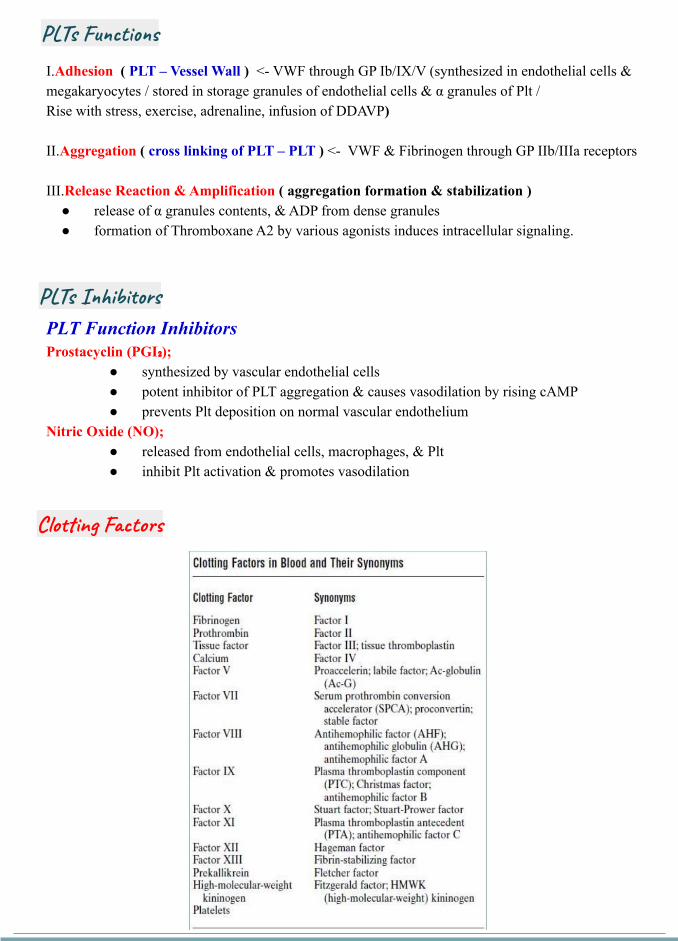
I.Adhesion ( PLT – Vessel Wall ) <- VWF through GP Ib/IX/V (synthesized in endothelial cells & megakaryocytes / stored in storage granules of endothelial cells & α granules of Plt / Rise with stress, exercise, adrenaline, infusion of DDAVP)
II.Aggregation ( cross linking of PLT – PLT ) <- VWF & Fibrinogen through GP IIb/IIIa receptors
III.Release Reaction & Amplification ( aggregation formation & stabilization ) ● release of α granules contents, & ADP from dense granules● formation of Thromboxane A2 by various agonists induces intracellular signaling.
PLT Function InhibitorsProstacyclin (PGI₂);
● synthesized by vascular endothelial cells ● potent inhibitor of PLT aggregation & causes vasodilation by rising cAMP ● prevents Plt deposition on normal vascular endothelium
Nitric Oxide (NO);● released from endothelial cells, macrophages, & Plt ● inhibit Plt activation & promotes vasodilation
Clotting Factors
PLTs Inhibitors
PLTs Functions

Fibrinolysis
DEPENDENT UPON: Vessel Wall Integrity (because we need vascularity. Vasculitis and CT disorders will affect that causing
bleeding tendency) Adequate Numbers of Platelets Proper Functioning Platelets Adequate Levels of Clotting Factors (Any defect of any of the factors will cause massive bleeding) Proper Function of Fibrinolytic Pathway
I. Vascular Phase: release of locally active vasoactive agents (Endothelin, Thromboxane A2, Fibrinopeptides) 🖝 Vasoconstriction at the site of injury 🖝 reduced blood flow
II. Platelet Phase: Plt Adhesion & Aggregation (via VWF, ADP, TXA2) 🖝 formation of PLT Plug
III. Plasma Coagulation Phase: Propagation of the clotting process by the coagulation cascade 🖝 formation of Fibrin Clot
IV. Fibrinolysis Phase: Termination of clotting by antithrombotic control mechanisms & removal of the clot
HEMOSTASIS
Hemostatic Phases
Great video explaining coagulation cascade:https://youtu.be/JoPQEDt1b0w

Primary Hemostasis:1. Endothelium Injury2. Platelet 3. Von Willebrand Factor
Secondary Hemostasis:1. Clotting Factors2. Soluble Protein Fibrinogen
converted to insoluble Fibrin
Primary Hemos.
Secondary Hemos.
Responsible for initial formation of platelet plug it’s affected in case of: Thrombocytopenia, von willebrand disease and also in platelet function disorder

● PLATELET COUNT● BLEEDING TIME (BT)● PROTHROMBIN TIME (PT) ● PARTIAL THROMBOPLASTIN TIME (PTT)● THROMBIN TIME (TT)
PROVIDES ASSESSMENT OF PLATELET COUNT AND FUNCTION
NORMAL VALUE 2-8 MINUTES
Measures Effectiveness of the Extrinsic Pathway
NORMAL VALUE 10-15 SECS
Prothrombin time tests the extrinsic and final common pathwaysProlongation in:
● Liver disease ● vitamin K antagonism (i.e. warfarin) and deficiency (In Bariatric surgery or removal of stomach)● Disseminated intravascular coagulation (In severe infection or malignancy)● Factor VII, X, V, II and fibrinogen defect
Measures Effectiveness of the Intrinsic Pathway
NORMAL VALUE 25-40 SECS
Activated partial thromboplastin time Tests the intrinsic and common pathways
Prolongation in: ● Liver disease ● Disseminated intravascular coagulation ● Heparin therapy ● Vitamin K antagonism or deficiency ● Factor XII, XI, IX, VIII, X, V, II, and fibrinogen defect
Taking Hx Congenital:
- Family Hx- Since Childhood (hemophilia &
clotting factors)Acquired:
- Middle age onset- Cause leading to DIC
Q) Pt on Heparin, what is followed is prolongation of PTT
LABORATORY EVALUATION (to know the source of bleeding)
PARTIAL THROMBOPLASTIN TIME
PROTHROMBIN TIME (PT):
BLEEDING TIME:
BLEEDING DISORDERS

● Time for Thrombin To Convert Fibrinogen to Fibrin ● A Measure of Fibrinolytic Pathway
NORMAL VALUE 9-13 SECS
Thrombin time evaluate fibrinogen and for inhibition of thrombin action
Prolongation in:● Hypofibrinogenaemia ● Dysfibrinogenaemia (http://www.fmshk.com.hk/hkabth)● Heparin therapy ● Disseminated intravascular coagulation
Not always available, and need accuracyTHROMBIN TIME
Hypofibrinogenaemia :Congenital deficiency which has problems with stabilizing clot, hence once they start to form the clot, bleeding happens again.
CONGENITAL BLEEDING DISORDERS
Hemophiliaan inherited bleeding disorder caused by deficiency of coagulation.
● Hemophilia A – Inherited deficiency of factor VIII (8); an X-linked recessive disorder. It is protected from proteolysis in the circulation by binding to vWF
● Hemophilia B – Inherited deficiency of factor IX (9); also called Christmas Disease; an X-linked recessive disorder.
● Hemophilia C – Inherited deficiency of factor XI (11); also called Rosenthal Syndrome; an autosomal recessive disorder. Rarely, heterozygotes may have bleeding (ie, autosomal dominant transmission, due to heterodimer binding). especially common in Ashkenazi Jews (ie, Jews from Eastern Europe).
It’s characterized based on the residual or baseline factor activity level (also referred to as "factor level"); expressed as a % of normal or in IU/mL.
Factor levels typically correlate with the degree of bleeding Symptoms. (Important)
• Severe Hemophilia – defined as <1 % factor activity (<0.01 IU/mL). • Moderate Hemophilia – defined as a factor activity level ≥1 % of normal and <5 % of normal ( ≥0.01 - <0.05 IU/mL).• Mild Hemophilia – defined as a factor activity level ≥5 % of normal and <40 % of normal (≥0.05 - <0.40 IU/mL).

Congenital >> genetic mutation in F8 & F9 located on the long arm of X chromosome. ▪ Observed commonly in males due to their hemizygous state▪ Rarely in females due to (Heterozygous females as result from nonrandom X
chromosome inactivation, skewed Lyonization, or the presence of other genetic abnormalities (Turner Syndrome or X autosomal translocations).
Acquired >> development of autoantibodies most commonly directed against FVIII – ass. with pregnancy, malignancy, advanced age.
Clinically >> hematomas, hemarthroses, bruising, bleeding (mucosal, GI, GU)Dx >> aPTT will be prolonged, Factor level will be low, Mixing study (corrected), Normal VWF & PT (the most accurate test is a specific assay for factor VIII or IX)Rx >> Replacement of the deficient coagulation Factor (recombinant or plasma derived) + Adjunctive therapy (Desmopressin (DDAVP levels of vwf will increase with this drug), Antifibrinolytic agents (Tranexamic Acid, Aminocaproic Acid), rFVIIa (with inhibitors)
According to the severity of hemophilia the treatment will differ:- Sever: Prophylaxis is needed- Mild: Give treatment if the pt bleeds, no need for prophylaxis
Von Willebrand Disease
Defect of Von Willebrand Factor:● Quantitative (type 1 & 3)● Qualitative (type 2)
Autosomal dominant (Important in MCQ)The Normal function of VWF:
● Mediate platelet adhesion● Stabilize factor VIII in circulation● Localize factor VIII to site of vessel injury
The most common bleeding disorder.

● control ● thrombasthenia
To distinguish you need VW antigen activity if its diminished with factor 8 diagnostic
To confirm test for platelet ristocetin activity
In case of VWF disease all will be normal except ristocetin
Platelet aggregometry: testing for platelet dysfunction

Congenital >> autosomal dominant (most types), recessive (rarely)
Acquired >> rare, caused by autoantibodies against vWF & immune complex formation, vWF binding to cancer cells, Congenital Heart Disease, Aortic Stenosis, Angiodysplasia. Rx (of the underlying disorder)
Dx >> normal aPTT in (Type 1 & 2), prolonged aPTT in (Type 2N, 2B, & 3), vWF:Ag, vWF:RCo, vWF multimers (to differentiate subtypes), FVIII assay (low in 2N & 3), Plt (low in 2M)
Rx >> Replacement of exogenous vWF concentrate, Desmopressin (DDAVP intranasal) Antifibrinolytic agents (Tranexamic Acid, Aminocaproic Acid), Conjugated Estrogens & oral contraceptive Agents (for menorrhagia) (best initial therapy is desmopressin if there is no response use factor VIII replacement or vWF concentrate)
Hemophilia A Factor IX deficiency Von Willebrand disease
inheritance sex-linked sex-linked Dominant (incomplete)
Main sites of hemorrhage
Muscle,joints,post-trauma or postoperative
Muscle,joints,post-trauma or postoperative
Mucous membranes, skin cuts, post-trauma or postoperative
Platelet count Normal Normal Normal
Bleeding time Normal Normal prolonged
Prothrombin time Normal Normal Normal
Partial thromboplastin time
Prolonged Prolonged Prolonged or normal
Factor VIII low Normal May be moderately reduced
Factor IX Normal low Normal
vWF Normal Normal low
Ristocetin-induced platelet aggregation
Normal Normal Impaired
Comparison between haemophilia and vWD
Von Willebrand Disease

PLATELET COUNT
● 100,000 - 400,000 CELLS/MM3 (NORMAL)● < 100,000 (Thrombocytopenia)
○ 50,000 - 100,000 (Mild Thrombocytopenia) (No need to give platelets, just follow up)
○ < 50,000 (Severe Thrombocytopenia) (Need intervention)
Causes of Thrombocytopenia
IMP
Portal HTN Cause back pressure on the spleen and increase spleen size, this will lead to decrease in All the cells including platelets so the pt if he develop esophageal varices on top ofthrombocytopenia it will lend to severe bleeding
This is the extrahepatic manifestation of Hepatitis:- one of them is thrombocytopenia - the other is EVAN Syndrome
Evan syndrome:- Autoimmune hemolytic cement- Autoimmune thrombocytopenia
Plt Disorders (Quantitative)
Platelets Disorders

Primary : isolated thrombocytopenia due to immune Plt destruction & reduce production (auto AB to megakaryocytes) Secondary : a/w disease/drug exposure 🖝 Viral (HIV, HCV, HBV, EBV, CMV, Parvovirus), SLE, APLS, H. Pylori Infection, Chronic Lymphocytic Leukemia (CLL), Hodgkin Lymphoma, AIHA
Dx >> Dx of exclusion, no robust clinical or Lab parameters, Typically CBC (Isolated 🖝 PLT < 100.000), 10% have ITP + AIHA (Evans Syndrome), PBS (Large Plts), Anti-Plt AB (not useful)
Clinically >> insidious onset of mucocutaneous bleed, M:F (3:1)
Rx >> rarely indicated if PLT > 50.000 unless bleeding, trauma/surgery, anticoag, comorbidities Steroids, IVIG, Splenectomy, TPO agonists (Romiplostim, Eltrombopag)
Immune Thrombocytopenic Purpura (ITP)
Approach to Thrombocytopenia
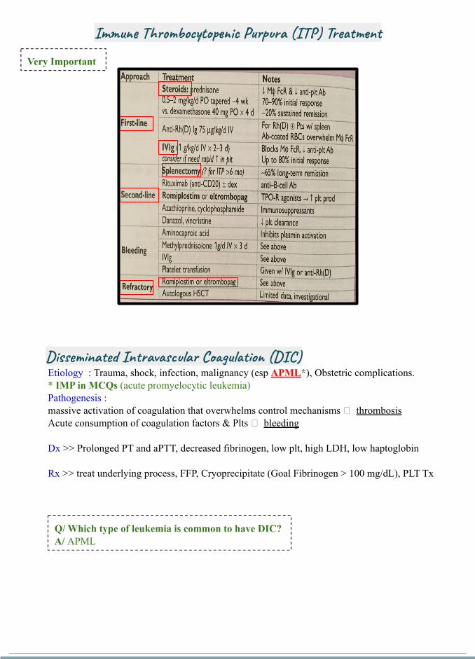
Etiology : Trauma, shock, infection, malignancy (esp APML*), Obstetric complications. * IMP in MCQs (acute promyelocytic leukemia)Pathogenesis : massive activation of coagulation that overwhelms control mechanisms 🖝 thrombosisAcute consumption of coagulation factors & Plts 🖝 bleeding
Dx >> Prolonged PT and aPTT, decreased fibrinogen, low plt, high LDH, low haptoglobin
Rx >> treat underlying process, FFP, Cryoprecipitate (Goal Fibrinogen > 100 mg/dL), PLT Tx
Very Important
Q/ Which type of leukemia is common to have DIC?A/ APML
Disseminated Intravascular Coagulation (DIC)
Immune Thrombocytopenic Purpura (ITP) Treatment

ACQUIRED PLT FUNCTIONAL DISORDERS
1. Liver Disease 2. Cardiopulmonary Bypass 3. Uremia (CKD)4. Dysproteinemia ( Multiple Myeloma or Waldenstrom Macroglobulinemia ) 5. Myeloproliferative Disorders (MPDs)6. Diabetes Mellitus 7. Acquired Glanzmann thrombasthenia
INHERITED DISORDERS OF PLT FUNCTION
1. Giant platelet disorders includes Plt GP abnormalities (eg, Bernard-Soulier Syndrome, Deficiency of Platelet Alpha granules (eg, Gray Platelet Syndrome), Deficiency May-Hegglin Anomaly (which also involves the presence of abnormal neutrophil inclusions (ie, Döhle-like bodies)), & some kindreds with type 2B vWD (Montreal Plt Syndrome)
2. Wiskott-Aldrich syndrome 3. Storage Pool Disorders such as Hermansky Pudlak Syndrome (HPS) (Hx of sore
throat+Purpura+Arthritis+Abdominal Pain+Nephropathy) (Deficiency of Dense Granules)
4. Glanzmann thrombasthenia (aggregate in response to ristocetin)5. Platelet release disorders 6. Glycoprotein VI defects 7. Sticky platelet syndrome 8. Congenital Deficiency of the ADP receptor P2Y129. Scott syndrome
Plt Disorders (Qualitative )

I. Detailed Pt & Family Medical History (Crucial & Vital regardless of the prior Lab testing) establish likelihood of a bleeding disorder guide laboratory Testing
• Early in the newborn period (circumcision) • After hemostatic Challenges ( Delivery, injury, trauma, surgery, invasive dental procedure,
menstruation )• Frequency & pattern • Duration
o Symptoms onset ( congenital vs. acquired )o time required for cessation
• Sites of bleeding (specific or multiple) (Very important)
Mucocutaneous Bleeding• Easy bruising• Epistaxis • Menorrhagia
Deep Tissue Bleeding• Joints (Hemarthrosis)
• Muscles • Central Nervous
System (intracerebral hemorrhage)
Primary Hemostasis Defects
( PLT or vW Factor )
Secondary Hemostasis Defects
( Clotting Factors Deficiencies )
• Current use of medications or herbal supplements • Use of Bleeding Assessment Tools (differentiate bleeding phenotypes, require validation by
prospective studies)
Two important points:I. Detailed Pt & Family Medical History (Crucial & Vital regardless of the prior Lab testing)II. Laboratory Testing
Approach to Pt with Potential Bleeding

Ant
ipla
tele
ts
Prostaglandin/COX Inhibitors
Glycoprotein IIb/IIIa Inhibitors
P2Y₁₂ ADP Inhibitors
Generic Name Trade Name Half Life
Dabigatran Pradaxa 12 – 28 hr
Argatroban Acova 39 – 51 min
Lepirudin Refludan 1.3 hr
Bivalirudin Angiomax 25 – 57 min
Unfractionated Heparin (UFH)
…………………………..
LMWH - Enoxaparin Clexan 4.5 – 7 hr
LMWH - Tinzaparin Innohep 3 – 4 hr
LMWH - Deltaparin
Fondaparinux Arixtra 17 – 21 hr
Warfarin Coumadin 7 – 11 hr
Rivaroxaban Xarelto 5 – 13 hr
Apixaban Eliquis 5 – 13 hr
Endoxaban Savaysa 10 – 14 hr
Ant
icoa
gula
nts
Direct Thrombin Inhibitors
Indirect Thrombin Inhibitors
Vitamin K epoxide reductaseInhibitor
Direct Xa Inhibitors
Generic Name Trade Name Half Life
Aspirin …………………..………
24 – 72 hr
Abciximab Reopro 72 hr
Eptifibatide Integrilin 4 hr
Tirofiban Aggrastat 4 hr
Clopidogrel Plavix 6 hr
Cangrelor Kengreal 3 – 6 min
Prasugrel Effient 7 – 15 hr
Ticlopidine Ticlid 13 hr
Ticagrelor Brilinta 7- 9 hr
-aprin
-xaban
Drugs Used for Clotting Disorders
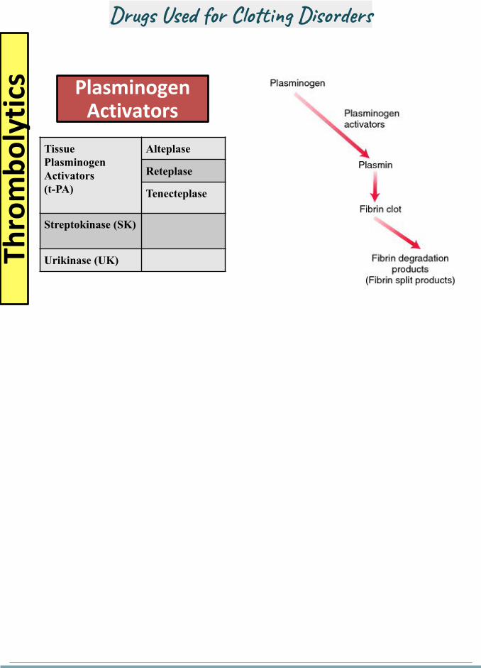
Thro
mb
oly
tics Plasminogen
Activators
Tissue Plasminogen Activators(t-PA)
Alteplase
Reteplase
Tenecteplase
Streptokinase (SK)
Urikinase (UK)
Drugs Used for Clotting Disorders

I. CBC (Platelet count) II. Prothrombin Time (PT) >> measures F VII, X, V, II, I - (N Time 10-14 secs)
III. International Nmalized Ratio (INR) >> the ratio of a pt's PT to a normal (control) sample, raised to the power of the ISI value for the control sample used.
IV. Activated Partial Thromboplastin Time (aPTT or PTT) >> measures F XII, XI, IX, VIII, X, V, II, I - (N Time 30 – 40 secs)
V. Thrombin (Clotting) Time (TT) >> sensitive to deficiency of Fibrinogen or inhibition of thrombin - (N Time 14 – 16 secs)
VI. Bleeding Time >> (3-8 secs) (not sensitive – not specific )
▪ Screening tests (not sensitive to all abnormalities ass. w a bleeding disorder)
Important MCQs Qs
Three patterns: - Extrinsic pathway - Intrinsic pathway- Common Pathway (both tests
prolonged)
Screening Tests
Causes of Prolonged Coagulation Profile
II. Laboratory Testing

Mixing Study (one to one mix of Pt’s plasma & known normal standard plasma, only if PT of aPTT prolonged)
● Corrected 🖝 clotting factor deficiency (risk of bleed)● Not corrected 🖝 inhibitors (directed against specific factor or global inhibitors “ Lupus
Inhibitor, risk of thrombosis “)1. PLT Function Assay (PFA - 100): assess PLT function
a. Specificity 🖝 90 % for severe PLT dysfunction of vWD (vWF plasma levels < 25%)b. Sensitivity 🖝 24 – 41 % (low) in mild PLT secretion defect or Storage Pool Disease 🖝🖝
( not screening tool ) 2. PLT Aggregation Tests: (5 external aggregating factors; ADP, Collagen, Ristocetin, Arachidonic
Acid, Adrenaline)3. Von Willebrand Factor ( Antigen & Activity )4. Factor XIII assay (F XIII Deficiency >> normal PT & PTT)5. Human Plasminogen Activator Inhibitor (PAI-1)6. Alpha 2 AntiPlasmin Inhibitor (α2 AP)
Specialized Tests

Although screening tests are used widely to identify hemostatic abnormalities associated with bleeding, they are NOT perfect The Clinical suspicion for a bleeding disorder is Critical to determine extent of the laboratory investigations
Take Home Message
Recommended Books by the doctorEssential Hematology (A. V. Hoffbrand, P. A. H. Moss)Uptodate Oxford Handbook of clinical hematology.
There are extra slides from the doctor that can be found on this link

Summary
,vancomycin
We recommend this approach when reading the question

Immune thrombocytopenia
Clinical -Antecedent Viral infection -Asymptomatic petechiae and ecchymosis -Nasal,GI,GU bleed
Lab Diagnosis of exclusion-Isolated thrombocytopenia-Peripheral smear: megakaryocyte
Rx if PLT > 50.000 and no bleeding: observeOther than that: 1st -Steroids, IVIG 2nd -Splenectomy Refractory- TPO agonists (Romiplostim, Eltrombopag)
Hemophilia
A BChristmas Disease
C (ashkenazi jews)Rosenthal Syndrome
8 9 11
X linked recessive autosomal recessive
Severity and conformation assessed by Factor Levels: Severe less than 1%
Moderate 1-5%Mild 5-40%
Von Willebrand Disease
Etiology Qual or Quan, Autosomal dominant (except type 3)
Clinical -Mostly asymptomatic-Ecchymosis,Petechiae-Other symptoms include epistaxis, gingival bleeding, menorrhagia , GI bleeding, and excessive bleeding during surgical procedures.
Lab -aPTT :normal (in Type 1 & 2) and prolonged (in Type 2N, 2B, & 3)-Factor 8 will be low in type 2N & 3-Quantitative assessment by vWF antigen assay: ↓ factor levels-Qualitative assessment by a ristocetin cofactor activity assay Failure of aggregation with a ristocetin assay or a ristocetin cofactor level < 30 IU/dL is considered definitive for vWD
Rx Recombinant von Willebrand factor (rVWF) concentrate,Desmopressin
Summary

DIC
Etiology Sepsis (most common),Trauma,malignancy(APML) ,Obstetric complications
Clinical -Bleeding + thrombosis
Lab - ↑PT, ↑aPTT, ↓ Fibrinogen (may be N b/c acute phase)-+ve D-Dimer/FDP-↓ PLT-+ve Schistocytes, ↑ LDH, ↓ Haptoglobin
Rx treat underlying process, FFP, Cryoprecipitate (Goal Fibrinogen > 100 mg/dL), PLT Tx
Questions
A. Low platelets B. Prolonged Partial thromboplastin time C. Prolonged Prothrombin time D. Elevated ESRE. Synovial fluid Leukocytosis
Answer B
A. ArgatrobanB. Tranexamic acidC. Protein C concentrateD. Hyperbaric oxygen
Answer C

A. Vitamin KB. Plasma exchange C. Intravenous ImmunoglobulinsD. Desmopressin E. Factor 8 Substitution
Answer D
A. Prolonged aPTTB. Prolonged PTC. Decreased level of vW FactorD. High TSH
Answer is D (not just because it's all heavy menstrual periods are Bleeding disorder be careful in the exam)


![[5] HEMA - Megaloblastic Anemia.pdf - KSUMSC](https://static.fdokumen.com/doc/165x107/631deac95ff22fc7450674ca/5-hema-megaloblastic-anemiapdf-ksumsc.jpg)