Female Perineum - KSUMSC
30
Female Perineum Color Code Important Doctors Notes Notes/Extra explanation Please view our Editing File before studying this lecture to check for any changes.
-
Upload
khangminh22 -
Category
Documents
-
view
4 -
download
0
Transcript of Female Perineum - KSUMSC

Female PerineumColor Code
ImportantDoctors NotesNotes/Extra explanation
Please view our Editing File before studying this lecture to check for any changes.
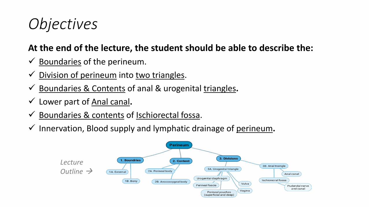
ObjectivesAt the end of the lecture, the student should be able to describe the:✓ Boundaries of the perineum.✓ Division of perineum into two triangles.✓ Boundaries & Contents of anal & urogenital triangles.✓ Lower part of Anal canal.✓ Boundaries & contents of Ischiorectal fossa.✓ Innervation, Blood supply and lymphatic drainage of perineum.
Lecture Outline
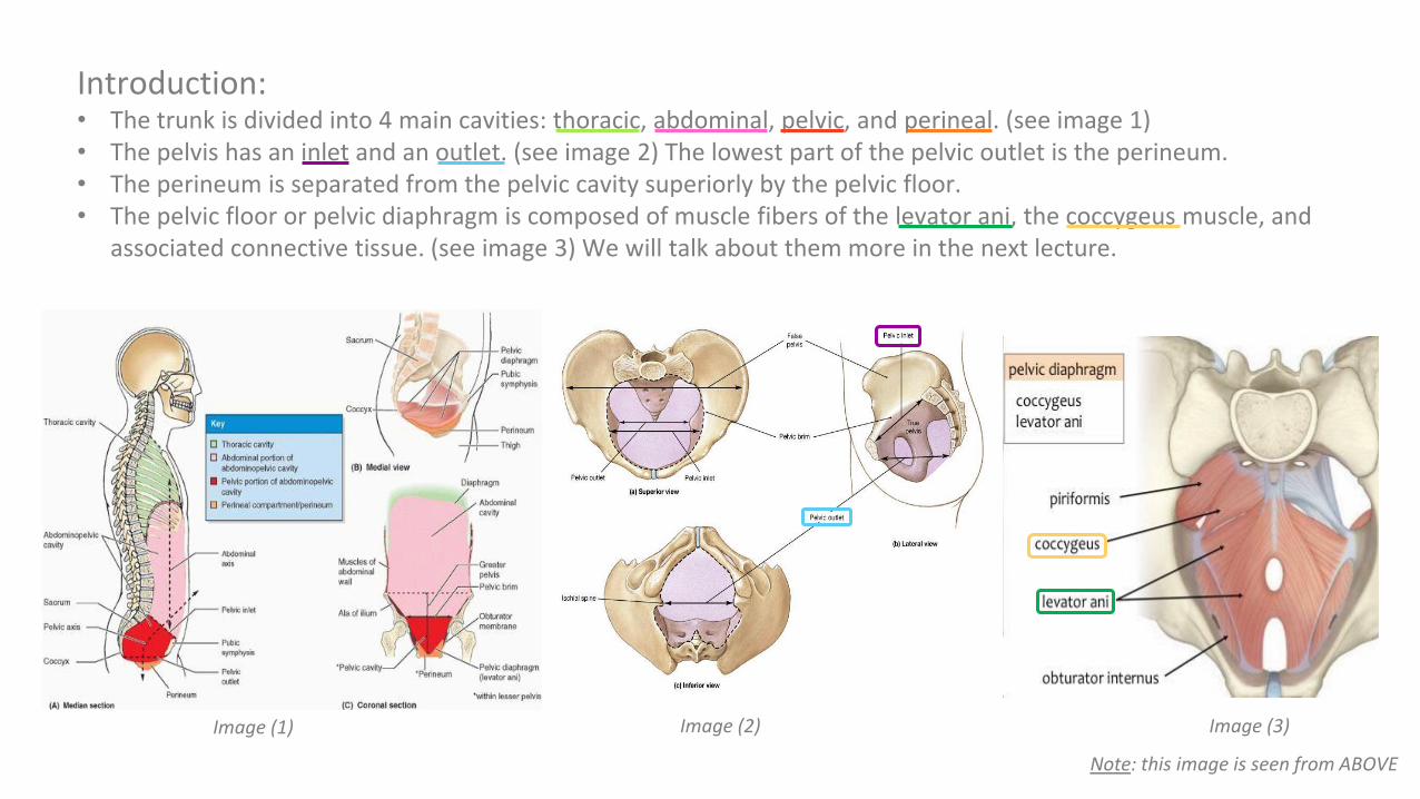
Introduction:• The trunk is divided into 4 main cavities: thoracic, abdominal, pelvic, and perineal. (see image 1)• The pelvis has an inlet and an outlet. (see image 2) The lowest part of the pelvic outlet is the perineum. • The perineum is separated from the pelvic cavity superiorly by the pelvic floor.• The pelvic floor or pelvic diaphragm is composed of muscle fibers of the levator ani, the coccygeus muscle, and
associated connective tissue. (see image 3) We will talk about them more in the next lecture.
Image (1) Image (2) Image (3)
Note: this image is seen from ABOVE
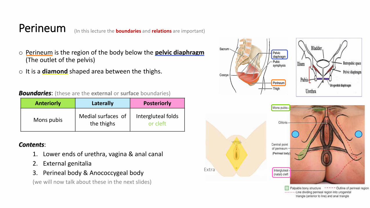
Perineum
o Perineum is the region of the body below the pelvic diaphragm (The outlet of the pelvis)
o It is a diamond shaped area between the thighs.
Boundaries: (these are the external or surface boundaries)
Contents: 1. Lower ends of urethra, vagina & anal canal2. External genitalia3. Perineal body & Anococcygeal body(we will now talk about these in the next slides)
Anteriorly Laterally Posteriorly
Mons pubis Medial surfaces of the thighs
Intergluteal folds or cleft
Extra
(In this lecture the boundaries and relations are important)

o Perineal body is an irregular fibromuscular mass of variable size and consistency, located atmidpoint of the line between the ischial tuberosities.
o Lies in the subcutaneous tissue, posterior to vaginal vestibule and anterior to the anal canal & anus
o Forms the central point of the perineum & blends anteriorly with the perineal membrane
Function:1. Gives attachment to perineal muscles2. Plays an important role in visceral support especially in female
Perineal membrane
PerineumPerineal body
Extra
Extra explanation:The perineal body is an irregularfibromuscular mass. It is located atthe junction of the urogenital andanal triangles – the central point ofthe perineum. This structure containsskeletal muscle, smooth muscle andcollagenous and elastic fibres.Anatomically, the perineal body liesjust deep to the skin. It acts as a pointof attachment for muscle fibres fromthe pelvic floor and the perineumitself

These pictures are extra.

o The anococcygeal body is a complex musculotendinous structure (more tendinous).o Situated between the anterior aspect of the coccyx and the posterior wall of the anorectal canalo Receives insertion of fibers of levator ani muscle (gives attachment of levator ani muscles).
PerineumAnococcygeal Body
:يكونبالترتيبيعنيVaginal vestibule
Perineal bodyAnal canal
Anococcygeal bodyCoccyx
Extra
Extra
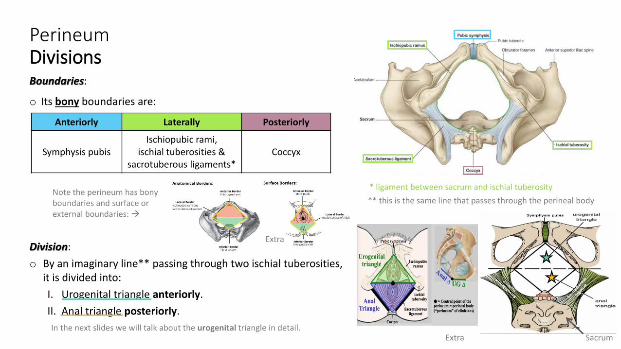
Boundaries:
o Its bony boundaries are:
Division:o By an imaginary line** passing through two ischial tuberosities,
it is divided into:I. Urogenital triangle anteriorly.II. Anal triangle posteriorly.
PerineumDivisions
Anteriorly Laterally Posteriorly
Symphysis pubisIschiopubic rami,
ischial tuberosities & sacrotuberous ligaments*
Coccyx
Note the perineum has bony boundaries and surface or external boundaries:
Extra
In the next slides we will talk about the urogenital triangle in detail.Extra
* ligament between sacrum and ischial tuberosity
Sacrum
** this is the same line that passes through the perineal body
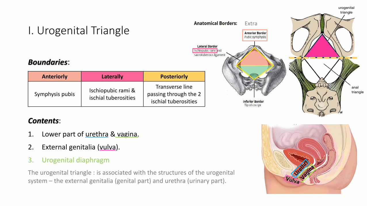
Extra I. Urogenital Triangle
Boundaries:
Contents: 1. Lower part of urethra & vagina.
2. External genitalia (vulva).
3. Urogenital diaphragmThe urogenital triangle : is associated with the structures of the urogenital system – the external genitalia (genital part) and urethra (urinary part).
Anteriorly Laterally Posteriorly
Symphysis pubis Ischiopubic rami &ischial tuberosities
Transverse line passing through the 2
ischial tuberosities

1. Mons pubis: a collection of fat overlying the pubes
2. Labia majora (labia means lips or flaps).
3. Labia minora.
4. Clitoris (the two labia minora meet at a point anteriorly and make the clitoris).
5. Vestibule of vagina: The interval or area between the two labia minora and has 2 openings:
6. Vagina & urethra open into the vestibule through urethral orifice anteriorly and vaginal orifice posteriorly.
I. Urogenital TriangleFemale External Genitalia (Vulva)
The external genital organs of the female are collectively known as the vulva (also called the pudendum). The vulva is comprised of many different structures:
Extra
04:24
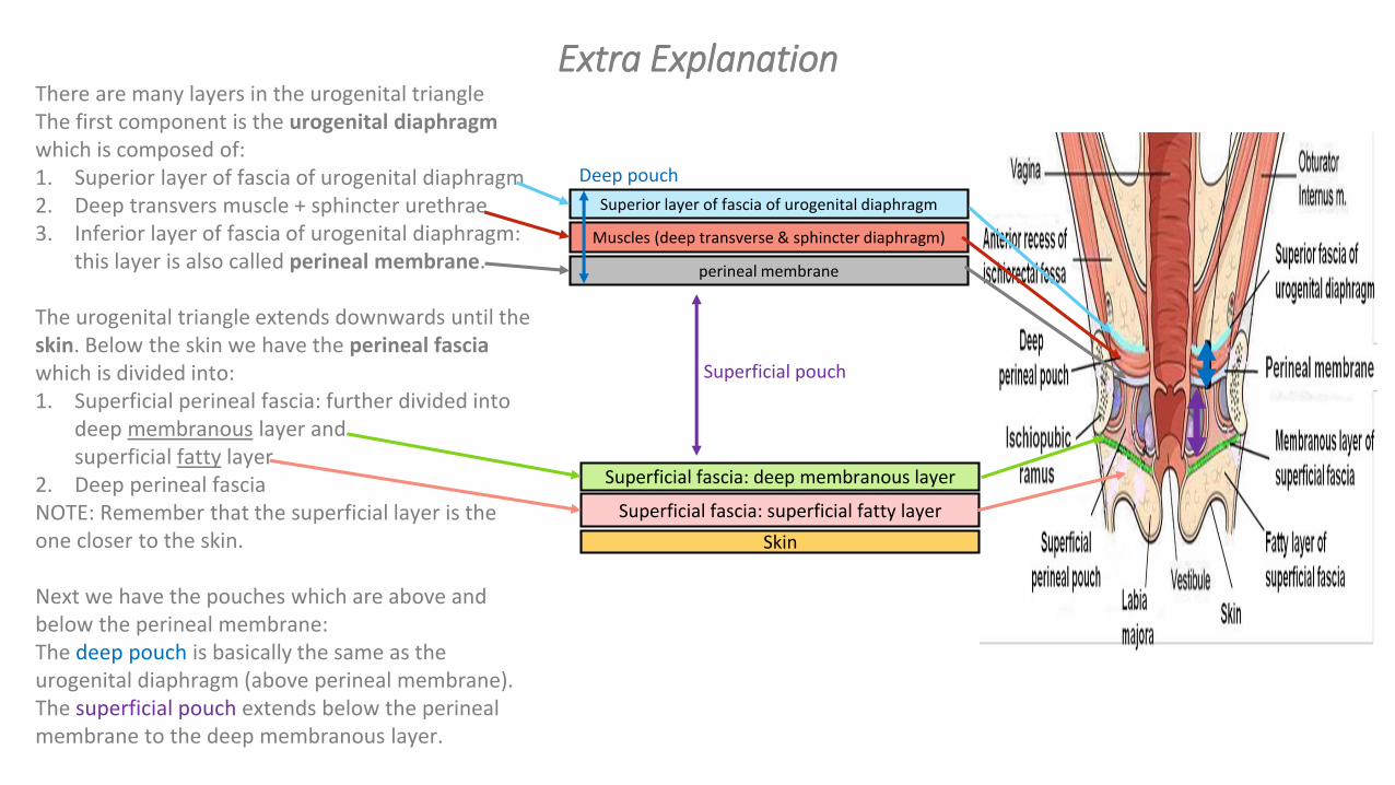
There are many layers in the urogenital triangleThe first component is the urogenital diaphragm which is composed of: 1. Superior layer of fascia of urogenital diaphragm2. Deep transvers muscle + sphincter urethrae3. Inferior layer of fascia of urogenital diaphragm:
this layer is also called perineal membrane.
The urogenital triangle extends downwards until the skin. Below the skin we have the perineal fascia which is divided into:1. Superficial perineal fascia: further divided into
deep membranous layer and superficial fatty layer .
2. Deep perineal fascia NOTE: Remember that the superficial layer is the one closer to the skin.
Next we have the pouches which are above and below the perineal membrane:The deep pouch is basically the same as the urogenital diaphragm (above perineal membrane).The superficial pouch extends below the perineal membrane to the deep membranous layer.
Superficial fascia: deep membranous layerSuperficial fascia: superficial fatty layer
Skin
Superior layer of fascia of urogenital diaphragm
Muscles (deep transverse & sphincter diaphragm)
perineal membrane
Superficial pouch
Deep pouch
Extra Explanation

• A triangular musculofascial diaphragm located in the anterior part of the perineum (in the urogenital triangle).
• Fills in the gap between the pubic arch.
• Composed of: musculo-fascial so it has muscles and fascia
(1) Sphincter urethrae and
(2) the deep transverse perineal muscles enclosed within
(3) the superior and (4) inferior layers of fascia of the urogenital diaphragm (The inferior layer of the fascia is formed by the perineal membrane)
Extra
Extra
I. Urogenital TriangleUrogenital Diaphragm
mus
clefa
scia
Note: we have 2 fascia: perineal fascia (superficial and deep) and fascia of urogenital diaphragm (superior and inferior)
The fascia cover the muscle from above and below

Perineal FasciaFascia of Urogenital Triangle (Perineal Fascia) is continuous anteriorly with the fascia of abdomen and consists of 2 layers: superficial and deep layers
Superficial perineal fasciaConsists of:
Deep perineal fascia invests the muscles in the superficial perineal pouch (the muscles will be mentioned
later)1. Superficial fatty layer (Camper’s fascia ) makes up the substance of mons pubis & labia majora and extends into the anal region.
2. Deep membranous layer (Colle’s fascia ): • Does not extend to anal region. • Becomes fused with the posterior margin
of the perineal membrane
I. Urogenital TrianglePerineal Fascia

I. Urogenital TrianglePerineal PouchesStructurally, the urogenital triangle is complex, with a number of fascial layers and pouches. Unlike the anal triangle, the urogenital triangle has an additional layer of strong deep fascia; the perineal membrane. This membrane has pouches on its superior and inferior surfaces: the superficial and deep pouches.

Perineal PouchesSuperficial Perineal Pouch
o It is the potential space between the deep membranous layer of superficial fascia and the perineal membrane (inferior fascia of urogenital diaphragm).
oBoundaries:Superiorly Laterally Inferiorly
Perineal membrane Ischiopubic rami
Membranous layer of
superficial fascia (colles fascia)
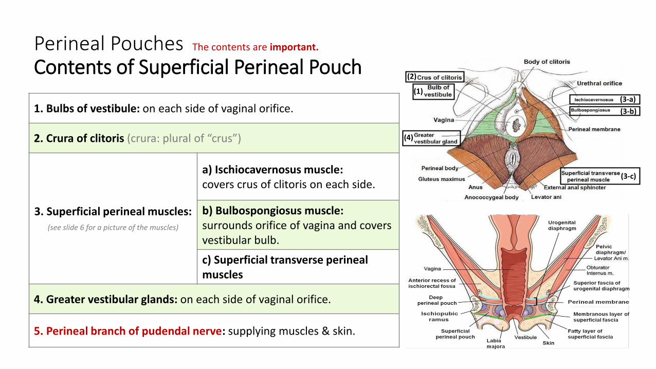
Perineal PouchesContents of Superficial Perineal Pouch
1. Bulbs of vestibule: on each side of vaginal orifice.
2. Crura of clitoris (crura: plural of “crus”)
3. Superficial perineal muscles: (see slide 6 for a picture of the muscles)
a) Ischiocavernosus muscle: covers crus of clitoris on each side.
b) Bulbospongiosus muscle: surrounds orifice of vagina and covers vestibular bulb.c) Superficial transverse perineal muscles
4. Greater vestibular glands: on each side of vaginal orifice.
5. Perineal branch of pudendal nerve: supplying muscles & skin.
(1)
(2)
(3-b)(3-a)
(3-c)
(4)
The contents are important.

Perineal PouchesDeep Perineal Pouch
o It is a completely closed space deep to the perineal membrane.
o It is the same as the urogenital diaphragm.
oBoundaries:Superiorly Laterally Inferiorly
Superior fascia of the urogenital
diaphragm
Inferior portion of obturator
internus fascia
Inferior fascia of the urogenital
diaphragm (perineal
membrane)

Perineal PouchesContents of Deep Perineal Pouch
1. Part of urethra
2. Part of vagina
3. Sphincter urethrae and Sphincter vaginae muscles; which is pierced by urethra & vagina.
4. Deep transverse perineal muscles
5. Internal pudendal vessels
6. Dorsal nerve of clitoris
(1)
(2)
(1)
(4)

o The vagina is a muscular canal that leads from the uterus to the external orifice of the genital canal
o It measures about 3 in. (8 cm) long. o It serves as the excretory duct for the menstrual flow & forms
part of the birth canal. o The vaginal orifice in a virgin possesses a thin mucosal fold,
called the hymen ( ةيرذالعغشاء ), which is normally perforated at its center to allow the menstrual blood to flow out.
Arteries: 1. Vaginal artery, a branch of the internal iliac artery 2. Vaginal branch of the uterine artery
Veins: 1. drain into the internal iliac veins.
Vagina
عدةعدبوالدات

Boundaries:
Contents:
1. Lower part of Anal canal2. Ano-coccygeal body3. Ischiorectal fossa on each side (will be discussed later)
II. Anal Triangle
Anteriorly Laterally PosteriorlyTransverse line
passing through the 2 ischial tuberosities
ischial tuberosity & sacrotuberous ligament. Coccyx

o It is about 1.5 in. long, descending from the rectal ampulla to the anus.
Relations (In female):
Division: Divided by pectinate line into:1. Upper half: derived from hindgut* (endoderm) 2. Lower half: derived from the proctodeum (ectoderm)
The two parts have different blood supply, nerve supply and lymphatic drainage.
II. Anal TriangleAnal Canal
Anteriorly Posteriorly LaterallyPerineal body,
urogenital diaphragm, lower part of vagina
Anococcygeal body
Ischiorectal fossae
Extra
*remember from GIT block: the artery of the hindgut is the inferior mesenteric artery

Anal canal Arterial supply Venous drainage Lymphatic drainage Nerves
Upper half (endoderm)
Superior rectal artery
(continuation of the inferior mesenteric
artery)
Superior rectal vein
drained into the inferior mesenteric vein
(portal circulation).
Para-rectal nodes
drained into inferior mesenteric
lymph nodes
(sensitive to stretch)Visceral^ sensory & motor
(Inferior Hypogastric Plexus)(sympathetic & parasympathetic).
Lower half (ectoderm)
Inferior rectal artery
(branch of internal pudendal artery)
Inferior rectal vein
drained into the internal pudendal vein
(Systemic circulation*) site of portal-systemic
anastomosis**.
Superficial inguinal lymph nodes
(sensitive to pain) Somatic^ motor & sensory nerves
(Inferior rectal N. branch of pudendal N.) supplies: external sphincter muscle of the
anus and the skin of the anal region.
This slide is important.
*Clinical importance:in children we give medication in the form of suppository (تحميله) We insert it in the lower half of the anal canal but we shouldn’t push it in too deeply because then it will go to the portal instead of the systemic circulation.
**In cases of portal hypertension this will lead to hemorrhoids (بواسير)
The name of the nerves are only mentioned on the girls slides.
^to review the difference between visceral and somatic look at this picture:

o A fascial lined wedge-shaped space on each side of the anal canal.
Boundaries:
Contents: 1. Dense fat.
2. Pudendal nerve & internal pudendal vessels within the pudendal canal.
3. Inferior rectal nerve & vessels crossing the fossa to reach anal canal.
II. Anal TriangleIschiorectal Fossa
Base Medial Wall Lateral Wall
Skin of the perineum.
Levator ani & anal canal.
Obturator internus, covered with fascia.

o The pudendal canal is a fascial canal formed by obturator fascia, located on the lateral wall of the ischiorectal fossa.
Contents:1. Pudendal nerve.2. Internal pudendal vessels (artery and vein).
II. Anal TrianglePudendal Canal
Extra
Mnemonic:VAN
VeinArteryNerve

II. Anal TrianglePudendal Nerve Block Important!
o Pudendal nerve block is used in providing analgesia for the second stage of labour to provide anesthesia (الوالدة) of the perineum in order to create or repair an episiotomy (An episiotomy is a surgically planned incision on the perineum and the posterior vaginal wall during second stage of labor to prevent perineal tear).
o Method: can be done by transvaginally or through perineal approach.Extra
Transvaginal method: • The needle is passed through
the vaginal mucous membrane toward the ischial spine.
• After the needle is passed through the sacrospinous ligament, the anesthetic solution is injected around the pudendal nerve
Perineal method: • The ischial tuberosity is palpated
subcutaneously through the buttock. (needs experience)
• The needle is inserted on the medial side of the ischial tuberosity to a depth of about 1 in. (2.5 cm) from the free surface of the tuberosity.
• The anesthetic is injected around the pudendal nerve.
لوالاهنااالثنينبينالفرقكتوردلامزالىوجنمنوكي .المهبلمنلخدي
منونتكالثانيةالطريقةبينمانيدعبمظعلاسحيرالدكتوابر
.طوللىعاإلبرةيدخل
The cut is made in an oblique line so we
don’t cut the perineal body
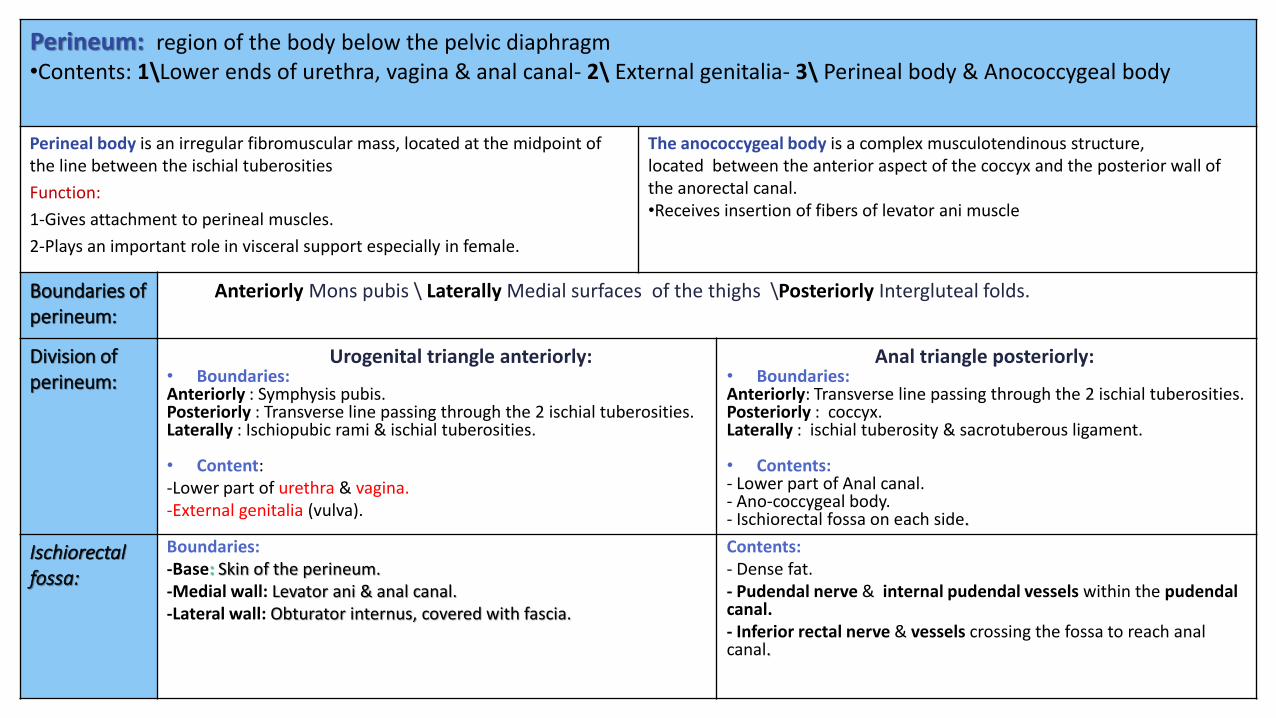
Perineum: region of the body below the pelvic diaphragm•Contents: 1\Lower ends of urethra, vagina & anal canal- 2\ External genitalia- 3\ Perineal body & Anococcygeal body
The anococcygeal body is a complex musculotendinous structure,located between the anterior aspect of the coccyx and the posterior wall of the anorectal canal.•Receives insertion of fibers of levator ani muscle
Perineal body is an irregular fibromuscular mass, located at the midpoint of the line between the ischial tuberositiesFunction:1-Gives attachment to perineal muscles.2-Plays an important role in visceral support especially in female.
Anteriorly Mons pubis \ Laterally Medial surfaces of the thighs \Posteriorly Intergluteal folds.Boundaries of perineum:
Anal triangle posteriorly: • Boundaries:Anteriorly: Transverse line passing through the 2 ischial tuberosities.Posteriorly : coccyx.Laterally : ischial tuberosity & sacrotuberous ligament.
• Contents: - Lower part of Anal canal.- Ano-coccygeal body.- Ischiorectal fossa on each side.
Urogenital triangle anteriorly:• Boundaries:Anteriorly : Symphysis pubis.Posteriorly : Transverse line passing through the 2 ischial tuberosities.Laterally : Ischiopubic rami & ischial tuberosities.
• Content: -Lower part of urethra & vagina.-External genitalia (vulva).
Division of perineum:
Contents: - Dense fat. - Pudendal nerve & internal pudendal vessels within the pudendal canal.- Inferior rectal nerve & vessels crossing the fossa to reach anal canal.
Boundaries:-Base: Skin of the perineum.-Medial wall: Levator ani & anal canal.-Lateral wall: Obturator internus, covered with fascia.
Ischiorectal fossa:

Pudendal Canal:Anal CanalVaginaPerineal PouchesPerineal FasciaFemale External Genitalia (Vulva)
Urogenital Diaphragm
-A fascial canal formed by obturator fascia, located on the lateral wall of the ischiorectal fossaContents:1-Pudendal nerve.2-Internal pudendal vessels.
- 1.5 in. long, descending from the rectal ampulla to the anus. Relations (In female):Anteriorly: Perineal body, urogenital diaphragm, and lower part of vaginaPosteriorly: Anococcygeal body. Laterally: Ischiorectalfossae.Division:Upper half: derived
from hindgut (endoderm)
Lower half: derived from the proctodeum(ectoderm)
*The two parts have different blood supply, nerve supply and lymphatic drainage.
-muscular canal that leads from the uterus to the external orifice of the genital canal.- 3 in. (8 cm) long.-It is the excretory duct for the menstrual flow & forms part of the birth canal. Arteries:-Vaginal artery, a branch of the internal iliac artery-Vaginal branch of the uterine arteryVeins: internal iliac veins
Superficial:It is the space between the deep membranous layer of superficial fascia and the perineal membrane.
Deep:It is a completely closed space deep to the perineal membrane
continuous anteriorly with the fascia of abdomen.consists of: Superficial perineal fascia: -Superficial fattylayer (Camper’s fascia). - Deep membranouslayer (Colle’s fascia ). Deep perineal fascia: invests the musclesin the superficial perineal pouch.
1. Mons pubis.2. Labia majora.3. Labia minora.4. Clitoris.5. Vestibule of
vagina. 6. urethral orifice &
vaginal orifice.
• A triangular musculofascial diaphragm located in in the urogenital triangle.
• Fills in the gap between the pubic arch.
Composed of: 1. Sphincter
urethrae 2. deep transverse
perineal muscles.3. Superior layer of
urogenital fascia4. Inferior layer of
urogenital fascia (perineal membrane)

Q1: All the following are contents of urogenital tringle except :
A) Urethra B) Vagina
C) Valva D) Anus
Q2:Which one of the following structures forms the center point of perineum?
A) Perineal body B) Ischial tuberosities
C) Anococcygeal body D) Coccyx
Q3: Which one of the following structures lie anterior to Urogenital Triangle?
A) Ischiopubic rami B) Ischial tuberosities.
C) External genitalia D) Symphysis pubis
Q4: Which of the following is a content of superficial perineal pouch ?
A) Dosal nerve of clitoris B) Pudendal artery
C) Pudendal vein D) Perineal branch of pudendal nerve
Answers: 1. D, 2. A, 3. D, 4. D, 5. A, 6. C, 7. B 8. C
MCQsQ5: Which of the following is a lateral boundary to deep perineal pouch?
A) Inferior portion of obturator internus fascia
B) Superior portion of obturator internus fascia
C) Ischiopubic rami
D) Superior fascia of the urogenital diaphragm
Q6: Vaginal branch originates from which of the following arteries?
A) Internal iliac artery B) Ovarian artery
C) Uterine artery D) Inferior mesenteric artery
Q7: Which of the following vein drain the inferior rectal vein?
A) Uterine vein B) Internal pudendal vein
C) Superior mesenteric vein D) Inferior mesenteric vein
Q8: The fascia canal of pudendal canal formed by which of the following?
A) Deep perineal fascia B) Superficial perineal fascia
C) Obturator fascia D) Superior fascia of urogenitaldiaphragm

Q1: Regarding the relation of the anal canal in female ?1-Anteriorly: perineal body, urogenital diaphragm, lower part of vagina2- Posteriorly: anococcygeal body3- Laterally: ischiorectal fossae
Q2: What’s the divisions of the anal canal and from where it’s derived ?- Upper half: Derived from hindgut (endoderm)- Lower half: Derived from proctodeum (ectoderm)
Q3: What are the contents of the superficial pouch?1. Bulbs of vestibule.2. Crura of clitoris 3. Superficial perineal muscles:
a) Ischiocavernosus muscle.b) Bulbospongiosus muscle.c) Superficial transverse perineal muscles
4. Greater vestibular glands.5. Perineal branch of pudendal nerve.
SAQ
Q4: What are the bony boundaries of the perineum?1-Anteriorly: Symphysis pubis2- Posteriorly: Coccyx3- Laterally: Ischiopubic rami, ischial tuberosities & sacrotuberous ligaments
Q5: What are the contents of the urogenital triangle?1. Lower part of urethra & vagina.2. External genitalia (vulva).
Q6: What are the contents of the anal triangle?1. Lower part of Anal canal2. Ano-coccygeal body3. Ischiorectal fossa on each side
Q6: What are the contents of the pudendal canal?1. Pudendal nerve.2. Internal pudendal vessels

Leaders:Nawaf AlKhudairyJawaher Abanumy
References:1- Girls’ & Boys’ Slides2- Greys Anatomy for Students 3- TeachMeAnatomy.com
@anatomy436
Feedback
Members:Alanoud AbuhaimedAnwar AlajmiGhaida AlsaeedLama AlfawzanLama AlTamimiRawan AlWadeeSafa Al-OsaimiShatha AlghaihbWejdan alzaid


![[5] HEMA - Megaloblastic Anemia.pdf - KSUMSC](https://static.fdokumen.com/doc/165x107/631deac95ff22fc7450674ca/5-hema-megaloblastic-anemiapdf-ksumsc.jpg)