Nasal Anatomy and Physiology - KSUMSC
20
433 ENT Team Nose I 1 | Page 8 [email protected] Color index: 432 Team – Important – 433 Notes – Not important Nasal Anatomy and Physiology
-
Upload
khangminh22 -
Category
Documents
-
view
1 -
download
0
Transcript of Nasal Anatomy and Physiology - KSUMSC
433 ENT Team Nose I
1 | P a g e
8
Color index:
432 Team – Important – 433 Notes – Not important
Nasal Anatomy and Physiology

433 ENT Team Nose I
2 | P a g e
Objectives:
Anatomy of the external nose, nose, nasal cavity and paranasal sinuses.
Physiology of the nose and paranasal sinuses.
Blood and nerve supply of the external nose, nose, nasal cavity and paranasal sinuses.
Functions of the nose and paranasal sinuses.
Congenital anomalies.
Choanal atresia.

433 ENT Team Nose I
3 | P a g e
Postnatal development of the nose
Chronology
At birth: Frontal sinus furrows appear, only two to three ethmoidal turbinates remain, Craniofacial ratio 8:1
Six months: Nares double their birth diameter.
Lateral Bony Wall
In neonate:
- The nasal and orbital floors are located at the same level.
- Lateral nasal wall serves as the medial orbital wall.
- Maxilla contributes minimally in fetus and neonate.
In adult:
- Only the upper half of the lateral nasal wall forms the medial orbital wall
- The nasal floor is at a lower level than the orbital floor.
The Nasal Pyramid
Bony constituents:
Support the upper part of the external nose:
1.Nasal processes of the frontal bones.
2.Nasal bones.
3.Ascending processes of the maxillae.
Cartilaginous constituents:
Support the lower part of the external nose:
1.Upper lateral cartilages.
2.Lower nasal cartilages.
3.Quadrilateral cartilages of nasal septum.
4.Alar cartilages.
The cartilages are connected with each other and with the bones by continuous perichondrium and periosteum.
The Piriform Aperture (Anterior Nasal Aperture):
It is a heart-shaped opening in the skull that is bounded by:
Inferior borders of the nasal bones superiorly.
Nasal surfaces of the maxilla laterally
The anterior nasal spine inferiorly
*The nasion is the midline bony depression between
eyes where the frontal and two nasal bones meet.
*Lower nasal cartilage makes the shape of the nose
(e.g. flat, long or straight).
433 ENT Team Nose I
4 | P a g e
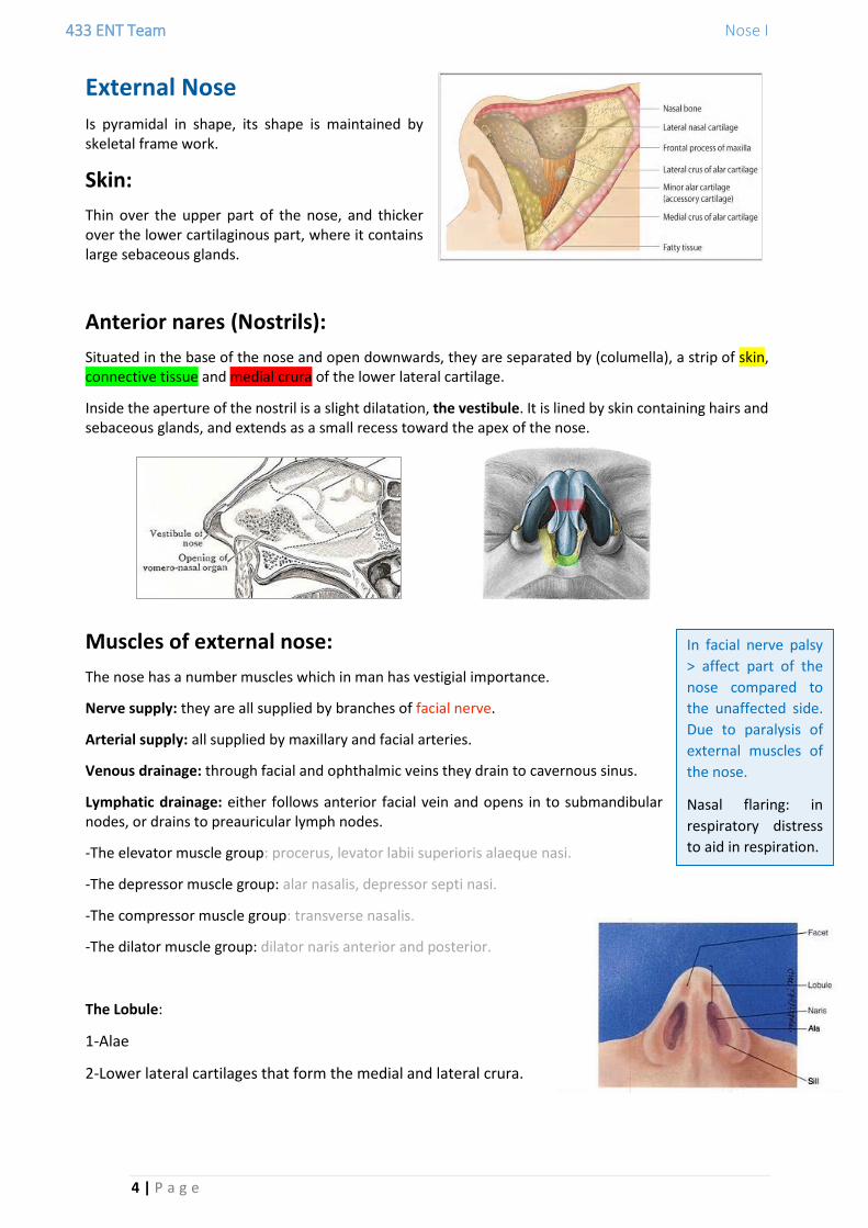
External Nose
Is pyramidal in shape, its shape is maintained by skeletal frame work.
Skin:
Thin over the upper part of the nose, and thicker over the lower cartilaginous part, where it contains large sebaceous glands.
Anterior nares (Nostrils):
Situated in the base of the nose and open downwards, they are separated by (columella), a strip of skin, connective tissue and medial crura of the lower lateral cartilage.
Inside the aperture of the nostril is a slight dilatation, the vestibule. It is lined by skin containing hairs and sebaceous glands, and extends as a small recess toward the apex of the nose.
Muscles of external nose:
The nose has a number muscles which in man has vestigial importance.
Nerve supply: they are all supplied by branches of facial nerve.
Arterial supply: all supplied by maxillary and facial arteries.
Venous drainage: through facial and ophthalmic veins they drain to cavernous sinus.
Lymphatic drainage: either follows anterior facial vein and opens in to submandibular nodes, or drains to preauricular lymph nodes.
-The elevator muscle group: procerus, levator labii superioris alaeque nasi.
-The depressor muscle group: alar nasalis, depressor septi nasi.
-The compressor muscle group: transverse nasalis.
-The dilator muscle group: dilator naris anterior and posterior.
The Lobule:
1-Alae
2-Lower lateral cartilages that form the medial and lateral crura.
In facial nerve palsy
> affect part of the
nose compared to
the unaffected side.
Due to paralysis of
external muscles of
the nose.
Nasal flaring: in
respiratory distress
to aid in respiration.

433 ENT Team Nose I
5 | P a g e
Nasal fossa (Nasal cavity)
The nasal cavity extends from the nostrils anteriorly to the choanae posteriorly.
The right and left nasal fossae (cavities) are separated by the nasal septum.
The nasal fossae are lined with mucous membranes.
Each fossa communicates with:
1.The paranasal sinuses, through their Ostia. 2.The nasopharynx, through the posterior choanae.
Boundaries of posterior choana (Posterior Nasal Aperture):
Boundaries of nasal fossa:
Floor: Roof (very narrow):
1.Palatine process of maxilla in the anterior
three quarters.
2.Horizontal part of palatine bone in posterior
one quarter.
*Maxilla bone and palatine bone set on the floor of the hard palate
*Olfactory epithelium is in the upper part of the nose. When removing polyps on the upper part you have to
be cautious not to injure the olfactory epithelium.
*One of the complications of sinus surgery is losing smell.
433 ENT Team Nose I
6 | P a g e
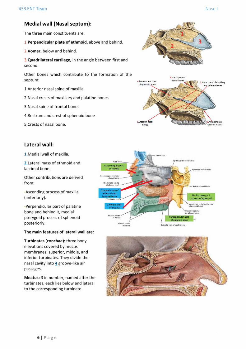
Medial wall (Nasal septum):
The three main constituents are:
1.Perpendicular plate of ethmoid, above and behind.
2.Vomer, below and behind.
3.Quadrilateral cartilage, in the angle between first and second.
Other bones which contribute to the formation of the septum:
1.Anterior nasal spine of maxilla.
2.Nasal crests of maxillary and palatine bones
3.Nasal spine of frontal bones
4.Rostrum and crest of sphenoid bone
5.Crests of nasal bone.
Lateral wall:
1.Medial wall of maxilla.
2.Lateral mass of ethmoid and lacrimal bone.
Other contributions are derived from:
-Ascending process of maxilla (anteriorly).
-Perpendicular part of palatine bone and behind it, medial pterygoid process of sphenoid posteriorly.
The main features of lateral wall are:
Turbinates (conchae): three bony elevations covered by mucus membranes; superior, middle, and inferior turbinates. They divide the nasal cavity into 4 groove-like air passages.
Meatus: 3 in number, named after the turbinates, each lies below and lateral to the corresponding turbinate.
433 ENT Team Nose I
7 | P a g e
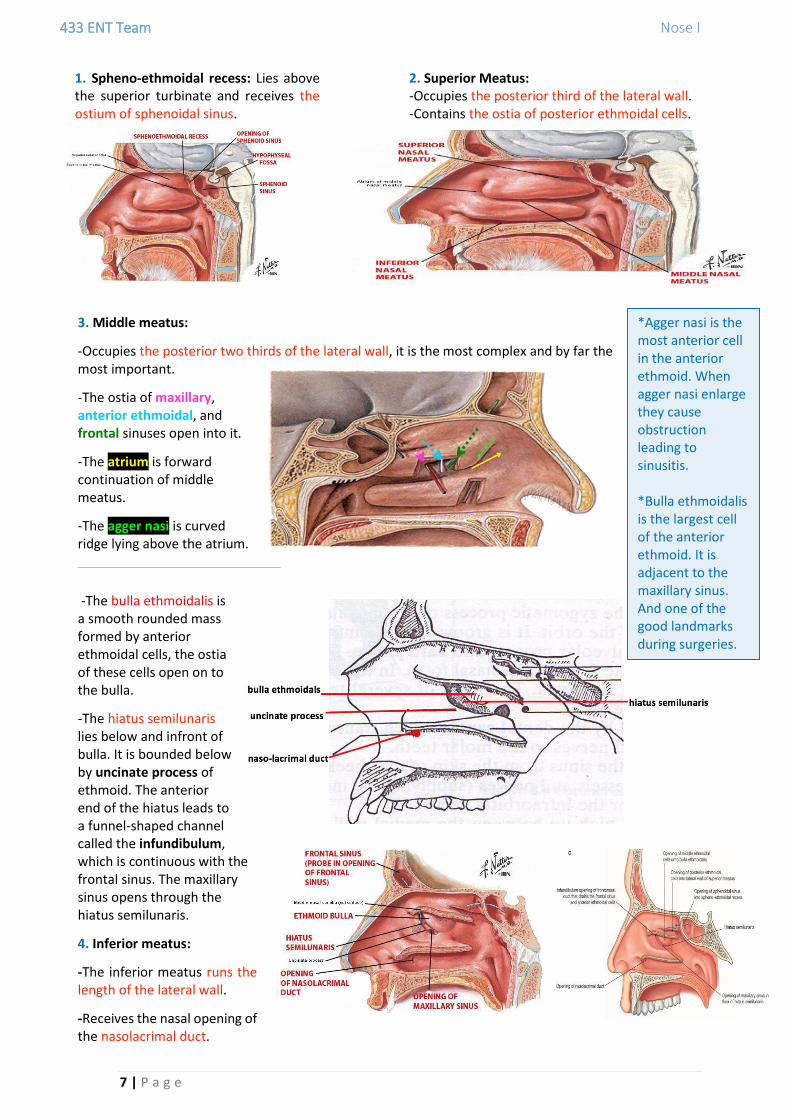
3. Middle meatus:
-Occupies the posterior two thirds of the lateral wall, it is the most complex and by far the most important.
-The ostia of maxillary, anterior ethmoidal, and frontal sinuses open into it.
-The atrium is forward continuation of middle meatus.
-The agger nasi is curved ridge lying above the atrium.
-The bulla ethmoidalis is a smooth rounded mass formed by anterior ethmoidal cells, the ostia of these cells open on to the bulla.
-The hiatus semilunaris lies below and infront of bulla. It is bounded below by uncinate process of ethmoid. The anterior end of the hiatus leads to a funnel-shaped channel called the infundibulum, which is continuous with the frontal sinus. The maxillary sinus opens through the hiatus semilunaris.
4. Inferior meatus:
-The inferior meatus runs the length of the lateral wall.
-Receives the nasal opening of the nasolacrimal duct.
1. Spheno-ethmoidal recess: Lies above the superior turbinate and receives the ostium of sphenoidal sinus.
2. Superior Meatus: -Occupies the posterior third of the lateral wall. -Contains the ostia of posterior ethmoidal cells.
*Agger nasi is the most anterior cell in the anterior ethmoid. When agger nasi enlarge they cause obstruction leading to sinusitis. *Bulla ethmoidalis is the largest cell of the anterior ethmoid. It is adjacent to the maxillary sinus. And one of the good landmarks during surgeries.

433 ENT Team Nose I
8 | P a g e
Ostiomeatal Complex:
A common channel that links the frontal sinus, anterior and middle ethmoid sinuses and the maxillary sinus to the middle meatus that allows air flow and mucociliary drainage. Composed of the following structures: Uncinate process, Ethmoid bulla, Middle turbinate, and The spaces between these structures (ethmoidal infundibulum, middle meatus, and hiatus semilunaris).
Mucosal Lining of the nasal cavity:
Modified Skin: Keratinized stratified squamous epithelium covering the vestibule. It contains sebaceous glands, sweat glands, and short, curved hair called vibrissae.
Olfactory: Specialized olfactory epithelium. Present in the olfactory cleft, which occupies the area between the superior turbinate, cribriform plate, and the corresponding area of the septum.
Respiratory mucosa: Ciliated pseudostratified columnar epithelium with goblet cells. It lines the lower two-thirds of the nasal septum, the lateral wall of the nose below the superior turbinate, and the floor of the nasal cavity. It extends into the sinuses through their Ostia and is thinner there. It is also continuous with the epithelia of the nasolacrimal duct and Eustachian tube.
Paranasal Sinuses
These are air spaces within certain bones of the skull. They are lined with a mucous membrane continuous with that of the corresponding nasal fossa through their ostia. The sinuses develop as outgrowths from the nasal cavity; hence they all drain directly or indirectly into the nose. The lining of the sinuses (muco-endosteum) is continuous with the nasal mucosa. The sinuses develop mostly after birth, and their degree of development varies greatly. Their exact functions are uncertain but they may have a role in:
Humidifying and warming the inspired air.
Regulation of intranasal pressure.
Increasing surface area for olfaction.
Lightening the skull.
Adding resonant effect to voice.
Absorbing shock.
Contributing to facial growth.
*Facial growth center is present in paranasal sinus.
*Maxillary sinus is the first to develop.
*Frontal sinus is the last sinus to develop during puberty.
*Most dangerous sinus during surgery is sphenoidal due to its relation with cavernous sinus contents, mainly
carotid > massive bleeding if injured.

433 ENT Team Nose I
9 | P a g e
1. Maxillary Sinuses:
It is the largest of the sinuses, with an average capacity of 15 ml in the adult. It is pyramidal in shape and occupies the body of maxilla. The base lies medially, the apex is in the zygomatic portion of the maxilla. Medial wall is the wall between the sinus and the nasal fossa.
The maxillary sinus drains into the middle meatus by means of the semilunar hiatus. The floor is formed by the alveolar process and hard palate.
In children, the floor lies at or above the level of the floor of the nasal fossa. In adults, it lies about 1.25 cm below the floor of the fossa.
The roots of several teeth may project into, or even perforate, the floor (sometimes the maxillary sinus gets penetrated during dental surgery).
Relations of maxillary sinus:
a.Orbit: Separated from the antrum by the thin roof of the sinus, which contains the infraorbital nerve (blue arrow).
b.Teeth (red arrow): May produce elevations in the floor of the sinus and the number of related teeth depends on the size of the antrum. The second premolar and first molar are usually related.
c.Middle meatus of the nose (green arrow): related to the upper part of the antrum.
d.Inferior meatus of the nose (orange arrow): Separated from the middle part of its medial wall by bone, which is usually thick in front and below but thinner above and behind.
e.Maxillary artery (yellow arrow): Related to the posterior wall, where it occupies the pterygopalatine fossa. It may be approached through the antrum for ligature.
f.Maxillary division of the 5th cranial nerve (purple arrow): also traverses the pterygopalatine fossa.
g.Nasolacrimal duct (black arrow): Passes downwards, medial to the antrum, to open into the inferior meatus.
*Infraorbital nerve (sensation of anterior part of the cheek) is a continuation of the
maxillary nerve. When a patient has paraesthesia in the cheek suspect mass around the
infraorbital nerve, common in RTA also when there is fracture of the floor.
*Lacrimal canaliculi > lacrimal sac > lacrimal duct > lacrimal open in the inferior meatus.
433 ENT Team Nose I
10 | P a g e
2. Ethmoidal Sinuses (Labyrinths):
Consists of a number (approximately 7-15) thin-walled cavities within the lateral masses of the ethmoid bones, in the agger nasi, and middle turbinate. The cells may invade any of the surrounding bones, including the frontal, sphenoid, and maxillary bones. There are two groups of cells:
1.Anterior: Usually small and numerous. They open into the upper part of the hiatus semilunaris or above the bulla ethmoidalis, ultimately draining into the middle meatus.
2.Posterior: Usually large and few, they open into the superior meatus.
Nasal septum divides the right and left ethmoid. The anterior and posterior groups of ethmoid are separated by the basal lamella (3rd part of the middle turbinates).
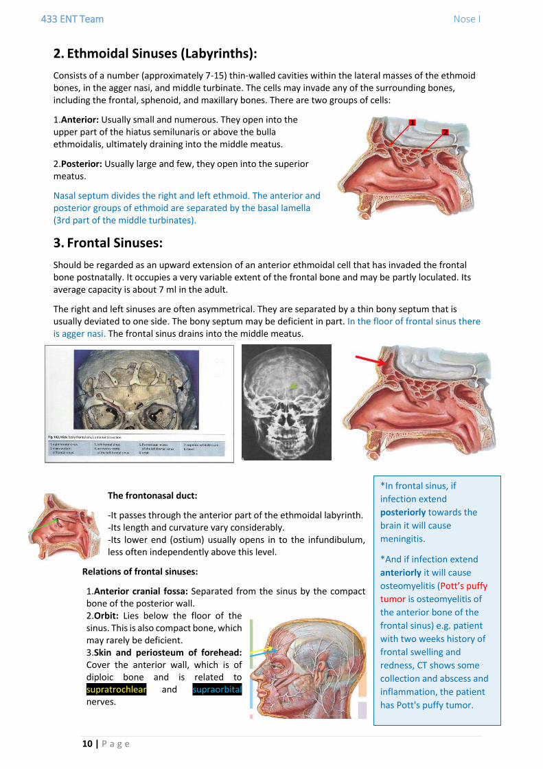
3. Frontal Sinuses:
Should be regarded as an upward extension of an anterior ethmoidal cell that has invaded the frontal bone postnatally. It occupies a very variable extent of the frontal bone and may be partly loculated. Its average capacity is about 7 ml in the adult.
The right and left sinuses are often asymmetrical. They are separated by a thin bony septum that is usually deviated to one side. The bony septum may be deficient in part. In the floor of frontal sinus there is agger nasi. The frontal sinus drains into the middle meatus.
The frontonasal duct:
-It passes through the anterior part of the ethmoidal labyrinth. -Its length and curvature vary considerably. -Its lower end (ostium) usually opens in to the infundibulum, less often independently above this level.
Relations of frontal sinuses:
1.Anterior cranial fossa: Separated from the sinus by the compact bone of the posterior wall. 2.Orbit: Lies below the floor of the sinus. This is also compact bone, which may rarely be deficient. 3.Skin and periosteum of forehead: Cover the anterior wall, which is of diploic bone and is related to supratrochlear and supraorbital nerves.
*In frontal sinus, if
infection extend
posteriorly towards the
brain it will cause
meningitis.
*And if infection extend
anteriorly it will cause
osteomyelitis (Pott’s puffy
tumor is osteomyelitis of
the anterior bone of the
frontal sinus) e.g. patient
with two weeks history of
frontal swelling and
redness, CT shows some
collection and abscess and
inflammation, the patient
has Pott's puffy tumor.

433 ENT Team Nose I
11 | P a g e
4. Sphenoidal Sinuses:
Lies behind the upper part of the nasal fossa. It occupies the body, and sometimes the wings and pterygoid processes of the sphenoid bone. The average capacity is about 7ml in the adult. The right and left sinuses are rarely symmetrical. They are separated by a septum (green arrow), which may be deficient in part and is often oblique.
The ostium (blue arrow) of sphenoid sinus is situated in the upper part of the anterior wall of the sinus. It communicates with the superior meatus indirectly through the sphenoethmoidal recess.
Relations: Laterally: the cavernous sinus containing: 1.Cranial nerves: 3rd, 4th, 5th (ophthalmic and maxillary divisions), and 6th. 2.Internal carotid artery 3.Optic nerve Above the sinus: Pituitary gland, optic chiasm, frontal lobe of brain, and olfactory tract. The pituitary gland may be approached surgically through the sinus.
Blood Supply of Nose and Paranasal Sinuses
Arterial supply: The nasal fossae and paranasal sinuses are supplied by branches of the external
and internal carotid arteries:
A.Derivatives of external carotid artery:
1.Sphenopalatine artery (the artery of epistaxis): via the maxillary artery supplies the turbinates and meatus of the nose and most of the septum. It passes through the sphenopalatine foramen.

433 ENT Team Nose I
12 | P a g e
2.Greater palatine artery: a branch of the maxillary artery contributes branches to the lateral nasal wall and (via the incisive canal) to the anterior part of the septum.
3.Superior labial artery: -A branch of the facial artery. It sends branches to the tip of the septum and the alae nasi. -Its anastomosis with a branch of the spheno- palatine artery and the greater palatine artery forms Kiesselbach's plexus, in Little's area (most common site for epistaxis).
B.Branches of internal carotid artery:
Anterior and posterior ethmoidal arteries: -Branches of the ophthalmic artery. -They supply the roof of the nose, anterior parts of the septum and lateral wall of the nose, and the ethmoidal and frontal sinuses. -Bleeding from these vessels is seen above the level of the middle turbinate.
431 Team:
Frontal Sinus: Supraorbital and supratrochlear divisions of ophthalmic artery. Ethmoid Sinus: Sphenopaltine and anterior and posterior ethmoidal arteries. Sphenoid Sinus: Posterior ethmoidal and sphenopalatine arteries. Maxillary Sinus: Superior alveolar and infraorbital arteries (divisions of maxillary).
Venous drainage: The veins form a cavernous plexus beneath the mucous membrane. They
open into:
1.Sphenopalatine vein and anterior facial vein, from the plexus. 2.Ophthalmic veins, from the ethmoidal veins. 3.Veins on the orbital surface of the frontal lobe of the brain, through the foramina in the cribriform plate. 4.Superior sagittal sinus, through the foramen caecum.
433 ENT Team Nose I
13 | P a g e
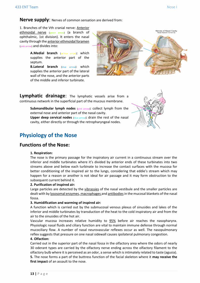
Nerve supply: Nerves of common sensation are derived from:
1. Branches of the Vth cranial nerve: Anterior ethmoidal nerve (green arrow) (a branch of ophthalmic, 1st division). It enters the nasal cavity through the anterior ethmoidal foramen (pink arrow) and divides into:
A.Medial branch (yellow arrow): which supplies the anterior part of the septum. B.Lateral branch (blue arrow): which supplies the anterior part of the lateral wall of the nose, and the anterior parts of the middle and inferior turbinate.
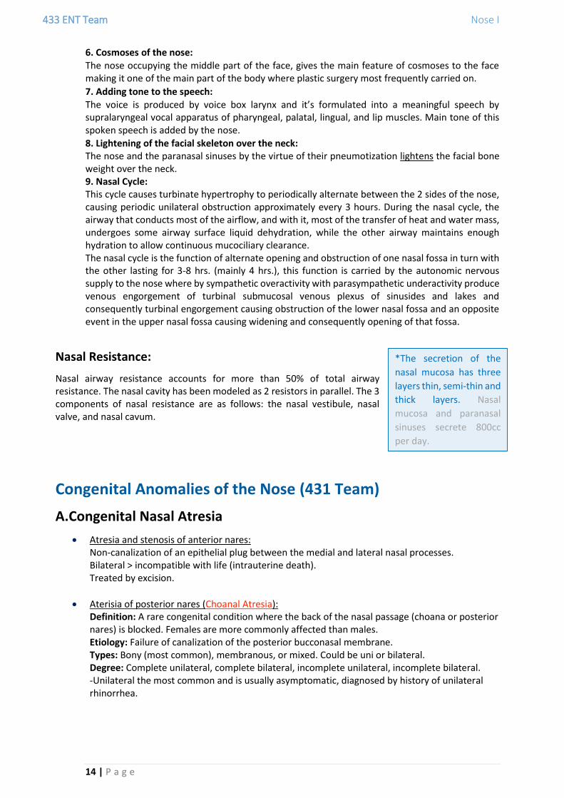
Lymphatic drainage: The lymphatic vessels arise from a
continuous network in the superficial part of the mucous membrane.
Submandibular lymph nodes (pink arrow): collect lymph from the external nose and anterior part of the nasal cavity. Upper deep cervical nodes (blue arrow): drain the rest of the nasal cavity, either directly or through the retropharyngeal nodes.
Physiology of the Nose
Functions of the Nose:
1. Respiration: The nose is the primary passage for the inspiratory air current in a continuous stream over the inferior and middle turbinates where it’s divided by anterior ends of these turbinates into two streams above and below each turbinate to increase the contact surfaces with the mucosa for better conditioning of the inspired air to the lungs, considering that eddie’s stream which may happen for a reason or another is not ideal for air passage and it may form obstruction to the subsequent current behind it. 2. Purification of inspired air: Large particles are detected by the vibrassies of the nasal vestibule and the smaller particles are dealt with by lysosomal enzymes, macrophages and antibodies in the mucosal blankets of the nasal fossa. 3. Humidification and warming of inspired air: A function which is carried out by the submucosal venous plexus of sinusides and lakes of the inferior and middle turbinates by transduction of the heat to the cold inspiratory air and from the air to the sinusides of the hot air. Vascular mucosa increases relative humidity to 95% before air reaches the nasopharynx. Physiologic nasal fluids and ciliary function are vital to maintain immune defense through normal mucociliary flow. A number of nasal neurovascular reflexes occur as well. The nasopulmonary reflex suggests that pressure on one nasal sidewall causes ipsilateral pulmonary congestion. 4. Olfaction: Carried out in the superior part of the nasal fossa in the olfactory area where the odors of nearly 30 oderant types are carried by the olfactory nerve ending across the olfactory filament to the olfactory bulb where it is perceived as an odor, a sense which is intimately related to taste (agusia). 5. The nose forms a part of the buttress function of the facial skeleton where it may receive the first impact of an assault to the nose.
433 ENT Team Nose I
14 | P a g e
6. Cosmoses of the nose: The nose occupying the middle part of the face, gives the main feature of cosmoses to the face making it one of the main part of the body where plastic surgery most frequently carried on.
7. Adding tone to the speech: The voice is produced by voice box larynx and it’s formulated into a meaningful speech by supralaryngeal vocal apparatus of pharyngeal, palatal, lingual, and lip muscles. Main tone of this spoken speech is added by the nose. 8. Lightening of the facial skeleton over the neck: The nose and the paranasal sinuses by the virtue of their pneumotization lightens the facial bone weight over the neck. 9. Nasal Cycle: This cycle causes turbinate hypertrophy to periodically alternate between the 2 sides of the nose, causing periodic unilateral obstruction approximately every 3 hours. During the nasal cycle, the airway that conducts most of the airflow, and with it, most of the transfer of heat and water mass, undergoes some airway surface liquid dehydration, while the other airway maintains enough hydration to allow continuous mucociliary clearance. The nasal cycle is the function of alternate opening and obstruction of one nasal fossa in turn with the other lasting for 3-8 hrs. (mainly 4 hrs.), this function is carried by the autonomic nervous supply to the nose where by sympathetic overactivity with parasympathetic underactivity produce venous engorgement of turbinal submucosal venous plexus of sinusides and lakes and consequently turbinal engorgement causing obstruction of the lower nasal fossa and an opposite event in the upper nasal fossa causing widening and consequently opening of that fossa.
Nasal Resistance:
Nasal airway resistance accounts for more than 50% of total airway resistance. The nasal cavity has been modeled as 2 resistors in parallel. The 3 components of nasal resistance are as follows: the nasal vestibule, nasal valve, and nasal cavum.
Congenital Anomalies of the Nose (431 Team)
A.Congenital Nasal Atresia
Atresia and stenosis of anterior nares: Non-canalization of an epithelial plug between the medial and lateral nasal processes. Bilateral > incompatible with life (intrauterine death). Treated by excision.
Aterisia of posterior nares (Choanal Atresia): Definition: A rare congenital condition where the back of the nasal passage (choana or posterior nares) is blocked. Females are more commonly affected than males. Etiology: Failure of canalization of the posterior bucconasal membrane. Types: Bony (most common), membranous, or mixed. Could be uni or bilateral. Degree: Complete unilateral, complete bilateral, incomplete unilateral, incomplete bilateral. -Unilateral the most common and is usually asymptomatic, diagnosed by history of unilateral rhinorrhea.
*The secretion of the
nasal mucosa has three
layers thin, semi-thin and
thick layers. Nasal
mucosa and paranasal
sinuses secrete 800cc
per day.
433 ENT Team Nose I
15 | P a g e
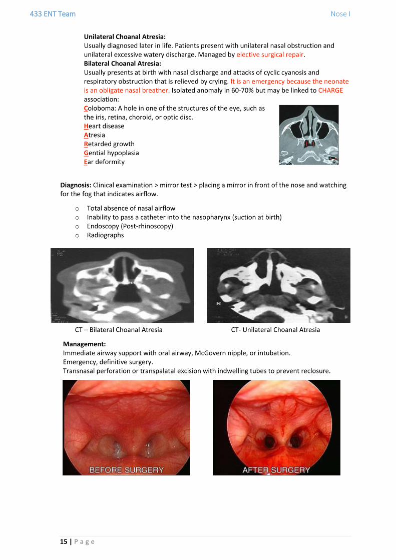
Unilateral Choanal Atresia: Usually diagnosed later in life. Patients present with unilateral nasal obstruction and unilateral excessive watery discharge. Managed by elective surgical repair. Bilateral Choanal Atresia: Usually presents at birth with nasal discharge and attacks of cyclic cyanosis and respiratory obstruction that is relieved by crying. It is an emergency because the neonate is an obligate nasal breather. Isolated anomaly in 60-70% but may be linked to CHARGE association: Coloboma: A hole in one of the structures of the eye, such as the iris, retina, choroid, or optic disc. Heart disease Atresia Retarded growth Gential hypoplasia Ear deformity
Diagnosis: Clinical examination > mirror test > placing a mirror in front of the nose and watching for the fog that indicates airflow.
o Total absence of nasal airflow o Inability to pass a catheter into the nasopharynx (suction at birth) o Endoscopy (Post-rhinoscopy) o Radiographs
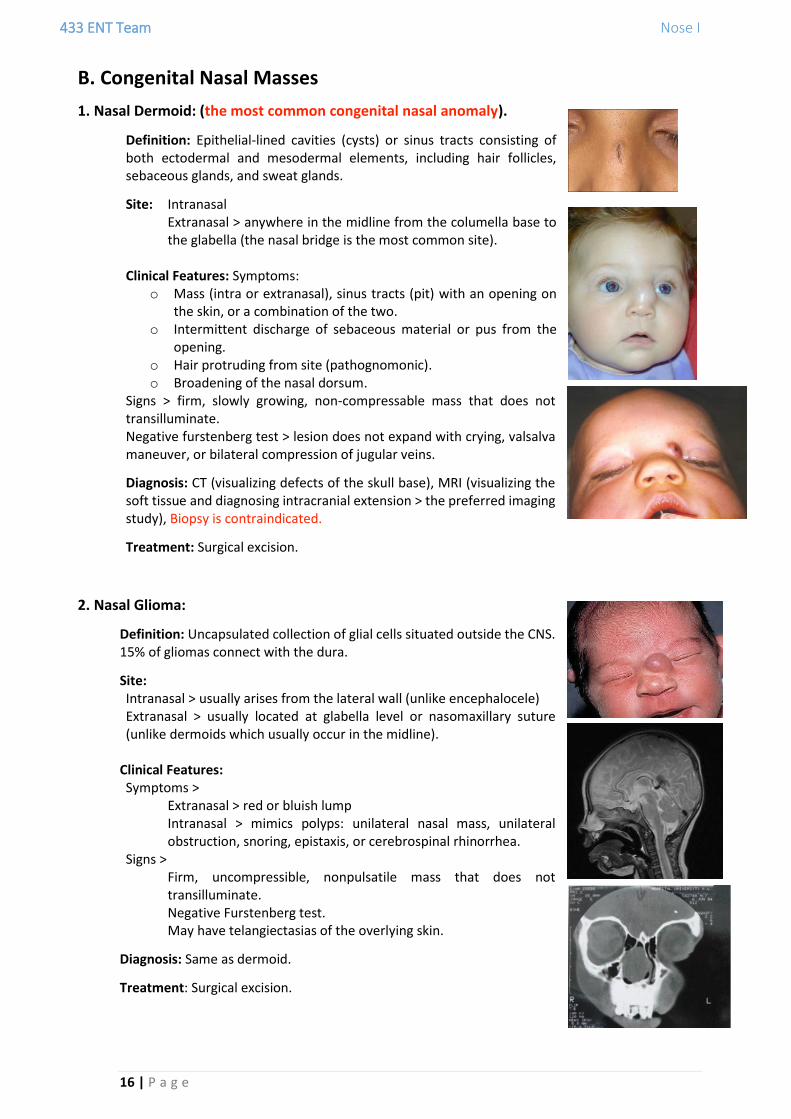
CT – Bilateral Choanal Atresia CT- Unilateral Choanal Atresia
Management: Immediate airway support with oral airway, McGovern nipple, or intubation. Emergency, definitive surgery. Transnasal perforation or transpalatal excision with indwelling tubes to prevent reclosure.
433 ENT Team Nose I
16 | P a g e
B. Congenital Nasal Masses
1. Nasal Dermoid: (the most common congenital nasal anomaly).
Definition: Epithelial-lined cavities (cysts) or sinus tracts consisting of both ectodermal and mesodermal elements, including hair follicles, sebaceous glands, and sweat glands.
Site: Intranasal Extranasal > anywhere in the midline from the columella base to the glabella (the nasal bridge is the most common site).
Clinical Features: Symptoms: o Mass (intra or extranasal), sinus tracts (pit) with an opening on
the skin, or a combination of the two. o Intermittent discharge of sebaceous material or pus from the
opening. o Hair protruding from site (pathognomonic). o Broadening of the nasal dorsum.
Signs > firm, slowly growing, non-compressable mass that does not transilluminate. Negative furstenberg test > lesion does not expand with crying, valsalva maneuver, or bilateral compression of jugular veins.
Diagnosis: CT (visualizing defects of the skull base), MRI (visualizing the soft tissue and diagnosing intracranial extension > the preferred imaging study), Biopsy is contraindicated.
Treatment: Surgical excision.
2. Nasal Glioma:
Definition: Uncapsulated collection of glial cells situated outside the CNS. 15% of gliomas connect with the dura.
Site: Intranasal > usually arises from the lateral wall (unlike encephalocele) Extranasal > usually located at glabella level or nasomaxillary suture (unlike dermoids which usually occur in the midline).
Clinical Features: Symptoms >
Extranasal > red or bluish lump Intranasal > mimics polyps: unilateral nasal mass, unilateral obstruction, snoring, epistaxis, or cerebrospinal rhinorrhea.
Signs > Firm, uncompressible, nonpulsatile mass that does not transilluminate. Negative Furstenberg test. May have telangiectasias of the overlying skin.
Diagnosis: Same as dermoid.
Treatment: Surgical excision.
433 ENT Team Nose I
17 | P a g e
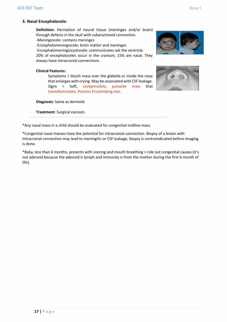
3. Nasal Encephalocele:
Definition: Herniation of neural tissue (meninges and/or brain) through defects in the skull with subarachnoid connection. -Meningiocele: contains meninges -Encephalomeningiocele: brain matter and meninges -Encephalomeningiocystocele: communicates wit the ventricle 20% of encephaloceles occur in the cranium, 15% are nasal. They always have intracranial connections. Clinical Features:
Symptoms > bluish mass over the glabella or inside the nose that enlarges with crying. May be associated with CSF leakage. Signs > Soft, compressible, pulsatile mass that transilluminates. Positive Frustenberg test.
Diagnosis: Same as dermoid. Treatment: Surgical excision.
*Any nasal mass in a child should be evaluated for congenital midline mass.
*Congenital nasal masses have the potential for intracranial connection. Biopsy of a lesion with intracranial connection may lead to meningitis or CSF leakage, biopsy is contraindicated before imaging is done.
*Baby, less than 6 months, presents with snoring and mouth breathing > role out congenital causes (it’s not adenoid because the adenoid is lymph and immunity is from the mother during the first 6 month of life).

433 ENT Team Nose I
18 | P a g e
(431 Team) Venous Drainage of the Nose:
The veins form a cavernous plexus beneath the mucous membrane. They open into:
o Sphenopalatine and greater palatine veins > Pterygoid plexus > Maxillary vein > External jugular vein.
o Anterior and posterior ethmoidal veins > Ophthalmic vein > Cavernous sinus. o Angular, lateral nasal, and superior labial veins > Facial vein > Internal jugular vein.
The pterygoid venous plexus and facial vein also communicate with the cavernous sinus:
Pterygoid venous plexus > Emissary vein > Cavernous sinus. Facial vein >
1. Angular vein > Nasofrontal vein > Superior ophthalmic vein > Cavernous sinus 2. Deep facial vein > Pterygoid venous plexus > Emissary vein > Cavernous sinus
The Dangerous Area of the Face (Bermuda Triangle): It is the area between the root of the nose and the 2 angles of the mouth. The veins that drain this region (mostly facial vein) are:
1. Connected to the cavernous sinus 2. Valveless, which facilitates retrograde flow of blood from the face
to the cavernous sinus. Any infection in this area may lead to cavernous sinus thrombosis and intracranial complications.
Nerve Supply of the Nose:
1. External Nose (Skin) It is innervated by the ophthalmic (V1) and maxillary (V2) divisions of the trigeminal nerve (CN V).
Ophthalmic (V1) > The superior aspect of the nose, including the tip > o Infratrochlear nerve o Supratrochlear nerve o External nasal branch of the anterior ethmoidal nerve
Maxillary (V2) > Inferior and lateral aspects of the nose > o Infraorbital nerve
2. Nasal Cavity
Olfactory Sensation:

433 ENT Team Nose I
19 | P a g e
Olfactory nerve > The roof and the uppermost parts of the medial and lateral walls.
General sensation: Trigeminal nerve (CN V) > Ophthalmic and maxillary divisions (via sphenopalatine ganglion)
o Lateral wall: 1. Anterior ethmoidal nerve (V1) > Lateral internal nasal branch. 2. Sphenopalatine nerve (V2) > Lateral posterior inferior branch. 3. Sphenopalatine ganglion (V2) > Lateral posterior superior nasal branch (Short
sphenopalatine nerve). o Nasal septum:
1. Sphenopalatine ganglion (V2) > a. Nasopaltine nerve (Long sphenopalatine nerve) b. Medial posterior superior branch.
2. Anterior ethmoidal nerve (V1) > Medial internal nasal branch.
Autonomic Fibers: Sensory branches of the sphenopalatine ganglion supplying the nasal mucosa carry postganglionic secretomotor fibers from the sphenopalatine fibers ganglions to the nasal glands. Autonomic fibers control the vascular tone and secretion of the nasal mucous glands. Sympathetic > vasoconstriction. Parasympathetic > vasodilation and increased nasal secretion.
Pathway of Autonomic Fibers:
Postganglionic sympathetic fibers pass from the superior cervical ganglion > deep petrosal nerve. Preganglionic parasympathetic fibers pass via the sensory root of the facial nerve > greater petrosal branch.
o The deep petrosal and great petrosal nerves merge to form the vidian nerve (Nerve of the pterygoid canal) > pterygopalatine ganglion > parasympathetic fibers synapse with the postganglionic secreto-motor fibers.
o Pterygopalatine ganglion gives terminal branches carrying the postganglionic sympathetic and parasympathetic fibers to their targets in the nasal cavity (blood vessels and nasal glands).

433 ENT Team Nose I
20 | P a g e
Othman Abid Rayan Alowaisheq
Done By: