Lecture XXVIII: Physiology and Modulation of Pain - KSUMSC
20
Lecture XXVIII: Physiology and Modulation of Pain MALE SLIDES IMPORTANT FEMALE SLIDES LECTURER’S NOTES EXTRA EDITING FILE GSLIDES
-
Upload
khangminh22 -
Category
Documents
-
view
7 -
download
0
Transcript of Lecture XXVIII: Physiology and Modulation of Pain - KSUMSC

Lecture XXVIII: Physiology and Modulation of Pain
MALE SLIDESIMPORTANT FEMALE SLIDES LECTURER’S NOTESEXTRAEDITING FILE GSLIDES

1 Lecture Twenty Eight
- Differentiate between pain & nociception - Describe the types of nerve fibres and receptor types that mediate pain •Describe - Different types of pain and pain pathways - Describe the role of thalamus and cerebral cortex in pain perception- Intensity of the pain can be altered by various extrinsic and intrinsic mechanisms- know gate-control hypothesis (spinal modulation)and role of body’s own morphines, the opioid peptides- Supra spinal modulation (Special pain control analgesic system).- know about opioid receptors and are formed in the midbrain, brainstem and spinal cord.- Appreciate that pain can also be facilitated- Know the sites & mechanism of pain relief
OBJECTIVES
PHYSIOLOGY OF PAIN
Pain Pain
classified by duration
Acute Chronic
Pain classified by
nature
Nociceptive Non-nociceptive
Somatic(Superficial or
deep)
Visceral Neuropathic Sympathetic
- Sensory receptors: Specialized peripheral endings of primary afferent neurons.- Nociceptors (pain receptors): Primary afferent receptors that respond selectively to
noxious stimuli.- Noxious stimuli: Stimulus (mechanical, chemical or thermal) that produces tissue
damage or threatens to do so - Polymodal nociceptors: Respond to various stimuli.- Reception: Response of nerve receptors in the skin and tissues to stimuli resulting from
actual or potential tissue damage.- Perception: the process by which pain is recognized and interpreted by the brain.
Figure 28-1
Basic Concepts

1) is a misnomer, as it is not actually a syndrome - it is a very generalized term that is often used to describe the condition of patients who have not had a successful result with back surgery or spine surgery and have experienced continued pain after surgery.
Nociception○ Sustained primarily by the nociceptive
system ○ Proportionate to the stimulation of the
nociceptor○ When acute
■ Serves a protective function ■ Normal pain
○ Pathologic when chronic ○ Responds to common analgesics
Eg:Acute burns, bone fracture, and other somatic and visceral pains
Idiopathic PainNo underlying lesion found yet, disproportionate to the degree of clinically discernible tissue injury
Mixed Pain❏ Failed low-back- surgery syndrome1
❏ Complex regional pain syndrome
2 Lecture Twenty Eight
FootnotesFOOTNOTES
PHYSIOLOGY OF PAIN
Pain & Nociception ● What is nociception? Refers to the transmission of signals evoked by
activation of nociceptors (pain receptors) from periphery to the CNS.
● What is pain? Is perception of unpleasant sensation that originates from a specific body region.
“Is an unpleasant sensory and emotional experience associated with actual or potential tissue damage.”
— International association for the study of pain (IASP)
Classification of Pain
○ Sustained by aberrant processes in PNS or CNS
○ Disproportionate to the stimulation of nociceptor
○ Serves no protective function ○ Pathologic pain ○ Resistant to common analgesics
Eg: Painful diabetic & peripheral neuropathies, deafferentation and sympathetically-maintained pains (BOX 28-1), nerve inflammation, compression.
Neuropathic Pain
BOX 28-1: CLINICAL RELEVANCESYMPATHETICALLY-MAINTAINED PAIN aka COMPLEX REGIONAL PAIN SYNDROME
Painful stimulus activates the sympathetic nervous system, after all, the ascending paleospinothalamic tract (discussed later), activates the reticular formations (which includes the locus coeruleus, major source of NE), thereby increasing sympathetic discharge.
- The increased sympathetic discharge activates alpha-receptors on nociceptive nerve endings of primary afferents, this activation leads to more activity of the primary nociceptive afferent, which will initiate new pain impulses after this activation, and will activate the locus coeruleus again which will increase sympathetic activity. This will lead to a loop of chronic pain, known as Sympathetically-Maintained Pain, or Complex Regional Pain Syndrome.

● Felt in 1 sec. or more following painful stimulus. ● It is associated with tissue damage & can be
referred to as burning pain, aching pain, throbbing pain “unbearable”, diffuse , dull , poorly localized or chronic pain
___________________________
Characteristics❖ Occurs SECOND upon stimulation of Polymodal
receptors
❖ Chronic type of pain, transmitted by C fibers
peripherally & centrally by paleospinothalamic Tract
❖ Characteristics of C fibers
➢ Non-Myelinated
➢ Diameter 0.4 – 1.2 μm
➢ conduction velocity 0.5 - 2 m/s
➢ Terminate in layer II and III of dorsal horn (substantia
gelatinosa)
➢ Associated with destruction of tissue
➢ Can occur in skin or any internal organ
❖ Slow, diffuse, dull, aching
❖ Neurotransmitter - P-Substance
❖ 80% of pain conduction
3 Lecture Twenty EightPHYSIOLOGY OF PAIN
Significance Pain is mainly a protective mechanism of the body, as it is not a pure sensation but a response to tissue injury. The response may be:
○ Motor/Behavioral responses – e.g. withdrawal, defense○ Emotional – e.g. anxiety, crying, depression, fear○ Autonomic reaction e.g. tachycardia, rise in B.P., sweating. (Mechanism in BOX 28-1)
So pain will help to:1. Avoid noxious stimuli 2. Remove body parts from danger 3. Promote healing by preventing further damage 4. Storage of painful experiences in memory helps us to avoid potentially
harmful event in the future
Types of Pain
● It is felt within 0.1 sec. after stimulation. ○ e.g. sharp, intense, pricking, cut
with knife, well localized._________________________
Characteristics❖ Occurs FIRST upon stimulation of Mechanical
and Thermal nociceptors
❖ Transmitted by Aδ(delta) fibers in the
peripheral nerves & centrally by
Neospinothalamic Tract, (discussed later in
lecture)
❖ Characteristics of Aδ fibers
➢ Myelinated ➢ Diameter fine 2 ➢ 5 μm➢ 12 - 30 m/sec. conduction velocity ➢ Terminated at I and V layer (mainly lamina
marginalis)➢ Associated with reflex withdrawal➢ Usually somatic , not visceral
❖ Fast pain, rapid, pricking and well localized ❖ Neurotransmitter - Glutamate ❖ 20% pain conduction
Slow pain
Pain is perceived at both the cortical &
thalamic levels.
The noxious stimuli activates:❏ 10-20% of the A-delta fibers (faster for localization) Fast pain
❏ 50-80% of the C-fibers (main carrier) Slow pain
Fast pain
- They are the most widely distributed and absent in brain tissue.- They are specific (have adequate stimulus), pain is not produced by overstimulation of other receptors.- They are high threshold receptors i.e. painful stimuli must be strong & noxious to produce tissue damage.- They do not adapt (or very little) to repetitive stimulation- It allows the pain to keep the person apprised of a tissue-damaging stimulus as long as it persists.- Pain is not produced by overstimulation of other receptors.
Nociceptors: Additional Characteristics

● special receptors that respond only to noxious stimuli and generate nerve impulses which the brain interprets as "pain". (Sherrington 1906)
● Pain receptors do not adapt at all or very slowly.● They are found in largest number & density in skin, periosteum joint
surface, arterial wall & dura mater (tentorium).● Three types of stimuli:
○ Mechanical ---> they elicit fast pain. respond to strong pressure (eg,from a sharp object).
○ Thermal ----->they elicit also fast pain.are activated by skin temperatures above 42°C or by severe cold.
○ Chemical ----> they produce slow pain. respond to various chemicals like bradykinin, histamine, high acidity, and environmental irritants .
○ Polymodal nociceptors: respond to combinations of these Stimuli.
● Distribution of Pain Receptors:○ Widespread in superficial layers of skin ○ Fewer in deep tissue ○ absent in brain tissue
● Nociceptors: are small and medium● Non-nociceptors: Large
4 Lecture Twenty EightPHYSIOLOGY OF PAIN
Pain Receptors Are Free Nerve Endings (Nociceptors)
Figure 28-2
Figure 28-3
Figure 28-4
Polymodal nociceptors
● The receptor that is activated by moderate cold is TRPM8.
○ The M refers to menthol, the ingredient in mint that gives it its “cool” taste.
● TRPV4 receptor are activated by warm temperatures up to 34 ºc;
● TRPV 3 receptors respond to slightly higher temperature of 35-39ºc.
❏ Innocuous cold receptors are on dendritic endings of Both Aδ fibers and C fibers.
❏ Innocuous warmth receptors are on Just C fibers.
❏ cold sensitive > heat-sensitive. (4- 10 times as many)
Thermoreceptors
Figure 28-5
TRPV1 TRPA1 ASIC P2X & P2Y❖ The V refers to a group of
chemicals called vanilloids
❖ Activated by intense heat, acids, and chemicals such as capsaicin1 (the active principle ingredient of hot peppers and an example of a vanilloid).
❖ A, for Ankyrin2
❖ Noxious mechanical, cold, and chemical stimuli may activate TRPA1 receptors on sensory nerve terminals.
❖ Acid sensing ion channel (ASIC)
❖ activated by pH changes within a physiological range
❖ May be the dominant receptors mediating acid-induced pain.
❖ nociceptive mechanical stimuli cause the release of ATP that acts on purinergic receptors➢ P2X, an ionotropic
receptor ➢ P2Y, a G protein-
coupled receptor
There are a variety of receptors located on the endings of nociceptive sensory nerves that respond to noxious thermal, mechanical, or chemical
stimuli (Figure 28-5)
Many of these are part of a family of nonselective cation channels called transient receptor potential (TRP) channels.
FootnotesFOOTNOTES
1) Capsaicin has a paradoxical clinical application, if applied in large quantities it can deplete substance P and induce analgesia! Therefore it is also used in treatment of arthritis
2) Ankyrins are proteins that link the cytoskeleton to membrane proteins, this maintains the integrity of the cell membrane and anchors ion channels to the cell membrane.

PHYSIOLOGY OF PAIN 5 Lecture Twenty Eight
Dual Pathways for Transmission of Pain Signals into the Central
Nervous System
● Neospinothalamic Tract for:○ Fast Pain ○ lamina I (lamina marginalis)
● Paleospinothalamic Tract ○ Slow Pain○ Almost entirely in laminae II and III of the dorsal
horns, which together are called the substantia gelatinosa
Figure 28-6
Nociceptors Stimulation ○ Pain receptors are depolarized either directly or through the production of pain producing substances
(inflammatory mediators) from damaged tissues or as a result of inflammation.■ Bradykinin (BOX 28-2), serotonin, Histamine, K+ ion, Acids, proteolytic enzymes (BOX 28-2). calcitonin
gene-related peptide (CGRP)1, interleukins2, PGs, Ach.○ PGs & substance P -> enhance the sensitivity of pain receptors.
Substance SourcePotassium Damaged cells
Serotonin Platelets
Bradykinins Plasma
Histamine Mast cells
Prostaglandins3 Damaged cells
Leukotrienes Damaged cells
Substance P Primary nerve Afferents
Table 28-1
Chemicals Released With Tissue DamageBOX 28-2: CLINICAL RELEVANCEinjured cells release proteases from their disrupted lysosomes, these proteases can activate extracellular kininogen to form bradykinin, bradykinin opens ion channels of nociceptors thus eliciting the sensation of pain. One more example is histamine release by mast cells in response to an allergen, such a bee sting, histamine can bind to ion channels on nociceptors. The mechanisms of pain are diverse and renewing, with many mediators activating the receptors. Hence why adaptation is difficult, there is really more than one way to activate nociceptors
FootnotesFOOTNOTES
1) CGRP: From the calcitonin family of peptides, synthesized in peripheral and central neurons, specifically DRG cells, plays an uncertain role in nociception2) ILs are the first cytokines to be discovered, inter = between, leukins = leukocytes, in response to injuries cells release IL-1 and IL-6, which acts on other
cells to trigger production of PGs, PGs then act on the hypothalamus, thermostat of the body, to trigger fever, and also acts on nociceptors to trigger pain.3) This is the basis of fever associated with inflammation and why analgesics are effective.
Figure 28-7

○ Transmit Slow Pain○ First order neurons:
- They are mainly type C fibers.- They enter spinal cord via dorsal roots, terminate at substantia
gelatinosa in laminae II & III of dorsal horn (substantia gelatinosa).
○ Second order neurons:- They start at SGR, cross to opposite side in front of central canal, Most
of the slow pain fibers project to reticular formation & the remaining proceed to thalamus (posterior nuclei). They ascend in lateral column of spinal cord & terminate at:
● Reticular formation of brainstem. ● Tectal area and the periaqueductal gray matter of the midbrain● Intralaminar nuclei of thalamus. ● Hypothalamus & adjacent region of basal brain. These lower regions of the brain appear to be important for feeling the suffering types of pain, therefore impulses arriving these regions have strong arousal effects and can be perceived. (poor localization)Only 1/10 to 1/4 of the fiber have passed all the way to the thalamus.
○ Third order neurons: - These start at thalamus. Few fibers project to cerebral cortex.
PHYSIOLOGY OF PAIN 6 Lecture Twenty Eight
Pain PathwaysPain sensation is carried by lateral spinothalamic tracts which includes 2 separate pathways:
1. Neospinothalamic Tract○ Transmit Fast Pain○ First order neurons:
- Are mainly A afferent nerves. - They terminate at lamina I & V of dorsal horn. (lamina
marginalis)○ Second order neurons:
- These constitute the tract.- They start at dorsal horn, cross to opposite side and ascend in
lateral column of spinal cord. The fibers ascend in brain stem to terminate in ventrobasal complex of thalamus.
○ Third order neurons: - These start at thalamus. Most fibers project to somatosensory
cortex and the basal areas of the brain.
2. Paleospinothalamic Tract
Figure 28-8
Figure 28-9

PHYSIOLOGY OF PAIN 7 Lecture Twenty Eight
Silent Nociceptors● In the skin and deep tissues there are additional nociceptors called "silent" or "sleep"
nociceptors.● These receptors are normally unresponsive to noxious mechanical stimulation, but become
“awakened” (responsive)to mechanical stimulation during inflammation and after tissue injury. One possible explanation of the "awakening"phenomenon is that continuous stimulation from the damaged tissue reduces the threshold of these nociceptors and causes them to begin to respond.
● This activation of silent nociceptors may contribute to the induction of hyperalgesia1, central sensitization, and allodynia1.
● Many visceral nociceptors are silent nociceptors.
Characteristics
Superficial Pain
● Arises from skin or other superficial structures (hair follicles, nails, and mucous membranes)
● Occurs in 2 phases (fast pricking, slow burning pain)● Well localized ● Associated with motor, autonomic, emotional reactions.
Deep Pain● Arises from muscles, joints, periosteum, tendons & ligaments● Diffuse, slow, and prolonged● conducted by type C fibers● May be referred (not well localized), initiate reflex contraction of nearby muscles.● Caused by: trauma, bone fracture, inflammation, arthritis, muscle spasm & ischemia
Visceral Pain
● There are few pain receptors in most viscera● Slow, diffuse, and poorly localized. Conducted by C fibers● Pain arising from parietal peritoneum, pleura, and pericardium is sharp, pricking type.● Often referred. Associated with rigidity of nearby muscles and autonomic reactions● Caused by: - Distension of a hollow organ- Inflammation of an organ - Ischemia, e.g. pain due to myocardial ischemia ❏ N.B: Cutting, crushing are not painful when applied to viscera● Aδ and fibers travel with autonomic afferent > Dorsal Horn of spinal cord (Lat.
spinothalamic tract) > Thalamus > Somatosensory Cortex● Some viscera are pain insensitive e.g. liver parenchyma, lung alveoli, brain tissue, visceral
layer of peritoneum, pleura and pericardium.
Types of Pain According To Site of Stimulation
The brain tissues themselves are almost totally insensitive to pain. - Tugging on the venous sinuses around the brain, damaging
the tentorium, or stretching the dura at the base of the brain can cause intense pain that is recognized as headache.
- Also, almost any type of traumatizing, crushing, or stretching stimulus to the blood vessels of the meninges can cause headache.
Headache
Figure 28-10
1) Hyperalgesia: Extreme sensitivity to pain. Allodynia: Pain response to a stimuli which do not normally provoke pain due to central sensitization.
FOOTNOTES

● Pain that is not felt in the diseased structure itself, but at another place in the body far away from the site of origin.
● Visceral and deep somatic pain are often referred, but superficial pain is not.
● Dermatomal rule: When visceral pain is referred to the surface of the body, the person generally localizes it in the dermatomal segment from which the visceral organ originated in the embryo, not necessarily where the visceral organ now lies.
● When pain is both localized and referred from its original site of localization it is called radiating pain.
● Examples of Referred Pain:- Cardiac Pain Is referred to the jaw, left shoulder & inner side of
left arm- Pain of appendicitis Is referred to periumbilical region- Pain from ureter Is referred to testicular region● Mechanism of referred pain:– Convergence:● Afferent pain fibers from skin area & diseased viscera that
develop from same embryonic segment converge on same 2nd order neuron and finally stimulate the same cortical neuron.
● The brain interprets the information coming from visceral nociceptors as having arisen from cutaneous nociceptors, because this is where nociceptive stimuli originate more frequently
– Facilitation theory: ● Pain fibers from skin are always carrying impulses, not enough
to produce pain.● Impulses from diseased viscus pass through afferents which
give collaterals to ST neurons receiving pain fibers from skin. ● As a result, ST neurons' excitability is raised (they are
facilitated) to reach a threshold level. ● The signals reaching the brain are projected to skin area and
pain is felt in skin dermatome
PHYSIOLOGY OF PAIN
REFERRED PAIN
BOX 28-3: CLINICAL RELEVANCEWHY DOES A HEART ATTACK CAUSE PAIN IN LEFT ARM? - DERMATOMAL RULE EXPLAINEDLet us try to explain why pain is referred from a visceral organ to another site according to the dermatomal rule.- Apparently, during embryological development, the pain afferents carrying impulses from visceral organs "sprout"
collaterals that synapse on the 2nd order neurons supplying the skin over the organ itself.- This serves as a protective mechanism to maximise the pain felt. However, after growth, the organ changes its place to
assume a more preferable position, the afferents carrying the pain impulses from viscera into the spinal cord elongate, supplying more 2nd order neurons which are placed further apart.
- Therefore signals are relayed and felt at different regions. This process causes the pain to be felt at different parts since the old collaterals -prior to elongations- are also retained.
.
Lecture Twenty Eight 8
Figure 28-11
Figure 28-12
Figure 28-13
Figure 28-14
Organ Site of referred pain Organ Site of referred pain
Meninges Back of head & neck Ureter Testicles
Heart Central chest, left arm Hip Knee
Stomach Epigastrium Appendix Umbilicus
Kidney Loin Uterus Low back

To modulate the pain, you should know firstly the grade or scale of the pain by whether numerical value or adjective chart. Because if you don't know the grade or scale of pain , how can you know that you have modulated the pain.● Visual Analog Scale, Locate area of pain on a picture, McGill pain
questionnaire (Evaluate sensory, evaluative, & affective components of pain: 20 subcategories, 78 words)
● What will happen if sensory area SI is removed? person's ability to interpret the quality of pain & precise location of pain will be affected.● Why patient with chronic pain syndrome have difficulty in sleeping?
Paleospinothalamic pathway sends information to reticular formation and thalamic nuclei which are part of brain activating / alerting system, therefore chronic pain syndrome causes difficulty in sleep.
PAIN MODULATION
PAIN MODULATION 9
It means pain perception variability (the degree to which a person reacts to pain) i.e. A decrease or an increase in the sensation of pain caused by inhibition or facilitation of pain signals.
● Spinal (segmental) inhibition: Gate control theory
● Supraspinal (descending) inhibition
● Peripheral sensitization (release of chemicals after tissue injury)● Central sensitization (Dis-inhibition)
FacilitationInhibition
Nociception
● Nociception consists of four basic processes:1. Transduction—nociceptors free nerve endings “Stimulation of nociceptors”2. Transmission3. Perception of Pain -At cortical Level 4. Modulation of Pain, Changing or inhibiting pain impulses in the descending tract (brain spinal cord) [norepinephrine and serotonin] or ascending tract.
Figure 28-15
Grading of Pain
Figure 28-16
Gate Control Theory of PainSpecial neurons in the the dorsal horn of spinal cord (Substantia gelatinosa of Rolando, second order neurons of Paleospinothalamic pathway) form the gate through which pain impulses must pass to reach brain.
Lecture Twenty Eight

Gate Opened Or Closed By Three Factors
Gate(SGR)
Messages from the brainTo the brain
Transmission nerve cells
Activity in pain nerves “C fibers” opens the gate
Activity in other sensory nerves “Aβ fibers” closes the gate (e.g. Rubbing the affected area)
Projection neuronneuron
Four Variables Control This Gate
Type C- fibres
(slow pain)
Type A-β fibres
(light touch)
Projection neuron(SGR)
Inhibitory interneurons
Inhibitory
The Gate Theory of Pain Control● The gate is closed by AB fibers & opened by C
nociceptors.● Projection neuron receives input from both C-fibers
and Aβ fibers.● Impulses coming along type C pain fibers cause the
release of substance P from these fibers and inhibits the inhibitory interneuron (open the gate).
● While impulses coming along Aβ fibers tend to keep the gate closed by activating the inhibitory interneuron.
● A-Delta fibres (sharp pain). ● C fibres (dull pain). ● A-Beta fibres (light touch).
● When pain and touch fibers are stimulated together, gate will be closed & pain is not felt.
● Implies a non-painful stimulus can block the transmission of a noxious stimulus.
● It Is based on the premise that the gate, located in the dorsal horn of the spinal cord, modulates the afferent nerve impulses.
Lecture Twenty Eight10 PAIN MODULATION
Figure 28-17
Figure 28-18
Figure 28-19

11
● Specialised nerve impulses arise in the brain itself and travel down the spinal cord to influence the gate.
● It can send both inhibitory and excitatory messages to the gate sensitising it to either C or A-β fibres.
● The inhibitory neurons make a pain blocking agent called encephalin.● Encephalin is an opiate substance which can block substance P, the
neurotransmitter from the C fibers, and this keeps the gate closed.
Central Control Trigger
The Gate Theory Explains The Pain Relief By:● Skin rubbing● Shaking the painful part
Transcutaneous Electrical Nerve Stimulation (TENS)- TENS uses electrodes to activate Aα and Aβ fibers in the vicinity of the injury. - TENS stimulates sensory nerves to block pain signals, stimulate endorphin production
to help normalize sympathetic function.● TRANSCRANIAL DIRECT CURRENT STIMULATION (TDCS):- It is a non-invasive procedure in which a device sends a small Direct Current (DC)
across electrodes in the areas of interest on the scalp to modulate brain function. - This current flow can increases or decreases the neuronal excitability in the specific
area being stimulated, based on which type of stimulation.- When the current passes from the anode to the cathode, it may increase the activity of
the brain at the anode site and decrease the activity of the brain near the cathode site.Interferential Stimulation- Interferential Stimulation differs from TENS because it allows a deeper penetration by
using two frequencies of the tissue with more comfort (compliance) and increased circulation. Stimulate parasympathetic nerve fibers for increased blood flow.
Acupuncture: - Stimulation of large type Aβ sensory fibers from peripheral tactile receptors depress
transmission of pain signals from the same body area by lateral inhibition in the spinal cord.
- The simultaneous physical and psychogenic excitation of the central analgesia system is the basis of pain relief by acupuncture.
- The analgesic effect of electroacupuncture may involve the release of endogenous opioids and activation of descending pain modulatory pathway
● All are supposed to stimulate mechanoreceptors (Aβ fibers) that activate neurons of dorsal column, the collaterals (inhibitory interneurons) relieve pain.
Conditions that open the gate Conditions that close the gatePhysical Conditions
Extent of the injury Medication
Inappropriate activity level(Overstimulation of C fibers)
Counterstimulation, e.g. massage
Emotional Conditions
Anxiety or worry Positive emotions
Tension Relaxation
Depression Rest
Mental Conditions
Focusing on the pain Intense concentration or distraction
Boredom Involvement and interest in life activities
Conditions That Open Or Close The Gate
Lecture Twenty EightPAIN MODULATION
Figure 28-20
Figure 28-21

BOX 28-4: CLINICAL RELEVANCE
Scientists were curious on how drugs like heroin and morphines the so-called opiates exert their effects and why people abuse them. - They hypothesized that they act on receptors, and after a certain experiment, receptors were found. - The next step was to found what the receptors responded to within the body, since there must have been endogenous
opiates! And this is exactly what they found in enkephalin, they called such endogenous morphine-like substances, endorphins.
Note that the term opiate and opioid differ, opioids refer any substance that bind to opioid receptors within the brain, either synthetic or natural. Whereas opiates refer to natural substances that bind to opioid receptors, hence all opiates are opioids and the opiates within the body are called endorphins.
Supra Spinal Modulation (Special Pain Control Analgesic System)This is a specific system that blocks pain transmission in CNS.
Its major constituents are:
Figure 28-22
Supra Spinal ModulationAnalgesia system of the brain and spinal cord, showing:(1) inhibition of incoming pain signals at the cord level.(2) presence of enkephalin-secreting neurons that suppress pain signals in both the cord and the brain stem.
Analgesia Occurs As FollowsPeri- ventricular nucleus projects to PAG.
PAG projects neurons containing aspartate & glutamate, stimulate (RMN)
They block pain signals by activating PIC.
RMN projects serotonin-ergic neurons to dorsal horn.
12
The Periventricular & Periaqueductal Gray Areas
Raphe Magnus Nucleus (RMN)
Pain inhibitory complex
Enkephalin Neurons In the mesencephalon and upper pons. It send signals to Raphe magnus nucleus.
These neurons surround portions of the third and fourth ventricles and the aqueduct of Sylvius.
A thin midline nucleus located in the lower pons and upper medulla.
From these nuclei, second-order neurons go down the dorsolateral columns in the spinal cord & secrete Serotonin which act on local neurons to secrete Enkephalin.
In dorsal horn of spinal cordAt this point, the analgesia signals can block the pain before it is relayed to the brain.
It consists of: multiple short enkephalinergic neurons that terminate on central endings of pain conducting afferent fibers. ● When stimulated the release enkephalin cause pre & postsynaptic inhibition of pain transmission. ● It prevents the release of substance P from pain nerve endings.
BODY OPIATE TRANSMITTERS, DIFFERENCE BETWEEN OPIOIDS AND OPIATES
Lecture Twenty EightPAIN MODULATION

Opioid Peptides and Pain Modulation● They are natural analgesic substances (morphine-like substances) present in
body. (Endorphine name is derived from endogenous morphine, since scientists initially thought that a similar substance to morphine existed within the body, they named them collectively endorphins)
● They act by binding to opiate receptors in analgesic system and dorsal horn of SC on central ending of pain conducting pain fibers.
E.g. endorphin, encephalin, dynorphin, endogenous morphine.Beta Endorphin is 50 times more potent than morphine and Dynorphin is 200 times more potent than pure morphine
Mechanism of actions of Opioid peptides on pain transmission:They exerts their analgesic effects by acting at various sites in peripheral & CNS
mechanism
direct indirectInhibiting discharge of nociceptor neurons.
Activating the descending inhibitory pathway be exciting periaqueductal grey neurons.
Inhibiting release of substance P from central terminal of nociceptor neurons
Activating neurons in the brain stem which release NE and serotonin which suppress pain transmission directly or indirectly via activation of enkephalinergic containing inhibitory interneurons.
Cause inhibition of dorsal horn spinothalamic neuron. Via K efflux causing hyperpolarization.
Cellular Actions Of Opioid Peptides- Reduction of cAMP synthesis by inhibiting Adenyl
cyclase. - Inhibition of transmitter release by inhibiting opening of
Ca++ channels. “Presynaptic inhibition”- Hyperpolarization by facilitating opening of voltage
gated K+ channels. (Postsynaptic inhibition)
● Activation of the presynaptic opioid receptor leads to a decrease in Ca++ influx, resulting in a decrease in release of glutamate and substance P.
● Activation of the postsynaptic opioid receptor hyperpolarizes the dorsal horn interneuron by causing an increase in K+ conductance.
● Decrease duration of the EPSP in the dorsal horn neuron.
● Activation of opioid receptor on dorsal root ganglia cell bodies also contributes to reduced transmission from nociceptive afferents.
Lecture Twenty Eight13 PAIN MODULATION
Figure 28-23
Figure 28-24 Figure 28-25
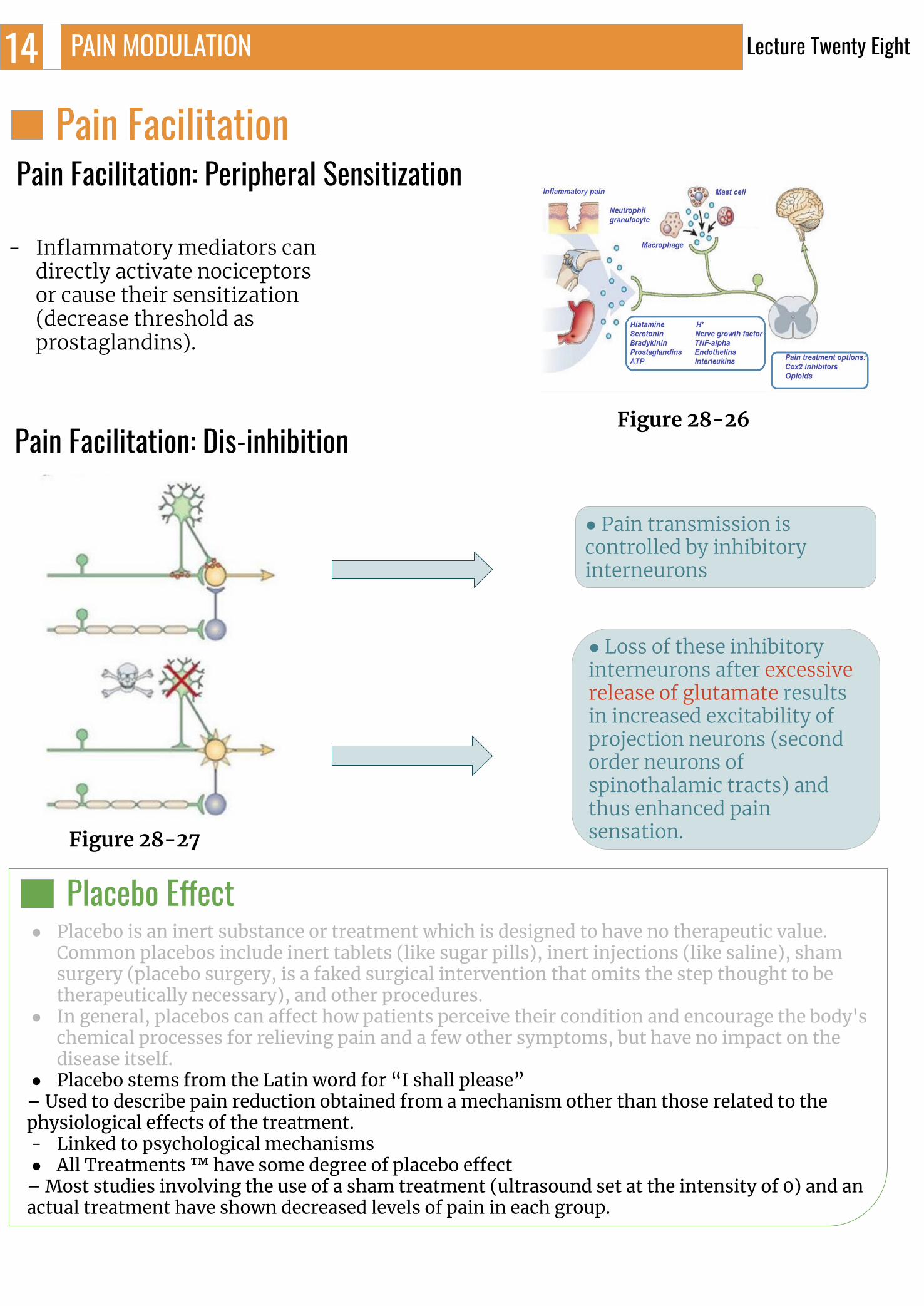
Pain Facilitation Pain Facilitation: Peripheral Sensitization
- Inflammatory mediators can directly activate nociceptors or cause their sensitization (decrease threshold as prostaglandins).
Pain Facilitation: Dis-inhibition
● Pain transmission is controlled by inhibitory interneurons
● Loss of these inhibitory interneurons after excessive release of glutamate results in increased excitability of projection neurons (second order neurons of spinothalamic tracts) and thus enhanced pain sensation.
Lecture Twenty Eight
Placebo Effect● Placebo is an inert substance or treatment which is designed to have no therapeutic value.
Common placebos include inert tablets (like sugar pills), inert injections (like saline), sham surgery (placebo surgery, is a faked surgical intervention that omits the step thought to be therapeutically necessary), and other procedures.
● In general, placebos can affect how patients perceive their condition and encourage the body's chemical processes for relieving pain and a few other symptoms, but have no impact on the disease itself.
● Placebo stems from the Latin word for “I shall please”– Used to describe pain reduction obtained from a mechanism other than those related to the physiological effects of the treatment. - Linked to psychological mechanisms● All Treatments ™ have some degree of placebo effect
– Most studies involving the use of a sham treatment (ultrasound set at the intensity of 0) and an actual treatment have shown decreased levels of pain in each group.
14 PAIN MODULATION
Figure 28-26
Figure 28-27

Fibromyalgia: Pain Without Injury
● Less blood flow in the muscles (ischemia)Muscular Pain
Mechanism Of Pain ReliefBlock production of inflammatory mediators .e.g. Aspirin & nonsteroidal anti-inflammatories.
Exogenously administration of opioid like drugs.
Sympathectomy can be useful.
Electrical stimulation of the dorsal column.
Selective activation of large diameter afferent fibers by transcutaneous electrical nerve stimulation. Stimulation of brainstem sites or
administration of drugs which can modify serotoninergic or adrenergic neurons e.g. antidepressants.
04
Causalgia03● Burning pain.● It is chronic burning pain condition seen after the section
(damage, cutting) of a nerve Triggered by a simple stimulus e.g. breeze or vibration.
Allodynia02● Pain caused by any other sensation (e.g. touch).● A person feels pain from stimuli that don't normally cause pain due to
central sensitization. For example, lightly touching your skin or brushing your hair might feel painful.
● clinical feature of many painful conditions, such as neuropathies, complex regional pain syndrome, postherpetic neuralgia, fibromyalgia, and migraine
Hyperalgesia01 ● Excessive Pain (e.g due to sunburn).● Increased sensitivity to Pain
Terms frequently used :
05
● The occurrence of body-wide pain in the absence of tissue damage, as in fibromyalgia, interferes with all aspects of a person's life and undermines their credibility.
● The problem is that normal activities can be exhausting, sleep is disturbed, the ability to concentrate is impaired, gastrointestinal function is often abnormal, persistent headaches are common, and the unrelenting pain that no one can see is often detrimental to their personal and professional lives--as it creates a "credibility gap."
Lecture Twenty Eight15 PAIN MODULATION

An interesting question to ask is what would happen to a sensory cortical area if a certain finger for instance was cut? Would the “finger area” for this particular finger go unused? Does it undergo atrophy?- The answer to these questions were answered in an experiment in which the third
finger of a monkey was amputated, months later the sensory cortex showed “remapping” that is, when finger 3 was surgically removed, finger 2 and 4 took over the area of finger 3 in the sensory cortex. As can be seen in the figure. Therefore the cortex that previously responded to digit or finger 3 is now responding to finger 2 and 4. This suggests an advanced ability of the cortex to rearrange itself and to maximize its functions.
- A similar phenomenon can be seen in people with amputated limbs, those people might say that they still feel their amputated limb, a phenomenon called “Phantom Limb Pain”, however what really happens is that areas adjacent to the amputated limb take over the sensory area of the amputated limb in the cortex. As in, for example, the thigh might take over the place of an amputated leg. Giving the sensation of phantom limb pain.
ContinuedCharacteristics
Thalamic syndrome
It is a neurological condition that results from a brain stroke affecting the thalamus.● Obstruction of the thalamogeniculate branch of the posterior cerebral artery.● Affects posterior thalamic nuclei● Prolonged severe pain with variable quality (Dejerine Roussy Syndrome)
Trigeminal neuralgia
It is excruciating intermittent pain by stimulation of trigger area in the face. ● e.g. Washing of face, combing hair, blast of air on face.● It results from compression of trigeminal nerve root by blood vessels.
Stress induced
analgesia
Pain suppression response that occurs during or following exposure to a stressful or fearful stimulus.● It’s a well known phenomenon seen when the soldier is wounded in battle field but
feels no pain until the battle is over.● Mild degree of pain is not felt if the other part of the body has excessive pain.● The cause is not known may be it is similar to gate control hypothesis.
Phantom pain sensations
Impression of pressure and pain that an individual experiences relating to a limb or an organ that is not physically part of the body.● Our brain can reorganize at the ventral posterior thalamic nucleus if sensory input is
cut off even after that part is amputated.● Paraplegic people experience phantom limbs. They can even experience continually
cycling legs.● It is the emotional and motivational systems that cause the phantom limb experience.
Congenital Analgesia
Congenital insensitivity to pain (CIP), also known as congenital analgesia, is one or more rare conditions in which a person cannot feel (and has never felt) physical pain due to gene mutations.● A well-known case of congenital insensitivity to pain is a girl
referred to as ‘Miss C' who was a student at McGill university in Montreal in the 1950s.● She was normal in every way, except that she could not feel pain. When she was a child
she had bitten off the tip of her tongue and had suffered third-degree burns by kneeling on a radiator.
● She did not feel any pain when she was given strong electric shocks or when exposed to very hot and very cold water. When these stimuli were presented to her she showed no change in heart rate, blood pressure or respiration.
● She died at the age of 29 as a result of her condition, because she damaged her knees, hips and spine.
BOX 28-5: CLINICAL RELEVANCEPHANTOM LIMB PAIN MECHANISM
Lecture Twenty Eight16 PAIN MODULATION

1. Pain receptors in the skin are typically classified as which of the following?A) Encapsulated nerve endingsB) A single class of morphologically specialized receptorsC) The same type of receptor that detects position senseD) Free nerve endings
2. Which of the following is an important functional parameter of pain receptors?A) Exhibit little or no adaptationB) Not affected by muscle tensionC) Signal only lexion at joint capsulesD) Can voluntarily be inhibited
3. Stimulation of which brain area can modulate the sensation of pain?A) Superior olivary complexB) Locus coeruleusC) Periaqueductal gray areaD) Amygdala
4. Which of the following is the basis for referred pain?A) Visceral pain signals and pain signals from the skin synapse with separate populations of neurons in the
dorsal hornB) Visceral pain transmission and pain transmission from the skin are received by a common set of neurons in
the thalamusC) Visceral pain signals are rarely of sufficient magnitude to exceed the threshold of activation of dorsal horn
neuronsD) Some visceral pain signals and pain signals from the skin provide convergent input to a common set of
neurons in the dorsal horn
5. A 10-year-old boy cuts his finger with a pocketknife and immediately applies pressure to the damaged area with his other hand to partially alleviate the pain. Inhibition of pain signals by tactile stimulation of the skin is mediated by which type of afferent neurons from mechanoreceptors?
A) α-type AB) β-type AC) δ-type AD) Type C
6. Impulses coming along type C pain fibers will:A) Close the gate. B) Open the gate. C) A&BD) None
ANSWER KEY: D, A, C, D, B, B

SHORT ANSWER QUESTIONS
1. What are the processes that Aβ fibers using to keep the gate closed?
2. What are the major constituents of the supraspinal modulating system?
3. What is the central control trigger?
7. Gait theory is:A) Spinal modulation of pain inputB) Opioid receptor modulationC) Supraspinal modulationD) None
8. Stimulation by touching or pulling on which structure is least likely to cause a painful sensation?A) The postcentral gyrus B) The dura overlying the postcentral gyrusC) Branches of the middle meningeal artery that lie superficial to the dura over the postcentral
gyrus D) Branches of the middle cerebral artery that supply the postcentral gyrus
9. Which statement concerning visceral pain signals is correct?A) They are transmitted along sensory fibers that course mainly with sympathetic nerves in the
abdomen and thoraxB) They are not stimulated by ischemia in visceral C) They are transmitted only by the lightly myelinated δ-type A sensory fibersD) They are typically well localized
10. Which of the following is a group of neurons in the pain suppression pathway that uses enkephalin as a neurotransmitter?A) Postcentral gyrusB) Nucleus raphe magnusC) Periaqueductal gray areaD) Type AB sensory fibers
ANSWER KEY: A, A, A, C
ANSWERS1. presynaptic inhibition of C fibers postsynaptic inhibition of secondary neurons in dorsal horn.2. The periaqueductal gray and periventricular
areas. Raph magnum nucleus and the nucleus reticularis paragigantocellularis. Pain inhibitory complex.3. It is a specialized nerve impulses arise in the
brain itself and travel down the spinal cord to influence the gate.

Omar Aldosari, Mohammed A. AlshehriTaif Alshammari, Taibah Alzaid
FEMALE PHYSIOLOGY CO-LEADERS Maha Alnahdi, Ghaliah Alnufaei
MALE PHYSIOLOGY CO-LEADERS Nayef Alsaber, Hameed M. Humaid
REFERENCES- Guyton and Hall Textbook of Medical Physiology
- Ganong’s Review of Medical Physiology