
Restrictive Lung Diseases - KSUMSC
20
Restrictive Lung Diseases Black: original content. Red: Important. Green: AlRikabi’s Notes. Grey: Explanation. Blue: Only found in boys slides. Pink: Only found in girls slides. OBJECTIVES ✓ ✓ ✓ ✓ ✓ ✓ ✓ Editing File
-
Upload
khangminh22 -
Category
Documents
-
view
5 -
download
0
Transcript of Restrictive Lung Diseases - KSUMSC
Restrictive Lung Diseases
Black: original content.Red: Important.Green: AlRikabi’s Notes.Grey: Explanation.Blue: Only found in boys slides.Pink: Only found in girls slides.
OBJECTIVES
✓
✓
✓✓
✓
✓
✓
Editing File

Restrictive Lung Disease :
●
●
●
●●
●
*kyphoscoliosis : both kyphosis and scoliosis.*Complication of RLD inc;ude Cor pulmonale (Pulmonary hypertension→25+mmHg in the pulmonary artery)
Note : The final stage of all CHRONIC restrictive lung disease is extensive fibrosis with honeycomb lung.
Honeycomb lung :
Definition: both alveoli and bronchioles coalescence ( to form cysts lined with cuboidal or columnar ( یلتحمونepithelium and separated by inflammatory fibrous tissue .
→ kyphosis→ scoliosis
→
Kyphoscoliosis :
Honeycomb lung Normal lung
DefinitionDefinition
● Group of diseases (of various etiologies) characterized by reduced expansion of lung parenchyma and decreased total lung capacity (decreased lung volume and compliance).
● Spirometry: FEV1 and FVC decreased (ratio is normal)● Symptoms: chronic dry cough & dyspnea● A heterogeneous group of disorders
characterized by bilateral, often patchy, pulmonary fibrosis mainly affecting the walls of the alveoli.“Group of diseases (of various etiologies) characterized by reduced expansion of lung parenchyma and decreased total lung capacity (decreased lung volume and compliance).”
● Spirometry: FEV1 and FVC decreased (ratio is normal)● Symptoms: chronic dry cough & dyspnea.
● Group of diseases (of various etiologies) characterized by reduced expansion of lung parenchyma and decreased total lung capacity (decreased lung volume and compliance).
● Spirometry: FEV1 and FVC decreased (ratio is normal)● Symptoms: chronic dry cough & dyspnea
Definition
یلتحمون
Note : The final stage of all CHRONIC restrictive lung disease is extensive fibrosis with honeycomb lung.

-Pneumonia .-Aspiration of gastric contents . -Pulmonary trauma .-Fat embolism .-Near drowning .-Post lung transplant .-Toxic inhalation injury (irritants such as chlorine,O2 toxicity).-Severe acute respiratory syndrome (SARS):The virus is a coronavirus that destroys the type II pneumocytes and causes diffuse alveolar damage.
Acute restrictive lung diseases (INTRINSIC TYPE)
1- Acute Respiratory Distress Syndrome (ARDS)
-Sepsis .-Shock .-Transfusion .-Uremia .-Severe trauma (e.g. bone fractures, head injury, burns, radiation)-Cardiopulmonary bypass .-Acute pancreatitis-Overdose with street drugs such as heroin .-Therapeutic drugs such as bleomycin .-Hematologic conditions e.g multiple transfusion, coagulation disorders.
Causes:All the causes lead to extensive bilateral injury to alveoli.
(Pneumonia and sepsis are the most common causes)
1
Damaging stimulus to
the lung
Damage to:A- Alveolar lining cells
2
Interstitial edema
High protein exudation into
the alveoli (hyaline
membrane)
3
Death in acute phase (70%)
Result in
4
Organization & Fibrosis
6
Mild focal Fibrosis (recovery
with minimal residual respiratory
dysfunction)10%
Either
Marked interstitial fibrosis
(honeycomb lung)&
Death due to respiratory impairment
20%
Or
B- alveolar capillary
endothelium
Respiratory failure occurring within one week of a known clinical insult with bilateral opacities on chest imaging.
Definition
Diffuse Alveolar Damage(DAD):●●●
Histology
Pathogenesis and outcomes:
Damaging stimulus to the
lung
Interstitial edemaDamage to:
Or:
Leads to: Death in acute phase (70%)
Direct injury to lung Indirect injury to lung
- Pneumonia .
-type II
- Sepsis,

Acute restrictive lung diseases (INTRINSIC TYPE)
CT scan→White lung syndrome Fibrin and
debris→form a hyaline membrane around the
alveoli
Atypical pneumonia - Could lead to interstitial pneumonitis
- Usually caused by Influenza virus - Edema in the interstitium and chronic
inflammatory infiltration -Inflammatory cells are lymphocytes (viral
infection), and not neutrophils
1- Acute Respiratory Distress Syndrome (ARDS)

Acute restrictive lung diseases (INTRINSIC TYPE)
2- Neonatal Respiratory Distress syndromes (NRDS)
Also called : Definition
Etiology
Pathogenesis and outcomes
● Inability of the immature lung to synthesize sufficient surfactant.*
● It is the same as ARDS except that it is caused by a deficiency of pulmonary surfactants in newborns, most often as a result of immaturity.
Immature and damaged type II
pneumocytes
Low level of surfactant
Alveolar collapse
Hypoxia
Alveolar lining cell damage
Pulmonary vasoconstriction
Endothelial damage
Fibrin hyaline membrane
*Surfactant is a complex of surface-active phospholipids, principally dipalmitoylphosphatidylcholine (lecithin) and at least two groups of surfactant-associated proteins. The importance of surfactant-associated proteins in normal lung function can be gauged by the occurrence of severe respiratory failure in neonates with congenital deficiency of surfactant caused by mutations in the corresponding genes. Surfactant is synthesized by type II pneumocytes

● Major categories of chronic interstitial lung diseases : They are categorized based on clinicopathologic features and characteristic histology
Chronic restrictive lung disease(INTRINSIC TYPE)
● Pathogenesis of chronic interstitial lung diseases :
Progression of Chronic interstitial lung disease could lead to:
● Respiratory failure ● Pulmonary
hypertension ● Cor pulmonale
Definition
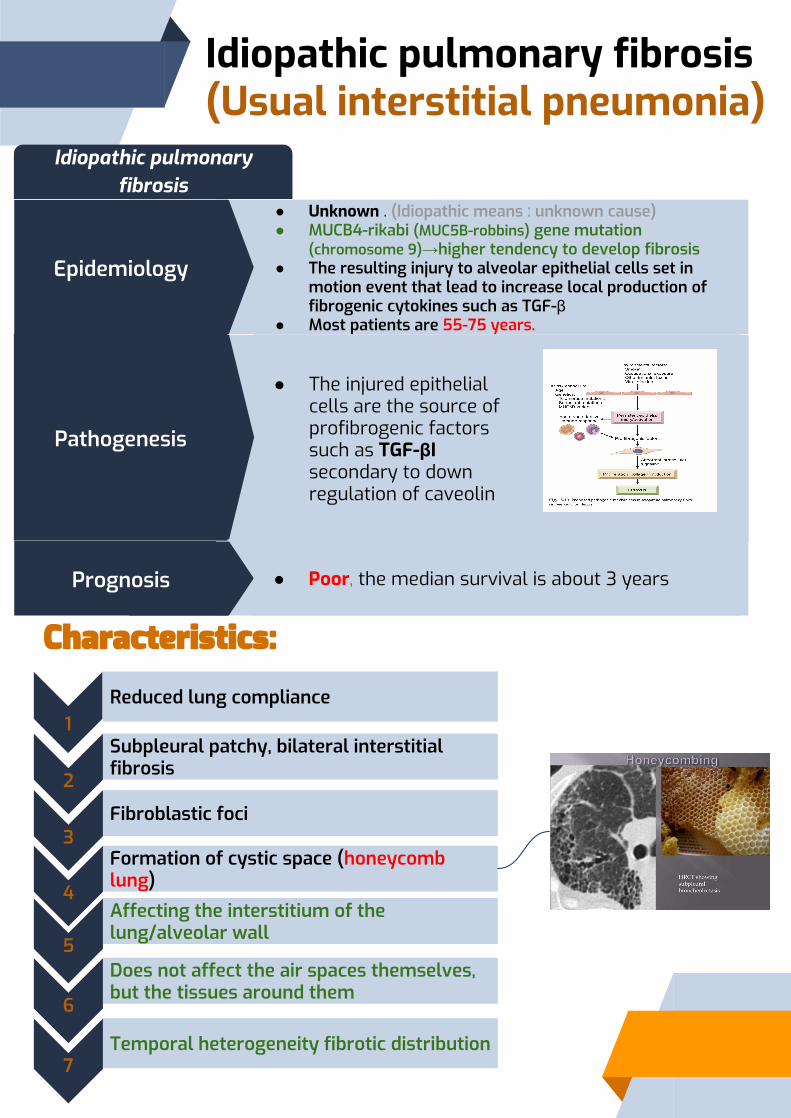
Idiopathic pulmonary fibrosis (Usual interstitial pneumonia)
Idiopathic pulmonary fibrosis
● Unknown . ●
→●
β●
● Poor, the median survival is about 3 years
● The injured epithelial cells are the source of profibrogenic factors such as TGF-βI secondary to down regulation of caveolin

Complications
Hypoxia, peripheral edema
Cyanosis, cor-pulmonale
Clubbing
Gradual deterioration in pulmonary status despite medical treatment
Morphology
Honeycomb change, Fibrosis in the subpleural region
Gross:
Interstitial fibrosis
Microscopic:
Dry cough
X ray: Early:
.Advanced:
Clinical features
Idiopathic pulmonary fibrosis (Usual interstitial pneumonia)
Treatment:To delay progression of the disease we may use:
-Pirfenidone (TGF-b1 antagonists) -Nintedanib (tyrosine kinase antagonist)
Note: The only definitive treatment is lung transplantation.

The development of pneumoconiosis is dependent on: 1. amount of dust
b. Long period
2. size (1-5) um
3. solubility physiochemical activity. 4. other irritants, tobacco smoking
1Coal worker’s pneumoconiosis (CWP)
2Silicosis (most common)
3Asbestosis
Pathogenesis: The pulmonary alveolar macrophage is a key cellular element in the initiation and perpetuation of inflammation, lung injury and fibrosis.
lung disorders caused by inhalation of mineral dusts leading to lung damage.
Definition
.
Etiology
Pneumoconiosis
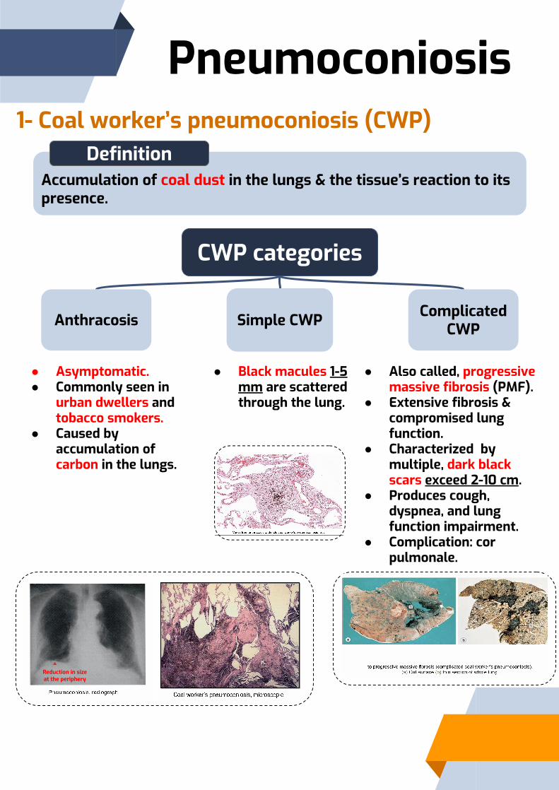
Accumulation of coal dust in the lungs & the tissue’s reaction to its presence.
Definition
CWP categories
Anthracosis
● Asymptomatic.● Commonly seen in
urban dwellers and tobacco smokers.
● Caused by accumulation of carbon in the lungs.
Simple CWP
● Black macules 1-5 mm are scattered through the lung.
Complicated CWP
● Also called, progressive massive fibrosis (PMF).
● Extensive fibrosis & compromised lung function.
● Characterized by multiple, dark black scars exceed 2-10 cm.
● Produces cough, dyspnea, and lung function impairment.
● Complication: cor pulmonale.
Reduction in size at the periphery
1- Coal worker’s pneumoconiosis (CWP)
Pneumoconiosis

2- Silicosis
Fibro-nodular lung disease caused by long term exposure to inhalation of crystalline silica particles (alpha-quartz or silicon dioxide).
Definition
1
Industrial exposure: mining of gold, tin, copper and coal, sandblasting, metal grinding, ceramic manufacturing.
2Stony-hard large fibrous scars
3Eggshell calcification
4Fibrous pleural plaques may develop
5
Predispose to lung cancer & tuberculosis1 for unknown reasons
1: silicosis depresses cell-mediated immunity, and crystalline silica may inhibit the ability of pulmonary macrophages to kill phagocytosed mycobacteria.
Scarring has contracted the upper lobe into a small dark
mass (arrow). Note the dense pleural thickening
concentrically arranged hyalinized collagen fibers surrounding an
amorphous center. The “whorled” appearance of the collagen fibers is quite distinctive for silicosis (HOW?
silica in sand contains quartz, which is fibrogenic)
Morphology
Pneumoconiosis

occupational exposure to asbestos is linked to parenchymal interstitial fibrosis.
Definition
Complicationslocalized fibrous plaques1, or, rarely,
diffuse fibrosis in the pleura
Pleural effusion
Pleural adhesions
Lung carcinoma (Bronchogenic carcinoma)
malignant pleural and peritoneal mesothelioma
Characterized by the presence of asbestos bodies (Ex:Ship-building industry), which are seen as golden brown, fusiform or beaded rods with a translucent center. Apparently they are formed when macrophages attempt to phagocytose asbestos fibers; the iron “crust” is derived from phagocyte ferritin.
Etiology
1: Are well-circumscribed plaques of dense collagen often containing calcium.2: A malignant tumor affecting the mesothelial cells in the pleura.
The risk for developing lung carcinoma is increased about 5-fold for asbestos workers; the relative risk for mesothelioma2, is more
than 1000 times greater. Concomitant cigarette smoking greatly increases the risk
for lung carcinoma but not for mesothelioma.
Asbestos bodies
severe interstitial fibrosis diffusely affecting the lower lobe of the lung.
Asbestos fibers causes bleeding > forms hemosiderin (prussian blue stain shows ferruginous bodies)
3- Asbestosis
Pneumoconiosis

Granulomatous diseases
1- Sarcoidosis
Immunological multisystem disease of unknown etiology (thought to be
autoimmune) characterized by noncaseating granulomatous inflammation in many tissues and organs.
Definition
Characteristics
● Lungs● Predominantly, Intrathoracic hilar
and paratracheal lymph nodes● Skin● Eyes
● Affecting all races & both sexes equally
● Unpredictable, It can be progressive & chronic.● It may present as episodes of activity. ● Majority of the patients respond well to treatment.
Morphology
Bilateral hilar lymphadenopathy
Non-Necrotizing interstitial granuloma
Symptoms include: 1- Uveitis
2- arthritis3- dryness of mouth4- lack of lacrimation
-Sarcoidosis is often confused with TB (the distinction is that there is no caseation.)

Associated with
Definition
● Micropolyspora faeni in hay● Thermophilic actinomycetes● microsporum→ extrinsic allergic alveolitis
● Birds● pigeons
Disease
● Thermophilic bacteria ● Source of antigen: Desert cooler
Antigen / Source of antigen
● Sugarcane bagasse
Granulomatous diseases
2-

Morphology
(Poorly defined granulomas)
●●●●●●
Granulomatous diseases
2-
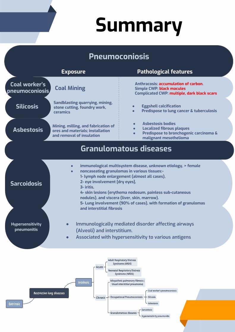
Exposure Pathological features
Coal MiningAnthracosis: accumulation of carbon.Simple CWP: black macules Complicated CWP: multiple, dark black scars
Sandblasting quarrying, mining, stone cutting, foundry work, ceramics
● Eggshell calcification● Predispose to lung cancer & tuberculosis
Mining, milling, and fabrication of ores and materials; installation and removal of insulation
● Asbestosis bodies● Localized fibrous plaques ● Predispose to bronchogenic carcinoma &
malignant mesothelioma
Summary
Coal worker’s pneumoconiosis
Silicosis
Asbestosis
Pneumoconiosis
●
●
Hypersensitivity pneumonitis
Granulomatous diseases
● immunological multisystem disease, unknown etiology, > female● noncaseating granulomas in various tissues:-
1- lymph node enlargement (almost all cases), 2- eye involvement [dry eyes], 3- iritis, 4- skin lesions (erythema nodosum, painless subcutaneous nodules), and viscera (liver, skin, marrow).5- Lung involvement (90% of cases), with formation of granulomas and interstitial fibrosis
Sarcoidosis

Diagnostics:- Radiology (x-ray, CT)- Spirometry- Cytology (sputum, bronchial brushing/washing, bronchoalveolar lavage) - Biopsy: Endobronchial, transbronchial, open lung biopsy, VAT (video assisted
thoracoscopic)
Definition: Group of diseases (of various etiologies) characterized by decreased lung volume and compliance. Spirometry: FEV1 and FVC decreased (ratio is normal)Symptoms:-Chronic dry cough-Dyspnea (varying in severity)
Complication:Cor pulmonale (Pulmonary hypertension→25+mmHg in the pulmonary artery)
Etiologies:1- Thoracic cage deformity: - decreased lung expansion -Severe kyphoscoliosis -Guillain-Barré syndrome ; weakens intercostal muscles2- Idiopathic pulmonary fibrosis:-Familial- Affecting the interstitium of the lung/alveolar wall- Do not affect the air spaces themselves, but the tissues around them -Honeycombed lung due to entrapped air (anthracosis→low po2 high pco2) -Affects lower part of the lung -Bilateral peripheral reticulation—>fibrosis shrinks the lung (trapped air→dilated alveoli) -Temporal heterogeneity fibrotic distribution- Histological: Blue stain indicating prevalent connective tissue (Masson’s trichrome stain) -Associated with usual interstitial pneumoniaPathogenesis: - Injury affecting macrophages leading to cytokine release - MUCB4 gene mutation (chromosome 9) mutation→higher tendency to develop fibrosis -shorter telomeres (Reduced genes that encode for telomerase)→shorter cell life (type I pneumocytes)→senescence + apoptosis→when they die they secrete: - TGF-b1→fibrogenic→stimulating fibroblasts and myofibroblasts > collagen - Low Caveolin (inhibits TGF-b1> there will be nothing to counteract it )Treatment: -Perfinidone (TGF-b1 antagonists) - -Nentedanib (tyrosine kinase antagonist)

3- RDS(Adult/Neonatal), DAD(diffuse alveolar damage), HMPD(hyaline membrane pulmonary disease)Causes:
-Very severe dyspnea and hypoxia- Very severe road traffic accident, major surgery, aspiration of gastric content, C section, severe acute pancreatitis, hypovolemic shock, septicemia-70% die
Effect: -Edema in the lung→atelectasis→collapseThe 30% who survive end up with chronic pulmonary fibrosis Morphology: CT scan→White lung syndrome Fibrin and debris→form a hyaline membrane around the alveoli
Risk factors of NRDS (surfactant deficiency):-Premature neonates (<36 weeks)- Multiple pregnancies -Maternal diabetes- C-section -Amniotic fluid aspiration
4- Atypical pneumonia - Could lead to interstitial pneumonitis
- Usually caused by Influenza virus - Edema in the interstitium and chronic
inflammatory infiltration -Inflammatory cells are lymphocytes (viral
infection), and not neutrophils
5- Pneumoconiosis - Caused by inhaling mineral dust>1-5mm in
diameterCoal→coal worker’s pneumoconiosis
Silica→Silicosis (most common) (silica in sand contains quartz, which is fibrogenic)
- Building industry -Concentric fibrosis
-Higher risk of TB for unknown reasonsAsbestos→Asbestosis
-Carcinogenic (mesothelioma) - Ship-building industry
- Asbestos fibers causes bleeding > forms hemosiderin (prussian blue stain shows
ferruginous bodies)
6- Drug addiction -Heroin
- Amiodarone (antiarrythmic)
7- Sarcoidosis -Idiopathic but now thought to be autoimmune -Symptoms include Uveitis, arthritis, dryness of
mouth, lack of lacrimation, etc - Often confused with TB
(the distinction is that there is no caseation)
8 -Hypersensitivity pneumonitis (extrinsic allergic alveolitis)
-Sensitivity to inhaled organic material -Ill defined granulomas (poor granulomas)
- Especially in upper lobesCauses:
- Pigeons - Desert cooler - Incense - Birds -Farmer’s lung→microsporum→ extrinsic allergic
alveolitis

Quiz1-D | 2- C | 3- B | 4- E
Answer Explanation File
4) A 62-year-old woman is rushed to the emergency room following an automobile accident. She has suffered internal injuries and massive bleeding and appears to be in a state of profound shock. Her temperature is 37°C (98.6°F), respirations are 42 per minute, and blood pressure is 80/40mmHg. Physical examination shows cyanosis and the use of accessory respiratory muscles. A CT scan of the chest is normal on arrival. Her condition is complicated by fever, leukocytosis, and a positive blood culture for staphylococci (sepsis). Two days later, the patient develops rapidly progressive respiratory distress, and a pattern of “interstitial pneumonia” can be seen on a chest X-ray. Which of the following is the most likely diagnosis?(A) Acutebronchiolitis(B) Alveolarproteinosis(C) Atelectasis(D) Desquamative interstitial pneumonitis(E) Diffuse alveolar damage

Thank you
●●●●●●●●●●
●●
●●●●●●●●●●●●


![[5] HEMA - Megaloblastic Anemia.pdf - KSUMSC](https://static.fdokumen.com/doc/165x107/631deac95ff22fc7450674ca/5-hema-megaloblastic-anemiapdf-ksumsc.jpg)