
Blistering Disorders - KSUMSC
16
Blistering Disorders Objectives were not in Dr. Hadeel’s slides. The lecture was given by Dr. Hend Al Otaibi and she only focused on the important points. Team leader: Ghada AlHadlaq Members: Rana Barasain Revised by: Shrooq Alsomali Template by group B Before you start.. CHECK THE EDITING FILE Sources: doctor’s slides and notes + 436 group B team [ Color index: Slides| Slides| Important|doctor notes|Extra]
-
Upload
khangminh22 -
Category
Documents
-
view
6 -
download
0
Transcript of Blistering Disorders - KSUMSC

Blistering Disorders
Objectives were not in Dr. Hadeel’s slides. The lecture was given by Dr. Hend Al Otaibi and she only focused on the
important points.
Team leader: Ghada AlHadlaq Members: Rana Barasain Revised by: Shrooq Alsomali Template by group B Before you start.. CHECK THE EDITING FILE Sources: doctor’s slides and notes + 436 group B team
[ Color index: Slides| Slides| Important|doctor notes|Extra]

Preview: ● Vesicle: an elevation that contains clear fluid ( < 5cm in diameter) ● Bulla: Localized fluid collection “large vesicle” ( > 5 cm in diameter)
This junction is important for blistering diseases. *Dr recommended watching a youtube video
Desmosome: Desmoplakin: between the walls of the cells - Desmoglein: intracellular between the cells Laminin 5: between the dermis and epidermis | Hemidesmosome: between anchoring filament & keratinocyte
Classification Of Vesiculo-bullous Diseases (based on location):
Subcorneal Blisters Intraepidermal Blisters Subepidermal Blisters
- Just beneath the stratum corneum. Very superficial. - Have thinner roofs. - Rupture easily. - Leave an oozing denuded surface. like bullous impetigo, it has subcorneal blisters that ruptures easily.
- Within the prickle cell layer of the epidermis. (spinous layer)
- Have thin roofs. - Rupture easily. Still superficial.
- Leave an oozing denuded surface. Like eczema
- Between the epidermis and dermis. basement
membrane, or dermo epidermal junction.
- Their roofs are relatively Thick - Tend to be tense - May contain blood
Blistering Disorders In Adults Blistering Disorders In Children
The main group of blistering disorders is associated with auto-antibody formation.
Genodermatosis, (epidermolysis bullosa), associated mainly with mechanical defects in and around the basement membrane zone.

Blistering Disorders (Diagnosis in General) - Accurate pathological diagnosis requires a biopsy of a small newly formed lesions and perilesional skin of immunopathological studies.
- In the case of blisters in children, electron microscopy may be required.
Diagnostic tests: 1. Routine Histology: lesional sample in formalin. 2. Direct immunofluorescence – Perilesional skin. 3. Indirect immunofluorescence – patient’s serum is added to specific substrates that express antigen of interest. 4. Electron microscopy.
Blistering Disorders In Children: Epidermolysis Bullosa (Read only) Group of mechanobullous genodermatosis Rare Usually present at birth or infancy Range from localized relatively mild trauma induced blisters to life threatening/debilitating conditions Diagnosis is made based on family history, clinical examination, light and electron microscopy
The main subsets are: 1-Epidermolysis bullosa simplex: mainly autosomal dominant 2-Junctional epidermolysis bullosa: autosomal recessive 3-Dystrophic epidermolysis bullosa: both autosomal dominant and autosomal recessive
1- Epidermolysis Bullosa Simplex: - Majority are autosomal dominant. - The pathological damage lies within the epidermis. - The main defect lies in defective genes coding for keratin 5
and 14 in the basal layer. - Blisters may be present at birth, or when the child starts to
walk or crawl, and they tend to develop mild blistering on the knees, hands, feet and other sites of friction.
- The blisters quickly rupture and heal with no scarring.
2- Junctional epidermolysis bullosa: - Autosomal recessive - The gene which is abnormal is:
1- Laminin 5, in 2 types of Junctional EB 2- 𝛂6 β4 in the third type
- Split is at the level of the lamina lucida - Clinical features may be present at birth either as blisters
(often around nails) or as raw denuded areas. - Mucous membranes may be severely involved - Teeth are commonly abnormal.

3- Dystrophic epidermolysis bullosa: - Autosomal dominant or autosomal recessive - All types are associated with defects in type 7 collagen gene
which causes defective anchoring fibrils - Squamous cell carcinoma may develop on scar sites - In the dominant variant, blisters develop later in infancy or
early childhood on friction sites and heal with scarring Hair and teeth develop normally - In the recessive variant, large bullae are present at birth and
they heal with scarring Mucous membranes, hair, nails and teeth may be abnormal
Treatment of Epidermolysis Bullosa: - Team management - Biopsies - Prevention of frictional bullae - Occupational therapy - Dental care - Skilled nursing care
Staphylococcal scalded skin syndrome (SSSS) Talked about it in prev. lecture - Caused by an epidermolytic toxin of certain types of Staph aureus, which splits the
epidermis at the level of the granular layer by cleaving desmoglein 1. pemphigus diseases also attack desmoglein.
- Commoner in children - Rapidly expanding shallow blisters which rupture quickly leaving painful raw areas - Swabs need to be taken and sent for bacterial cultures - Treatment is with systemic anti staphylococcal antibiotics - Dressings - Heal without scarring

Autoimmune Bullous Diseases Classification and Overview Lecture actually starts here.
Pemphigus group:
▪ Loss of intraepidermal adhesion
Pemphigoid group:
▪ Loss of subepidermal adhesion
1. Pemphigus vulgaris (PV): a. Classic b. Pemphigus vegetans
sometimes present to dentists because of painful oral ulcers
1. Pemphigoid a. Bullous pemphigoid b. Cicatricial pemphigoid c. Pemphigoid gestationis
2. Pemphigus foliaceus: a. Classic b. Fogo selvagum rare c. Pemphigus erythematosus (Senear- Usher Syndrome)
2. Dermatitis herpatiformis
3. Drug Induced pemphigus 3. Linear IgA disease a. Of Childhood b. Adult form
4. Paraneoplastic pemphigus 4. Epidermolysis bullosa aquisita
5. IgA pemphigus
Pemphigus Bullous Pemphigoid Dermatitis Herpetiformis
Age middle age old primarily adults
General health poor good itchy
Circulating antibodies
IgG to intercellular adhesion proteins
IgG to basement membrane region
IgG to endomysium and transglutaminase
Fixed antibodies IgG in intercellular space IgG at basement membrane IgA granular deposits in papillary dermis
Nature of blisters superficial, and flaccid tense, and blood filled Small, excoriated and grouped
Site of blisters trunk, flexures, and scalp often flexural elbows, knees, upper back, buttocks
Blisters in mouth common rare rare
Treatment -Steroids -Immunosuppressives
-Steroids -Immunosuppressives
-Dapsone -Sulfapyridine -Gluten free diet
Pemphigus vulgaris Bullous pemphigoid Appearance
Age Younger Older
Mucus membrane involvement yes Rare
Autoantibodies Against desmoglein 3 Against hemidesmosomes
Blister location Intraepidermal (superficial) Subepidermal (deep)
Blister quality Flaccid, rupture easily Tense and firm
Nikolsky’s sign Nikolsky positive Nikolsky negative
prognosis poor Favorable
The Pemphigus Group:
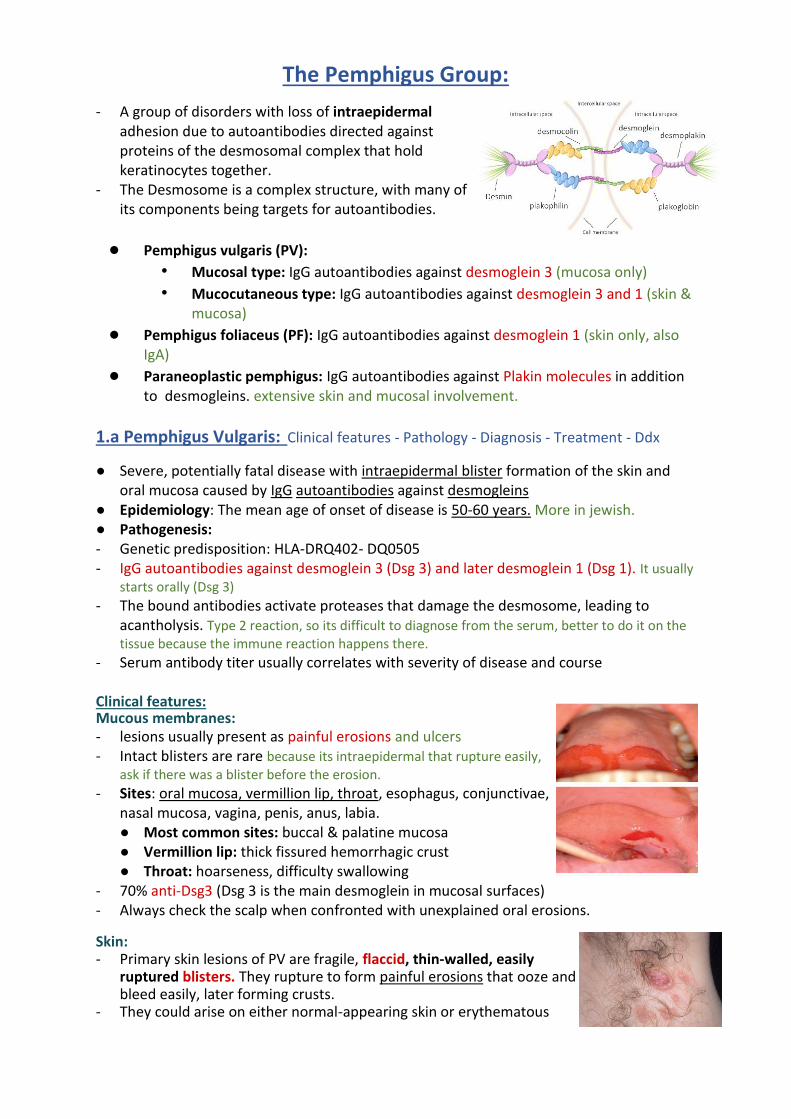
- A group of disorders with loss of intraepidermal adhesion due to autoantibodies directed against proteins of the desmosomal complex that hold keratinocytes together.
- The Desmosome is a complex structure, with many of its components being targets for autoantibodies.
● Pemphigus vulgaris (PV):
• Mucosal type: IgG autoantibodies against desmoglein 3 (mucosa only)
• Mucocutaneous type: IgG autoantibodies against desmoglein 3 and 1 (skin & mucosa)
● Pemphigus foliaceus (PF): IgG autoantibodies against desmoglein 1 (skin only, also IgA)
● Paraneoplastic pemphigus: IgG autoantibodies against Plakin molecules in addition to desmogleins. extensive skin and mucosal involvement.
1.a Pemphigus Vulgaris: Clinical features - Pathology - Diagnosis - Treatment - Ddx
● Severe, potentially fatal disease with intraepidermal blister formation of the skin and oral mucosa caused by IgG autoantibodies against desmogleins
● Epidemiology: The mean age of onset of disease is 50-60 years. More in jewish. ● Pathogenesis: - Genetic predisposition: HLA-DRQ402- DQ0505 - IgG autoantibodies against desmoglein 3 (Dsg 3) and later desmoglein 1 (Dsg 1). It usually
starts orally (Dsg 3) - The bound antibodies activate proteases that damage the desmosome, leading to
acantholysis. Type 2 reaction, so its difficult to diagnose from the serum, better to do it on the tissue because the immune reaction happens there.
- Serum antibody titer usually correlates with severity of disease and course
Clinical features: Mucous membranes: - lesions usually present as painful erosions and ulcers - Intact blisters are rare because its intraepidermal that rupture easily,
ask if there was a blister before the erosion. - Sites: oral mucosa, vermillion lip, throat, esophagus, conjunctivae,
nasal mucosa, vagina, penis, anus, labia. ● Most common sites: buccal & palatine mucosa ● Vermillion lip: thick fissured hemorrhagic crust ● Throat: hoarseness, difficulty swallowing
- 70% anti-Dsg3 (Dsg 3 is the main desmoglein in mucosal surfaces) - Always check the scalp when confronted with unexplained oral erosions.
Skin: - Primary skin lesions of PV are fragile, flaccid, thin-walled, easily
ruptured blisters. They rupture to form painful erosions that ooze and bleed easily, later forming crusts.
- They could arise on either normal-appearing skin or erythematous

base. - Can become generalized or localized. - Lesions that heal often leave hyperpigmented patches with NO
scarring. - More generalized disease due to the development of IgG
autoantibodies against Dsg1 in the skin along with Dsg3.
Pathology:
- Intraepidermal blister formation due to loss of cell-cell adhesion of keratinocytes (acantholysis) without keratinocyte necrosis. This is how we differentiate between it and steven/TEN.
- They maintain their attachment to the basement membrane via hemidesmosomes, this giving the appearance of “row of tombstones”
- Mild dermal perivascular infiltrates
Diagnostic Approach: - History: always ask medication Hx - Physical examination: skin, mucous membranes, nails
● Nikolsky sign: because of an absence of cohesion within the epidermis, its upper layers easily move laterally with slight pressure or rubbing in active patients with pemphigus. Its positive, but not due to detachment of the dermo epidermal junction.
● Asboe-Hansen sign: “bulla- spread phenomenon” gentle pressure on an intact bulla forces the fluid to spread under the skin away from the site of pressure
- Investigation ● Skin biopsy: from lesional skin, intact
vesicles if found ● DIF: from perilesional skin shows
deposition of IgG (100%), C3 (80%) ● Indirect IF ● ELISA: to identify anti-Dsg3,1
Treatment: - Systemic corticosteroids are the mainstay of therapy for pemphigus and
immunosuppressive agents or immunomodulators are often used for their steroid sparing effect in order to reduce the side effects of the corticosteroids
- Prednisone at 1.0 mg/Kg/day (usually 60 mg/day) is a typical initial dosage - The therapeutic effects are clinically estimated by the number of new blisters per day
and the rate of healing of new lesions, and then the prednisone is gradually tapered
Immunosuppressive agents in combination with oral prednisone:
Azathioprine IVIG Rituximab for severe cases Cyclosporine
Pulse methylprednisolone
Cyclo - phosphamide
Mycophenolate mofetil Extracorporeal photopheresis
Topical treatments: Corticosteroids Antibiotics Immunomodulators (e.g. topical tacrolimus) Differential Diagnosis:
When skin is involved When mucous membranes is involved
• Bullous impetigo • Denture intolerance

• Dyskeratotic acanthoytic disorders: - Hailey-Hailey - Grover disease
• Erosive candidiasis
• Chronic recurrent aphthae
• Erythema multiforme
• Erosive lichen planus
• Herpetic ginigivitis
1.b. Pemphigus Vegetans: - Is a rare vegetative variant of pemphigus vulgaris
Clinical Features: The difference is in the location and primary lesion.
- Characterized by flaccid blisters that become erosions and then form fungoid vegetations, especially in intertriginous areas, the scalp and face.
- Early lesions start as pustules (rather than vesicles), then they soon progress to vegetative plaques, with thick crust. All types other than vulgaris have crusts.
Treatment: same as pemphigus vulgaris
2.a Pemphigus Foliaceus:
- Is a form of pemphigus in which patients develop scaly, crusted cutaneous erosions often on an erythematous base.
- Disease of middle-aged and older patients
Clinical Features:
- In this form of pemphigus they do not have mucosal involvement even with widespread disease because the IgG autoantibodies are against desmoglein 1.
- Lesions have a seborrheic distribution (face, scalp, and upper trunk) and they form crusts. The picture is extra.
- More often drug induced than pemphigus vulgaris. - Patients with pemphigus foliaceus are not severely ill.
Diagnostic Approach: - History: always ask medication Hx - Physical examination: skin, mucous membranes, nails
● Nikolsky sign: present - Investigation
● DIF: from perilesional skin shows superficial deposition of IgG ● ELISA: to identify IgG antibodies against Dsg 1
Treatment: - Same as pemphigus vulgaris but usually more responsive to therapy - Dapsone may be helpful which is directed against neutrophils.
Extra pic

2.b. Pemphigus Erythematosus: (Senar-Usher Syndrome)
- Localized variant of Pemphigus foliaceus. Ddx of rosacea, or SLE - Scaly, crusted lesions of pemphigus foliaceus over the malar
region and in other seborrheic areas. - Patients have immunologic features of both Lupus erythematosus
and pemphigus (i.e. IgG and C3 deposition on cell surface of keratinocytes as well as the basement membrane zone, in addition to circulating ANA), but It does not meet criteria of Diagnosis of lupus.
- Very rare, only reported in few patients.
3. Drug Induced Pemphigus: - Most patients go into remission after the offending drug is discontinued.
Drugs that induce pemphigus can be divided into two groups:
Agents with the sulfhydryl group Agents without the sulfhydryl group
• Penicillamine: PF is seen more than PV, ratio 4:1
• Captopril
• Piroxicam
• Sulfhydryl group of these drugs interacts with the sulfhydryl groups of Dsg1 & Dsg 3 (acantholysis without antibody formation)
• Beta- blockers
• Cephalosporines
• Penicillins
• Rifampin
• Induce acantholysis via immune mechanisms
4. IgA Pemphigus: All other pemphigus and caused by IgG, and its pemphigus because of
its intra epidermal location. Represents a group of autoimmune intraepidermal blistering diseases presenting with:
1. Vesiculopustular eruption
2. Neutrophilic infiltration of the skin so we can use dapsone!
3. Circulating IgA autoantibodies against the cell surface of keratinocytes, but NO IgG autoantibodies.
Two distinct types: 1- Subcorneal pustular dermatosis (SPD) 2- Intraepidermal neutrophilic type (IEN) - Both types present with flaccid vesicles or
pustules that coalesce to form an annular pattern with central crusting
- Sunflower-like configuration of pustules is a characteristic sign of the IEN type - Most common site: axilla, groin, trunk - NO mucous membrane involvement - Pruritus is a significant symptom
Diagnostic Approach:

- History and Physical examination (skin, mucous membrane, nails) - Investigation ● DIF: IgA autoantibodies directed against keratinocyte cell surface
Treatment: Most cases are responsive to dapsone because of neutrophilic infiltration. If not: corticosteroids & other immunosuppressive agents. 5. Paraneoplastic Pemphigus:
- Associated with underlying neoplasms, both benign and malignant - Most commonly associated neoplasms: rare cancer types
○ Non-Hodgkin lymphoma
○ Chronic lymphocytic leukemia
○ Castleman’s disease
○ Malignant and benign thymomas - Not associated with common tumors such as adenocarcinomas and SCC
Clinical features: extensive, severe, hemorrhagic
- The most constant clinical feature is the presence of intractable stomatitis.
- The Stomatitis consists of erosions and ulcerations that affect all layers of the oropharynx and characteristically extend onto the Vermilion lip
- Stomatitis is usually the earliest presenting sign, severe, the one that persists after treatment and is extremely resistant to therapy.
- Pseudomembranous conjunctivitis: scarring, blindness. - Could also affect: esophagus, nasopharynx, vagina, labia, penis. - Cutaneous findings are polymorphic always a hint for paraneoplastic: ● Erythematous macules ● Flaccid blisters and erosions (resembling pemphigus) ● Tense blisters (resembling pemphigoid) ● Erythema multiforme like lesions ● Lichenoid eruptions
- Histology is rarely helpful
Treatment:
- Treat the underlying tumor - Benign tumors: it may take 6-18 months to see complete resolution of lesions after
excision of benign neoplasms - Malignant tumors: ● No consensus on a standard effective therapeutic regimen ● Cutaneous lesions respond more rapidly than the stomatitis, which is refractory to
treatment - Prognosis of paraneoplastic pemphigus is poor due to its resistant nature to treatment

Pemphigoid Group
1.a Bullous Pemphigoid (BP): - The most common autoimmune subepidermal blistering disease, caused by autoantibodies
against hemidesmosomes in the basement membrane zone (BMZ). The blisters are tense. - Predominantly affects the elderly.
Pathogenesis: Tissue-bound and circulating autoantibodies directed against two hemidesmosomal proteins:
○ BPAG 1“ BP230” (bullous pemphigoid antigen 1)
○ BPAG 2 “BP180” (bullous pemphigoid antigen 2) is most likely to be more involved in the initial immune response, since it is transmembrane.
Drug-induced bullous pemphigoid:
Diuretics (furosemide)
D-penicillamine Antibiotics (amoxicillin, ciprofloxacin)
Potassium iodide
Clinical features: - Intensely pruritic eruption with widespread blister formation, remember
pemphigus was painful not pruritic. - In early stages and atypical variant: excoriated, eczematous, urticarial lesions,
can stay a long time without blistering. - Always keep BP in mind when confronted with an elderly patient with
persistent urticarial lesions. - Mucosal involvement in < 20 %. Cicatricial: skin is 25% involved (opposite) - Non-bullous phase: cutaneous manifestations are non-specific & polymorphic
(pruritus, excoriations, eczematous, urticarial lesions) - Bullous phase: characterized by the development of vesicles and bullae on
normal or erythematous skin along with urticarial lesions. Blisters are stable and tense.
- Bullae predominate on the flexural aspects of the limbs and the lower trunk (extremities)
Diagnostic Approach:
-The diagnosis of BP is based upon the clinical presentation, histological features and positive findings on direct and indirect immunofluorescence - History and Physical examination - Investigations: The following are elevated in 60% of patients with BP ● Increased eosinophils on CBC, and increased ESR and IgE.
These also are increased in eczema or urticaria, that's why we do biopsy.
● Skin biopsy: ● Non-bullous phase: non-specific, eosinophilic inflammatory
infiltrate ● Bullous phase: subepidermal blister, accompanied by a dermal
inflammatory infiltrate composed of eosinophils.
● DIF: from perilesional, uninvolved skin, linear, continuous deposits of IgG and C3 along the epidermal basement membrane
● Indirect IF: using salt-split human skin ● ELISA: identifies antibodies against both BP 230 & 180 in 60-80% of patients
Treatment
Mild/localized Disease Extensive/persistent Cutaneous Disease
• Superpotent topical corticosteroids
• Dapsone
• Topical immunomodulators (tacrolimus)
• Superpotent topical corticosteroids
• Oral corticosteroids
• Azathioprine
• Methotrexate
1.b Cicatricial Pemphigoid: Cicatricial because it causes scarring.
- Is a chronic, autoimmune, subepithelial blistering disorder characterized by a predominant involvement of the external mucosal surfaces (mainly oral & conjunctival mucosa, but it could affect any mucosal site) and a tendency for scarring.
- Patients > 65 years
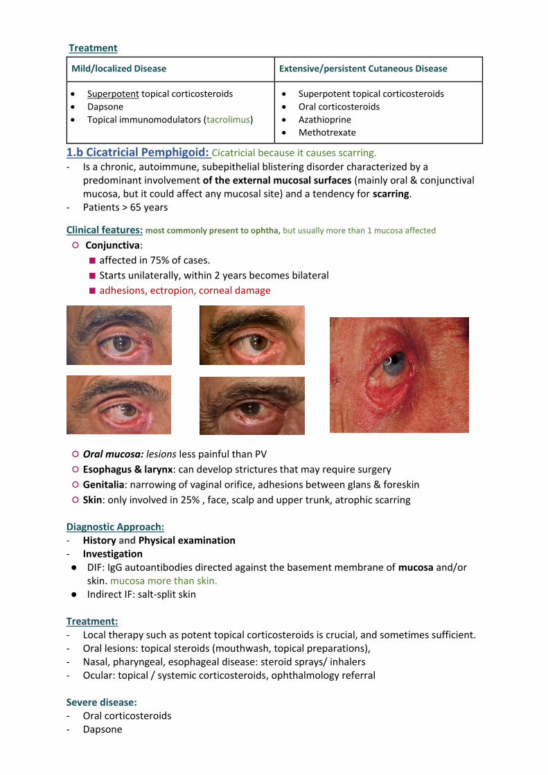
Clinical features: most commonly present to ophtha, but usually more than 1 mucosa affected
○ Conjunctiva:
■ affected in 75% of cases.
■ Starts unilaterally, within 2 years becomes bilateral
■ adhesions, ectropion, corneal damage
○ Oral mucosa: lesions less painful than PV
○ Esophagus & larynx: can develop strictures that may require surgery
○ Genitalia: narrowing of vaginal orifice, adhesions between glans & foreskin
○ Skin: only involved in 25% , face, scalp and upper trunk, atrophic scarring Diagnostic Approach: - History and Physical examination - Investigation ● DIF: IgG autoantibodies directed against the basement membrane of mucosa and/or
skin. mucosa more than skin. ● Indirect IF: salt-split skin
Treatment: - Local therapy such as potent topical corticosteroids is crucial, and sometimes sufficient. - Oral lesions: topical steroids (mouthwash, topical preparations), - Nasal, pharyngeal, esophageal disease: steroid sprays/ inhalers - Ocular: topical / systemic corticosteroids, ophthalmology referral Severe disease: - Oral corticosteroids - Dapsone

- Cyclophosphamide - Azathioprine - Surgical therapy to treat the ectropion and adhesions.
1.c Pemphigoid Gestationis (Herpes gestationis): - A form of BP occuring during pregnancy - Occurs in 1/10000-40000 pregnancies. Rare - No maternal risk, no increase in birth defects. - However, pregnancy complications and fetal death occurs in 15-30% mostly IUGR.
Clinical features: - Erythematous urticarial plaques, alone or with papules, vesicles, blisters in sub-epidermal
area, erosions. Tense vesicle/bullae on erythematous plaque or urticaria, annular shaped. - Intense pruritus. - Sites: Abdomen, proximal extremities. - Rarely appears postpartum, resolve within 3 months. - Occasionally recurs with menses or ingestion of OCP, tends to be worse in next
pregnancy. - The antibodies cross the placenta, the newborn can have blisters for a few weeks. If the mother got the disease early in her pregnancy, the chances of the newborn being affected is higher than if she got it later on.
Diagnostic Approach: - History and Physical examination - Investigation
● Cbc & differential: eosinophilia ● DIF & indirect IF
Treatment: - Topical steroids - Systemic steroids: avoid in 1st trimester - Skin care: to prevent infection - Antihistamines: for tx of pruritus
2. Dermatitis Herpetiformis (DH): Herpetiformis because its a group of vesicles.
- Pruritic vesicular disease caused by IgA autoantibodies directed against epidermal transglutaminase. It is a differential for any patient that complains of itching.
- Characterized by granular IgA deposition at the basement membrane zone (BMZ). - It is also a cutaneous manifestation of celiac disease and is associated with gluten
sensitivity in virtually all cases. - DH and celiac disease are genetic disorders strongly associated with HLA-DQ2 genotype,
in which IgA antiendomysial antibodies are directed against tissue transglutaminases (in the skin: epidermal transglutaminase)
Clinical features: - Grouped ’herpetiform’ papules/vesicles/urticarial wheals over an erythematous base,
associated with intense pruritus, burning, stinging and excoriations. - Sites: extensor surfaces of elbows/knees, sacrum, buttocks, scalp. - Spontaneous remissions may occur, but disease is often lifelong.

Diagnostic Approach: - History and Physical examination - Investigations ● Skin biopsy: subepidermal blister, with neutrophilic micro-abscesses in the papillary
dermis is the hallmark of the disease. bullous pemphigoid: eosinophils. ● DIF: Granular deposits of IgA in the dermal papillae ● Indirect IF ● ELISA: 80% identifies IgA against transglutaminase. ● Jejunal Biopsy: flattening of the villi
Treatment - Gluten free diet - Dapsone. IgA always comes with neutrophils.
3. Linear IgA Disease: - Subepidermal tense blistering disease characterised by linear IgA deposition at the
basement membrane zone (BMZ). It affects children unlike the previous diseases. - May be Identical to DH but without GI involvement (GI disease is rare). - Resembles BP but the difference is IgA. - Over 50% have mucosal involvement.
If it resolved quickly, we call it Linear IgA, but if it persists, we call it Chronic bullous disease of childhood.
3.a The childhood form: - Occurs in children ”preschool”. - Characterized by annular erythema and tense
subepidermal blisters “crown of jewels”. - They develop predominantly in flexural areas.
(lower trunk, thigh, groin), axillae, face, mucous membranes.

- Usually resolves spontaneously, remits within 2-4 years.
Diagnostic Approach: - History and Physical examination - Investigations: ● DIF: linear IgA deposits along the basement membrane ● Indirect IF
Treatment: - Dapsone Remember: igA and neutrophils - Sulfapyridine - Antibiotics: Tetracycline, erythromycin, dicloxacillin. In the childhood age group, they’re
a good treatment and were found to be better than dapsone.
Questions: 1) From Where you will take a biopsy of Bullous pemphigoid? A. Erythematous area at periphery B. The blister itself C. Normal Skin D. Mucous Membranes 2) A case about pemphigus vulgaris then what drug cause this condition A. Digoxin B. Captopril C. Ibuprofen D. 3) 30-year-old male present s with erosion and crust on lips. He was diagnosed with lymphoma a month ago. What is the most likely diagnosis? A. Paraneoplastic pemphigus B. Scurvy C. Herpes labialis. 4) 12-year-old boy presented with vesicles and erosion look like cluster of jewels, what's the most likely diagnosis? A. Linear IgA dermatosis B. Bullous pemphigoid C. Pemphigus vulgaris D. Impetigo 5) Pt with erosions and vesicles, DIF was done and showes IgG & C3 deposition in the epidermis pattern, what’s most likely diagnosis A. Rosacea B. Dermatitis herpetiformis C. Pemphigus vulgaris D. Bullous pemphigus 6) A pregnant female with papulovesicular eruption involving abdomen and extremities, suspected to have herpes gestationis. Which of the following is a feature of Pemphigoid gestationis? A. It's a viral disease-

B. It's non itchy eruption- C. It starts in first trimester- D. It relapses with contraceptive pills- 7) A 30 year old lady referred urgently by the obstetrician with severe and extensive itching associated with widespread urticated plaques and two tense blisters on her left axilla, mucosal membranes are not involved- She is at 21 weeks of gestation with her second pregnancy- What is the most likely diagnosis? A. Acute urticaria B. Scabies infection C. Erythema multiforme D. Pemphigoid gestationis 8) A patient with pruritus (forgot the scenario) With neutrophilic microabscesses in the papillary dermis What's the management? A. Topical steroid B. Systemic steroid C. A diet containing rice D. A diet containing wheat Dx: Dermatitis herpetiformis 9) 21 year old male complaining of grouped itchy vesicles at his extensors, immunofluorescent shows positive granular IgA deposition in dermal papilla. Which of the following is the most likely diagnosis? A. Rosacea B. Dermatitis herpetiformis C. Pemphigus vulgaris D. Bullous pemphigus 10) Direct immunofluorescence of skin biopsy from 54 years old patient with blistering disease revealed intracellular deposit of IgG and C3. In which one of the following blistering diseases this immunopathology finding is seen? A. Bullous pemphigoid B. Pemphigus vulgaris C. Cicatricial pemphigoid D. Dermatitis herpetiformis
1 2 3 4 5 6 7 8 9 10
A B A A C D D C B B


![[5] HEMA - Megaloblastic Anemia.pdf - KSUMSC](https://static.fdokumen.com/doc/165x107/631deac95ff22fc7450674ca/5-hema-megaloblastic-anemiapdf-ksumsc.jpg)

