gametogenesis & female cycles - KSUMSC
31
Prof. Saeed Abuel Makarem
-
Upload
khangminh22 -
Category
Documents
-
view
2 -
download
0
Transcript of gametogenesis & female cycles - KSUMSC

Prof. Saeed Abuel Makarem
OBJECTIVES
By the end of the lecture, you should be able to:
Describe the female cycles (Ovarian & Uterine).
Define gametogenesis.
Differentiate the types of gametogenesis.
Describe the process of spermatogenesis.
Describe the process of oogenesis.
Health is a great valuable thing, we never
have a true idea of its value until we lose it.

Female Reproductive Cycles• The reproductive
cycles start at
puberty !.• Normally continues
until menopause.
• Reproductive cycles depend upon activities & coordination of:
1. Hypothalamus,
2. Pituitary gland,
3. Ovaries,
4. Uterus,
5. Uterine tubes,
6. Vagina and
7. Mammary glands.
OVARIAN AND UTERINE CYCLES
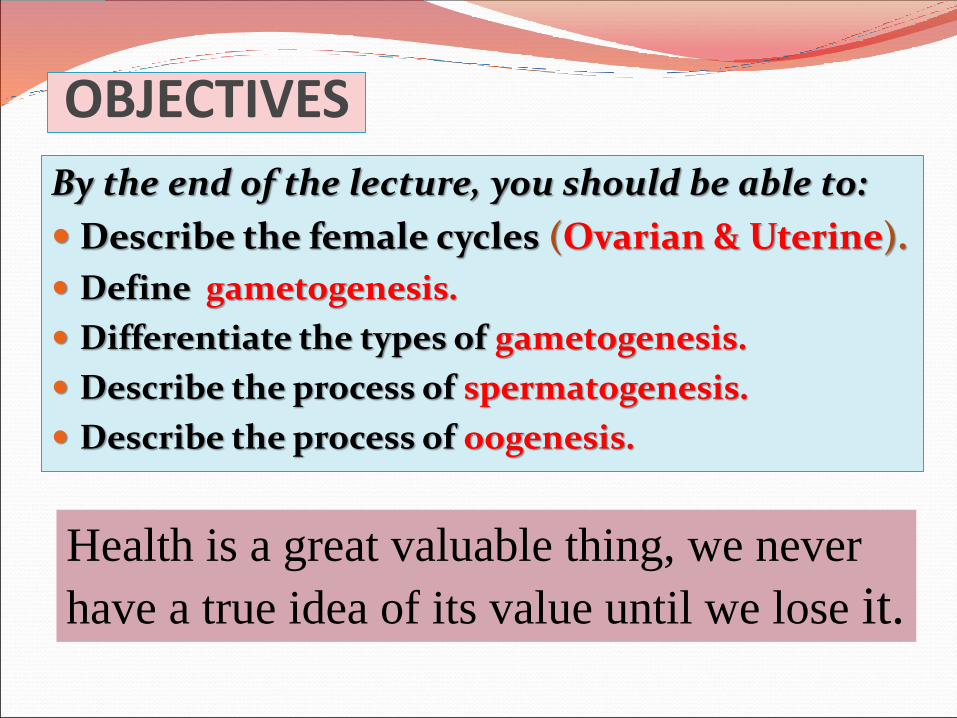
GnRH
• Gonadotrophin-releasing hormone (GnRH) is synthesized by neurosecretory cells of the Hypothalamus.
• Carried to the Pituitary gland (anterior lobe).
• It stimulates the pituitary to release Two Hormones that act on Ovaries (FSH& LH)

OVARIAN CYCLE • The ovarian cycle is under the control of the
Pituitary Gland.
• It is divided into 3phases: (FOL)
1- Follicular, phase.
2- Ovulatory, phase.
3- Luteal phase.• The ovarian cortex
contains hundreds of thousands of primary follicles (400,000 t0 500,000).
• Each consists of one primary oocyte encircled by single layer of flat follicular cells.
After puberty the simple flat follicular cells
become cuboidal, then columnar then
forming many layers around the oocyte.
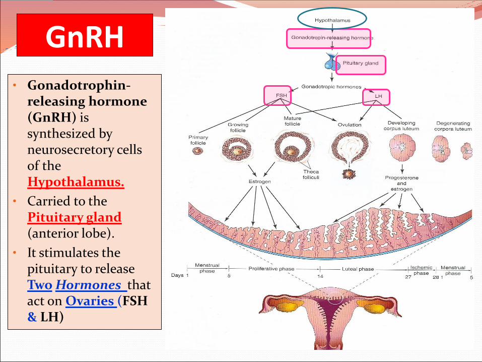
FSH• Follicle-
Stimulating Hormone .
• It is secreted by the pituitary gland.
• FUNCTIONS:
It stimulates the primary (ovarian) follicles:
1- To develop & mature.
2- To produce Estrogen by its follicular cells.

• The follicle becomes enlarged until it gets full maturity.
• It produces swelling on the surface of the ovary.
• Early development of ovarian follicle is induced by FSH.
• Final stages of maturation require LH (luteinizing hormone).
• LH. Also secreted by the pituitary gland.
• It causes ovulation(rupture of the mature follicle).
Growing follicles produce estrogen which
regulates the development and functions of
the reproductive organs.

Corpus Luteum• Now the remaining part of the ruptured follicle is called corpus luteum, (CL).
• It secretes Progesterone and small amount of Estrogen.
• These 2 hormones stimulate
endometrial glands to secrete and prepare the endometrium for implantation of the fertilized Ovum (Blastocyst).
• If the oocyte is fertilized the Corpus Luteum enlarges and remains till the 4th month of pregnancy.
• If the oocyte is not fertilized the corpus luteum involutes and degenerates in 10-12 days and called corpus albicans.
CL
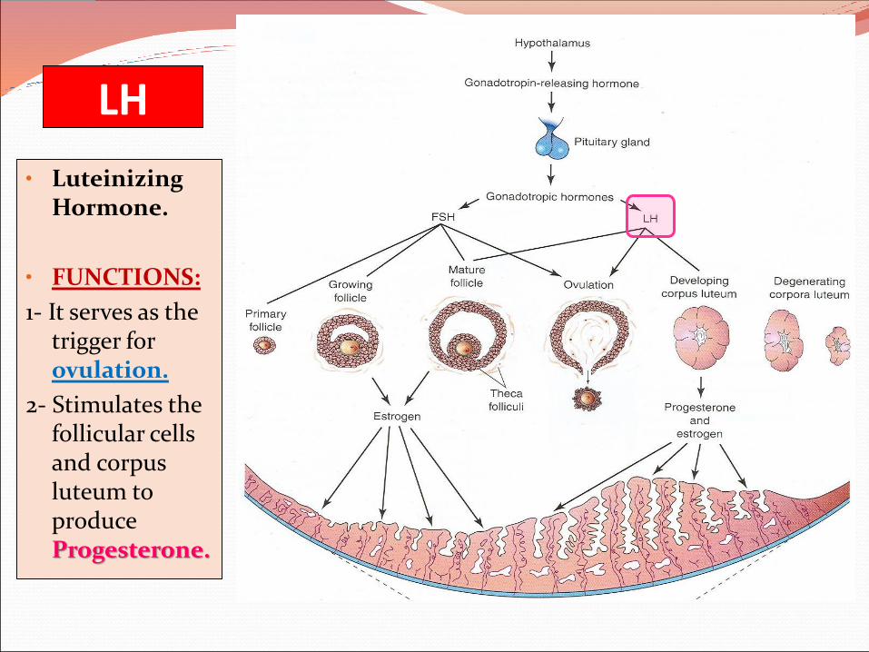
LH
• Luteinizing Hormone.
• FUNCTIONS:
1- It serves as the trigger for ovulation.
2- Stimulates the follicular cells and corpus luteum to produce Progesterone.

Uterine or Menstrual Cycle• It is the cyclic changes
which occur in the endometrium of the uterus by the effect of estrogen & progesterone.
• Average menstrual cycle is about 28 days.
• Day One is the day when menstrual blood flow begins.
• It varies by several days in normal women.
• Ranges between 23 and 35 days in 90 % of women.
• It sometimes varies in the same woman.

Phases of Menstrual CycleFour phases:
1. MenstrualPhase, (5 days).
2. Proliferative or FollicularPhase. (9 days).
3. Luteal Phase, (13 days).
4. Ischemic Phase, (1 day).
5 9 13ONE
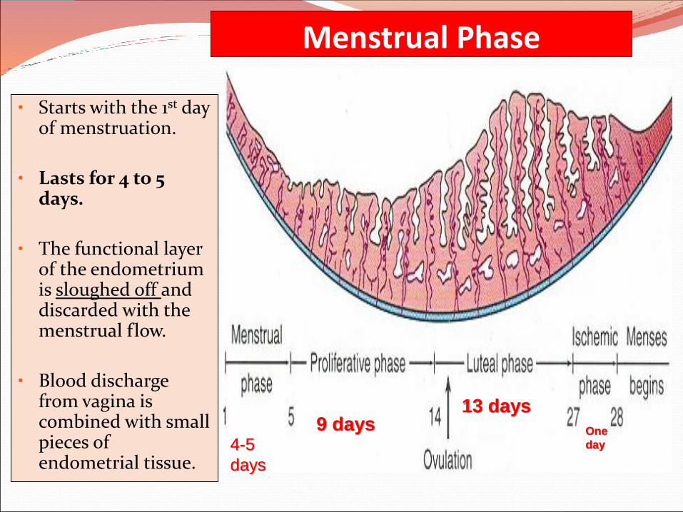
Menstrual Phase
• Starts with the 1st day of menstruation.
• Lasts for 4 to 5 days.
• The functional layer of the endometrium is sloughed off and discarded with the menstrual flow.
• Blood discharge from vagina is combined with small pieces of endometrial tissue.
4-5
days
9 days13 days
One
day

Proliferative Phase• It is a phase of repair
and proliferation.
• It lasts about 9 days.
• Coincides with growth of ovarian follicle.
• So it is controlled by Estrogen secreted by the follicular cells.
• Thickness of the endometrium is increased 2-3 times.
• The glands increase in number and length and the spiral arteries elongate.

Luteal Phase• It is a Secretory or Progesterone phase.
• It lasts about 13 days.
• Coincides with the formation, growth and functioning of the Corpus Luteum.
• Glandular epithelium secrete a material rich in glycogen.
• Endometrium increase in thickness under the influence of progesterone and estrogen.

Luteal Phase
• Spiral arteries grow into the superficial layer of the endometrium.
• Arteries become increasingly coiled.
• Large venous network develops.
• Direct arterio-venous anastomosisis a prominent features.

Ischemic Phase• Degeneration of the corpus
luteum leads to decrease in the levels of progesterone and estrogen which lead to:
1. Loss of interstitial fluid.
2. Marked shrinking of the endometrium.
3. Spiral arteries become constricted.
4. Venous stasis.
5. Ischemic necrosis.
6. Rupture of damaged vessel wall.
7. Blood seeps into the surrounding connective tissues.
8. Loss of 20-80 ml of blood.
9. Entire compact layer and most of the spongy layer of endometrium is discarded

It is the production of mature male & female gametes (Sperms & Ova).
Spermatogenesis:
It is the series of changes by which the primitive germ cells (spermatogonia) are transformed into mature sperms.
Oogenesis:
Sequence of events by which the primitive germ cells (oogonia) are transformed into mature oocytes.

It is the cell division that takes place in the germ cells to produce male & female gametes.
It consists of two cell
divisions, meiosis I & meiosis II,during which theDiploid number of chromosomes (46) is reduced to Haploid number (23).

At the beginning of meiosis I, (prophase) male & female germ cells replicate their DNA so that each of the 46 chromosomes is duplicated into sister Chromatid.

By the end of the first meiotic division, each new cell formed (Secondary Spermatocyte or Secondary Oocyte) has haploid (half) number of chromosome.
It is half number of chromosomes of the Primary Spermatocyte or primary Oocyte.

WHAT IS THE DIFFERENCE BETWEEN MITOSIS & MEIOSIS?
DIPLOID
HAPLOID

AIM:
Formation of sperms with haploid number of chromosomes.
SITE:
Seminiferous tubules of the testis.
TIME:
From puberty till old age.
DURATION:
About two months.
N.B.
Sperms are stored and become functionally mature in the Epididymis.
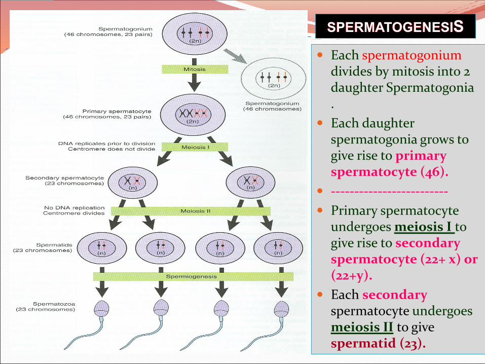
Each spermatogoniumdivides by mitosis into 2 daughter Spermatogonia .
Each daughter spermatogonia grows to give rise to primary spermatocyte (46).
-------------------------
Primary spermatocyte undergoes meiosis I to give rise to secondary spermatocyte (22+ x) or (22+y).
Each secondaryspermatocyte undergoes meiosis II to give spermatid (23).

It is change in shape
(metamorphosis) through which the Spermatids are transformed into mature Sperms:
1. Nucleus is condensed and forms most of the head.
2. Golgi apparatus forms the Acrosome, (acrosomal cap).
3. Mitochondria forms a spiral sheath.
4. Centriole elongates to form the axial filament.

OOGENESIS AIM:
Formation of secondary oocytes with haploid number of chromosomes.
SITE:
Cortex of the ovary
TIME:
Starts very early during fetal life becomes completedafter puberty & continues until menopause.
NB. It occurs monthly Except during pregnancy.

Before Birth: During early fetal life, primitive ova (Oogonia) proliferate by mitotic division and enlarge to form Primary Oocytes (46).
At Birth all primary oocytes have completed the prophase of the 1st
meiotic division and remain arrested at prophase and do not finish their first meiotic division until puberty.

After Puberty
Shortly before ovulation, the Primary Oocyte completes its first meiotic division (which was arrested at prophase) and give rise to Secondary oocyte (23) & First Polar Body.
The Secondary Oocyte receives almost all the cytoplasm.
The First Polar Body receives very little amount of cytoplasm.
It is small nonfunctional cell that soon degenerates.

At ovulation, the nucleus of the secondary oocyte begins the second meiotic division but progresses only to metaphasewhere division is arrested.

If the secondary oocyte is fertilized, the second meiotic division is completed otherwise it degenerates 24 hours after ovulation.
Most of the cytoplasm is retained by the Mature Oocyte(Fertilized Oocyte).
The rest is in the 2nd
Polar Body which soon degenerates.

During Fetal Life After puberty during each ovarian cycle
After fertilization
Proliferation: Each oogonium is divided by mitosis into 2 daughter oogonium with diploid number of chromosome,(44+XX
1st meiotic division is completed shortly before ovulation
2nd meiotic division is completed as the sperm penetrates the zona pellucida.
Growth:Oogonia enlarge to form primary oocyte with diploid number of chromosomes, (44+XX).
A reduction division by which the primary oocyte divided into 2ry oocyte (haploid number of chrmosome22+x, and first polar body which degenerates.
The secondary oocyte divides into mature ovum and 2nd polar body which degenerates.
Primary oocyte begins its 1st meiotic division witch arrest at prophase.
2nd meiotic division begins: at ovulation but stops at metaphase.
NB. NO PRIMARY OOCYTES ARE FORMED AFTER BIRTH

GOOD LUCK