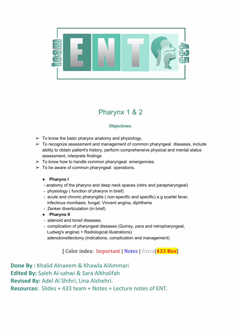
Pharynx 1 & 2 - KSUMSC
19
Pharynx 1 & 2 Objectives: ➢ To know the basic pharynx anatomy and physiology. ➢ To recognize assessment and management of common pharyngeal diseases, include ability to obtain patient's history, perform comprehensive physical and mental status assessment, interprets findings ➢ To know how to handle common pharyngeal emergencies. ➢ To be aware of common pharyngeal operations. ● Pharynx I - anatomy of the pharynx and deep neck spaces (retro and parapharyngeal) - physiology ( function of pharynx in brief) - acute and chronic pharyngitis ( non-specific and specific) e.g scarlet fever, infectious moniliasis, fungal, Vincent angina, diphtheria - Zenker diverticulation (in brief) ● Pharynx II - adenoid and tonsil diseases. - complication of pharyngeal diseases (Quinsy, para and retropharyngeal, Ludwig's angina) + Radiological illustrations) adenotonsillectomy (indications, complication and management) [ Color index: Important | Notes | Extra|433 Box ] Done By : Khalid Alnaeem & Khawla AlAmmari Edited By: Saleh Al-sahwi & Sara Alkhalifah Revised By: Adel Al Shihri, Lina Alshehri. Resources: Slides + 433 team + Notes + Lecture notes of ENT.
-
Upload
khangminh22 -
Category
Documents
-
view
3 -
download
0
Transcript of Pharynx 1 & 2 - KSUMSC
Pharynx 1 & 2
Objectives:
➢ To know the basic pharynx anatomy and physiology. ➢ To recognize assessment and management of common pharyngeal diseases, include
ability to obtain patient's history, perform comprehensive physical and mental status assessment, interprets findings
➢ To know how to handle common pharyngeal emergencies. ➢ To be aware of common pharyngeal operations.
● Pharynx I
- anatomy of the pharynx and deep neck spaces (retro and parapharyngeal) - physiology ( function of pharynx in brief) - acute and chronic pharyngitis ( non-specific and specific) e.g scarlet fever, infectious moniliasis, fungal, Vincent angina, diphtheria - Zenker diverticulation (in brief)
● Pharynx II - adenoid and tonsil diseases. - complication of pharyngeal diseases (Quinsy, para and retropharyngeal, Ludwig's angina) + Radiological illustrations) adenotonsillectomy (indications, complication and management)
[ Color index: Important | Notes | Extra|433 Box]
Done By : Khalid Alnaeem & Khawla AlAmmari Edited By: Saleh Al-sahwi & Sara Alkhalifah Revised By: Adel Al Shihri, Lina Alshehri. Resources: Slides + 433 team + Notes + Lecture notes of ENT.

Anatomy of the pharynx:
● It extends from the base of the skull to the level 6 cervical vertebra at the lower border of
cricoid cartilage.
● Funnel shaped, 10 cm length.
● Widest portion (5cm) at hyoid.
● Narrowest portion(1.5cm) at caudal end.
❖❖ Parts of the pharynx:
1- Nasopharynx (from the base of the skull till the soft palate)
● Opens anteriorly to the nose, ● Above: the base of skull ● Below: soft palate ● Laterally: ○ Opening of the Eustachian tube ○ Torus tubarius (the elevated edge of the Eustachian tube opening).
○ Pharyngeal recess (fossa of rosenmuller) (Is a depression in the pharyngeal wall behind the torus tubarius) (very important to examine nasopharynx in smoker adult complaining of nasal obstruction because nasopharyngeal cancer commonly occurs in this fossa). COMMON SITE FOR NASOPHARYNGEAL CARCINOMA. An adult pt with hearing problems, upon examination > fluids accumulation found BEHIND the tympanic membrane > IT IS MANDATORY to look if there is nasopharyngeal cancer or not.
○ Adenoid a mouth breather child with obstructive sleep apnea due to enlarged adenoid could result in
eustachian tube obstruction > eustachian tube dysfunction and fluids accumulation.
○ Nasopharyngeal isthmus (opening in the floor between the soft palate and the posterior pharyngeal wall).
○ Nasopharyngeal mass in child think adenoid, in adult think cancer.
2. Oropharynx
● Opens anteriorly to the mouth and divided from the oral cavity by
Tonsillar pillar.
● Above: soft palate.
● Below: the upper border of epiglottis.
● Palatine tonsils: between the anterior and posterior pillar we grade the
tonsils, an imaginary line from uvula and we see how big are the tonsils. <25→ grade 1 <50% → grade 2 <75% grade 3, 100% (kissing tonsils) grade 4
3. Laryngopharynx (Hypopharynx)
● Opens anteriorly to the larynx
● Above: the upper border of the epiglottis
● Below: lower border of cricoid

● Pyriform fossa: (Is a depression in the mucous membrane on each side of the laryngeal inlet).
● Valleculae: Is a depression on each side of the median glossoepiglottic. It is the area between the epiglottis and base of the tongue. Important landmark for intubation and traps saliva to prevent the initiation if the swallowing reflex
● When the patient presents with halitosis, check the oral hygiene and make sure that the patient is cleaning the tongue, and exclude other causes like reflux and diverticula.
❖❖ Structures of pharynx: Fibromuscular tube, four layers:
1-Mucous membrane:
● Nasopharynx – Ciliated columnar epithelium
● Oro and hypopharynx –Stratified squamous epithelium
● Transitional epithelium
● Subepithelial lymphoid tissue of the pharynx (waldeyer’s ring)
2-Submucosa
● Nerves, blood vessels,and lymphatics.
● Mucous and salivary glands .
● Subepithelial lymphoid tissue (Waldeyer’s Ring). Characteristics of Waldeyer’s Ring:
○ No afferents
○ Efferent to deep cervical nodes
○ No capsule except the palatine tonsils
● structures of Waldeyer’s Ring:
○ Adenoid (No capsule.)
○ Lingual tonsils.
○ Tubal tonsils.
○ Lateral pharyngeal bands.
○ discrete nodules.
3-Muscle layer
● External: Three constrictor muscles:
1. Superior constrictor: Arises from pterygoid, pterygomandibular ligament post end of
mylohyoid fibers
2. Middle constrictor: Arises from the hyoid bone and stylohyoid ligament.
3. Inferior constrictor: Thyropharyngeus, Cricopharyngeus.
Killian’s dehiscence: Potential gap between the thyropharyngeus and cricopharyngeus (Zenker’s diverticulum occurs in this weak area and diagnosed by barium swallow).
Superior constrictors 4 parts :
● pterygo-pharyngeal : from pterygoid
● buccopharyngeal: from pterygomandibular ligament
● Mylo-Pharyngeal: mylohyoid

● Glossopharyngeal: from tongue
● internal: Three muscles:
1. Stylopharyngeus 2. Salpingopharyngeus 3. Palatopharyngeus
Pharyngeal aponeurosis:
● Incomplete connective tissue coat in the lateral and posterior walls of the pharynx between
the muscular layers.
● Pharyngobasilar fascia
Bucco-Pharyngeal fascia
● Thin layer covers the muscular layer of pharyngeal wall.
Relations of pharynx:
● Posteriorly: prevertebral fascia
● Anteriorly: Parapharyngeal space
❖❖ Palatine tonsils:
● 12--15 crypts.
● The deep surface is separated from the constrictor muscles of the pharynx by connective
tissue (capsule).
● Tonsils can never grow back after tonsillectomy
because the capsule is removed unlike adenoid which
doesn’t have a capsule and could grow back.
● When tonsillectomy is performed you have to make
the incision in the connective tissue, if the surgeon
goes more medially he will enter the tonsils, if more
lateral he will enter the muscles
● Food may be trapped in the crypts ⇒ Halitosis
❖❖ Parapharyngeal Space: (space of collection of pus)
Potential space lies outside the pharynx.Triangular in cross section, it extends from the base of
the skull above to the sup mediastinum and apex of hyoid bone.
● Anteromedial wall: Buccopharyngeal fascia ● Posteromedial wall: Cervical vertebrae, prevertebral muscle and fascia
● Lateral wall:
○ (Up) the mandible, pterygoid muscle, parotid gland
○ (Lower) Sternomastoid muscle
➔➔ Compartment:

Prestyloid internal maxillary artery, fat, inferior alveolar, lingual, and auriculotemporal
nerves.
Poststyloid (more dangerous)
neurovascular bundle (carotid artery,, internal jugular vein, sympathetic chain ,CN IX,X and,XI).
● (if the patient has tonsillitis and on examination there is bulge in lateral pharyngeal wall,
on CT there is poststyloid abscess so I have to do incision and drainage since this is a
dangerous area, they could have carotid rupture)
❖❖ Retropharyngeal Space:
It extend from the base of skull to superior mediastinum. Lies behind the pharynx
● Anterior: posterior pharyngeal wall and its covering buccophayngeal fascia.
● Posterior: cervical vertebrae and muscles and fascia
● Contents: Retropharyngeal lymph nodes
● (If a child has tonsillitis and on examination you found a bulge in the posterior wall (in front of you) you do a CT scan. It might be an abscess. an adult with a posterior bulge without acute infection, think of TB).
● Posterior to the retropharyngeal space, there is a Danger Area, any infection in this area will
lead to mediastinitis.
● Also there is the Prevertebral Space. any infection in this space will be transmitted all the way
down to the coccyx.

TEAM 433 Nerve Supply
Sensory: Each of the three sections of the pharynx have a different innervation:
• The nasopharynx is innervated by the maxillary branch of the trigeminal nerve (CN V).
• The oropharynx by the glossopharyngeal nerve (CN IX). • The laryngopharynx by the vagus nerve (CN X). Motor: All the muscles of the pharynx are innervated by the vagus nerve (CN X), except for the stylopharyngeus, which is innervated by the glossopharyngeal nerve (CN IX).
Also the Sympathetic fibers of the superior cervical ganglia play a role in the innervation.
Blood supply
Arterial from the external carotid
artery:
• Ascending pharyngeal • The lingual artery • The facial artery • The maxillary artery Venous drainage to the internal
jugulaR
Lymphatics
• Retropharyngeal nodes.
• Deep cervical (jugular) nodes.
Physiology of the pharynx:
❖❖ Functions of the pharynx:
● Respiration ● Speech
● Resonating cavity
● Articulation
● Taste: taste buds

❖❖ Functions of the subepithelial lymphoid tissue:
● Protective functions : ○ Formation of lymphocytes
○ Formation of antibodies
○ Acquisition of immunity
○ Localization of infection
○ Salivation.
❖❖ Deglutition:
A. Oral stage
➢ voluntary
➢ closure of mouth
➢ cessation of respiration
➢ raising of larynx
➢ sudden elevation of the tongue to press against the palate, and
pushes it backwards towards the oropharynx
➢ Mixing food with saliva
B. Pharyngeal stage
➢ reflux ➢ contraction of nasopharynx sphincter ➢ larynx rises more, ➢ laryngeal inlet closure ➢ epiglottis diverts the food into cricopharyngeal sphincter ➢ contraction of constrictor muscles ➢ relaxed cricopharyngeal sphincter
C. esophageal stage
Adenoid (IMPORTANT):
VERY IMPORTANT, YOU WILL BE ASKED ABOUT IT
● Hypertrophy of the nasopharyngeal tonsils due to infections and other causes which can cause
symptoms of airway obstruction.
● Most commonly between the age of 3-7 years.
Pathological types ● Inflammatory.
● Tuberculosis.
Clinical features: ● Mouth breathing and snoring.
● Hyponasality (loss of normal resonance).

● Adenoid face (long and open-mouthed face of children with adenoid hypertrophy).
● Nasal discharge and Eustachian tube obstruction.
Main Adverse
effects (433)
● Nasal obstruction.
● Pharyngitis (due to dry mouth).
● Otitis media.
● Rhinosinusitis.
● Recurrent upper respiratory tract infections.
● Obstructive sleep apnea.
● Lateral x ray shows enlarged adenoid (IMP)
Diagnosis 1) X-ray. (should be done with the neck extended in order to fully visualize the
adenoid)
2) Flexible fiberoptic. (now used instead of x-ray)
Using fiberoptic, adenoid hypertrophy is graded based on the degree of
obstruction:
➢➢ Grade 1: <25%obstruction
➢➢ Grade 2: 25-50% obstruction
➢➢ Grade 3: 50-75% obstruction
➢➢ Grade 4 : 75-100% (complete obstruction)
➢ fiberoptic inserted through the nose showing
grade 3 adenoid
Treatment ➔ Conservative if small. ➔ Surgical: adenoidectomy. ➔ Indications: recurrent / persistent otitis media, recurrent/chronic sinusitis, and
obstructive sleep apnea.
Sleep apnea:
● Snoring is a sign of partial obstruction of the upper airway during sleep.
● Snoring is always present during obstructive sleep apnea.
● Sleep apnea: Cessation of airflow at the mouth and nostrils lasting 10 seconds for at least 30
apneic episodes.
➔➔ Types:
1. Central sleep apnea: Failure of respiratory drive from the brain. No chest movement.
2. Obstructive sleep apnea (OSA): Due to anatomical narrowing of the upper airway. “For
example: deviated nasal septum, large inferior turbinate, polyp, adenoid, large tongue,
large tonsils and retrognathia (posterior positioning of the maxilla or mandible)”.
Differentiate central from peripheral by sleep study.
3. Mixed.
➔➔ Stages of sleep (skipped by the doctor he said not important):
Slow wave sleep:
● Brain waves are slow in deep restful sleep.
● There’s a decrease in vascular tone and respiratory rate and basal metabolic rate.
Rapid eye movement:
● Brain quite active. ● Active dreaming.
➔➔ Pathophysiology of OSA:
● During REM or deep sleep, obstruction occurs resulting in decrease arterial oxygen and increased arterial carbon dioxide pressure.
● Nocturnal desaturation arouses patient and causes increase pulmonary and systemic arterial
pressure.
● Leads to hypersomnolence (excessive sleeping or sleepiness).
● Predisposes to hypertension and stroke.
● Nocturnal enuresis in children
➔➔ Predisposing Factors:
● Obesity, nasal or pharyngeal obstruction by tonsils or adenoids in children, increasing age, alcohol, and smoking.
➔➔ Investigations: (doctor didn't skip this part)
Sleep study:
● EEG
● EKG
● EOG
● pulse oximeter
● respiration rate
● nasal and oral air flow
➔➔ Treatment:

● Nonsurgical: behavior modification: reduce weight and coffee intake - reflux management and avoid alcohol at night. Or use positive pressure ventilation. Obese → lose weight.
Large turbinate → nasal spray. Deviated septum → fix it. Large tongue → coblation.
● medical treatment
● CPAP (continuous positive airway pressure).
● Surgical :UPPP (Uvulopalatopharyngoplasty) : a procedure that is done when the soft
palate is redundant or if big tonsils or adenoids are present. Remove uvula, tonsils, suture
pillars. Studies say it’s temporary for 1 year.
Acute infections of the oropharynx:
Acute tonsillitis
CAUSES ● Viral (most common cause).
● Bacterial (group A β-hemolytic streptococcus) moraxella, H. influenza,
bacteroides).
Signs and
symptoms
● Fever. there should be fever in order to call it (acute)
● Sore throat.
● Pain on swallowing (odynophagia.
● Jaw stiffness (trismus).
● Halitosis (bad breath).
● Phases: erythema, exudative, follicular tonsillitis.
Complicatio
ns
● Peritonsillar abscess (Quinsy).
● Parapharyngeal or retropharyngeal abscess.
● Otitis media.
● Rheumatic fever, glomerulonephritis, scarlet fever. associated with group A
streptococcus (GAS).
Treatment ● Oral antibiotics (penicillin), bed rest, hydration, analgesia.
● If the symptoms are severe : admit the patient and give IV fluids, IV antibiotics and
analgesia.
★ What are the indications for tonsillectomy? 1) Recurrent, 6 attacks in 1 year OR 4 times per year in 2 years OR 3 times per year in 3 years. 2) Grade 3 or 4 tonsils → (OSA) 3) Asymmetrical tonsillar enlargement + smoker > you have to remove it to take biopsy 4) Peritonsillar abscess.

Membranous Tonsillitis Follicular Tonsillitis
Catarrhal Tonsillitis Parenchymatous Tonsillitis
INFECTIOUS MONONUCLEOSIS IMP
Pathogen ● Epstein barr virus. Adolescents are especially susceptible (kissing disease).
Signs and
symptoms
● Fever.
● Lymphadenopathy.
● Malaise.
● Exudative tonsillitis.
● Hepatosplenomegaly.
● Membrane on tonsils (membranous tonsillitis)
Diagnosis ➔Monospot test.
➔Paul bunnel test (heterophile antibodies in serum) 80% mononuclear and 10%
atypical lymphocytes on smear.
Complications ● Involvement of cranial nerves.
● Meningitis.
● Autoimmune hemolytic anemia.
● Splenic rupture (activity restriction may be necessary to prevent splenic rupture in
patients with splenic enlargement).
Treatment ● Hydration, analgesia and oral hygiene. avoid ampicillin causes maculopapular rash.

Scarlet fever (Scarlatina) IMPORTANT
Cause ➔ The rash of scarlet fever is caused by the streptococcal pyrogenic exotoxins (ie, SPE
A, B, C, and F).
Signs and
symptoms
● Red pharynx
● Strawberry tongue
● Perioral skin erythema and desquamation
● Dysphagia
● Malaise
● Severe cervical lymphadenopathy.
Diagnosis ➔ Dick test a test to determine susceptibility or immunity to scarlet fever by an
injection of scarlet fever toxin.
Treatment ● Antibiotic.
Diphtheria
Pathogen ● Corynebacterium diphtheriae. The incidence has fallen markedly because of
immunization.
Signs and
symptoms
● Sore throat.
● Fever.
● Green (book: gray) plaques friable membrane on uvula and tonsils bleeds with
scrapping. ● Onset can be rapid and characterized by a grey membrane (difficult to remove) on the
tonsils, fauces and uvula. Pyrexia is usually low and diagnosis is confirmed by examination and culture of a swab.
● Systemic symptoms due to the exotoxins: Toxemia , Mild fever , Tachycardia and
Paralysis.
● unilateral mostly.
Diagnosis ➔Culture
Complications ● Myocarditis.
● Nephritis.
● Airway obstruction death.
Treatment ● Antibiotics (penicillin or erythromycin), antitoxin.

Vincent’s angina
Cause ● Gram negative fusiform bacillus and a spirillum with anaerobic.
● Causative organisms: Bacillus fusiformis and Borrelia vincentii
● Acute ulcerative lesion.
● Big ulcers on the tonsils
Signs and
symptoms
● Sudden in onset.
● Pain.
● Fever.
● Cervical adenitis.
● The base of the deep ulcers bleed when the membranous slough is removed.
● The symptoms subside in 4-7 days.
Treatment ● Metronidazole (flagyl), antiseptic, mouthwash.
Tonsillar hypertrophy grading:
➔ Grade 0 : Tonsils are found confined to the space between the anterior and posterior pillars
➔ Grade 1 : Tonsils are enlarged and is just seen coming out of the anterior pillar. (cover 25% of
the space between the pillars)
➔ Grade 2 : The enlarged tonsil reaches to about half the distance of uvula. (cover 50% of the
space between the pillars)
➔ Grade 3 : The enlarged tonsil comes into contact with the uvula. (cover 75% of the space
between the pillars)
➔ Grade 4 : The enlargement of tonsil is so much that both tonsils lie virtually in contact with each
other i.e. kissing tonsils
Indications of Tonsillectomy:
1. Recurrent tonsillitis: 6 attacks or more during 1 year or 4 attacks per year for 2 years, or 3 attacks
per year for 3 years.
2. Hypertrophied tonsils causing airway obstruction.
3. Unilateral tonsillar enlargement: tonsillar enlargement suspicious of malignancy (firm unilateral
enlargement in an adult smoker).
4. Peritonsillar abscess (Quinsy) -treated by incision and drainage - wait for 6 weeks then book the
patient for tonsillectomy.

Tonsillectomy complications:
1. Hemorrhage:
➢ Primary
➢ Reactionary
➢ Secondary
2. Respiratory obstruction. (because of uvular edema, hematoma, aspirated material).
3. Injury to near-by structures.
4. Pulmonary and distant infections.
❖❖ Primary hemorrhage
➢ Bleeding occurring during the surgery ➢ Causes:
■ Bleeding tendency ■ Acute infections ■ Bad technique
➢ Management: ■ General supportive measures ■ Diathermy, ligature or stitches ■ Packing
❖❖ Reactionary hemorrhage
➢ Bleeding occurring within the first 24 hours postoperative period ➢ Causes:
■ Bleeding tendency ■ Slipped ligature
➢ Diagnosis: ■ Rising pulse & dropping blood pressure ■ Rattle breathing ■ Blood trickling from the mouth ■ Frequent swallowing ■ Examination
➢ Treatment: ■ General supportive measures ■ Take patient back to OR ■ Control like reactionary hemorrhage
❖❖ Secondary hemorrhage
➢ Occur 5-10 days postoperatively

➢ Due to infection
➢ Treated by antibiotics
➢ May need diathermy or packing
● Notes:
○ Encourage eating/drinking after tonsillectomy to reduce slough tissue
○ Slough tissue is a good media for infection this is why it’s important to reduce it
○ It usually takes children 1 week to heal after tonsillectomy. Adults → 2 weeks.
Moniliasis:
●
● White patches caused by candida albicans fungus.
● In bronchial asthma patients due to usage of inhaled steroids
(patients are instructed to wash mouth with water after
usage) or immunocompromised patients like patients on renal
dialysis.
● RX: nystatin.
Parapharyngeal abscess
Source ➢Source of the infection: odontogenic ,tonsils,parotid
Signs and
symptoms
● Trismus
● fever
● Neck mass
● muffled voices (hot potato voice)
● intraoral bulge
● Investigations:
● Laboratory and bacteriology
● CT (best modality)
● MRI
Complications ➢Aspiration
➢Cranial nerve palsy
➢Airway compromise
➢Septic thrombophlebitis
➢Carotid blowout
➢Endocarditis
Treatment ● EXTERNAL drainage
● IV ABX
● Airway management
● You have to mention all three

Retropharyngeal abscess (more in children) (IMP)
Signs and
symptoms
● Odynophagia
● Hot potato voice
● Drooling
● Stiff neck
● Fever
● Stridor
Complications ➢Mediastinitis
➢Respiratory distress
➢Rupture abscess
Treatment ● Drainage (TRANSORAL) it is important to know that retropharyngeal abscess drainage
is transoral while in parapharyngeal abscess it is external
● IV ABX
● Make sure to secure the airway. MENTION ALL 3!
Peritonsillar abscess (quinsy)(IMP) : An abscess between the tonsil capsule and
the adjacent lateral pharyngeal wall
Signs and
symptoms
● Fever
● Otalgia
● Odynophagia
● Uvular deviation
● Trismus
● Drooling of saliva
● Hot potato voice
● On one side
Complications ➢Para and retropharyngeal abscess
➢ Aspiration pneumonia
Treatment ● I&D
● Aspiration
● Iv ABX
Ludwig’s angina: Bilateral cellulitis of submandibular and sublingual spaces.
occurs in diabetics after dental procedure⇒ difficulty in breathing

Signs and
symptoms
● Wooden floor of the mouth
● Neck swelling and indurations
● Drooling
● Respiratory distress
● Swollen tongue
● Dysphagia
● Trismus
● Often seen with teeth abscess
Complications ➢Airway distress the abscess pushes the tongue upwards -> blocks airway
➢Sepsis
Treatment ● Tracheotomy
● External drainage
● IV ABX
Chronic pharyngitis
Pathogenesis ● Postnasal drip
● Irritant ( dust. Dry heat, smoking, alcohol)
● Reflux esophagitis
● Chronic mouth breathing
● Allergy
● Granulomatous disease
● Connective tissue disease
● Malignancy
Signs and
Symptoms
➢Constant mouth clearing
➢Dry throat
➢Pharyngeal crusting
➢Thick granular wall
Treatment ● Address underlying etiology
Zenker's diverticulum: Herniation of the mucosa at killian’s triangle due to increased intraluminal
pressure
Signs and
Symptoms
● Dysphagia
● Regurgitation of undigested food
● Aspiration
Diagnosis ➢Barium swallow
Treatment ● Cricopharyngeal myotomy.
● Diverticulectomy
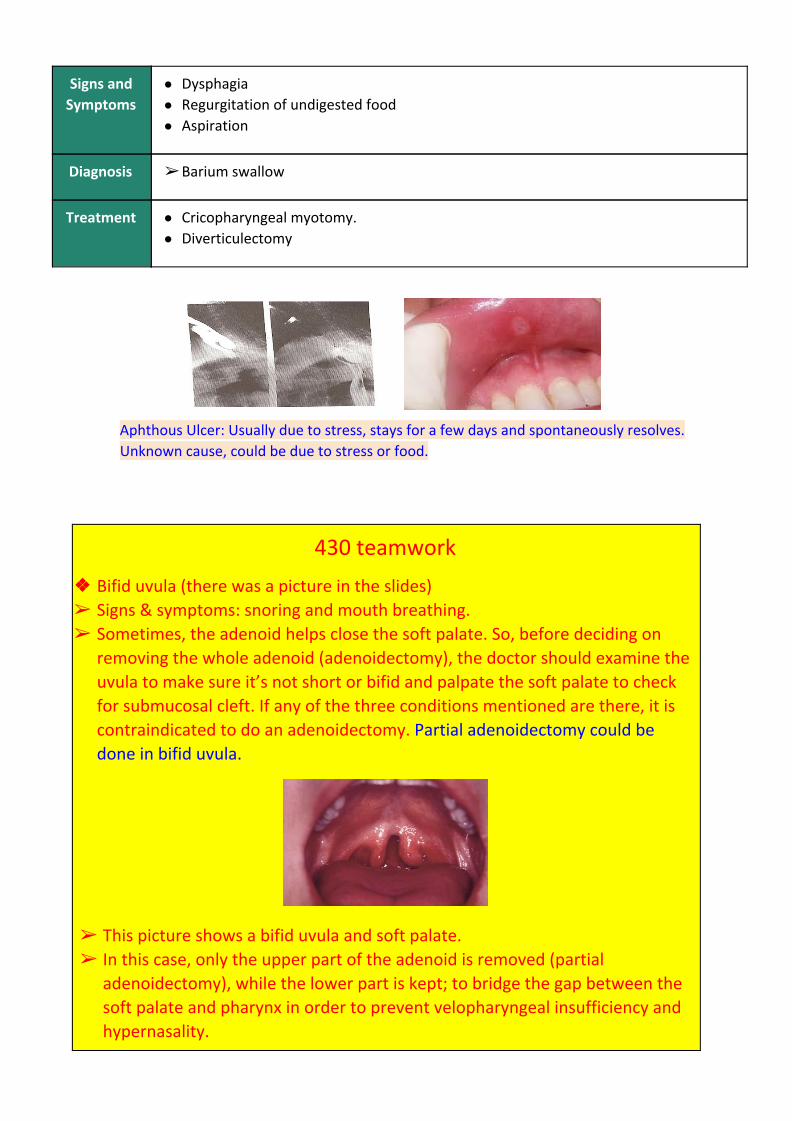
Aphthous Ulcer: Usually due to stress, stays for a few days and spontaneously resolves.
Unknown cause, could be due to stress or food.
430 teamwork
❖❖ Bifid uvula (there was a picture in the slides) ➢ Signs & symptoms: snoring and mouth breathing. ➢ Sometimes, the adenoid helps close the soft palate. So, before deciding on
removing the whole adenoid (adenoidectomy), the doctor should examine the uvula to make sure it’s not short or bifid and palpate the soft palate to check for submucosal cleft. If any of the three conditions mentioned are there, it is contraindicated to do an adenoidectomy. Partial adenoidectomy could be done in bifid uvula.
➢ This picture shows a bifid uvula and soft palate. ➢ In this case, only the upper part of the adenoid is removed (partial
adenoidectomy), while the lower part is kept; to bridge the gap between the soft palate and pharynx in order to prevent velopharyngeal insufficiency and hypernasality.

➢ Velopharyngeal insufficiency (VPI) is a disorder resulting in the improper closing of the velopharyngeal sphincter (soft palate muscle in the mouth) during speech, allowing air to escape through the nose instead of the mouth which results in hypernasality.


![[5] HEMA - Megaloblastic Anemia.pdf - KSUMSC](https://static.fdokumen.com/doc/165x107/631deac95ff22fc7450674ca/5-hema-megaloblastic-anemiapdf-ksumsc.jpg)