Development Of Renal system - KSUMSC
14
Development Of Renal system Editing file Renal block-Anatomy-Lecture 2,4
-
Upload
khangminh22 -
Category
Documents
-
view
1 -
download
0
Transcript of Development Of Renal system - KSUMSC
DevelopmentOf
Renal system
Editing file
Renal block-Anatomy-Lecture 2,4

Color guide :Only in boys slides in GreenOnly in girls slides in Purpleimportant in RedNotes in Grey
ObjectivesAt the end of the lecture, students should be able to:
Part (1) :● Identify the embryological origin of kidneys & ureters.● Differentiate between the 3 systems of kidneys during development.● Describe the development of collecting & excretory parts of permanent kidney.● Describe the fetal kidney & identify the pre- and postnatal changes that occur in the kidney.● Enumerate the most common anomalies of kidneys & ureters.
Part (2) :● Describe the cloaca and the formation of the urogenital sinus. ● Discuss the division of the urogenital sinus into various parts and name the adult organs that are derived
from each part.● Describe how the caudal parts of the mesonephric ducts and ureters are absorbed into the urogenital sinus
and the significance of this embryonic event. ● Discuss the position of the urachus and its significance and fate.● Describe the various anomalies concerned with the urinary bladder and urethra.

3Development of Kidney
The embryonic ORIGIN of kidney and ureters is from intermediate mesoderm intermediate mesoderm Divides into:
1. Nephrogenic ridge (cord): forms kidneys & ureters2. Gonadal ridge: forms gonads (testes or ovaries)
❖ Three systems of kidneys develop:
Week 5
Metanephric system:● Appears pelvis ● Starts to function at 9th week
Start of week 4
Pronephric system: ● Appears in cervical region● Analogous to kidney of fish● Formed of tubules & a duct● Not function in human● Disappears End of
week 4
Mesonephric system: ● Appears in thoracic & abdominal regions● Analogous to kidney of amphibians (البرمائیات)● Formed of tubules & a duct● Function temporarily● In male: forms genital duct● In both sexes: forms ureteric bud
Pronephric
Pronephric
Mesonephric
Metanephric
Mesonephric
Metanephric
intermediate mesoderm

4Metanephros (Permanent Kidney)
Formed of 2 origins:1) Ureteric bud (derived from mesonephric
duct): gives collecting part of kidney
2) Metanephric blastema (mass): gives excretory part of kidney
Ureteric bud elongates & penetrates metanephric mass
B
Stalk of ureteric bud forms ureter & cranial end forms renal pelvis.
C
Continuous branching gives straight then arched collecting tubules
E
Branching of renal pelvis gives 3 major calyces.Branching of major calyces gives minor calyces.
D
Collecting part

5Metanephros (Permanent Kidney)
Each arched collecting tubule is surrounded by a cap of metanephric mass.
The metanephric cap forms the metanephric vesicle,which is elongates to form an S-shaped metanephric tubule.
-The tubule lengthens to form: proximal & distal convoluted tubules + loop of Henle-The end of each tubule forms glomerular (Bowman’s) capsule,Which is then invaginated by capillaries (glomerulus).
Excretory part The Nephron● It’s the functional units of the kidney● The nephron is formed by fusion of:
1. Excretory tubule formed of metanephric mass (cap).
2. Arched collecting tubule formed of ureteric bud.
● At full term, each kidney contains: 800000 – 1000000 nephrons.
Excretory tubule
collecting tubule

6Changes in The Fetal Kidney
By week 9
A. Change in position: The kidney ascends from pelvis to abdomen & attains its adult position, caudal to suprarenal gland.B. Change in blood supply: As the kidney ascends, its blood supply changes from renal branches of common iliac arteries into renal
branches of abdominal aorta.C. Rotation: Initially, hilum (site of entry & exit of vessels & nerves) is ventral then rotates medially about 90° & becomes medial.● Glomerular filtration begins ● Kidney is subdivided into lobes that are visible externally.
In full term (end of fetal period) After birth
● Lobulation diminishes at the end of fetal period.● Nephron formation is complete
● Increase in size: due to elongation of tubules and increase in connective tissue between tubules (not due to increase in number of nephrons)
● Disappearance of kidney lobulation (usually disappear by the end of the first postnatal year)
Lobes of kidney

7Anomalies of kidney development
Pelvic kidney: failure of ascent of one kidney (ureter is short)
Horseshoe kidney: the poles of both kidneys (usually the lower poles) fuse: the kidneys have a lower position than normal but have normal function
Unilateral renal agenesis: due to absence of one ureteric bud
Supernumerary kidney: due to development of 2 ureteric buds
Right side: malrotation of kidney Left side: bifid ureter & supernumerary kidney
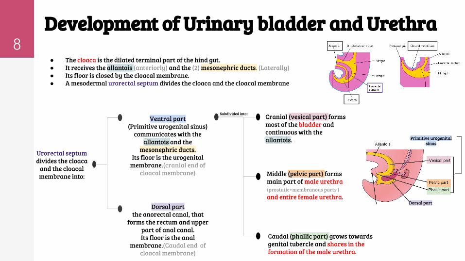
8Development of Urinary bladder and Urethra ● The cloaca is the dilated terminal part of the hind gut. ● It receives the allantois (anteriorly) and the (2) mesonephric ducts. (Laterally) ● Its floor is closed by the cloacal membrane.● A mesodermal urorectal septum divides the cloaca and the cloacal membrane
Middle (pelvic part) forms main part of male urethra (prostatic+membranous parts ) and entire female urethra.
Cranial (vesical part) forms most of the bladder and continuous with the allantois.
Caudal (phallic part) grows towards genital tubercle and shares in the formation of the male urethra.
Dorsal part the anorectal canal, that
forms the rectum and upper part of anal canal. Its floor is the anal
membrane.(Caudal end of cloacal membrane)
Ventral part(Primitive urogenital sinus)
communicates with the allantois and the
mesonephric ducts. Its floor is the urogenital
membrane.(cranial end of cloacal membrane)
Subdivided into :
Urorectal septum divides the cloaca
and the cloacal membrane into:
lllllllllkkv
lllllllllkkc
lllllllllkkc
Primitive urogenital sinus
Dorsal part

9Urinary bladder
❖ It develops mainly from the vesical part of the urogenital sinus. ❖ The trigone is derived from the absorbed distal parts of the mesonephric ducts.❖ The epithelium is endodermal in origin, of the urogenital sinus. ❖ The other layers are derived from the splanchnic mesoderm
❖ The allantois is at first continues with the bladder , then it becomes a thick fibrous cord called urachus which extends from apex of the bladder to the umbilicus, in adult it is represented by the median umbilical ligament.
❖ After absorption of the mesonephric ducts to form the trigone, the ureters open separately in the bladder.
❖ In infants and children the bladder is an abdominal organ. ❖ It starts to enter the greater pelvis at about 6 years and becomes a pelvic organ after puberty.
Trigones formation
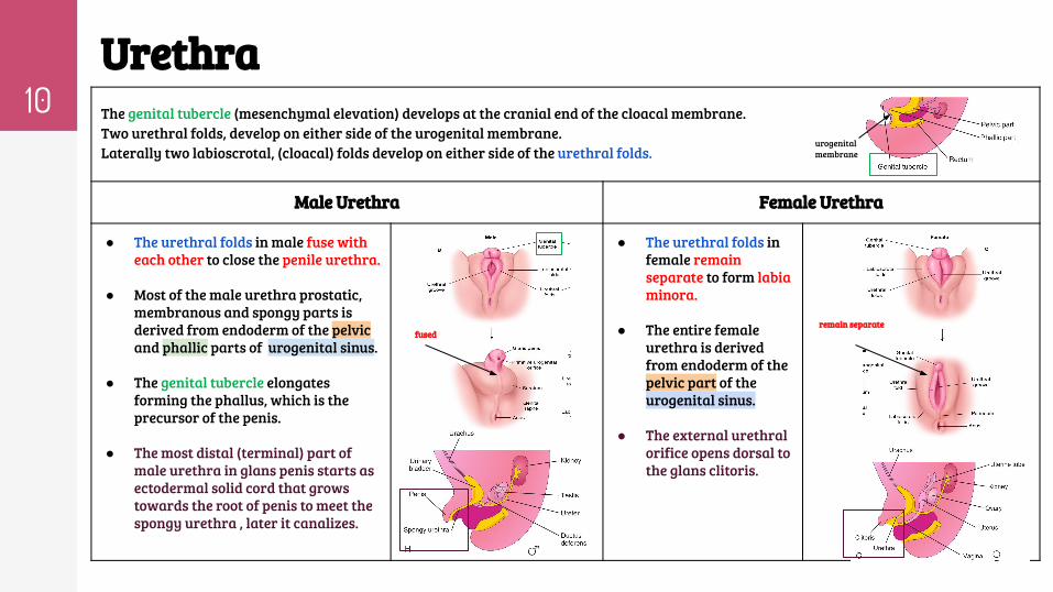
10Urethra The genital tubercle (mesenchymal elevation) develops at the cranial end of the cloacal membrane. Two urethral folds, develop on either side of the urogenital membrane.Laterally two labioscrotal, (cloacal) folds develop on either side of the urethral folds.
Male Urethra Female Urethra
● The urethral folds in male fuse with each other to close the penile urethra.
● Most of the male urethra prostatic, membranous and spongy parts is derived from endoderm of the pelvic and phallic parts of urogenital sinus.
● The genital tubercle elongates forming the phallus, which is the precursor of the penis.
● The most distal (terminal) part of male urethra in glans penis starts as ectodermal solid cord that grows towards the root of penis to meet the spongy urethra , later it canalizes.
● The urethral folds in female remain separate to form labia minora.
● The entire female urethra is derived from endoderm of the pelvic part of the urogenital sinus.
● The external urethral orifice opens dorsal to the glans clitoris.
remain separatefused
urogenital membrane
11Anomalies of u.bladder and urethra development
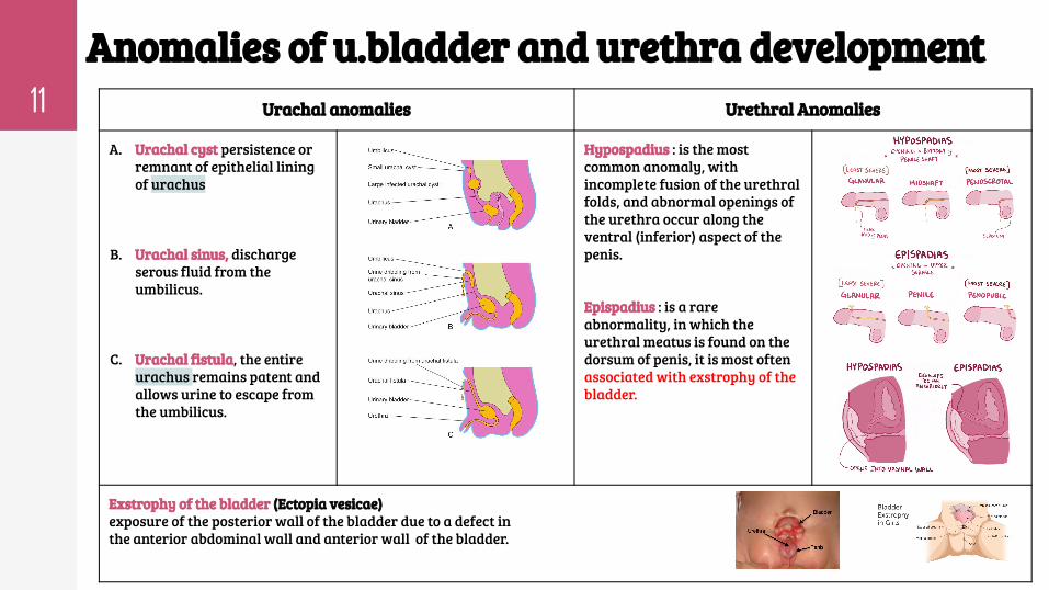
Urachal anomalies Urethral Anomalies
A. Urachal cyst persistence or remnant of epithelial lining of urachus
B. Urachal sinus, discharge serous fluid from the umbilicus.
C. Urachal fistula, the entire urachus remains patent and allows urine to escape from the umbilicus.
Hypospadius : is the most common anomaly, with incomplete fusion of the urethral folds, and abnormal openings of the urethra occur along the ventral (inferior) aspect of the penis.
Epispadius : is a rare abnormality, in which the urethral meatus is found on the dorsum of penis, it is most often associated with exstrophy of the bladder.
Exstrophy of the bladder (Ectopia vesicae)exposure of the posterior wall of the bladder due to a defect in the anterior abdominal wall and anterior wall of the bladder.
12Summary
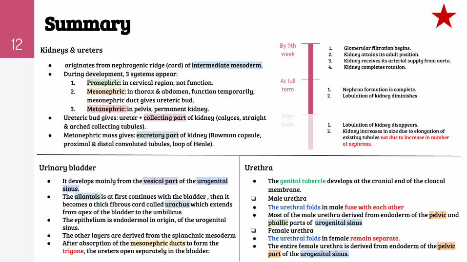
Kidneys & ureters
● originates from nephrogenic ridge (cord) of intermediate mesoderm.● During development, 3 systems appear:
1. Pronephric: in cervical region, not function. 2. Mesonephric: in thorax & abdomen, function temporarily,
mesonephric duct gives ureteric bud.3. Metanephric: in pelvis, permanent kidney.
● Ureteric bud gives: ureter + collecting part of kidney (calyces, straight & arched collecting tubules).
● Metanephric mass gives: excretory part of kidney (Bowman capsule, proximal & distal convoluted tubules, loop of Henle).
1. Glomerular filtration begins. 2. Kidney attains its adult position.3. Kidney receives its arterial supply from aorta.4. Kidney completes rotation.
By 9th week
1. Nephron formation is complete.2. Lobulation of kidney diminishes
At full term
After birth 1. Lobulation of kidney disappears.
2. Kidney increases in size due to elongation of existing tubules not due to increase in number of nephrons.
Urinary bladder● It develops mainly from the vesical part of the urogenital
sinus. ● The allantois is at first continues with the bladder , then it
becomes a thick fibrous cord called urachus which extends from apex of the bladder to the umbilicus
● The epithelium is endodermal in origin, of the urogenital sinus.
● The other layers are derived from the splanchnic mesoderm● After absorption of the mesonephric ducts to form the
trigone, the ureters open separately in the bladder.
Urethra ● The genital tubercle develops at the cranial end of the cloacal
membrane.❏ Male urethra ● The urethral folds in male fuse with each other ● Most of the male urethra derived from endoderm of the pelvic and
phallic parts of urogenital sinus❏ Female urethra ● The urethral folds in female remain separate.● The entire female urethra is derived from endoderm of the pelvic
part of the urogenital sinus.

13
MCQsQuestion 1: Which one of the following events happens by 9th week?
A. Nephron formation is complete
B. Disappearance of kidney lobulation
C. Kidney attains its adult position
D. Metanephric system appears
Question 2: Which one of the following structures is a derivative of the
ureteric bud?
A.Major calyces
B. Loop of Henle
C. Proximal convoluted tubule
D.Glomerulus
Question 3: the blood supply of the kidney before week 9 ?
A. Common iliac arteries
B. Suprarenal artery
C. Branches of common iliac arteries
D. Branches of abdominal aorta
Question 4: Supernumerary kidney happen due to ?
A. absence of one ureteric bud
B. development of 2 ureteric buds
C. failure of ascent of one kidney
D. short ureter
Question 5: In infants and children the bladder is an ---------------organ.
A. thoracic
B. abdominal
C. pelvic
D. central
Question 6: The trigone is derived from ?
A. splanchinic mesoderm.
B.absorbed proximal parts of the mesonephric ducts
C. urogenital sinus
D.absorbed distal parts of the mesonephric ducts
Question 7: female urethra is derived from ?
A.endoderm of the phallic part of the urogenital sinus.
B. ectoderm of the pelvic part of the urogenital sinus.
C. mesoderm of the pelvic part of the urogenital sinus.
D.endoderm of the pelvic part of the urogenital sinus.
Question 8 : which of the following Anomalies is most often associated with
exstrophy of the bladder.
A.Hypospadius
B. Epispadius
C. Urachal cyst
D. Urachal fistula Answers: Q1.C- Q2.A -Q3.C -Q4.B- Q5.B- Q6.D- Q7.D- Q8.B
Team leaders
● Abdulrahman Shadid● Ateen Almutairi
Team members
Girls team :
● Ajeed Al Rashoud● Taif Alotaibi● Noura Al Turki● Amirah Al-Zahrani● Alhanouf Al-haluli● Sara Al-Abdulkarem● Rawan Al Zayed● Renad Al Haqbani● Nouf Al Humaidhi● Jude Al Khalifah● Nouf Al Hussaini● Rahaf Al Shabri● Danah Al Halees● Rema Al Mutawa● Amirah Al Dakhilallah● Maha Al Nahdi ● Ghaida Al Braithen
Boys team:
● Faisal Alqifari ● Salman Alagla● Ziyad Al-jofan● Ali Aldawood● Khalid Nagshabandi● Omar Alammari● Sameh nuser
Contact us: THANKS!
Editing file