
Vasculitis - KSUMSC
12
Vasculitis Objectives: (1) Know the common causes of vasculitis with special emphasis on the clinic-pathological features and mechanism of: - Giant cell arteritis. - Polyarteritis nodosa. - Wegener's granulomatosis. - Cutaneous hypersensitivity vasculitis. - Thromboangiitis obliterans (Buerger’s disease) Editing file Black: original content. Green: Boy‘s doctor notes . Red: Important. Dark orange: Girl’s Doctor notes. Light Purple:From Robbin’s. Grey: Explanation. Blue:only found in boys slides. Pink: Only found in girls slides.
-
Upload
khangminh22 -
Category
Documents
-
view
7 -
download
0
Transcript of Vasculitis - KSUMSC

Vasculitis
Objectives:(1) Know the common causes of vasculitis with special emphasis
on the clinic-pathological features and mechanism of:- Giant cell arteritis.- Polyarteritis nodosa. - Wegener's granulomatosis.- Cutaneous hypersensitivity vasculitis.- Thromboangiitis obliterans (Buerger’s disease)
Editing file
Black: original content. Green: Boy‘s doctor notes . Red: Important. Dark orange: Girl’s Doctor notes. Light Purple:From Robbin’s. Grey: Explanation.Blue:only found in boys slides. Pink: Only found in girls slides.
Vasculitis
It is inflammation of vessel walls with many possible symptoms.
Definition
Etiology
Usually immune mediated
Subtopic
Etiology
Usually immune mediated
Infection, physical or chemical injury
● Immune complex deposition ● Antineutrophil cytoplasmic antibodies (ANCAs)● Anti-endothelial cell antibodies ● Autoreactive T cells
Vessel Disease Comment
LargeGiant-cell arteritis >50yrs and women are at high risk, it
affects the head Arteries.
Takayasu arteritis3 F <40yrs. “Pulseless disease”
MediumPolyarteritis nodosa Young adults. Widespread.
Kawasaki disease4 <4yrs. Coronary disease. Lymph nodes.
Small
Wegener granulomatosis Lung, kidney. c-ANCA.
Churg-Strauss syndrome Lung. Eosinophils. Asthma. p-ANCA.
Microscopic polyangiitis Lung, kidney. p-ANCA.
Cutaneous leukocytoclastic vasculitis
Idiopathic, infectious, drugs, chemicals, cancer and systemic disease like HNP
Overview
EtiologyUsually immune mediated Infection, physical or chemical injury
● Immune complex deposition1
● Antineutrophil cytoplasmic antibodies (ANCAs)
● Anti-endothelial cell antibodies● Autoreactive T cells2
● including that due to radiation, mechanical trauma, and toxins
● Infections can indirectly precipitate immune-mediated vasculitis
(1): such as the arthus phenomenon and serum sickness (2): cause injury in some forms of vasculitides characterized by formation of granulomas (3): is a granulomatous vasculitis of medium- and large-sized arteries characterized principally by ocular disturbances and marked weakening of the pulses in the upper extremities. Manifests with thickening of the aorta. occur in those younger than 50 years. associated with Japanese ethnicity. (4): an acute, febrile, usually self-limited illness of infancy and childhood associated with an arteritis of mainly large- to medium-sized vessels.

already mentioned
Giant-cell (Temporal) arteritis
● Chronic, granulomatous inflammation of large to medium sized arteries, especially the branches of the carotid artery in the head (temporal artery and branches of the ophthalmic artery)
● Involvement is segmental1, acute and chronic.
Definition
PathogenesisChronic, granulomatous inflammation of large to medium sized
arteries, especially the branches of the carotid artery in the head (temporal artery and branches of the ophthalmic artery)
Involvement is segmental, acute and chronic.
● The diagnosis depends on biopsy (from the temporal artery) and histologic confirmation.
● Treatment: corticosteroids 4 or anti-TNF therapies.
Giant cells
Disruption and fragmentation
of internal elastic lamina
Granulomatous inflammationof the blood vessel wall
The healed stage reveals collagenous thickening of the vessel wall and the
artery is transformed into a fibrous cord
Proliferation of the intima with
associated occlusion of the
lumen.
Morphology:
fever, facial pain or headache, often most intense along the
course of the superficial temporal artery
Jaw pain
Thickened and painful temporal
artery2
Symptoms
Visual problems and acute vision loss3
● It’s too crowded here. you can insert page if you need
● Please use one font size. Different sizes in one page can reduce the quantity of the work
●● Pathogenesis no need for
mind map
Pathogenesis:● occurs as a result of a T-cell– mediated immune response to antigen.● Pro-inflammatory cytokines (especially TNF) and anti-EC antibodies.● The characteristic granulomatous inflammation, an association with certain MHC class II haplotypes, and
the excellent therapeutic response to steroids, all strongly support an immune etiology.
Epidemiology:● Most common type of vasculitis● Patients older than 50yrs.● Female: Male = 2:1
Most common in elderly, complain of chronic headache, if untreated it will lead to blindness (because it effect the ophthalmic artery).
1:Only a portion is involved and not the entire artery2: can feel it 3: can lead to blindness, can see 2 of everything 4: Must give the treatment ASAP5: Fibrosis always come with healing
(1):Only a portion is involved and not the entire artery(2): palpable(3): can lead to blindness, can see 2 of everything, because it effect the ophthalmic artery.(4): Must give the treatment ASAP to decrease the immune system activity Dr note: the case will most likely be a +50 years old female complaining of severe headache and fever.

polyarteritis nodosa1
Necrotizing vasculitis involving multiple organs, lungs are spared.(Cutaneous only or systemic)
Definition
Disease of young adultsEpidemiology
Characteristics: There is segmental necrotizing2 inflammation of arteries of
medium to small size, in any organ(especially kidney and skin) except the lungs.
Most frequently kidneys (most common), skin,heart, liver, and gastrointestinal tract.
Polyarteritis nodosa has been associated with hepatitis B or hepatitis C virus infection.(because the immune system will attack
the endothelial cells confusing it with HBV)
Weakening of the arterial wall due to the inflammatory process may cause aneurysmal dilation3 or localized rupture.
Renal arterial involvement is often prominent and is a major cause of death.
Particularly characteristic of PAN is that all the different stages of activity ( i.e. active and chronic stages) may coexist in same
artery or in different artery at the same time.6
5
4
3
2
1
Polyarteritis nodosa with segmental inflammation
and fibrinoid necrosis and occlusion of the lumen of
this artery. Note that part of the vessel wall at the left
side is uninvolved.
Fatal if untreated, but steroids and cyclophosphamide are curative.
1: more than one artery forming nodulesPolyarteritis nodosa can cause blood in the urine which means UTI infection
(1): more than one artery forming nodulePolyarteritis nodosa can cause blood in the urine.(2): fibrinoid necrosis and luminal thrombosis (3): check “lecture 2” for more details
Clinical manifestationsSome clinical manifestations are due to ischemia and
infarction of affected tissues/ organs
abdominal pain
Fever muscular pain
NeuritisMelena (bloody stool)
Weight loss

Thromboangitis obliterans Buerger disease
Microscopically
Thrombus obstructing the blood vessels.
Acute and chronic inflammation. luminal thrombosis.
The inflammatory process extends into adjacent veins and nerves (rare with other forms of vasculitis), and in time all three structures become encased in fibrous tissue.
Buerger disease (Thromboangitis obliterans)
Buerger disease1 is a condition that known as :severe vascular insufficiency and gangrene of extremities.
● characterized by segmental, thrombosing, acute and chronic inflammation of medium-sized and small arteries of the leg and hands (tibial and radial arteries), with secondary extension into adjacent veins and nerves.
● occurs almost exclusively in heavy smokers of cigarettes (Abstinence from cigarette smoking in the early stages of the disease brings relief from further attacks ).
● Tobacco either leads to direct toxicity to endothelium, or induces an immune response.● usually beginning before age 35.
Cat
egor
y Pi
ctur
es
pain in the affected hand or foot induced by
exercise (called instep claudication)Even at rest due to the neural involvement.
Chronic ulcerations of the toes, or fingers may appear, followed in time by gangrene.
Clinical features include
1: starts in an artery progresses to a vein then nerve (patient will be in a very aggressive pain)
● segmental, thrombosing, acute and chronic inflammation of medium-sized and small arteries of the leg and hands (tibial and radial arteries), with secondary extension into adjacent veins and nerves.
Characterized
● occurs almost exclusively in heavy smokers of cigarettes (Abstinence from cigarette smoking in the early stages of the disease brings relief from further attacks ).
● Tobacco either leads to direct toxicity to endothelium, or induces an immune response.
● usually begins before age 35
Risk factors
Chronic ulcerations of the toes, or fingers may appear, followed in
time by gangrene.
Pain in the affected hand or foot induced by exercise (called instep
claudication) Even at rest due to the neural involvement
Clinical features includeClinical features include
Pain in the affected hand or foot induced by exercise (called instep
claudication) Even at rest due to the neural involvement
Chronic ulcerations of the toes, or fingers may appear, followed in time by
gangrene.
already mentioned
Morphology
Subtopic Subtopic Subtopic
Morphology
Acute & chronic Inflammation The inflammatory process extends into adjacent
veins and nerves (rare with other forms of vasculitis), and in time all three structures become encased in
fibrous tissue.
luminal thrombosis
Definition
EpidemiologyMales are affected more often than females, at an average age of about 40 years.Classic presentation is a middle-aged male with sinusitis or nasopharyngeal ulceration, hemoptysis with bilateral nodular lung infiltrates, and hematuria due to rapidly progressive glomerulonephritis.
Necrotizing granulomatous vasculitis involving nasopharynx, lungs, and kidneys.Also known as Granulomatosis with Polyangiitis.
Necrotizing granulomas of the upper and lower respiratory tract.2
Necrotizing or granulomatous
vasculitis of small to medium-sized vessels.
Renal disease in the form of necrotizing,
crescentic, glomerulonephritis.
Characterized by the triad of
Clinical features
C-ANCAs (antineutrophilic cytoplasmic antibodies) is positive in serum of more than 95% of patients.
Persistent pneumonitis, chronic sinusitis, mucosal ulcerations of the nasopharynx, and evidence of renal disease.
May lead to death within 2 years if not treated.
Prognosis
I think it’s better if you use another mind map and change the slant lines to straight
(1) The patient usually complains of 2 things: - respiratory problems: like epistaxis (nose bleeding).- Renal problems: like hematuria (blood in the urine).(2) : it involves the lung(3): causes a hole in the nasal septum due to the ulceration
Clinical features(1):C-ANCAs 4 (antineutrophilic cytoplasmic antibodies) is positive in serum of more than 95% of patients.
Persistent pneumonitis, chronic sinusitis, mucosal ulcerations3 of the nasopharynx, and evidence of renal disease.
(1) The patient usually complains of 2 things: 1- respiratory problems: like epistaxis (nose bleeding). 2-Renal problems: like hematuria (blood in the urine).(2) : unlike polyarteritis nodosa, Wegener involves the lung(3): causes a hole in the nasal septum (Palatal ulceration and destruction)(4): ANCAs are a heterogeneous group of autoantibodies directed against constituents (mainly enzymes) of neutrophil primary granules, monocyte lysosomes, and ECs. classified according to their antigen specificity:
● Anti-proteinase-3 (PR3-ANCA), previously called c-ANCA. PR3 is a neutrophil azurophilic granule constituent that shares homology with numerous microbial peptides
● Anti-myeloperoxidase (MPO-ANCA), previously called p-ANCA. (next slide)
Wegener granulomatosis(Granulomatosis with polyangiitis)

Microscopic polyangitis/polyarteritis
Churg-Strauss syndrome (additional reading)
It is a systemic small vessel vasculitis associated with glomerulonephritis1.
Definition
Eosinophil-rich and granulomatous inflammation involving the respiratory tract and necrotizing vasculitis affecting small vessels.
Definition
Clinical features:P-ANCA2 is characteristically present.
In the past it has been confused with leukocytoclastic vasculitis.
Clinical features:Associated with P-ANCAs 2
Associated with asthma and blood eosinophilia.
CHECK
(1): with no necrosis(2): is a lysosomal granule constituent involved in oxygen free radical generation
EXTRA but importantMicroscopic polyangiitis is very similar to Granulomatosis with polyangiitis but there are 3 key differences between them:1- Microscopic polyangiitis doesn’t affect nasopharynx(only kidney,lungs).2- No granulomas in microscopic polyangiitis.3- We will find P-ANCA in microscopic polyangiitis instead of C-ANCA.
Cutaneous leukocytoclastic (Hypersensitivity vasculitis/Angiitis)
Necrotizing vasculitis of arterioles, capillaries, venules. (Can be Cutaneous only or systemic)
What is Leukocytoclasis1?The nuclear debris of infiltrating neutrophils (karyorrhexis of neutrophils) in and around the vessels.
Definition
Characteristics
● Inflammation of small blood vessels (commonly seen in the post-capillary venules in the dermis).
● Characterized by palpable purpura. (Extravasated RBC).● All lesions tend to be of the same age.● It affects many organs e.g. skin (most common), mucous
membranes, lungs , brain, heart, GI , kidneys and muscle.● The most common vasculitis seen in clinical practice.
▪ Drugs e.g penicillin, foreign proteins (streptokinase). ▪ Infectious microorganisms e.g. strept. and other infections. ▪ Heterologous proteins. ▪ Food products and toxic chemicals ▪ Tumor antigens in various cancers
▪ collagen vascular diseases (lupus erythematosus, rheumatoid arthritis), ▪ Henoch-Schonlein purpura2 (skin rash, abdominal pain, hematuria)Et
iolo
gy
Idiopathic
It may be part of a systemic disease
eg:
Immunological reaction to an
antigen that may present as:
(1): Leukocytoclasis: WBC breakdown(2): Purpura: hemorrhagic spots on the skin (red and purple discoloration)
What is Henoch-Schonlein purpura?It’s an IgA-mediated, autoimmune systemic disease, in which the small vessels show leukocytoclastic vasculitis of childhood.
Henoch-Schonlein purpura:
Etiology Unknown
Characteristics
● Serum levels of IgA are high in HSP● Skin biopsy will show necrotizing leukocytoclastic vasculitis of capillaries in
the dermis.● The immunofluorescence shows IgA immunoglobulin and complement 3
(C3) deposition on the wall the affected arterioles, capillaries and venules.● It causes Skin purpura, arthritis, abdominal pain, GIT bleeding, orchitis and
nephritis.

Cutaneous leukocytoclastic (Hypersensitivity vasculitis/Angiitis)
Investigations● Skin biopsy is often diagnostic
Histologically:Infiltration of vessel wall with neutrophils, which become fragmented called as leukocytoclasia or nuclear dust
Direct immunofluorescence: Will show deposits of IgA immunoglobulin in the wall the capillaries
Leukocytoclastic vasculitis in a skin biopsy showing fragmentation of neutrophil nuclei in and around
vessel walls.
Summary (Robbins)

Summary
VasculitisIt is inflammation of vessel walls with many possible symptoms
Giant-cell arteritis
Polyarteritis Nodosa
Wegener granulomatosis
Thromboangiitis obliterans (Buerger disease)
Cutaneous hypersensitivity
vasculitis (Cutaneous leukocytoclastic)
Vessel size Epidemiology Organ affected Clinical features
Large to medium vessels
Medium to small vessels
Small to medium vessels
Medium & small
vessels
Small vessels
Branches of the carotid artery (temporal & ophthalmic)
Systemic: any organ except
lungs.Mostly Kidney
- Necrotizing granulomas: URT &
LRT.- Renal disease
Leg and hands (tibial & radial
arteries)
Most common: the dermis of skin
Females>Males. Ages
Above 50
Young adults
Males > Females
Age of 40
Heavy smokers
before 35
All ages
- Fever, facial pain, headache.
- Visual problems
Fever, weight loss, abdominal pain, melena &
neuritis.
- Persistent pneumonitis &
mucosal ulcerationsof nasopharynx.
C-ANCAs → +ve in serum
- Instep claudication- Chronic ulceration of
toes → Gangrene- sever pain
Henoch-Schonlein purpura
Elevated IgA
T
Microscopic polyangiitis
Small vessels
Kidney (Glomerulonephritis) Elevated P-ANCAIdiopathic
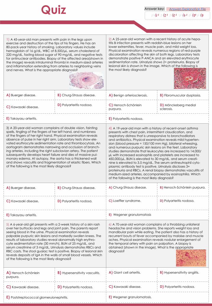
1) A 45-year-old man presents with pain in the legs upon exercise and destruction of the tips of his fingers. He has an 80-pack-year history of smoking. Laboratory values include hemoglobin of 16 g/dL, WBC of 8,500/µL, serum cholesterol of220 mg/dL, fasting blood sugar of 90 mg/dL, and negative tests for antinuclear antibodies. Biopsy of the affected area(shown in the image) reveals intraluminal thrombi in medium-sized arteries and inflammation extending from arteries to neighboring veins and nerves. What is the appropriate diagnosis?
2) A 25-year-old woman with a recent history of acute hepa-titis B infection presents with reddish-blue lesions on herlower extremities, fever, muscle pain, and mild weight loss.Physical examination reveals numerous regions of red-purplediscoloration affecting the skin of both legs. Laboratory testsdemonstrate positive P-ANCA and an elevated erythrocytesedimentation rate. Urinalysis shows 2+ proteinuria. Biopsy oflesional skin is shown in the image. Which of the following isthe most likely diagnosis?
A) Buerger disease. B) Churg-Strauss disease. A) Benign arteriosclerosis. B) Fibromuscular dysplasia.
C) Kawasaki disease. D) Polyarteritis nodosa. C) Henoch-Schönlein purpura.
D) Mönckeberg medial sclerosis.
E) Takayasu arteritis. E) Polyarteritis nodosa.
3) A 20-year-old woman complains of double vision, faintingspells, tingling of the fingers of her left hand, and numbnessof the fingers of her right hand. Physical examination revealsabsence of pulse in her right arm. Laboratory tests show ele-vated erythrocyte sedimentation rate and thrombocytosis. Anaortogram demonstrates narrowing and occlusion of branch-ing arteries, including the right subclavian artery. The patientsubsequently develops heart failure and dies of massive pul-monary edema. At autopsy, the aorta has a thickened wall and shows vasculitis and fragmentation of elastic fibers. Which of the following is the most likely diagnosis?
4) A 19-year-old man with a history of recent-onset asthmapresents with chest pain, intermittent claudication, andrespiratory distress that is unresponsive to bronchodilatorsand antibiotics. Physical examination reveals mild hyperten-sion (blood pressure = 150/100 mm Hg), bilateral wheezing,and numerous purpuric skin lesions on the feet. Laboratorystudies demonstrate that leukocytes are increased to 14,000/µL with increased eosinophils and platelets are increased to450,000/µL. BUN is elevated to 30 mg/dL, and serum creati-nine is elevated to 3.5 mg/dL. The serum antineutrophil cyto-plasmic antibody test is positive. Urinalysis discloses 3+ proteinuria and RBCs. A renal biopsy demonstrates vasculitis ofmedium-sized arteries, accompanied by eosinophilia. Whichof the following is the most likely diagnosis?
A) Buerger disease. B) Churg-Strauss disease. A) Churg-Strauss disease. B) Henoch-Schönlein purpura.
C) Kawasaki disease. D) Polyarteritis nodosa. C) Loeffler syndrome. D) Polyarteritis nodosa.
E) Takayasu arteritis. E) Wegener granulomatosis
5) A 6-year-old girl presents with a 2-week history of a skin rashover her buttocks and legs and joint pain. The parents report seeing blood in the urine. Physical examination reveals palpable purpuric skin lesions and markedly swollen knees. The results of laboratory studies reveal abnormally high erythro-cyte sedimentation rate (30 mm/h), BUN of 25 mg/dL, and serum creatinine of 3 mg/dL. Urinalysis demonstrates RBCs and RBC casts. The stool guaiac test is positive. Biopsy of lesional skin reveals deposits of IgA in the walls of small blood vessels. Which of the following is the most likely diagnosis?
6) A 70-year-old woman complains of a throbbing unilateralheadache and vision problems. She reports weight loss andmandibular pain while eating. The patient also has a history ofrecurrent bouts of fever accompanied by malaise and muscleaches. Physical examination reveals nodular enlargement of the temporal artery with pain on palpation. A biopsy is obtained (shown in the image). What is the appropriate diagnosis?
A) Henoch-Schönlein purpura.
B) Hypersensitivity vasculitis. A) Giant cell arteritis. B) Hypersensitivity angiitis.
C) Kawasaki disease. D) Polyarteritis nodosa. C) Kawasaki disease. D) Polyarteritis nodosa.
E) Poststreptococcal glomerulonephritis. E) Wegener granulomatosis.
Quiz 6) A ;5) A ; 4) A; ; 3) E ; 2) E ; 1) A
Answer key: Answers Explanation File

Team leaders● Raghad AlKhashan● Mashal Abaalkhail
Team Members
● Alhanouf Alhaluli● Amirah Alzahrani● Danah Alhalees● Deana Awartani● Elaf AlMusahel● Lama Alassiri ● Lama Alzamil ● Leena Alnassar● Leen Almazroa ● Njoud Alali ● Noura Alturki● Reema Alserhani● Rema Almutawa ● Taibah Alzaid
● Abdulaziz Alghamdi● Alwaleed Alarabi● Alwaleed Alsaleh● Faisal Almuhid● Jehad Alorainy ● Khalid Alkhani● Mohammad Alhamoud ● Mohammad Aljumah● Mohanad makkawi● Muath Aljehani● Nawaf AlBhijan● Suhail Basuhail● Abdulla Alhawamdeh● Hani Alhudhaif● Tariq Aloqail


![[5] HEMA - Megaloblastic Anemia.pdf - KSUMSC](https://static.fdokumen.com/doc/165x107/631deac95ff22fc7450674ca/5-hema-megaloblastic-anemiapdf-ksumsc.jpg)


