Orthopedic Teams 430 & 429 - KSUMSC
240
Orthopedic Teams 430 & 429 Lecture Page Number 01 Orientation, History Taking, And Examination Not included 02 Introduction to orthopedics 1 03 Bone Infections new 14 04 Xray interpretation last version 28 05-06 Principles of Fractures and Common Adult Fractures 42 07 Orthopaedic Emergencies , Open Fractures 64 08 Compartment Syndrome and Acute Joint Dislocation 79 09 Common Spinal Disorders 89 10 spinal injuries 98 11 common pediatric fractures 111 12 Common Pediatric Hip Problem ( 429 team) 140 13 Common Pediatrics Lower Limb Deformities 148 14 Osteoarthrosis 161 15 Peripheral Nerve injury 171 16 Common Shoulder Disorders 179 17 metabolic bone disorders 197 18 soft tissue and spots injuries 211 I9 Musculoskeletal Tumors (429 team) 223
-
Upload
khangminh22 -
Category
Documents
-
view
8 -
download
0
Transcript of Orthopedic Teams 430 & 429 - KSUMSC
Orthopedic Teams
430 & 429
Lecture Page Number
01 Orientation, History Taking, And Examination Not included
02 Introduction to orthopedics 1
03 Bone Infections new 14
04 Xray interpretation last version 28
05-06 Principles of Fractures and Common Adult Fractures 42
07 Orthopaedic Emergencies , Open Fractures 64
08 Compartment Syndrome and Acute Joint Dislocation 79
09 Common Spinal Disorders 89
10 spinal injuries 98
11 common pediatric fractures 111
12 Common Pediatric Hip Problem ( 429 team) 140
13 Common Pediatrics Lower Limb Deformities 148
14 Osteoarthrosis 161
15 Peripheral Nerve injury 171
16 Common Shoulder Disorders 179
17 metabolic bone disorders 197
18 soft tissue and spots injuries 211
I9 Musculoskeletal Tumors (429 team) 223
Isn't it funny how someone can say "I believe in Allah " but still follow the Satan who by the way also, " believes " in
Allah…
430 ORTHOPEDICS TEAM
Lecture: Introduction to orthopedics.
Team Members:
Faten Al-‐Mohideb. Hanan Al-‐Salman. Aliya Al-‐Awaji.
Hadeel Al-‐Ghamdi.
Ghadeer Al-‐Wuhayd.
Hissa Al-‐Balla.
Lujain Al-‐Yousef.
Nouf Al-‐Hammad.
Jawaher Al-‐Faraydi.
Hiba Al-‐Rahiem.
Nour Al-‐Enezi.
Wejdan Al-‐Swayyid.
Arwa Abudawood.
Leena Al-‐Shaman.
Areej Al-‐Qunaitir.
Team Leader:
Ayedah Al-‐Ruhaimi.
-‐‐The slides were provided by the doctor.
-‐‐Important notes in Red. –Medical dictionary in blue.
-‐‐Copied slides in Black.
-‐‐Doctor's notes in green.
1 Introduction to orthopedics
Page 1 of 236

ORTHO = Straight, Upright, Correct. Paios = Child. First used by Nicolas Andry a French doctor (1841) in a book titled Orthopedia : the art to
correct and prevent deformities in children.
Orthopedic Surgery = Not only Bone Surgery
• Orthopedic specialty is the branch of medicine which manage trauma and disease of
Musculoskeletal system.
• It includes: bones, muscles, tendons, ligaments, joints, peripheral nerves, vertebral
column and spinal cord and its nerves.
Orthopedic Specialty: Also Known as: Trauma and Orthopedic Surgery
Sub-‐Specialties in orthopedic include: Pediatric Orthopedic, Sport and Reconstructive
Orthopedic, Orthopedic Trauma, Arthroplasty (It is a surgery to relieve pain and restore
range of motion by realigning or reconstructing a joint, it has a rule in prosthetic joints),
Spinal Surgery, Oncology orthopedic and Foot and Ankle surgery.
Red Flags: (It means be careful this case has something strange & need an immediate
treatment).
• Red Flags = Warning Symptom or Sign
• Red flags should always be looked for and remembered
• Presence of a red flag means the necessity for urgent or different
action/intervention
E.g. Patient has back pain but recently he can't pass urine this is red flag. We have to
give him attention different from other patients with only back pain.
Examples of Red Flags: very imp
• Open Fractures: more serious and very high possibility of infection and
complications because bone naturally is well saved in sterile environment but in open
fracture there will be communication with outside (bacteria) to inside (bone) means
this fracture is susceptible to sever possible communication and have to be treated
immediately and in a special way other than closed fractures.
• Complicated Fractures: fracture with injury to major blood vessel, nerve or
nearby structure. E.g. Patient who came with fracture but when we check the pulse
there is no pulse. This mean that he has either: compression on the artery or there is a
damage to the artery .So, we have to take him to the OR immediately within 2-‐‐3 hours
not like other fractures which we used to treat them within 24 hours.
Case: Child who came with supracondylar fracture of the elbow due to falling on his arm, his arm was
swollen & we can't feel the radial pulse.
2 Introduction to orthopedics
Page 2 of 236

• Compartment Syndrome: increase in intra-‐‐compartment pressure which
endangers the blood circulation of the limb and may affect nerve supply.
Muscles, nerves and blood vessels they all covered by fascia which has the ability to
expand when there is a pressure but to very little extend. So, when there is a swelling in
the lower limb usually below the knee (very rarely in upper limb), the fascia will expand
a little bit but pressure still there and it will increase slowly until the blood can't go in or
out & this will lead to more swollen. With time the muscles around will become
infracted due to the absence of the blood supply & this may lead to lose the limb
(necrosis). That's why when we have a fracture with too much swelling and too much
pain & in advanced cases no pulse, pallor this mean this case require a treatment
priority over the other cases.
• Cauda Equina Syndrome: compression of the nerve roots of the Cauda Equina at
the spinal canal which affect motor and nerve supply to lower limbs and bladder
(also saddle or peri-‐‐anal area).
-‐‐When there is a back pain + difficulty in passing urine or numbness or reduced
sensation (altered or absence) at sacral or peri-‐‐anal area this mean a very serious
condition & we have to do urgent surgery.
• Infection of Bone, Joint and Soft Tissue: All the infections especially the acute
once are considered as Red Flag.
-‐‐ Osteomyelitis: Infection of the bone (from inside & outside). Osteo: bone myle:
bone marrow.
-‐‐Septic Arthritis: Infection of the joint. If you don't treat it very quickly, it will destroy
the structure of the bone. The cartilage & the articular surface will destroyed & the
ligaments may tear & the capsule may punctured.
-‐‐Cellulitis: spreading (not localized) Infection of the soft tissue, skin May cause
septicemia or irreversible damage.
• Multiple Trauma or Pelvic Injury: more than one fracture or injury sustained at
the same time consider massive blood loss and associated injuries.
Why pelvic bone fracture is important? Because it's a big bone and bleeds a lot
(could lead to blood loss about 45 units of blood inside). That's why we press the
pelvic from outside (Pelvic tamponade) to stop the bleeding.
-‐‐Patient with multiple fractures or pelvic fracture has the
priority of treatment over the one who have single fracture.
3 Introduction to orthopedics
Page 3 of 236
• Acute joint Dislocations: requires urgent reduction or may cause serious
complications.
Dislocation means that the two ends of the joint are no longer in contact. This
requires immediate treatment & if we don't reduce it, the joint will deprive from
blood supply (because the joint out of its place here) & this can lead to necrosis
or infarction.
Alignment terminology: Valgus: when the limb and the
joint goes away from the
midline.
Varus: when the limb and the
joint goes toward the midline.
In the upper limb the elbow is
called Cubitus. So, the same as
the lower limb we call it either
cubitus valgus or varus.
االررةبك االووحشیةی
In examination you have to
describe the alignment before
you describe the swelling or
small wound for example.
Congenital or Acquired:
-‐‐Acquired conditions include :
• Trauma
• Developmental: E.g. baby born normal but after that while he is growing, he start to
develop the abnormality e.g. genu valgum or develop limping.
• Inflammation
• Infection.
What is the different between inflammation and infection? Infection caused
by organisms but in inflammation not necessary there is organisms e.g.
rheumatic disease (autoimmune or body rejection).
• Neuromuscular: e.g. cerebral palsy (1-‐‐2% of Saudi child have it) and muscular
atrophy.
• Degenerative: Because of loss of content of hydration.
• Metabolic: The bone normally active, every day millions of bon cells
destroyed by osteoclast and millions of cells built back by osteoblast.
4 Introduction to orthopedics
Page 4 of 236
• Tumor.( either: Benign or malignant & it's either primary or secondary or
metastatic".
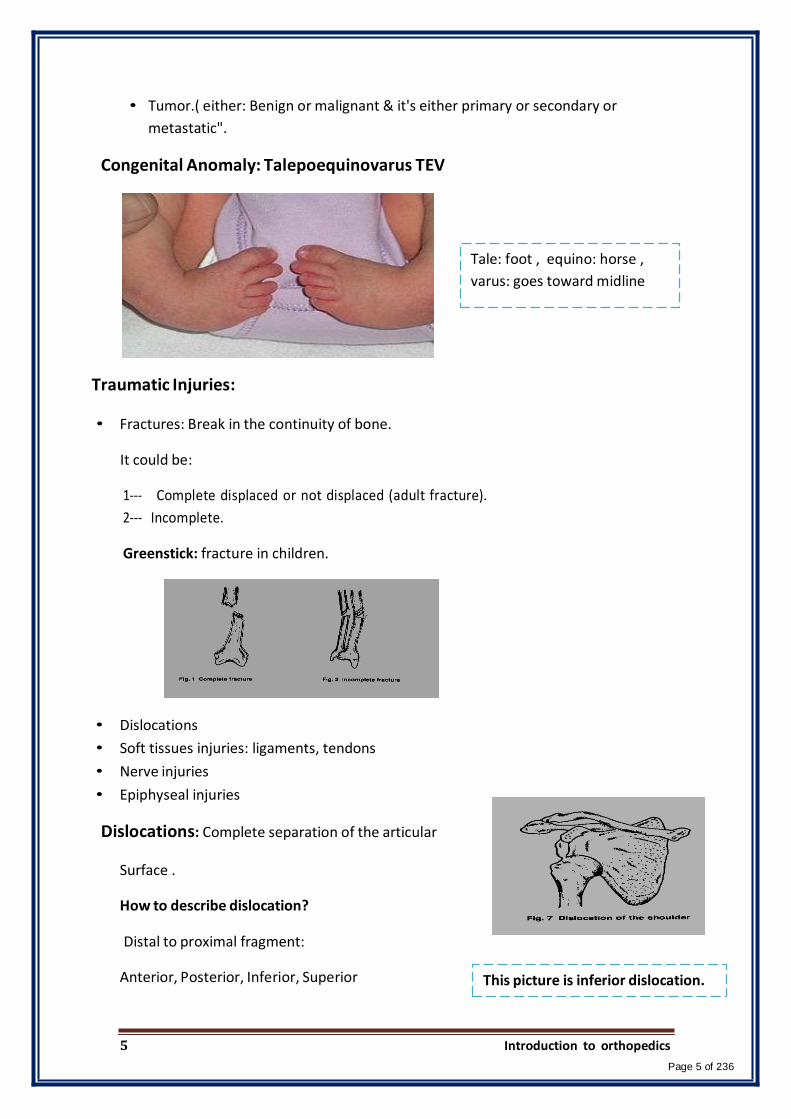
Congenital Anomaly: Talepoequinovarus TEV
Tale: foot , equino: horse ,
varus: goes toward midline
Traumatic Injuries: • Fractures: Break in the continuity of bone.
It could be:
1-‐‐ Complete displaced or not displaced (adult fracture).
2-‐‐ Incomplete.
Greenstick: fracture in children.
• Dislocations
• Soft tissues injuries: ligaments, tendons
• Nerve injuries
• Epiphyseal injuries
Dislocations: Complete separation of the articular
Surface .
How to describe dislocation?
Distal to proximal fragment:
Anterior, Posterior, Inferior, Superior
This picture is inferior dislocation.
5 Introduction to orthopedics
Page 5 of 236
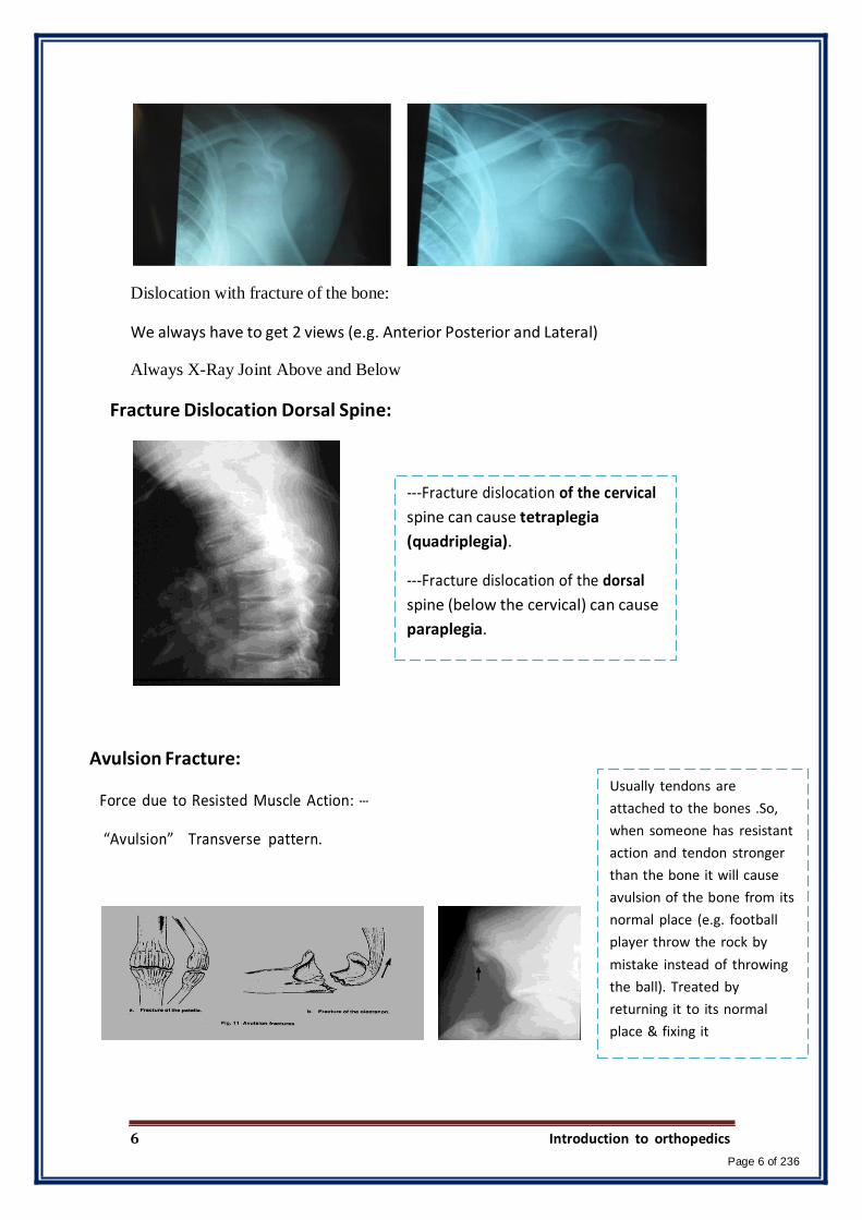
Dislocation with fracture of the bone:
We always have to get 2 views (e.g. Anterior Posterior and Lateral)
Always X-Ray Joint Above and Below
Fracture Dislocation Dorsal Spine:
-‐‐Fracture dislocation of the cervical
spine can cause tetraplegia
(quadriplegia).
-‐‐Fracture dislocation of the dorsal
spine (below the cervical) can cause
paraplegia.
Avulsion Fracture:
Force due to Resisted Muscle Action: -‐‐
“Avulsion” Transverse pattern.
Usually tendons are
attached to the bones .So,
when someone has resistant
action and tendon stronger
than the bone it will cause
avulsion of the bone from its
normal place (e.g. football
player throw the rock by
mistake instead of throwing
the ball). Treated by
returning it to its normal
place & fixing it
6 Introduction to orthopedics
Page 6 of 236
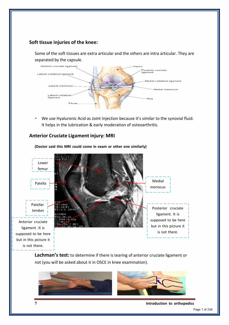
Soft tissue injuries of the knee:
Some of the soft tissues are extra articular and the others are intra articular. They are
separated by the capsule.
• We use Hyaluronic Acid as Joint Injection because it's similar to the synovial fluid.
It helps in the lubrication & early moderation of osteoarthritis.
Anterior Cruciate Ligament injury: MRI
(Doctor said this MRI could come in exam or other one similarly)
Lower
femur
Patella Medial
meniscus
Patellar
tendon
Anterior cruciate
ligament .It is
supposed to be here
but in this picture it
is not there.
Posterior cruciate
ligament. It is
supposed to be here
but in this picture it
is not there.
Lachman’s test: to determine if there is tearing of anterior cruciate ligament or
not (you will be asked about it in OSCE in knee examination).
7 Introduction to orthopedics
Page 7 of 236

Medial Collateral Ligament (MCL): Extra articular
We do stress test for the knee to determine if there is injury of the collateral
ligaments or not. It appears normal in x-‐‐ray.
(Developmental Dislocation of Hip) DDH:
"Shallow"
flat
Acetabulum
Dislocated
hip
Normal hip
-‐‐The mother complains of difficulty in changing the baby diaper. At the beginning we
do US because it's easier but if the baby age become few months, we do x-‐‐ray.
Orthosis : Pavlick Harness
Foot deformity: Hallux Valgus (big toe goes away from midline) (acquired)
Forefoot become wide
then when patient
wearing tight shoes it
becomes more deviation.
8 Introduction to orthopedics
Page 8 of 236

Epiphysis
SCFE (Slipped Capital Femoral Epiphysis):
Scoliosis:
This is a rare condition but it's
important to know how to diagnose
it.
The children when they reach the
puberty age, they feel hip pain
(sometimes they feel it in the knee
also) which cause restriction of hip
movement. So, what happen is that
the ligaments which attach the
epiphysis with the main shaft
become affected by the hormones &
become lax. Along with the effect of
weight this will move the epiphysis
from its place.
This condition will lead to an early
joint replacement.
-‐It is the lateral deviation of the spin from the
mid line.
-‐‐ Most of the time it's painless and
developmental (but it could be congenital).
-‐Mostly affect female more than male.
Degenerative Disorders: the most common cause is losing of hydration.
• Occur at any joint
• Can be primary (due to aging) or secondary (other problems e.g. infection).
• Increased wear and tear
• Can lead to pain and/or deformity and/or loss of function
• Increase with advancing age
• Management depends on type and age
Osteoarthritis of Hip:
The changes which can happen in
osteoarthritic hip:
-‐Decrease in the joint space because the
cartilage becomes thinner.
-‐‐ There maybe sclerosis of the hip.
-‐There will be an osteophyte.
9 Introduction to orthopedics
Page 9 of 236
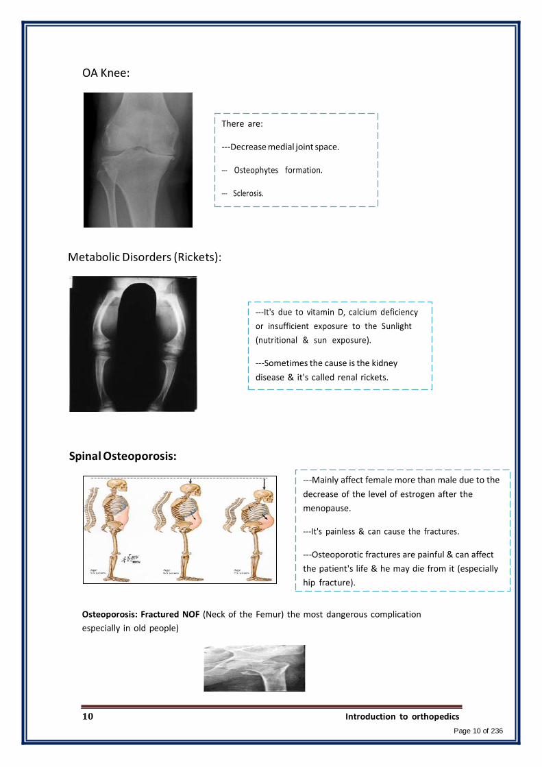
OA Knee:
There are:
-‐Decrease medial joint space.
-‐‐ Osteophytes formation.
-‐‐ Sclerosis.
Metabolic Disorders (Rickets):
Spinal Osteoporosis:
-‐It's due to vitamin D, calcium deficiency
or insufficient exposure to the Sunlight
(nutritional & sun exposure).
-‐Sometimes the cause is the kidney
disease & it's called renal rickets.
-‐Mainly affect female more than male due to the
decrease of the level of estrogen after the
menopause.
-‐It's painless & can cause the fractures.
-‐Osteoporotic fractures are painful & can affect
the patient's life & he may die from it (especially
hip fracture).
Osteoporosis: Fractured NOF (Neck of the Femur) the most dangerous complication
especially in old people)
10 Introduction to orthopedics
Page 10 of 236
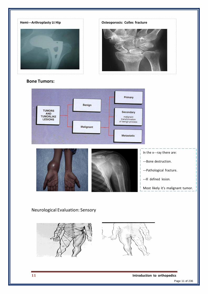
Hemi-‐Arthroplasty Lt Hip Osteoporosis: Colles fracture
Bone Tumors:
Neurological Evaluation: Sensory
In the x-‐ray there are:
-‐Bone destruction.
-‐Pathological fracture.
-‐Ill defined lesion.
Most likely it's malignant tumor.
11 Introduction to orthopedics
Page 11 of 236

Muscle wasting:
There are differences between the R &L side:
-‐Difference in the shoulder's shape (the left one has normal
contour while the right one is slipped).
-‐There is atrophy of the muscle at the right side due to nerve
damage.
Muscle Power Testing: Iliopsoas
Muscle Power Testing : Quadriceps
Muscle Power testing:
0 = no power.
1= simple contracting.
2= slight contraction within the
gravity.
3= muscle power against gravity
4= against gravity with resistance
5= against gravity with normal resistance
Neuromuscular disorder: Polio Chronic Osteomyelitis : discharging sinus
Spinal Infection: Tuberculosis Chronic Osteomyelitis : Sequestrum
12 Introduction to orthopedics
Page 12 of 236
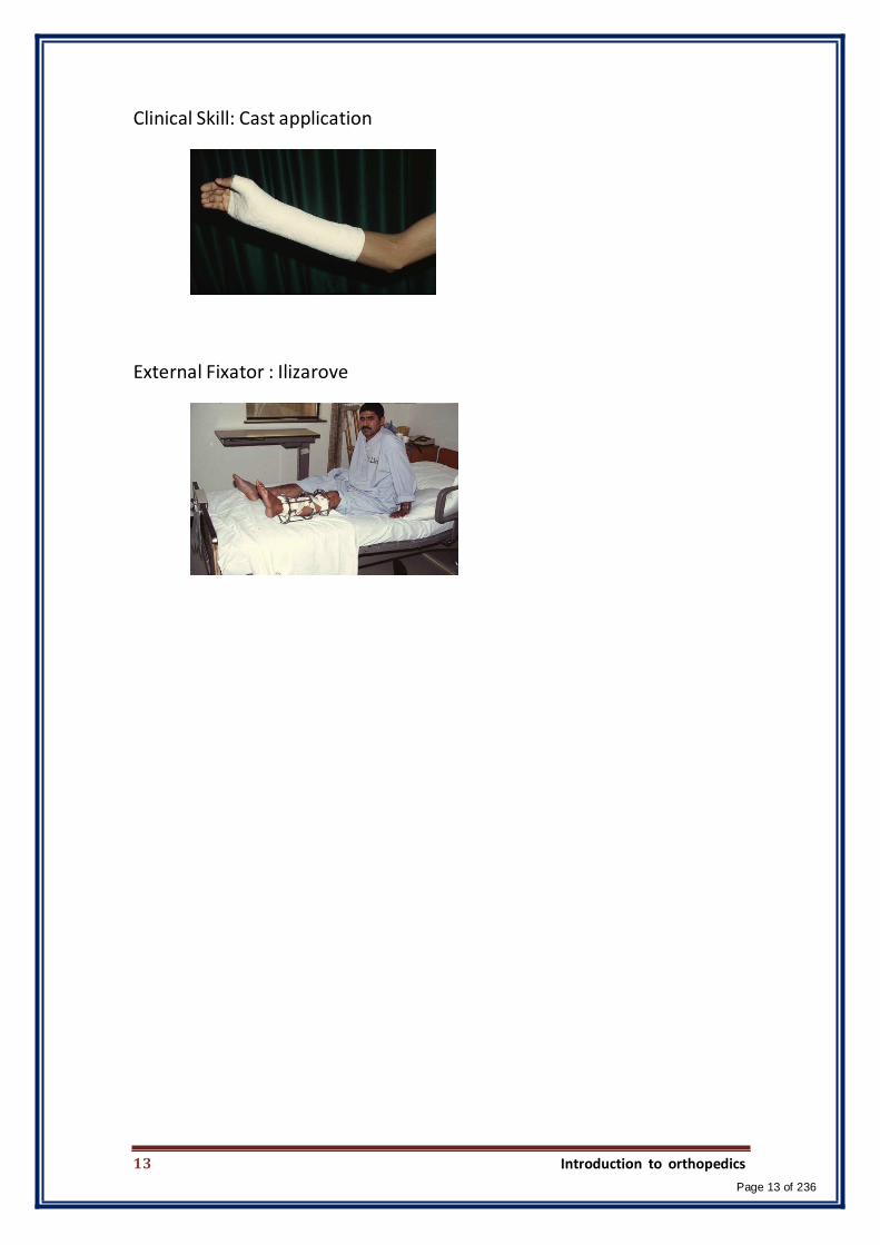
Clinical Skill: Cast application
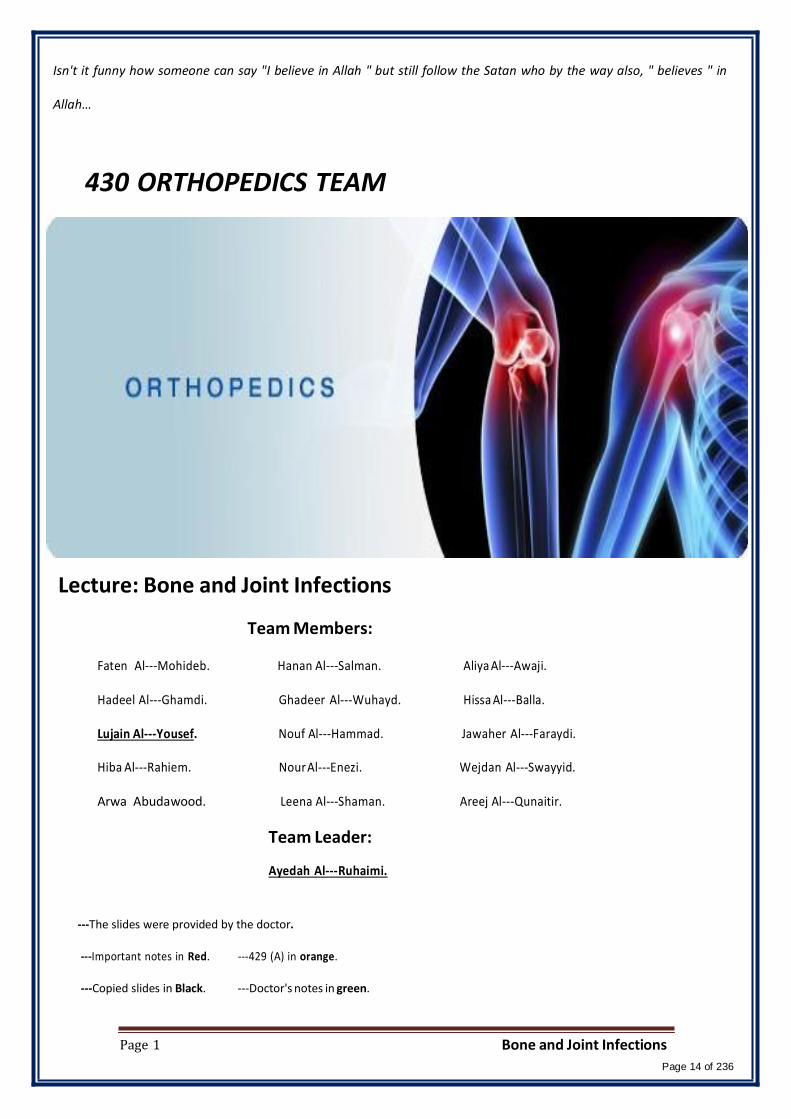
External Fixator : Ilizarove
13 Introduction to orthopedics
Page 13 of 236
Isn't it funny how someone can say "I believe in Allah " but still follow the Satan who by the way also, " believes " in
Allah…
430 ORTHOPEDICS TEAM
Lecture: Bone and Joint Infections
Team Members:
Faten Al-‐Mohideb. Hanan Al-‐Salman. Aliya Al-‐Awaji.
Hadeel Al-‐Ghamdi.
Ghadeer Al-‐Wuhayd.
Hissa Al-‐Balla.
Lujain Al-‐Yousef.
Nouf Al-‐Hammad.
Jawaher Al-‐Faraydi.
Hiba Al-‐Rahiem.
Nour Al-‐Enezi.
Wejdan Al-‐Swayyid.
Arwa Abudawood.
Leena Al-‐Shaman.
Areej Al-‐Qunaitir.
Team Leader:
Ayedah Al-‐Ruhaimi.
-‐The slides were provided by the doctor.
-‐‐Important notes in Red. -‐‐429 (A) in orange.
-‐‐Copied slides in Black. -‐‐Doctor's notes in green.
Page 1 Bone and Joint Infections
Page 14 of 236
Introduction:
-‐‐ Bone infections are considered one of the red flags because if you ignore them, they
may affect patient's life (he may die). Also, they are easily preventable just by giving the
right treatment at the right time & they are very obvious (clear to identify).
• Initial treatment based on presumed infection type clinical findings and symptoms.
• Definitive treatment based on final culture.
Glycocalyx: [It's a polysaccharide formed by the bacteria around metal implants (e.g.
prosthetic knee) to protect themselves against the immunity, usually the treatment is
not completed unless the metal is removed.
– exopolysaccharide coating.
– envelops bacteria.
– enhances bacterial adherence to biologic implants.
Bone Infections:
1. Osteomyelitis.[oste= bone , myel= bone marrow , itis= inflammation]
2. Septic arthritis.
3. Infected Total Joint Arthoplasty.
1] Osteomyelitis [OM]:
• Infection of bone and bone marrow.
• Route of infection.
-‐Mostly OM is treated medically by Abx unless there is pus.
-‐While waiting for the culture results (take 3-‐5 days) always
start with broad spectrum empirical Abx depending on the
most common organism in this area.
– Direct inoculation Open fractures, e.g. inserting the bacteria while you are
giving an injection to a patient with tendonitis.[ from the environment ]
– Blood-‐‐borne organisms Haematogenous.[most common]
• Determination of the offending organism
A. NOT a clinical diagnosis.[It's Microbiological diagnosis , that is why deep culture
is essential. So, before you start giving the empirical Abx, you have to take a
sample for culture because after 2-‐‐3 days if the patient isn't responding, you can
adjust your Abx according to the culture that you send 5 days ago ]
B. DEEP CULTURE is essential.
• Classification:[ It's important to know the severity & to choose the right treatment]
A. Acute hemotagenous OM. D.Chronic OM
B. Acute OM.
C. Subacute OM.
Page 2 Bone and Joint Infections
Page 15 of 236

A. Acute Hematogenous OM: [No Hx of trauma , open wound or injections (nothing
from outside) before 2 weeks ]
Case: child, Hx of sore throat 2 weeks ago. He was ok but 5 days
ago he started to limp then unable to walk. He is febrile & look
sick [acute presentation]
Clinical Features:
• Caused by blood-‐‐borne organisms.
• More common in children. b/c they o Boys > girls. are rich
o Most common in long bone metaphysis or blood
supplied epiphysis. areas. o Lower extremity >> upper extremity.
• Pain.[localized]
• Loss of function of the involved extremity. (e.g. if it's in
the upper extremity he will not be able to shake hands,
eat,wave &move…)
• Soft tissue abscess.[ Redness/swelling][ localized to the
site of infection especially at the beginning ].
• Fever.
Radiographic Changes:[ x-‐‐ray is not a diagnostic tool]
• Soft tissue swelling (early sign).[ the only sign you’re
going to see in acute presentation]
• Bone demineralization (10-‐‐14 days).
• Sequestra dead bone with surrounding granulation tissue. which looks in the X-
ray as a white area within a black area. The black area is the infection while the
white area is the necrotic bone late sign.[It has to be removed in the OR b/c mostl
it won’t resolve by itself otherwise they will become a source of infection]
• Involucrum periosteal new bone. which looks in the X-ray as if the cortex of the
bone is doubled with a black space, separates the 2 layers late sign.
Taking a sample from the throat to know the organism doesn’t help b/c
it is usually different organism. Also, the culture will take 5 days either
you take it from the bone or throat. So, there is no benefit.
Page 3 Bone and Joint Infections
Page 16 of 236
y

Diagnosis:
• Elevated WBC count.
• Elevated ESR.[ takes almost 3 weeks to peak up]
• Blood cultures may be positive.
• C-‐‐reactive protein (CRP):[ very important for diagnosis & follow up of treatment, it
takes 5 days to peak up]
o Most sensitive monitor of infection course in children.
o Short half-‐‐life.
o Dissipates in about 1 week after effective treatment
Case about CRP: If you have a young patient with a history of frequent travelling abroad (these people
have tendency to have multiple organisms) & you gave him a broad spectrum first generation
cephalosporin, in 5 days the patient still spiking fever & he doesn't look well (this mean that you don't
treat the right organism with the right Abx) & the clinical examination suggesting that he isn't responding
, what you are going to do??
-‐Request CRP & it will peak up within 5 days & if the patient isn't responding to Abx , you have to change
it otherwise he will go into septic shock & die.
•Nuclear medicine studies ( bone scan)may help when not sure.[there will be high
uptake(reactive area)but it is not specific (it just give a clue) b/c it could be soft
tissue, bone infection or pathology other than infection like tumor]
• MRI:[the best diagnostic/sensitive tool but it takes time to arrange, children need to
be anesthetized][MRI is the most sensitive for infection]
– Shows changes in bone and bone marrow before plain films
– Decreased T1-‐‐weighted bone marrow signal intensity.
– Increased postgadolinium fat-‐‐suppressed T1-‐‐weighted signal intensity
– Increased T2-‐‐weighted signal relative to normal fat.
Page 4
There is increase signal intensity
of the proximal tibia which
indicates OM of proximal tibia.
Bone and Joint Infections
Page 17 of 236

Treatment Outline: [Mostly with broad spectrum Abx at first then change to
specific Abx after the culture except in some indications. So, it's treated medically
most of the time not surgery].
• Take samples for culture.
• Start empirical broad-‐‐spectrum Abx.
• Observe improvement with clinical parameters
(Temp, pain) and blood tests (ESR,CRP).
• Review culture results within 3-‐‐5 days, if you find discrepancy between what you
thought is the organism & the culture result then you have to think either you
need to adjust your Abx or not and proceed accordingly.
Case: Pt is suspected to have OM and you admit him & started to give him empirical Abx because you
thought that it is S.aureus as it's the most common organism. However the culture showed that
the organism is not covered by your Abx>> what should you do?
If the pt is improving clinically(symptoms) + Lab test ( CRP going down) to your Abx don’t change
it even if the sensitivity test shows that the organism isn't sensitive to the ABx. If there is not
improvement change it.
• Decide on duration of Abx (IV vs oral).
[Duration mostly complete 6 weeks, sometimes start with IV for few
weeks and then orally and sometime all the 6 weeks IVdepends on
the severity ,immunocompromised pt,…ect]
e.g.: young ,no medical problems barely acute presentation3 weeks is
enough. While if he is old with HTN,DM and renal failure max
(6weeks).
o Empirical Treatment:[ is the one you give w/o definitive diagnosis or organism]
• Before definitive cultures become available.
• Based on patient’s age and other circumstances.
-‐‐The most common organism in all age groups is S.aureus. So, what you need to
think about is the second organism in each age.
Page 5 Bone and Joint Infections
Page 18 of 236

Organism Empirical Tx Notes
Newborn [0-‐‐4 months]
Staphylococcus aureus(most common) Gram-‐‐negative bacilli. Group B streptococcus.
Broad Spectrum Abx
Immunity is not fully Developedso they may be Afebrile(don't have fever) ,Cry.[difficult to dx] 70% positive blood Culture before Abx, not aspiration from bone or deep tissue. .[other age groups are less]
-You may find some swelling. -can't localize the pain, wherever you touch the baby, he will cry.
Children [ >4 months]
S. aureus (most common).
Coliforms (uncommonVaccnine)
Broad Spectrum Abx
Haemophilus influenza Boneinfectionsalmost Completely eliminated due to Vaccination.
Adults [≥21 years old
S. aureus(most common). Wide variety of other organisms have been isolated [especially
in people who have
abnormal life style.]
Broad Spectrum Abx
Sickle Cell Anemia
Salmonella is a characteristic (most specific) organism – but not the mo common S.aureus is still the most common.
Broad Spectrum Abx
Hemodialysis and IV drug abuser
S. aureus S. epidermidis[b/c usually the
problem come from the skin]
Pseudomonas aeruginosa
Broad Spectrum Abx
They are treated aggressively
(combining 2-‐3 Abx or one but
Very tough Abx)b/c they are
considered as immunocimpramised
patients.
Page 6 Bone and Joint Infections
Page 19 of 236

o Operative Treatment:
• Indications for operative intervention:
Drainage of an abscess.
Débridement of infected and necrotic tissues sequestrumprevent further
destruction.
Refractory cases that show no improvement or the patient is getting worse after
nonoperative treatment.
B. Acute OM:
• After open fracture or open reduction with internal fixation.[injections]
Clinical findingssimilar to acute hematogenous OM.[same presentation the only
difference is the treatment]
Treatment:
Radical I&D [Irrigation & Debridement],remove anything looks like dirty or died
tissueSURGERY.[ b/c infection after open fractures tends to be chronic also you
want to prevent the infection before its happening that why you start aggressively
by surgery].
Removal of orthopaedic hardware if necessary.
Soft tissue coverage for open wounds if needed. [The bone must be covered to
prevent infection and if it’s infected it will get worse].
Most common offending organisms are:
S. aureus.
P. aeruginosa.
Coliforms.
Empirical therapy Broad-‐‐spectrum Abx.
Page 7 Bone and Joint Infections
Page 20 of 236
C. Subacute OM:
Diagnosis [Usually]:
– Painful limp.[mild]
– No systemic and often no local signs or symptoms.
– Signs and symptoms on plain radiograph.
• May occur in:
– Partially treated acute osteomyelitis.
– Occasionally in fracture hematoma.
• Frequently normal tests: [usually results appear normal]
– WBC count.
– Blood cultures.
Usually useful tests:
ESR.
Bone cultures.
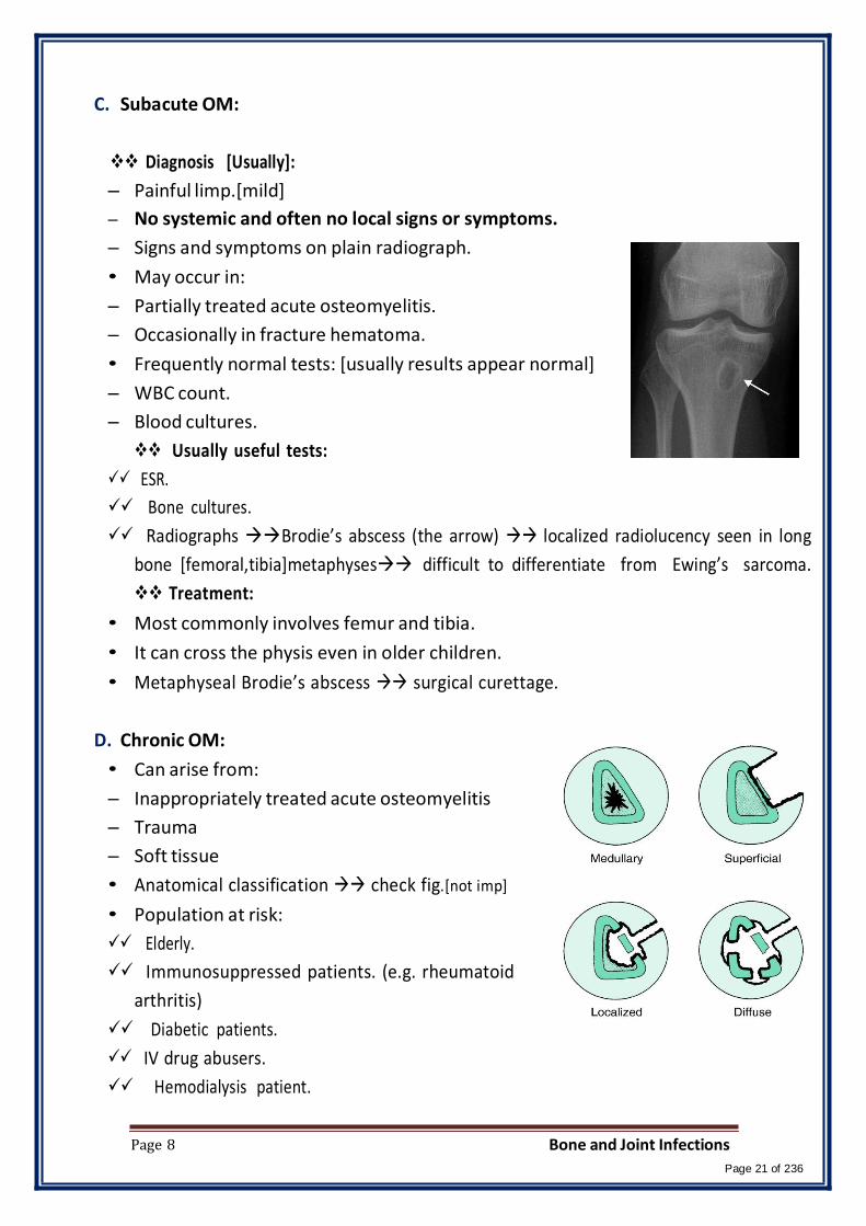
Radiographs Brodie’s abscess (the arrow) localized radiolucency seen in long
bone [femoral,tibia]metaphysesdifficult to differentiate from Ewing’s sarcoma.
Treatment:
• Most commonly involves femur and tibia.
• It can cross the physis even in older children.
• Metaphyseal Brodie’s abscess surgical curettage.
D. Chronic OM:
• Can arise from:
– Inappropriately treated acute osteomyelitis
– Trauma
– Soft tissue
• Anatomical classification check fig.[not imp]
• Population at risk:
Elderly.
Immunosuppressed patients. (e.g. rheumatoid
arthritis)
Diabetic patients.
IV drug abusers.
Hemodialysis patient.
Page 8 Bone and Joint Infections
Page 21 of 236

Most common organisms:
S. aureus.
Enterobacteriaceae.
P. aeruginosa.
Clinical Features: [ not acute presentation & mostly no fever/some pain/some loss
of function]
• Skin and soft tissues involvement.[pus discharge]
• Sinus tract may occasionally develop squamous cell carcinoma.[b/c of the chroni
irritation]
• Periods of quiescence followed by acute exacerbations.[means they don’t
present the same for the whole period, they recover and then relapse …so on]
Sonogram: It is a special X-‐‐ray procedure that is
done with contrast dye to visualize any
abnormal opening (sinus) in the body. If the
dye reaches the bone that means it is chronic
OM.
Diagnosis:
• Nuclear medicine activity of the disease
• Best test to identify the organismsOperative sampling of deep specimens from
multiple foci.
Treatment: [Should Be Based on the Culture]
o Empirical Therapy is not indicated. MCQ [wait for the culture unless it is
acute
in-‐‐top if chronic]
• IV antibiotics must be based on deep cultures.
Page 9 Bone and Joint Infections
Page 22 of 236
c

o Surgical Debridement: [ imp to remove the implant]
• Complete removal of compromised bone and soft tissue
• Hardware:
– Most important factor.
– Almost impossible to eliminate infection without removing implant. [ If you can't
remove the implant e.g. patient with fractured ankle . You wait until the fractured
heal & then take it out & then do your aggressive treatment & Abx. If you can't
&the patient stared to develop septicemia or septic shock, you need to transform
your implant by removing it & put external fixture but don't leave the fracture
loose & mobile].
– Organisms grow in a glycocalyx (biofilm) shields them from antibodies and
antibiotics.
• Bone grafting and soft tissue coverage is often required.
• Amputations are still required in certain cases.
-‐‐Treatment is to open & clean the abscess & if you can take culture.
2] Septic Arthritis: [mostly treated surgically b/c the cartilage is very sensitive to
infection, So if it was leaved for a few hours to a day the cartilage will be gone
forevertake the patient to the OR and wash it out]
• Route of infection:
– Hematogenous spread.
– Extension of metaphyseal osteomyelitis in children.
– Complication of a diagnostic or therapeutic joint procedure.
• Most commonly in infants (hip) and children.
• Metaphyseal osteomyelitis can lead to septic arthritis in: [ areas where it’s near
the cartilage (within the joint capsule].
Proximal femur [ e.g. greater trochanter]most common in this category.
Proximal humerus.[suspect the shoulder is also affected].
Radial neck.
Distal fibula. (ankle).
Page 10 Bone and Joint Infections
Page 23 of 236

• Adults at risk for septic arthritis are those with:
RA Due to joint effusion, Synovium is always inflamed & the immunity is compromised.
– Tuberculosis most characteristic
– S. aureus most common
Case: Pt has septic artharitis in the elbow
Suspect RA
IV drug abuse Pseudomonas most characteristic , but not the most common.
Treatment Outline: 1stOR (surgery : open or orthoscopic & take sample for
culture) 2nd Empirical Abx & after 3-‐‐4 days you will get the result & adjust
according to it.
o Empirical therapy: [After the surgery][if the patient has OM and SA is suspected
take him to the OR, SA has the priority}
• Prior to the availability of definitive cultures.
• Based on the patient's age and/or special circumstances.
Newborn (up to 3 months of age):
Most common organisms:
S. aureus.
Group B streptococcus.
Less common organisms:
Enterobacteriaceae.
Neisseria gonorrhoeae.
– 70% with adjacent bony involvement.
– Blood cultures are commonly positive.
– Initial abx after sugical wash out broad-‐‐spectrum Abx.
Children (3 months to 14 years of age)
Most common organisms:
S. aureus.
Streptococcus pyogenes.
S. pneumonia.
H. influenzae markedly decreased with vaccination.
Gram-negative bacilli.
– Initial treatment broad-‐‐spectrum Abx.
Page 11 Bone and Joint Infections
Page 24 of 236

Acute monarticular septic arthritis in adults: [severity depends on how many
joint is involved, if monarticular = less serious while if polyarticular = more
serious].
Most common organisms:
S. aureus
Streptococci
gram-negative bacilli
– Antibiotic treatment broad-‐‐spectrum Abx
Chronic monarticular septic arthritis
Most common organisms:
Brucella
Nocardia
Mycobacteria
fungi
Polyarticular septic arthritis:
Most common organisms:
Gonococci
B. burgdorferi
acute rheumatic fever
viruses
o Surgical Treatment:
• Mainstay of treatment.
– Surgical drainage open or arthroscopic.[Orthoscopic is used more b/c it is less
invasive, can reach difficult places, same results as open]when you are asked what
is better? Say the results are comparable and you can do either.
– Daily aspiration[not recommended].
• Tuberculosis infections pannussimilar to that of inflammatory arthritis.[not
imp]
• Late sequelae of septic arthritis soft tissue contractures It's a problem even
after cleaning the jointmay require soft tissue procedures (such as a
quadricepsplasty to be able to bend the knee).[not imp].
-‐‐Just remember that the treatment of choice for SA is surgery ( either
open or arthroscopic) & give empirical Abx after the surgery.
Page 12 Bone and Joint Infections
Page 25 of 236
3] Infected Total Joint Arthoplasty [TJA]: [always when there is a metal you are
afraid of infection, most common knee then hip & shoulder].
Prevention: [measures to avoid infection] [the best Treatment is prevention].
• Perioperative intravenous antibiotics most effective method for decreasing its
incidence.
• Good operative technique
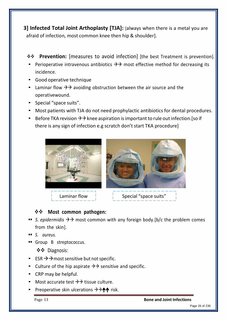
• Laminar flow avoiding obstruction between the air source and the
operativewound.
• Special “space suits”.
• Most patients with TJA do not need prophylactic antibiotics for dental procedures.
• Before TKA revision knee aspiration is important to rule out infection.[so if
there is any sign of infection e.g scratch don’t start TKA procedure]
Laminar flow Special “space suits”
Most common pathogen:
S. epidermidis most common with any foreign body.[b/c the problem comes
from the skin].
S. aureus.
Group B streptococcus.
Diagnosis:
• ESR most sensitive but not specific.
• Culture of the hip aspirate sensitive and specific.
• CRP may be helpful.
• Most accurate test tissue culture.
• Preoperative skin ulcerations risk.
Page 13 Bone and Joint Infections
Page 26 of 236

Treatment Outline:
• Acute infections [within 2-‐‐3 weeks of arthroplasty]: [Just wash it out & take the
insert out & replace it].
– Prosthesis salvage stable prosthesis.
– Exchange polyethylene [a plastic material] components.
– Synovectomybeneficial.
• chronic TJA infections [>3 weeks of arthroplasty]:
– Implant and cement remova.[to replace the metal , take it out & put a new one].
– Staged exchange arthroplasty.
– Glycocalyx:
Formed by polymicrobial organisms.
Difficult infection control without removing prosthesis and vigorous debridemen
– Helpful steps:
Use of antibiotic-‐‐impregnated cement.
Antibiotic spacers/beads.
1 -‐‐Take out all the implant. b 2 -‐‐Put a. Polyethylene (to prevent softtissue contraction)and
b. Cement is mixed with Abx and placed in the joint. a 3 -‐‐After infection is over.
4 -‐‐Place a new implant. b
The doctor mentioned these scenarios at the end of the lecture:
1-‐‐ You have a patient who is 30 years old , drug abuser came to the ER because of
limping , inability to walk, fever & pain. What you are going to do?
-‐‐ Hx. -‐‐ Physical EX. BP=80/40. The doctor mention that he is in septic shock .
So, he will start very aggressive Abx & IV fluid & take the patient to the OR ASAP.
Let us assume that the patient was taken to the OR , it was septic arthritis with distal
femur OM , you washed it out & you debris the joint . everything was good & the
patient stabilized a little bet & you took him back to his room . what you are going to
do now??
-‐continue on Abx & monitor his response by clinical exam , vital signs , WBC, ESR,
CRP.
-‐‐ The second day you find the patient looks better than before but the vital sign is
still spiking fever & CRP is still high . So, you give broad spectrum ABx or add another
Abx to the one you already gave.
Page 14 Bone and Joint Infections
Page 27 of 236
t.

Isn't it funny how someone can say "I believe in Allah " but still follow the Satan who by the way also, " believes " in
Allah…
430 ORTHOPEDICS TEAM
Lecture: X-‐‐ray interpretation.
Team Members:
Faten Al-‐Mohideb. Hanan Al-‐Salman. Aliya Al-‐Awaji.
Hadeel Al-‐Ghamdi. Ghadeer Al-‐Wuhayd. Hissa Al-‐Balla.
Lujain Al-‐Yousef. Nouf Al-‐Hammad. Jawaher Al-‐Faraydi.
Hiba Al-‐Rahiem. Nour Al-‐Enezi. Wejdan Al-‐Swayyid.
Arwa Abudawood. Leena Al-‐Shaman. Areej Al-‐Qunaitir.
Team Leader:
Ayedah Al-‐Ruhaimi.
-‐The slides were provided by the doctor.
-‐Important notes in Red.
-‐Copied slides in Black.
-‐Doctor's notes in green.
Hello, hope
you enjoy it!
Page1 X-‐ray Interpretation
Page 28 of 236

Objectives
• Review a systematic approach to interpreting orthopedic x-‐‐rays • Review the language of fracture description
Medical Decision Making is a Triad of: • History – from patients/records • Physical Examination • Confirming Studies – Imaging, Labs, etc.
Imaging • X-‐‐ray • Ultrasound • CT Scan • MRI • Nuclear Medicine
X-‐‐RAY • Radiation Source • Patient Exposed • Capture Image • Interpret Image • Ionizing Radiation • Radiation damages cells • Patient Blocks Transmission of Radiation
• Soft tissues Less • Bones More
• Capture Image • Films • Digital
• Interpret Image • Radiologist • Orthopaedist
• Best for: • Hard tissue • Bones • Often combined with other imaging
Ionizing radiation hazard:
1) DNA damage incompatible with
life apoptosis.
2) DNA damage which is fixable
cell return normal.
3) DNA damage which leads to
transformation either non harmful
or harmful, if harmful tumor.
ABCs APPROACH: Pre ABC: identify pt, read provided info (the most important to confirm the
x-‐‐ ray belongs to him/her and avoid mistakes).
A -‐‐Adequacy. -‐‐Alignment.
B -‐Bones. C -‐‐Cartilage. S -‐‐Soft tissues
• Apply ABCs approach to every orthopedic film you evaluate.
Page2 X-‐ray Interpretation
Page 29 of 236

ADEQUACY
• All x-‐‐rays should have an adequate number of views.
– Minimum of 2 views—AP and lateral (optimally two orthogonal views
'' 2 perpendicular views'' at least).
– 3 views preferred (to enhance our brain to reconstruct a 3D image).
– Joint above and joint below (if x-‐‐ray of joint: distal half of the proximal
bone and proximal part of the distal bone. If x-‐‐ray of bone: the
proximal and distal joints should be included).
• All x-‐‐rays should have adequate penetration (not the concern in this lect.)
Inadequate: 2 orthogonal views (A-‐P
and lateral). The distal half of femur is
not shown. So, it's not adequate.
Note: in A-‐P view fibula is lateral. In
lateral view fibula is posterior.
ALIGNMENT
• Alignment: Anatomic relationship between bones on x-‐‐ray
– Bone alignment vs other side
– Bone alignment relative to proximal and distal bones
• 2 things to comment on alignment, first the distal part of the bone
relative to the proximal (e.g. tibiovarameans the distal part of
tibia is medial in relative to the proximal part of tibia) , second at the
level of the joint, the distal bone relative to proximal bone (e.g.
genu valgustibia is lateral in relative to femur. Genu=knee).
• Normal x-‐‐rays should have normal alignment
• Fractures and dislocations may affect the alignment on the x-‐‐ray
• In alignment you describe the distal part in reference to the
proximal part.
(Example: valgus means the distal part is lateral to the proximal part)
• 5-‐‐10 degree valgus in the knee is normal.
Page3 X-‐ray Interpretation
Page 30 of 236

BONES
1. Identify bone
2. Examine the whole bone for
1. Discontinuity fractures or lytic
changes
2.Change in bone shadow
consistency change in density
3. Describe bone abnormality
1. Location (because treatment
differ).
2. Shape
In deformity we describe two elements:
1) angulation
2) translation which has 2 components:
-‐‐ magnitude
-‐direction, if A-‐P view medial or lateral
translation. If lateral view ant. & post.
remember: we describe distal relative to
proximal.
In AP view: deformity is described as either
varus or valgus, if apex of angle lateral
varus deformity. If apex medial valgus
deformity.
In lateral view: deformity is described as
either extension or flexion. If apex of angle
anterior extension deformity, if apex
post. flexion deformity.
The Mid Line
Pre ABCs: Patient identity unknown. No history or
examination information. X-‐ray of right tibia
A: Not adequate because only one view(AP). And joint
above is not fully visible.
Fibula
B: Transverse fracture (one cortices is opposite to the
other cortices) in mid shaft of tibia,
Distal part of tibia is going away from the mid line
valgus deformity, angulation 30 (always proximal is the
reference & distal is the one which we are checking).
Apex of the angle is medial.
Translation (It's the displacement of distal fragment
relative to the proximal one. If it's AP view we say either
medial or lateral while if its lateral view we say either
anterior or posterior): medial displacement 75-‐80%.
C: insignificant S: insignificant
Page4 X-‐ray Interpretation
Page 31 of 236
2
Landmarks:
1) Femur
curve.
2) Gluteal
line.
3) Medial
epicondyle
larger than
3
1
Pre ABCs: Patient identity unknown. No history or
examination info. X-‐ray of right femur.
A: not adequate because only one view and the
knee and hip joints are not visible.
B: short oblique fracture in mid shaft of right
femur, apex of the angle is lateral, varus
deformity, angulation 40-‐50.Translation: medial
displacement of 90% approximately (or 10%
apposition),
C: insignificant S: insignificant
Pre ABCs: Patient identity unknown. No history or
examination information. X-‐ray of the pelvis.
A: not adequate because only one view and the
distal femur is not fully visible.
B: short oblique fracture in the upper third of right
femur, apex of angle is lateral, varus deformity,
angulation 600.
Translation: 100% medial displacement.
C: insignificant S: insignificant
Pre ABCs: Patient identity unknown. No history or
examination information. X-‐ray of left humerus.
A: not adequate because only one view.
B: complete spiral fracture at shaft of left humerus.
Apex of angle anterior . Extension deformity,we
can't say here varus or valgus because its lateral
view not AP view.
Translation: 100%posterior displacement.
C: insignificant S: insignificant
Page5 X-‐ray Interpretation
Page 32 of 236

7)
Pre ABCs: Patient identity unknown.No history or
examination information. X-‐ray of forearm.
A: adequate x-‐ray
The Mid
Line
B: transverse fracture in lower third of radius,
A) A-‐P apex of angle medial. Valgus deformity (
distal part away from midline). Angulation of 20-‐‐
300.
B) lateral viewapex of angle is anterior,
extention deformity, angulation 300.
N.B. in lateral view you describe the deformity as
extention/flexion not varus/valgus.
C: insifnificant S: insignificant
Landmarks: thumb and coronoid process (red
circle) is always anterior. Olecranon is posterior.
it’s a growth plate not a fracture. Pre ABCs: Patient identity unknown. No history
or examination information. X-‐ray of proximal
humerus.
A: not adequate because only one view and no
joint above joint below.
B: Hypodense well circumscribed lesion with
septae and ballooning, not clear if there is a
fracture or not.
C: Cartilage can't be seen in x-‐ray because it's less
dense. So, instead of that we look to the joint
space if its decrease this means that the cartilage
is gone (e.g. in osteoarthritis). But if you see
increase in the joint space this means there is
instability (the collateral ligaments aren't there.
S: insignificant
Page6 X-‐ray Interpretation
Page 33 of 236
CARTILAGE
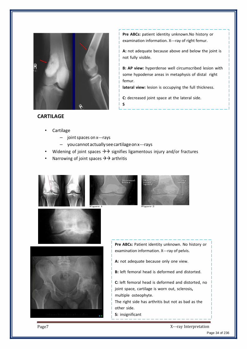
Pre ABCs: patient identity unknown.No history or
examination information. X-‐ray of right femur.
A: not adequate because above and below the joint is
not fully visible.
B: AP view: hyperdense well circumscribed lesion with
some hypodense areas in metaphysis of distal right
femur.
lateral view: lesion is occupying the full thickness.
C: decreased joint space at the lateral side.
S
• Cartilage
– joint spaces on x-‐‐rays
– you cannot actually see cartilage on x-‐‐rays
• Widening of joint spaces signifies ligamentous injury and/or fractures
• Narrowing of joint spaces arthritis
Page7
Pre ABCs: Patient identity unknown. No history or
examination information. X-‐ray of pelvis.
A: not adequate because only one view.
B: left femoral head is deformed and distorted.
C: left femoral head is deformed and distorted, no
joint space, cartilage is worn out, sclerosis,
multiple osteophyte.
The right side has arthritis but not as bad as the
other side.
S: insignificant
X-‐ray Interpretation
Page 34 of 236
SOFT TISSUES
• Soft tissues implies to look for soft tissue swelling and joint effusions
• These can be signs of
– Trauma
– occult fractures
– Infection
– Tumors
REVIEW: ABCs
A -‐‐ Assess adequacy of x-‐‐ray which includes proper number of views and penetration. -‐‐ Assess alignment of x-‐‐rays.
B -‐‐ Examine bones throughout their entire length for fracture lines and/or distortions.
C -‐‐ Examine cartilages (joint spaces) for widening. S -‐‐ Assess soft tissues for swelling/effusions.
EXAMPLES:
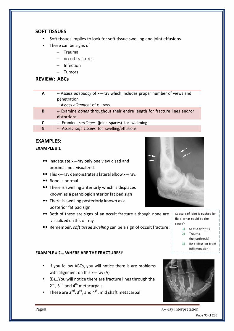
EXAMPLE # 1
Inadequate x-‐‐ray only one view disatl and
proximal not visualized.
This x-‐‐ray demonstrates a lateral elbow x-‐‐ray.
Bone is normal
There is swelling anteriorly which is displaced
known as a pathologic anterior fat pad sign
There is swelling posteriorly known as a
posterior fat pad sign
Both of these are signs of an occult fracture although none are
visualized on this x-‐‐ray
Remember, soft tissue swelling can be a sign of occult fracture!
EXAMPLE # 2… WHERE ARE THE FRACTURES?
• If you follow ABCs, you will notice there is are problems
with alignment on this x-‐‐ray (A)
• (B)…You will notice there are fracture lines through the
2nd, 3rd, and 4th metacarpals
• These are 2nd, 3rd, and 4th, mid shaft metacarpal
Capsule of joint is pushed by
fluid: what could be the
cause?
1) Septic arthritis
2) Trauma
(hemarthrosis)
3) RA ( effusion from
inflammation)
Page8 X-‐ray Interpretation
Page 35 of 236

fractures.
• A teaching point: Notice the ring on this film. Always remove rings of
patients with fractured extremities because swelling may preclude removal
later.
LANGUAGE OF FRACTURES
• Important for use to describe x-‐‐rays in medical terminology.
• Improves communication with orthopedic consultants
• Things you must describe (clinical and x-‐‐ray):
– Open vs Closed fracture
– Anatomic location of fracture
– Fracture line
– Relationship of fracture fragments
– Neurovascular status
OPEN VS CLOSED
Must describe to a consultant if fracture is open or closed
Closed fracture
◦ Simple fracture
◦ No open wounds of skin near fracture
Open fracture
◦ Compound fracture
◦ Cutaneous (open wounds) of skin near fracture site. Bone may
protrude from skin
◦ Open fractures are open complete displaced and/or comminuted
OPEN FRACTURES
• Orthopedic emergency
• Requires emergency orthopedic consultation
• Bleeding must be controlled
• Management
– IV antibiotics
– Tetanus prophylaxis
– Pain control
– Surgery for washout and reduction
ANATOMIC LOCATION
• Describe the precise anatomic location of the fracture
• Include if it is left or right sided bone
• Include name of bone
Page9 X-‐ray Interpretation
Page 36 of 236
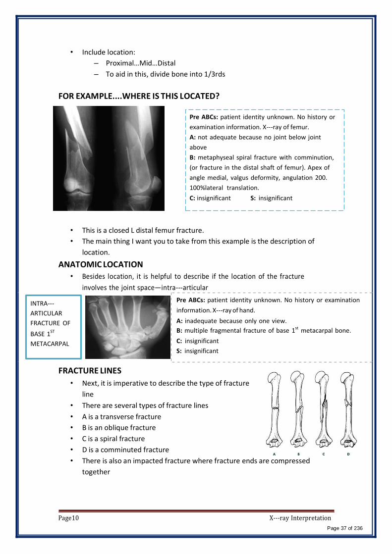
• Include location:
– Proximal…Mid…Distal
– To aid in this, divide bone into 1/3rds
FOR EXAMPLE....WHERE IS THIS LOCATED?
Pre ABCs: patient identity unknown. No history or
examination information. X-‐ray of femur.
A: not adequate because no joint below joint
above
B: metaphyseal spiral fracture with comminution,
(or fracture in the distal shaft of femur). Apex of
angle medial, valgus deformity, angulation 200.
100%lateral translation.
C: insignificant S: insignificant
• This is a closed L distal femur fracture.
• The main thing I want you to take from this example is the description of
location.
ANATOMIC LOCATION
• Besides location, it is helpful to describe if the location of the fracture
involves the joint space—intra-‐‐articular
INTRA-‐‐
ARTICULAR
FRACTURE OF
BASE 1ST
METACARPAL
Pre ABCs: patient identity unknown. No history or examination
information. X-‐ray of hand.
A: inadequate because only one view.
B: multiple fragmental fracture of base 1st metacarpal bone.
C: insignificant
S: insignificant
FRACTURE LINES
• Next, it is imperative to describe the type of fracture
line
• There are several types of fracture lines
• A is a transverse fracture
• B is an oblique fracture
• C is a spiral fracture
• D is a comminuted fracture
• There is also an impacted fracture where fracture ends are compressed
together
Page10 X-‐ray Interpretation
Page 37 of 236

WHAT TYPE OF FRACTURE LINE IS THIS???
ANS: TRANSVERSE FRACTURE
• Transverse fractures occur perpendicular to the long
axis of the bone.
• To fully describe the fracture, this is a closed midshaft
transverse humerus fracture.
ANOTHER EXAMPLE OF FRACTURE LINE…
ANS: SPIRAL FRACTURE
• Spiral fractures occur in a spiral fashion along the long
axis of the bone
• They are usually caused by a rotational force
• To fully describe the fracture, this is a closed distal
spiral fracture of the fibula
ONE MORE EXAMPLE…
ANS: COMMINUTED FRACTURE
• Comminuted fractures are those with 2 or more bone fragments
are present
• Sometimes difficult to appreciate on x-‐ray but will clearly
show on CT scan
• To fully describe the fracture, this is a closed R comminuted
intertrochanteric fracture
FRACTURE FRAGMENTS • Terms to be familiar with when describing the relationship of fracture
fragments
– Alignment
– Angulation
– Apposition
– Displacement
– Bayonette apposition
– Distraction
– Dislocation
Page11 X-‐ray Interpretation
Page 38 of 236
ALIGNMENT/ANGULATION
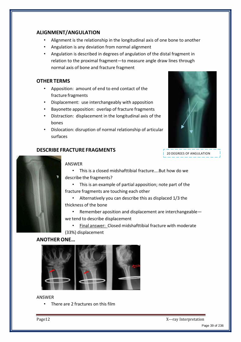
• Alignment is the relationship in the longitudinal axis of one bone to another
• Angulation is any deviation from normal alignment
• Angulation is described in degrees of angulation of the distal fragment in
relation to the proximal fragment—to measure angle draw lines through
normal axis of bone and fracture fragment
OTHER TERMS
• Apposition: amount of end to end contact of the
fracture fragments
• Displacement: use interchangeably with apposition
• Bayonette apposition: overlap of fracture fragments
• Distraction: displacement in the longitudinal axis of the
bones
• Dislocation: disruption of normal relationship of articular
surfaces
DESCRIBE FRACTURE FRAGMENTS 20 DEGREES OF ANGULATION
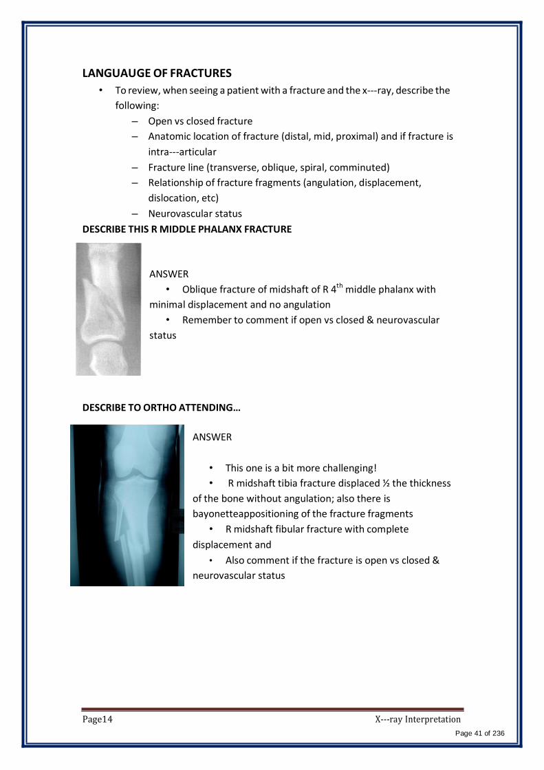
ANSWER
• This is a closed midshafttibial fracture….But how do we
describe the fragments?
• This is an example of partial apposition; note part of the
fracture fragments are touching each other
• Alternatively you can describe this as displaced 1/3 the
thickness of the bone
• Remember aposition and displacement are interchangeable—
we tend to describe displacement
• Final answer: Closed midshafttibial fracture with moderate
(33%) displacement
ANOTHER ONE…
ANSWER
• There are 2 fractures on this film
Page12 X-‐ray Interpretation
Page 39 of 236

• Closed distal radius fracture with complete displacement. Also there is an
ulnar styloid fracture which is also displaced
• The displacement is especially prominent on the lateral view highlighting the
importance of multiple views.
• There may be intra-‐‐articular involvement as joint space is close by
• Remember, remove all jewelry from extremity fractures
BAYONETTE APPOSITION
DISLOCATION
• Note the dislocation; the articular surfaces of the
knee no longer maintain their normal relationship
• Dislocations are named by the positioin of the distal
segemnt
• This is an Anterior knee dislocation.
Landmarks to help identify the direction of dislocation:
Fibula always posterior, patella anterior & condyles
posterior.
NEUROVASCULAR STATUS
• Finally when communicating a fracture, you will want to describe if the
patient has any neurovascular deficits
• This is determined clinically
Page13 X-‐ray Interpretation
Page 40 of 236
LANGUAUGE OF FRACTURES
• To review, when seeing a patient with a fracture and the x-‐‐ray, describe the
following:
– Open vs closed fracture
– Anatomic location of fracture (distal, mid, proximal) and if fracture is
intra-‐‐articular
– Fracture line (transverse, oblique, spiral, comminuted)
– Relationship of fracture fragments (angulation, displacement,
dislocation, etc)
– Neurovascular status
DESCRIBE THIS R MIDDLE PHALANX FRACTURE
ANSWER
• Oblique fracture of midshaft of R 4th middle phalanx with
minimal displacement and no angulation
• Remember to comment if open vs closed & neurovascular
status
DESCRIBE TO ORTHO ATTENDING…
ANSWER
• This one is a bit more challenging!
• R midshaft tibia fracture displaced ½ the thickness
of the bone without angulation; also there is
bayonetteappositioning of the fracture fragments
• R midshaft fibular fracture with complete
displacement and
• Also comment if the fracture is open vs closed &
neurovascular status
Page14 X-‐ray Interpretation
Page 41 of 236
Isn't it funny how someone can say, "I believe in Allah " but still follow the Satan who by the way also, " believes " in
Allah…
430 ORTHOPEDICS TEAM
Lecture: Principles of Fractures and Common Adult Fractures.
Team Members:
Faten Al-‐Mohideb. Hanan Al-‐Salman. Aliya Al-‐Awaji.
Hadeel Al-‐Ghamdi. Ghadeer Al-‐Wuhayd. Hissa Al-‐Balla. Lujain
Al-‐Yousef. Nouf Al-‐Hammad. Jawaher Al-‐Faraydi.
Hiba Al-‐Rahiem. Nour Al-‐Enezi. Wejdan Al-‐Swayyid.
Arwa Abudawood. Leena Al-‐Shaman. Areej Al-‐Qunaitir.
Team Leader:
Ayedah Al-‐Ruhaimi.
-‐The slides were provided by the doctor.
-‐Important notes in Red.
-‐Copied slides in Black.
-‐Doctor's notes in green.
[Principles of Fractures and Common Adult Fractures] Page1
Page 42 of 236
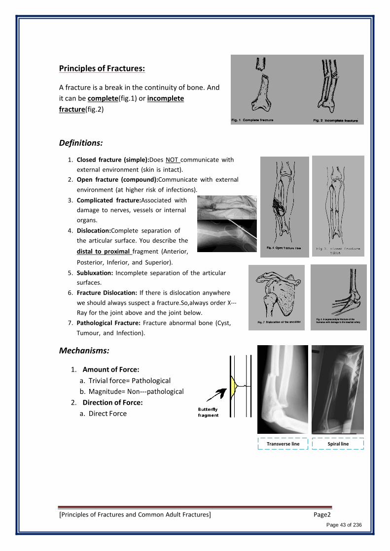
Principles of Fractures:
A fracture is a break in the continuity of bone. And
it can be complete(fig.1) or incomplete
fracture(fig.2)
Definitions:
1. Closed fracture (simple):Does NOT communicate with
external environment (skin is intact).
2. Open fracture (compound):Communicate with external
environment (at higher risk of infections).
3. Complicated fracture:Associated with
damage to nerves, vessels or internal
organs.
4. Dislocation:Complete separation of
the articular surface. You describe the
distal to proximal fragment (Anterior,
Posterior, Inferior, and Superior).
5. Subluxation: Incomplete separation of the articular
surfaces.
6. Fracture Dislocation: If there is dislocation anywhere
we should always suspect a fracture.So,always order X-‐‐
Ray for the joint above and the joint below.
7. Pathological Fracture: Fracture abnormal bone (Cyst,
Tumour, and Infection).
Mechanisms:
1. Amount of Force:
a. Trivial force= Pathological
b. Magnitude= Non-‐‐pathological
2. Direction of Force:
a. Direct Force
Transverse line Spiral line
[Principles of Fractures and Common Adult Fractures] Page2
Page 43 of 236
b. Indirect Force:
i. On Long Bones:
• Twisting Force →Spiral Line
• Angulating Force→Transverse pattern
• Angulating + Axial compression→
Transverse line + Triangular “Butterfly”
• Angulating + Axial compression + Twisting forces→ Short oblique Comminuted
Short oblique
pattern
• Vertical compression→ Comminuted
ii. On CancellousBones(Spongy bones):
a. Direct force
b. Indirect force:
• Comminuted Pattern
• Burst
iii. Force due to Resisted Muscle Action:
fracture
pattern
• Avulsion→ Transverse pattern
Diagnosis:
i. History: a. Trauma:
• Pathological (trivial)
• Non-‐‐pathological ( magnitude) b. Mechanism:
• Fall from height,
• RTA, pedestrian, Driver….? c. Complaint:
• Pain sharp, increase by movement, Not radiating
• Loss of Function
• Deformity
• Symptoms of complications
• Other organs: head, chest, abdomen ii. Examination:
a. General examination:
• Signs resulting from fracture or trauma:
o Vital signs, Shock A,B,C
o Associated Head, Chest, Abdomen
[Principles of Fractures and Common Adult Fractures] Page3
Page 44 of 236
• Signs related to cause of fracture:
o Pathological fractures …CA Lung, Prostate.
b. Local examination:
• LOOK: Skin damage (open fracture) , deformity, swelling
• FEEL: Localized tenderness
• MOVE: Abnormal movement, crepitus
• DO:
o Special tests: neurovascular status
o Measurements: shortening [Always compare]
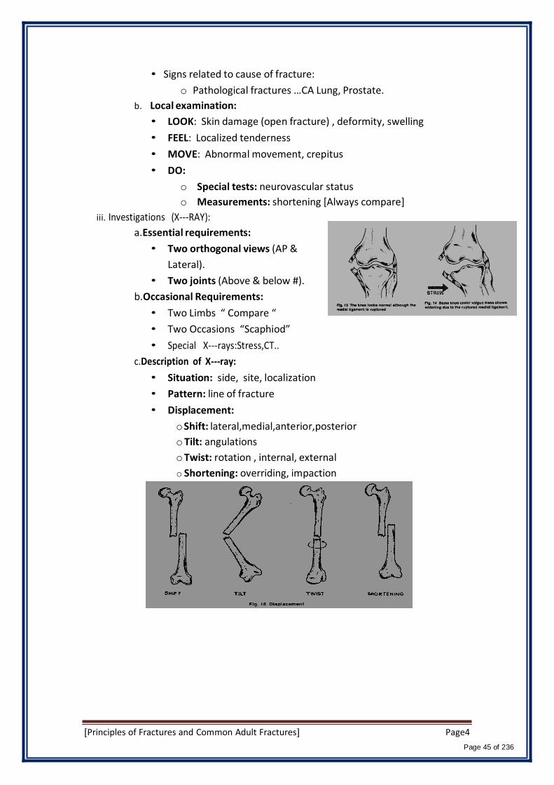
iii. Investigations (X-‐‐RAY):
a.Essential requirements:
• Two orthogonal views (AP &
Lateral).
• Two joints (Above & below #).
b.Occasional Requirements:
• Two Limbs “ Compare “
• Two Occasions “Scaphiod”
• Special X-‐‐rays:Stress,CT..
c.Description of X-‐ray:
• Situation: side, site, localization
• Pattern: line of fracture
• Displacement:
o Shift: lateral,medial,anterior,posterior
o Tilt: angulations
o Twist: rotation , internal, external
o Shortening: overriding, impaction
[Principles of Fractures and Common Adult Fractures] Page4
Page 45 of 236

Repair of Fracture:
a. Primary repair:
• With Rigid Internal Fixation (open the fracture take out the
hematoma and fix the bone in place)
• No Callus formation
• Active Haversian remolding
• Long time of healing
b.Secondary repair:
• Without rigid fixation(keep the hematoma and just do back
slab or full cast and allow for healing)
• Commonest type even with internal fixation (In lower limp fractures, they
usually put an implant in the medullary canal without opening the fracture
“keep the hematoma”allowing secondary healing)
• Stages:
Time Factor-‐‐ Perkin’s formula:
Union Consolidation
Upper limb Spiral
Transverse
Lower Limb Spiral
Transverse
3 weeks
6 weeks
6 weeks
12 weeks
6 weeks
12 weeks
12 weeks
24 weeks
Fracture in children
• Different from those in adults.
• Children's bones are more malleable, allowing a plastic type of "bowing" injury.
• The periosteum is thicker than in adults and usually remains intact on one side of the fracture (greenstick fracture) which helps to:
1. stabilize any reduction, 2. decreases the amount of displacement, and 3. Lower incidence of open fractures in children than in adults.
• Healing is more rapid.
[Principles of Fractures and Common Adult Fractures] Page5
Page 46 of 236

• Open reduction is rarely indicated.
• High remolding rate.
• Growth disturbance.
• Often missed (poor communication).
• X-‐‐rays of both limbs for comparison.
Physeal Injuries:
• 30% of the fractures
• Salter-‐Harris classification: based on the radiographic appearance of the
fracture
Meta-‐‐physis
Epi-‐‐physis
Birth Fractures:
• Most commonly in the clavicle, humerus, hip, and femur.
• They rarely require surgery but frequently are diagnosed as pseudopalsy,
infection, or dislocation.
Fractures Caused by Child Abuse
• Between birth and 2 years of age.
• Multiple fractures in different stages of healing are almost always indicative of child
abuse.
• Multiple areas of large ecchymoses in different
stages of resolution (from black and blue to brown
and green) also are pathognomonic of child abuse.
• The most common sites of fractures caused by child
abuse are the humerus, tibia, and femur.
• Bone scan or a skeletal survey generally is indicated.
Pathological Fractures
• Fracture within an abnormal bone structure due to:
1. Congenital diseases (Osteogenesisimperfecta).
2. Infection (osteomyelitis).
3. Fracture through a cyst.
4. Metabolic diseases (Osteoporosis,Osteomalacia, Pagets disease).
5. Bone tumours (primary or metastatic).
[Principles of Fractures and Common Adult Fractures] Page6
Page 47 of 236
• Diagnosis: o History:
1. Insignificant amount of trauma. 2. Constitutional symptoms. 3. History of malignancy.
• Examination: o General: signs and symptoms of malignancy or infection.
o Local: 1. Tenderness, pain, swelling. 2. Muscle spasm and deformity is minimal.
• Investigation: o Radiology:
1. X-‐‐rays of the lesion, MRI, CT-‐‐scan. 2. X-‐‐ray / CT-‐‐chest (pulmonary Mets.) 3. Bone Scan.
o Laboratory: 1. CBC w/ dif., ESR, C-‐‐RP. 2. Acid phosphatase P, B J P 3. LDH, etc.
• Management: o The aim is to make patient more functional and pain free for the
remaining life span. o Early operative stability should be carried out.
o Chemotherapy, Radiation, Hormonal. • Indications for prophylactic fixation(surgical) due to metastasis:
o Involvement of the cortex.
o Increased pain. o Pure lysis. o Weight bearing area.
Fracture Management[Save life, save limb, save function] • GENERAL AIM: To save the patient’s life
• LOCALAIM:Rapid recovery of the injured part and its function
• GENERAL management: o Life threatening Injuries (Shock , Head, Chest, Abdomen)
• LOCALmanagement Dangers to viability: o Ischemia
o Infection • How to save function?
1. Reduction 2. Immobilization 3. Soft tissue treatment 4. functional activity & rehabilitation
Complications of fractures Boney Complications:
o Delayed Union: 1. Healing Slow but Active, Remove the cause! 2. Fracture Site Tender
[Principles of Fractures and Common Adult Fractures] Page7
Page 48 of 236

3. X-‐‐ Ray little Callus, Medulla Open o Nonunion:
1. Reparative process Stopped, Need Intervention 2. Painless, Abnormal Movement, Psudoarthrosis! 3. X-‐‐ Ray: Sclerosis, Blocked Medulla.
Delayed Union & Nonunion Causes: Local:
1. Poor Blood Supply 2. Soft Tissue Interposition 3. Infection 4. Inadequate Immobilization 5. Over-‐‐Distraction 6. Pathology, Tumors
General: 1. Nutritional status 2. Bone Disease 3. Old Age
Malunion:not healing properly in growth plate fractures is called coxavara, in order to avoid it you have to detect it early.
Causes: 1. Primary Neglected fractures 2. After Reduction! Watch and get X-‐‐Ray after 10 days. 3. Epiphyseal Growth plate cause deformities with time.
Avascular Necrosis: 1. Death of bone tissue from; impairment or loss of blood Supply.
Coxavara
2. Most common anatomical sites are the scaphoid, head of tallus and head of
femur.
3. Plane radiographs shows bone sclerosis and none or delayed union.
Myositis Ossificans:“not inflammation of the muscle! “
Sustaining a blunt injury that causes deep tissue bleeding
which lead to bone formationoutside the bone. The patient
complains of pain and limitation of movement at the site of
previous trauma or fractures. (Usually present after 6 months
to a year)
[Principles of Fractures and Common Adult Fractures] Page8
Page 49 of 236
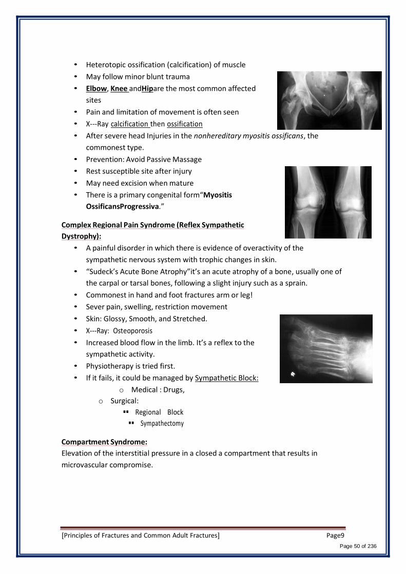
• Heterotopic ossification (calcification) of muscle
• May follow minor blunt trauma
• Elbow, Knee andHipare the most common affected
sites
• Pain and limitation of movement is often seen
• X-‐‐Ray calcification then ossification
• After severe head Injuries in the nonhereditary myositis ossificans, the
commonest type.
• Prevention: Avoid Passive Massage
• Rest susceptible site after injury
• May need excision when mature
• There is a primary congenital form“Myositis
OssificansProgressiva.”
Complex Regional Pain Syndrome (Reflex Sympathetic
Dystrophy):
• A painful disorder in which there is evidence of overactivity of the
sympathetic nervous system with trophic changes in skin.
• “Sudeck’s Acute Bone Atrophy”it’s an acute atrophy of a bone, usually one of
the carpal or tarsal bones, following a slight injury such as a sprain.
• Commonest in hand and foot fractures arm or leg!
• Sever pain, swelling, restriction movement
• Skin: Glossy, Smooth, and Stretched.
• X-‐‐Ray: Osteoporosis
• Increased blood flow in the limb. It’s a reflex to the
sympathetic activity.
• Physiotherapy is tried first.
• If it fails, it could be managed by Sympathetic Block:
o Medical : Drugs,
o Surgical:
Regional Block
Sympathectomy
Compartment Syndrome:
Elevation of the interstitial pressure in a closed a compartment that results in
microvascular compromise.
[Principles of Fractures and Common Adult Fractures] Page9
Page 50 of 236

Common Adult Fractures
Upper limb fractures:
1. Clavicle
2. Humeral(Proximal, shaft)
3. Both bone forearm(Radius, ulna)
4. Distal radius
Mechanism of Injury of the Upper Limb:
1. Mostly indirect (due to fall)
2. Commonly described as “fall on outstretched hand “or FOOSH injury.
3. Type of injury depends on
– position of the upper limb at the time of impact
– force of injury
– Age
Fracture of the clavicle:
• Common fracture (2.6%-‐‐12% of all fractures, 44%-‐‐66% of
fractures about the shoulder)
• Commonest site is the middle one third (80%)
• Mainly due to indirect injury
• Direct injury leads to comminuted fracture
Clinical evaluation:
• splinting of the affected extremity, with the arm
adducted (the patient is trying to support it with the
other arm)
• Assess for skin integrity
• neurovascular examination is necessary
• The chest should be auscultated (pleura are near to
fracture site→ pneumothorax)
Radiographic evaluation:
• Anteroposterior radiographs
Treatment (depending on the x-‐‐ray findings):
o Conservative:
– arm sling or figure of
eight sling
o Operative fixation:
– indicated if there is:
1. tenting of
the skin
2. open fracture
3. neurovascular injury
4. Nonunion
[Principles of Fractures and Common Adult Fractures] Page10
Page 51 of 236
– Plates and screws are used in adult upper limb injuries, nails almost
always used in lower limb injuries.
Complications:
1. Neurovascular compromise
2. Malunion
3. Nonunion( 85% occurring in the middle third)
4. Posttraumatic arthritis (AC or acromioclavicular joint , SC or Sternoclavicular
joint)
Proximal Humerus Fractures:
• Proximal Humerus ( includes surgical and anatomical neck )
• comprise 4% to 5% of all fractures
• Represent the most common humerus fracture (45%)
Clinical evaluation:
• Pain, swelling, tenderness, painful range of motion, and variable
crepitus. (any patient with fracture will present with those
symptoms)
• A careful neurovascular examination is essential, axillary nerve function.
Radiographic evaluation:
• AP and lateral views
• Computed tomography (it is the best modality
to assess intra-‐articular fractures) MCQ!
• Rule out Fracture-‐‐dislocation (Four-‐‐ part)
• Neer’s classification, has four parts:
1. humeral shaft
2. humeral head
3. Greater tuberosity
4. Lesser tuberosity
– A part is defined as displaced if (>0.5cm) of
fracture displacement or
(>45 degrees) of
angulation.
Treatment:
o Conservative:
– Non-‐‐ or minimally displaced fractures (less than 5 mm)
– 85% of fractures are minimally displaced or nondisplaced.
– Sling immobilization.
– Early shoulder motion at 7 to 10 days to prevent stiffness of the
joint.
[Principles of Fractures and Common Adult Fractures] Page11
Page 52 of 236

o Operative fixation:
– Displaced more than 5 to 10 mm.
– Three-‐‐ and four-‐part fractures.
– Replacement of humeral head for four-‐part in elderly.
Complications:
• Osteonecrosis: four-‐‐part (13%-‐‐34%), three-‐‐part (3% to 14%), anatomic neck
fractures.
• Vascular injury (5% to the axillary artery)
• Neural injury(Brachial plexus injury, Axillary nerve injury)
• Shoulder stiffness
• Nonunion, Malunion, Heterotopic ossification.
Fractures Shaft of the Humerus:
• 3% to 5% of all fractures
• Commonly Indirect injury(Spiral or Oblique)
• Direct injuries(transverse or comminuted )
• May be associated with Radial Nerveinjury
Clinical evaluation:
• Rule out open fractures
• Careful NV examination, with particular attention to radial nerve
function.
Radiologic evaluation:
• AP and lateral radiographs of the humerus
including the shoulder and elbow joints on each
view.
Classification (Descriptive):
• Open vs. closed.
• Location: proximal third, middle third, distal
third.
• Degree: nondisplaced, displaced.
• Direction and character: transverse, oblique,
spiral, segmental, comminuted
• Articular extension.
Treatment:
Conservative:
• Most of the time is conservatively because the bone is
covered and stabilized by muscles.
• Closed Reduction in upright position.
• U-‐‐shaped Slab from the elbow to the shoulder and after one
week change it to functional splint to prevent joints stiffness
[Principles of Fractures and Common Adult Fractures] Page12
Page 53 of 236
• Few weeks later Functional Brace may be used
Surgical:
• Multiple trauma (fractures)
• Inadequate closed reduction
• Pathologic fracture
• Associated vascular injury
• Floating elbow (freely moving elbow because both humerous and
forearm are fractured)
• Segmental fracture (fracture above or below. i.e the segment is not
stable)
• Intraarticular extension
• Bilateral humeral fractures
• Neurologic loss following
penetrating trauma
• Open fracture
Complications:
• Radial Nerve Injury (Wrist drop):
12% of fractures
• 2/3( 8%) Neuropraxia
• 1/3 ( 4%) lacerations or transection
• In open fractures; immediate exploration and ± repair
• closed injuries treated conservatively
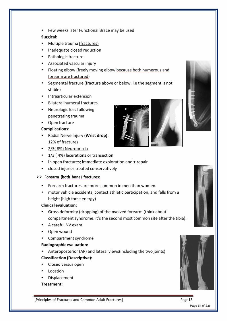
Forearm (both bone) fractures:
• Forearm fractures are more common in men than women.
• motor vehicle accidents, contact athletic participation, and falls from a
height (high force energy)
Clinical evaluation:
• Gross deformity (dropping) of theinvolved forearm (think about
compartment syndrome, it’s the second most common site after the tibia).
• A careful NV exam
• Open wound
• Compartment syndrome
Radiographic evaluation:
• Anteroposterior (AP) and lateral views(including the two joints)
Classification (Descriptive):
• Closed versus open
• Location
• Displacement
Treatment:
[Principles of Fractures and Common Adult Fractures] Page13
Page 54 of 236

• Surgical treatment is the rule because of instability. No role of conservative
treatment here.
Complications:
• Nonunion
• Compartment Syndrome
• Post-‐‐traumatic radioulnar synostosis radius and
ulnar bones unite together and become one bone
(unique for forearm fractures but not common)→(3% to 9% )
• malunion
• Infection
• Neurovascular injury
Distal Radius Fractures:
• Distal radius fractures are among the most common fractures of
the upper extremity.
• one-‐‐sixth of all fractures treated in emergency departments.
Clinical evaluation:
• Swollen wrist with ecchymosis, tenderness, and painful range of
motion.
• neurovascular assessment: median nerve function(Carpal tunnel
compression symptoms are common, 13%-‐‐23%)
• Look for open fracture.
Radiographic evaluation:
• Posteroanterior and lateral views.
• Radial inclination: averages 23 degrees (range, 13 to 30
degrees)
• Radial length: averages 11 mm (range, 8 to 18 mm).
• Palmar (volar) tilt: averages 11 degrees (range, 0 to 28
degrees).
Classifications:
• Articular extension:
o Extraarticular Vs.intraarticular
• Displacement:
o Colles’ fracture (most common of extra articular)
Vs.Smith fracture
Colles’ fracture:
– Extraarticular fractures.
– 90% of distal radius fractures
– Fall onto a hyperextended wrist with the forearm in pronation.
– dorsal displacement and angulation (apex volar) dinner fork
deformity
[Principles of Fractures and Common Adult Fractures] Page14
Page 55 of 236

– Radial shift, and radial shortening.
Smith’s fracture (reverse Colles’ fracture):
“Rare”
– A volar displacement
– volar angulation (apex dorsal) of the
distal radius (garden spade deformity).
– a fall onto a flexed wrist with the forearm
fixed in supination.
Barton’s fracture:
– Intraarticular fracture with dislocation or subluxation of the wrist (distal
radius)
– Treated surgically to prevent stiffness, OA of the joint.
Colles’ # Smith’s #
– Dorsal or volar rim of the distal radius is displaced with the hand and
carpus.
– Volar involvement is more common
– fall onto a dorsiflexed wrist with the forearm fixed in
pronation.
Barton’s (lateral) Barton’s (AP)
Treatment: Concevative:
• Acceptable radiographic parameters:
o Radial length: within 2 to 3 mm of the contralateral wrist.
o Palmar tilt: neutral tilt (0 degrees).
o Intraarticular step-‐off: <2 mm.
o Radial inclination: <5degrees.
• Below elbow cast
Surgical:
• Unacceptable reduction
• Secondary loss of reduction
• Articular comminution, step-‐off, or gap
• Barton’s fracture
Complications:
• Median nerve dysfunction
[Principles of Fractures and Common Adult Fractures] Page15
Page 56 of 236
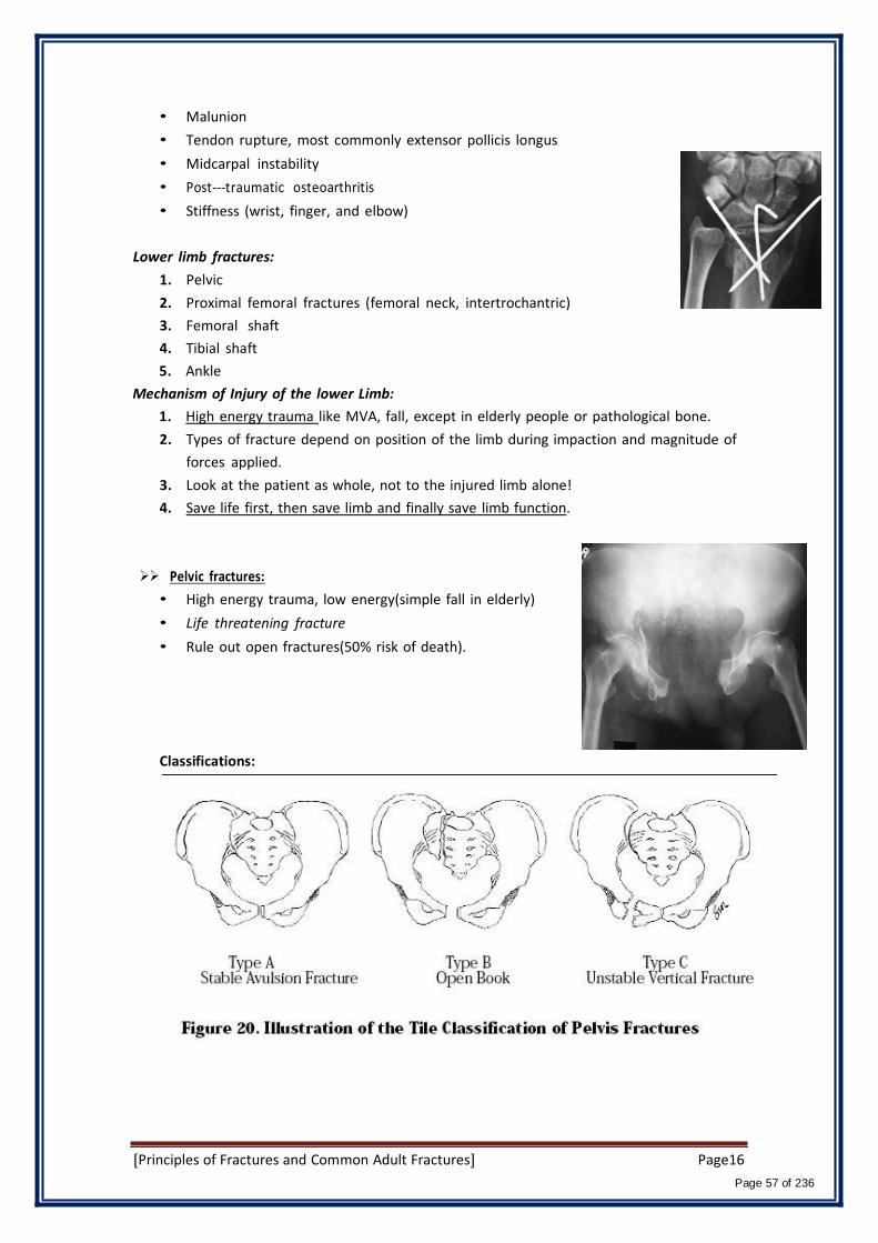
• Malunion
• Tendon rupture, most commonly extensor pollicis longus
• Midcarpal instability
• Post-‐traumatic osteoarthritis
• Stiffness (wrist, finger, and elbow)
Lower limb fractures:
1. Pelvic
2. Proximal femoral fractures (femoral neck, intertrochantric)
3. Femoral shaft
4. Tibial shaft
5. Ankle
Mechanism of Injury of the lower Limb:
1. High energy trauma like MVA, fall, except in elderly people or pathological bone.
2. Types of fracture depend on position of the limb during impaction and magnitude of
forces applied.
3. Look at the patient as whole, not to the injured limb alone!
4. Save life first, then save limb and finally save limb function.
Pelvic fractures:
• High energy trauma, low energy(simple fall in elderly)
• Life threatening fracture
• Rule out open fractures(50% risk of death).
Classifications:
[Principles of Fractures and Common Adult Fractures] Page16
Page 57 of 236

Radiographic evaluation:
• AP of the pelvis
• Inlet radiograph to see the ring from inside
• Outlet radiograph
• CT
Management:
• ATLS guidelines
• Type A: Conservative treatment
• Type B: Anterior fixation
• Type C: Both anterior &
posterior fixation
Complications:
Type B Type C
• Hemorrhage (hypovolemic shock, life threatening)
• Infection up to 25%
• Thromboembolism
• Bladder (15% )/bowel injuries
[Principles of Fractures and Common Adult Fractures] Page17
Page 58 of 236

• Neurological damage (L5-‐S1)
• Persistent sacro-‐iliac joint pain
• Malunion
Intertrochanteric fractures:
• Extracapsular (low risk for AN)
• Heals well, low risk for
osteonecrosis
• elderly, osteoporotic women
• Simple fall
Clinical evaluation:
• Inability to bear-‐weight
• Limb is short, abducted and externally
rotated
Radiological evaluation:
• AP and lateral(cross-‐‐table)
Treatment:
• Usually operative:
o Dynamic hip screw (DHS)
o Proximal femoral nail
DHS DHS Nail
Femoral neck (transcervical) fractures:
• Risk of osteonecrosis
• High and low mechanism of injuries (young Vs. elderly)
• Evaluation as inintertrochanteric fractures.
• There is a capsule that surrounds the neck of the femur,
and is attached, in front, to the intertrochanteric line;
carrying blood supply to the bone.
• Fracture distal to the capsule will compromise the blood
[Principles of Fractures and Common Adult Fractures] Page18
Page 59 of 236
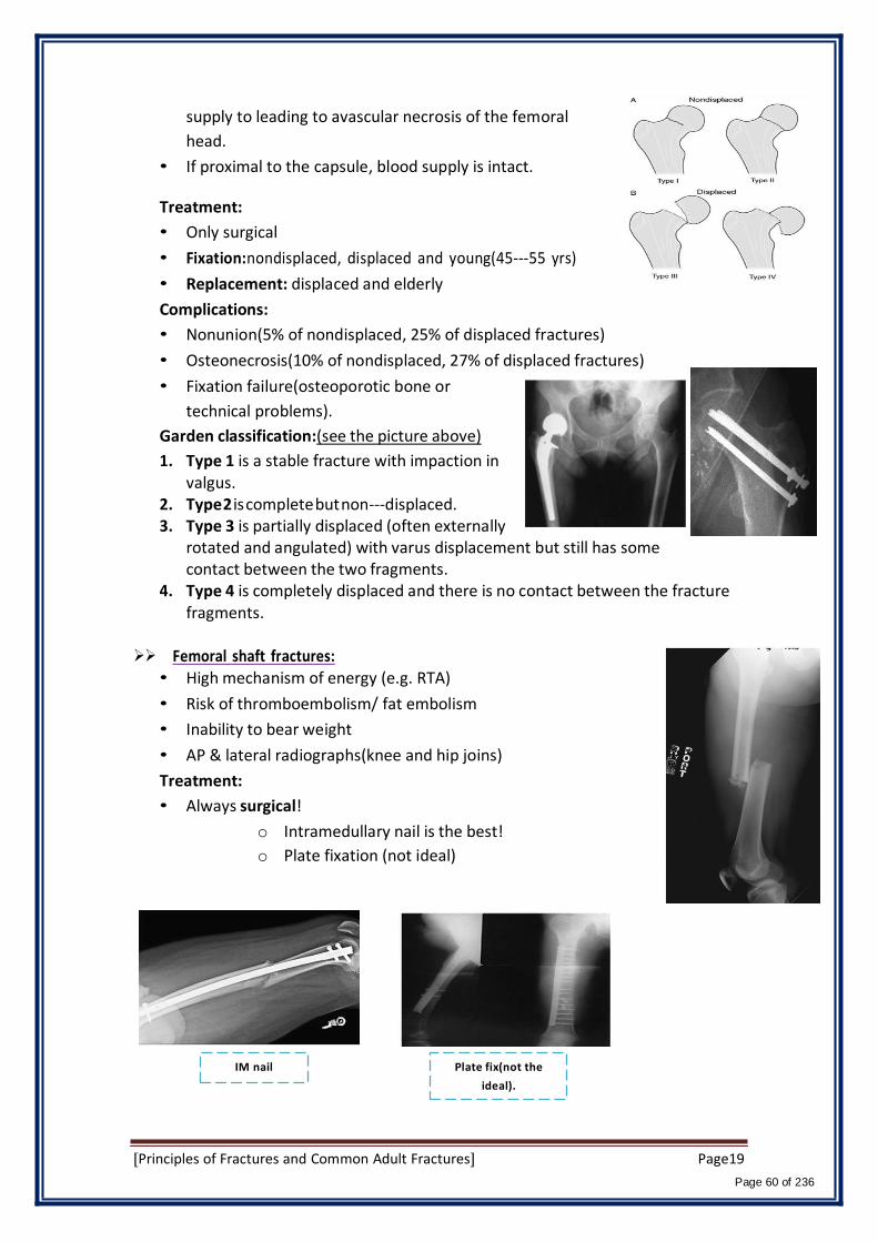
supply to leading to avascular necrosis of the femoral
head.
• If proximal to the capsule, blood supply is intact.
Treatment:
• Only surgical
• Fixation:nondisplaced, displaced and young(45-‐55 yrs)
• Replacement: displaced and elderly
Complications:
• Nonunion(5% of nondisplaced, 25% of displaced fractures)
• Osteonecrosis(10% of nondisplaced, 27% of displaced fractures)
• Fixation failure(osteoporotic bone or
technical problems).
Garden classification:(see the picture above)
1. Type 1 is a stable fracture with impaction in valgus.
2. Type 2 is complete but non-‐‐displaced. 3. Type 3 is partially displaced (often externally
rotated and angulated) with varus displacement but still has some contact between the two fragments.
4. Type 4 is completely displaced and there is no contact between the fracture fragments.
Femoral shaft fractures:
• High mechanism of energy (e.g. RTA)
• Risk of thromboembolism/ fat embolism
• Inability to bear weight
• AP & lateral radiographs(knee and hip joins)
Treatment:
• Always surgical!
o Intramedullary nail is the best!
o Plate fixation (not ideal)
IM nail Plate fix(not the
ideal).
[Principles of Fractures and Common Adult Fractures] Page19
Page 60 of 236
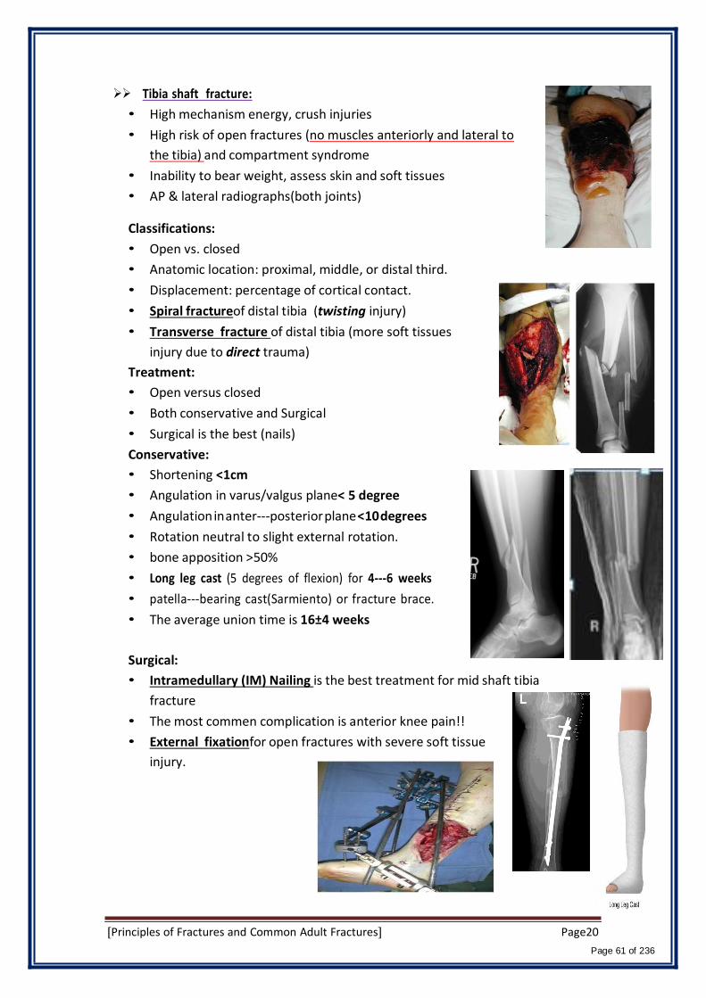
Tibia shaft fracture:
• High mechanism energy, crush injuries
• High risk of open fractures (no muscles anteriorly and lateral to
the tibia) and compartment syndrome
• Inability to bear weight, assess skin and soft tissues
• AP & lateral radiographs(both joints)
Classifications:
• Open vs. closed
• Anatomic location: proximal, middle, or distal third.
• Displacement: percentage of cortical contact.
• Spiral fractureof distal tibia (twisting injury)
• Transverse fracture of distal tibia (more soft tissues
injury due to direct trauma)
Treatment:
• Open versus closed
• Both conservative and Surgical
• Surgical is the best (nails)
Conservative:
• Shortening <1cm
• Angulation in varus/valgus plane< 5 degree
• Angulation in anter-‐‐posterior plane <10 degrees
• Rotation neutral to slight external rotation.
• bone apposition >50%
• Long leg cast (5 degrees of flexion) for 4-‐‐6 weeks
• patella-‐‐bearing cast(Sarmiento) or fracture brace.
• The average union time is 16±4 weeks
Surgical:
• Intramedullary (IM) Nailing is the best treatment for mid shaft tibia
fracture
• The most commen complication is anterior knee pain!!
• External fixationfor open fractures with severe soft tissue
injury.
[Principles of Fractures and Common Adult Fractures] Page20
Page 61 of 236
• Plate fixation:
– 97% success rates
– Complication: infection, wound breakdown, nonunion
– increase with higher-‐‐energy injury patterns.
Ankle Fractures:
• Incidence increased in elderly women
• Most ankle fractures are isolated malleolar fractures
• Open fractures are rare < 2%.
• Mechanism of injury: position of the foot at time of injury, the
magnitude, direction, and rate of loading.
Clinical evaluation:
• A dislocated ankle should be reduced and splinted immediately
(before radiographs if clinically evident)
Radiographic evaluation:
• AP, Lateral and mortise views (the only joint that should have 3
views, beside the shoulder if you’re suspecting dislocation)
AP view:
• Tibiofibula overlap of <10 mm is abnormal: syndesmotic injury.
• Tibiofibula clear space of >5 mm is abnormal: syndesmotic
injury
• Talar tilt
Lateral view:
• The dome of the talus should be centered under the tibia and
congruous with the tibial plafond
• Posterior tibialmalleolous fractures can be identified
Mortise view:
• Foot in 15 degrees of internal rotation
• A medial clear space
• Tibiofibular overlap
• Talar shift
Denis –Weber classification:
A. Infra-‐syndesmotic. (No problem to the joint) conservative
treatment.
[Principles of Fractures and Common Adult Fractures] Page21
Page 62 of 236
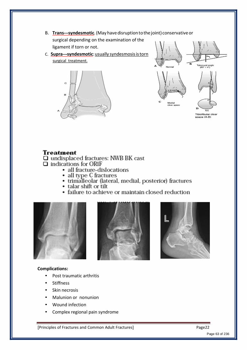
B. Trans-‐syndesmotic. (May have disruption to the joint) conservative or
surgical depending on the examination of the
ligament if torn or not.
C. Supra-‐syndesmotic: usually syndesmosis is torn
surgical treatment.
Complications:
• Post traumatic arthritis
• Stiffness
• Skin necrosis
• Malunion or nonunion
• Wound infection
• Complex regional pain syndrome
[Principles of Fractures and Common Adult Fractures] Page22
Page 63 of 236
Page 64 of 236
1Page Orthopaedic Emergencies 2
Isn't it funny how someone can say "I believe in Allah " but still follow the Satan who by the way also,
" believes " in Allah…
430 ORTHOPAEDICS TEAM
7- Orthopaedic Emergencies 2/ Red Flags (Open fracture, Fracture with
Neurovascular compromise, Pelvic fracture)
Team Members:
Hissah Alballa
Team Leader:
Ayedah Al-Ruhaimi.
Sources:
• Dr.Ahmad Bin Nasser’s slides.
• Lecture’s recording.
• 429 team work (group A1).
• Wikipedia
• Notes from clinical skills: Management of Open Fracture with Dr.Zaheer
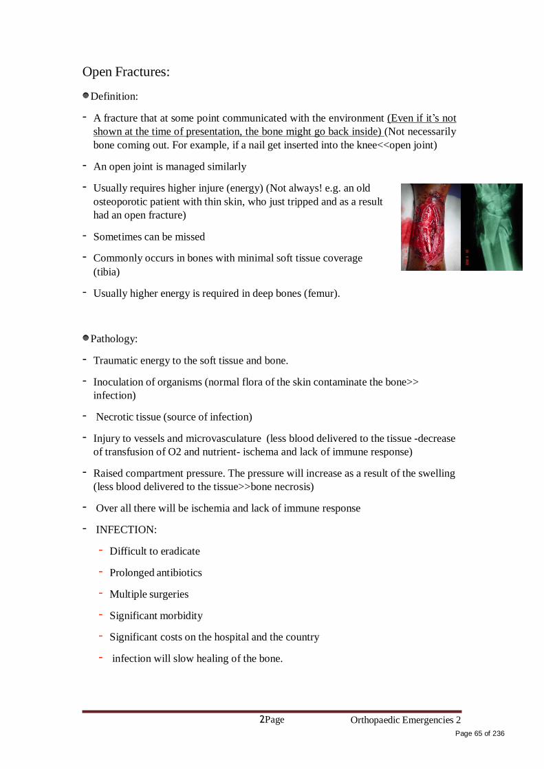
2Page Orthopaedic Emergencies 2
Page 65 of 236
Open Fractures:
Definition:
- A fracture that at some point communicated with the environment (Even if it’s not
shown at the time of presentation, the bone might go back inside) (Not necessarily
bone coming out. For example, if a nail get inserted into the knee<<open joint)
- An open joint is managed similarly
- Usually requires higher injure (energy) (Not always! e.g. an old
osteoporotic patient with thin skin, who just tripped and as a result
had an open fracture)
- Sometimes can be missed
- Commonly occurs in bones with minimal soft tissue coverage
(tibia)
- Usually higher energy is required in deep bones (femur).
Pathology:
- Traumatic energy to the soft tissue and bone.
- Inoculation of organisms (normal flora of the skin contaminate the bone>>
infection)
- Necrotic tissue (source of infection)
- Injury to vessels and microvasculature (less blood delivered to the tissue -decrease
of transfusion of O2 and nutrient- ischema and lack of immune response)
- Raised compartment pressure. The pressure will increase as a result of the swelling
(less blood delivered to the tissue>>bone necrosis)
- Over all there will be ischemia and lack of immune response
- INFECTION:
- Difficult to eradicate
- Prolonged antibiotics
- Multiple surgeries
- Significant morbidity
- Significant costs on the hospital and the country
- infection will slow healing of the bone.

3Page Orthopaedic Emergencies 2
Page 66 of 236
- Atrophy of the muscles
the patient will be depressed because of the long stay at the hospital
- An open fracture is a usually a “red flag” warning of significant trauma. Detailed
assessment of the patient is necessary
- An open fracture is associated with significant morbidity. Must act quickly take
fast detailed history.
- A delay in management is proven to increase the likelihood of complications. Give
urgent priority while triaging (open fracture is code 1 while closed 3) , provide
initial management and consult urgently. patient most be seen at least with in 20
min, if surgery is needed best with in 1 h maximum.
Diagnosis:
- Sometimes obvious!
- Other times, settle,,, be observant. The wound could be very
small (need high suspension )
- A wound close to a fracture is an open fracture until proven
otherwise.
- Whenever a fracture is diagnosed, go back and check the skin.
- A small wound continuously oozing blood bones are very
vascular, especially, if you see fat droplets within the blood (diagnostic), is an open
fracture!
- Not always close to the fracture, it may be around it. Especially the femur (the
deeper the bone more coverage of soft tissue around the bone the far the opening)
- Don’t probe!!
- If in doubt, use good light, if there is a break in the dermis or fat is seen, call it an
open fracture
- If unsure that the fracture is open consider it open. Better to overcall than miss it!
Algorithm:
- Assess and stabilize the patient, ATLS principles
- Assess the condition of the soft tissue and bone to help grade the open fracture.
Look for other injuries.
- Manage the wound locally

4Page Orthopaedic Emergencies 2
Page 67 of 236
- Stabilize the fracture
- IV antibiotics
- Tetanus status
Assessment:
- if polytrauma, apply ATLS principles
- if isolated injury:
- Hx:
• Mechanism and circumstances of injury. Detailed Hx must be taken. If it was
a RTA u need to know where was the patient siting (front or back), wearing
belt?, or pedestrian.
• Time since injury
• PMH/PSH/Allergy/Drugs/Smoking
• Tetanus vaccination status
- Examine the affected region for:
• Soft tissue: skin loss may require skin grafting
• Degree of contamination ( look for dirt )
• Necrotic and devitalized tissue
• Size of wound
• Coverage loss, muscle and skin
• Compartment syndrome
• Bone:
• Comminution
• Stripping of bone periosteum
• Examine away from injury to joint above and below
• X-rays to joint above and below
• Neurovascular status distally:
• On arrival and post reduction and splinting later.
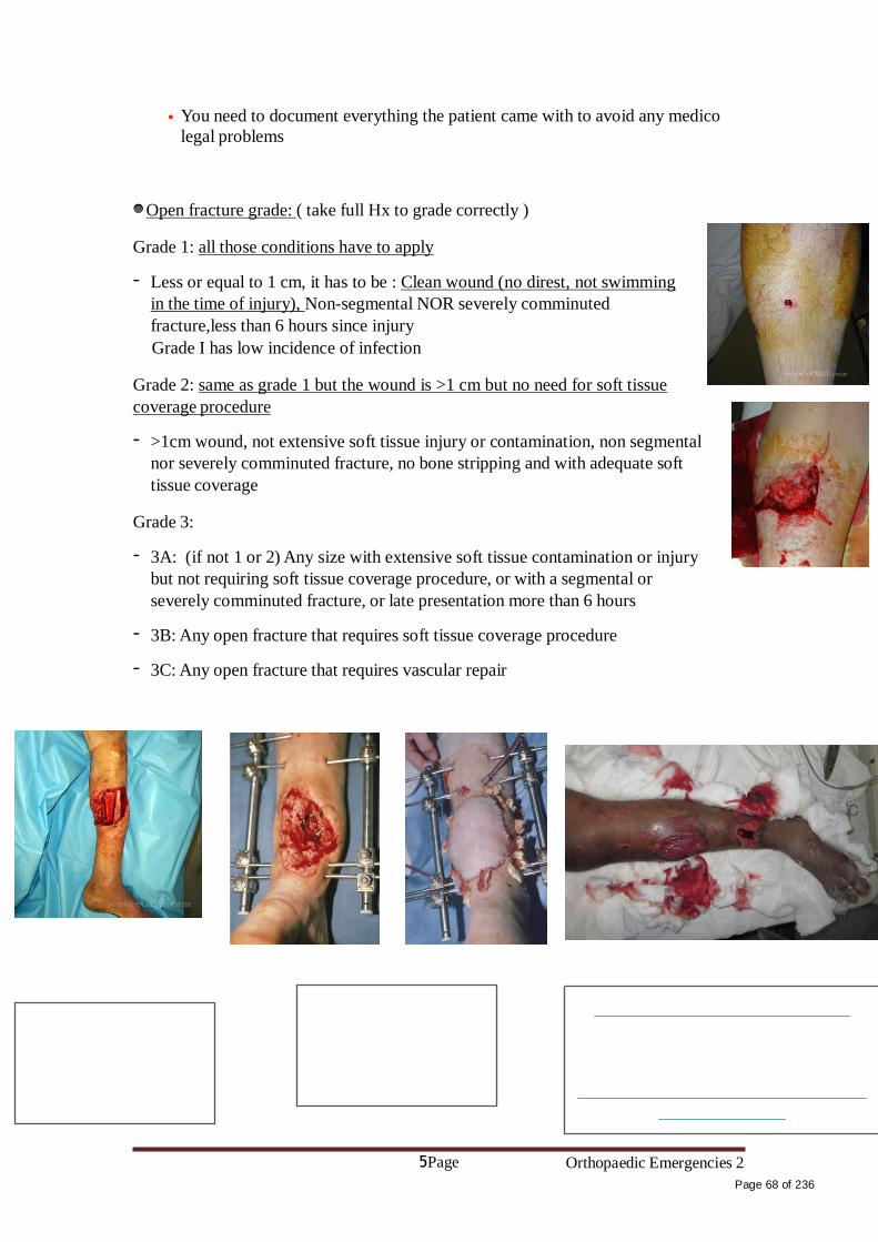
5Page Orthopaedic Emergencies 2
Page 68 of 236
• You need to document everything the patient came with to avoid any medico
legal problems
Open fracture grade: ( take full Hx to grade correctly )
Grade 1: all those conditions have to apply
- Less or equal to 1 cm, it has to be : Clean wound (no direst, not swimming
in the time of injury), Non-segmental NOR severely comminuted
fracture,less than 6 hours since injury
Grade I has low incidence of infection
Grade 2: same as grade 1 but the wound is >1 cm but no need for soft tissue
coverage procedure
- >1cm wound, not extensive soft tissue injury or contamination, non segmental
nor severely comminuted fracture, no bone stripping and with adequate soft
tissue coverage
Grade 3:
- 3A: (if not 1 or 2) Any size with extensive soft tissue contamination or injury
but not requiring soft tissue coverage procedure, or with a segmental or
severely comminuted fracture, or late presentation more than 6 hours
- 3B: Any open fracture that requires soft tissue coverage procedure
- 3C: Any open fracture that requires vascular repair

6Page Orthopaedic Emergencies 2
Page 69 of 236
Management:
I. Local: 1st step : Analgesics
- Take a picture. you don’t want to open it every time another doctor (intern,
registrar, consultant, ortho team) wants to examine
- If dirty, irrigate with normal saline to remove gross contamination. Don’t use
water even if sterile, the osmolarity of the water is more so the tissue will loss
fluid due to the concentration gradient. Just irrigate, if there is a big piece u
can remove it by sterile equipment.
- If bone sticking out try to reduce gently then immobilize and re-check
neurovascular status. Realignment in the ER to: decrease further damage.
decrease pain, decrease fat emboli.
- Cover with sterile wet gauze. If it becomes bloody use another one above it.
Wet because u don’t want to cause any injury when removing it.
- If bleeding apply direct pressure on wound. Never use a tourniquet, it will
compromise the blood supply.
- No culture swabs in ER. There are studies that say it’s useless.
II. Antibiotics:
- First generation Cephalosporin for gram positives (Ex: Cefazolin) in all open
fractures
- Aminoglycoside to cover gram negatives ( Ex: Gentamicin) sometimes not
required in grade 1 but in general it is safer to give in all grades, gram + is
most common but u can’t guarantee it’s only gram +. If it’s grade 1 and u said
I will only cover gram +, or u said I’ll cover both + -, both answers are
considered correct.
- Add penicillin or ampicillin or clindamycin for clostridium (will cause
gangrene) in grade 3 open fractures and all farm and soaked wounds. Also, if
>6 h
III. Tetanus prevention: Tetanus vaccine is a vaccine composed of deactivated tetanus
toxins. Passive immunity
Wound types:
- Clean wounds:
1. <6 hours from injury

7Page Orthopaedic Emergencies 2
Page 70 of 236
2. Not a farm injury
3. No significant devitalized tissue
4. Non immersed (soaked, e.g. if the injury accrued while swimming) wound
5. Non contaminated wound
- Other wounds
Clean wounds
Other wounds
Completed vaccination
Not completed or
unknown (pt doesn’t know
or unconscious)
Completed vaccination
Not completed or unknown
Booster
< 10
Booster
>10
Td 0.5ml IM
Booster
< 5years
Booster
> 5
TIG 250U
And Td 0.5ml IM
nothing Td 0.5
ml IM
nothing Td 0.5ml IM
- As soon as patient is stable and ready, alert the OR, and talk to the pt get consent
for surgery
- Plan: Irrigation w normal saline, debridement and fracture stabilization
- The sooner the less risk of further morbidity (more time>>more complications)
IV. In the OR: move to the OR as soon as the pt is stable enough.
- Extend wound if necessary. If small wound & the surgeon is unable to reach the
bone.
- Thorough irrigation to the bone.
- Debride all necrotic tissue (source of infection). start layer by layer from the skin,
subcutaneous tissue then muscle. How will you know the muscle in dead? from
the difference in colour, the muscle will not contract with mechanical or electrical
stimulation .
- Remove bone fragments without soft tissue attachment ( it will die>>source of
infection) except articular fragments. Deal with it later, the most imp thing now is
to stablize the fracture and prevent infection.
- Usually requires second look or more every 48-72 hours. Necrotic tissue may not
appear the first time.

8Page Orthopaedic Emergencies 2
Page 71 of 236
- Generally do not close open wounds (Except clean wound with <6 h) on first
look. Only approximate it to allow drainage so there won’t be a sudden infection.
But actually most of the time pts will come<6 h, the wound will be clean
properly, so the doctors will close the wound. Safe answer is to not close the
wound completely.
V. Fracture management:
- Generally avoid internal fixation (plate and screw)
- Generally external fixator is used.
- Femur and tibia shaft fractures can usually be treated immediately with IM nail
except severe injuries and contamination except severe injuries and
contamination
- Observe for compartment syndrome post- operatively
Results:
If all principles applied, in ideal senario where the pt was transferred immediately to
the hospital, the management was immediate and proper:
- 2% complication rate in grade 1
- 10% complication rate in grade 2
- Up to 50% complication rate in grade 3
Fractures with nerve or vascular injuries
- Don’t miss it !!!!
- Always perform an accurate (distal to the fracture) assessment at presentation, post
manipulation and reduction, post surgical fixation, serially until condition stabilizes
- Serial examination helpful in deciding line of treatment
- Serial examination helps avoid confusion
- High correlation between vascular injury and nerve injury. Proximity (anatomically
near each other)
Mechanisms:
- Penetrating trauma
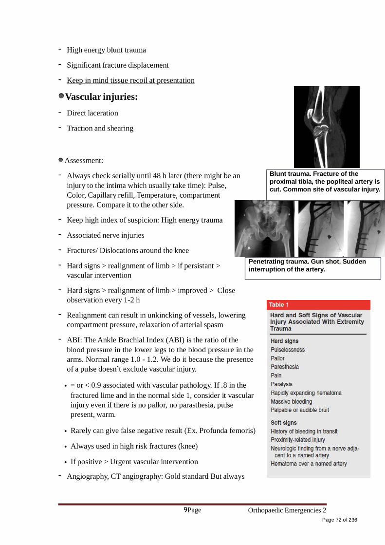
9Page Orthopaedic Emergencies 2
Page 72 of 236
- High energy blunt trauma
- Significant fracture displacement
- Keep in mind tissue recoil at presentation
Vascular injuries:
- Direct laceration
- Traction and shearing
Assessment:
- Always check serially until 48 h later (there might be an
injury to the intima which usually take time): Pulse,
Color, Capillary refill, Temperature, compartment
pressure. Compare it to the other side.
- Keep high index of suspicion: High energy trauma
- Associated nerve injuries
- Fractures/ Dislocations around the knee
- Hard signs > realignment of limb > if persistant >
vascular intervention
- Hard signs > realignment of limb > improved > Close
observation every 1-2 h
- Realignment can result in unkincking of vessels, lowering
compartment pressure, relaxation of arterial spasm
- ABI: The Ankle Brachial Index (ABI) is the ratio of the
Blunt trauma. Fracture of the
proximal tibia, the popliteal artery is
cut. Common site of vascular injury. Penetrating trauma. Gun shot. Sudden
interruption of the artery.
blood pressure in the lower legs to the blood pressure in the
arms. Normal range 1.0 - 1.2. We do it because the presence
of a pulse doesn’t exclude vascular injury.
• = or < 0.9 associated with vascular pathology. If .8 in the
fractured lime and in the normal side 1, consider it vascular
injury even if there is no pallor, no parasthesia, pulse
present, warm.
• Rarely can give false negative result (Ex. Profunda femoris)
• Always used in high risk fractures (knee)
• If positive > Urgent vascular intervention
- Angiography, CT angiography: Gold standard But always

10Page Orthopaedic Emergencies 2
Page 73 of 236
start with simple tests
• Not without risks
• Vascular surgeon to arrange with interventional radiologist
Management:
- Once vascular injury is confirmed:
• Coordination between: Vascular surgeon, Orthopedic surgeon, General surgeon. ( All AIM to save the Limb )The principle is that fixing the artery should be first.
But what if after after fixing the artery the bone moves (it’s unstable) and damage it
again? so there should be collaboration between the surgeons. most of the time the
orthopaedic surgeon will quickly stabilise the fracture fast by an general fixator.
- To emergently re-establish perfusion and protect repair with skeletal stabilization
- Warm ischemia (the hand is still attached to the body. cold ishemia is when the
amputated part was in a bag ice. In cold ishemia can wait longer)time dictates
treatment (Ex, the injury was 30 min ago, the orthopaedic surgeon can start before
the vascular)
- Most times, a quick external fixator is applied, followed by vascular repair
- Avoid prolonging warm ischemia to do
- Prolonged warm ischemia >6 hours —>Prophylactic fasciotomy. Sometime after
vascular repeat there will be re perfusion reaction>> edema in the muscle>>
compartment syndrome.
- Grade 3C open fractures have the worst outcome

11Page Orthopaedic Emergencies 2
Page 74 of 236
- Amputation may be necessary in severe cases
Nerve Injury:
- Cause of medico-legal concern
- Accurate assessment and documentation at presentation, post
reduction, post surgery is essential
- Remember to examine for motor and sensation prior to sedation
- Closed fractures not requiring surgery with nerve injuries
high chance that the nerve injury will be reversed without
intervention:
• Usually good outcome >80%
• Usually managed conservatively in the early stages
• Recovery may take more than 6 months. If not, do something.
- Intact nerve before reduction, absent after reduction:
Radial nerve run in the spiral groove
• Controversial management
• Usually observe
• It’s acceptable if u say u will redisplace the fracture. But it
been proven that it’s not always useful, just leave it. For us, go
with redisplacement.
- Fracture requiring surgery with nerve injury, what dictate the need
of surgery is the fracture not the nerve injury:
• Limited exploration. Don’t perform another incision.
- Open fracture with nerve injury:
• Here we have to explore the nerve, tag nerve ends for later
repair.
Distal redial fracture, cause radial nerve palsy(common)
Follow up:
- If no surgery was done.
- Clinically
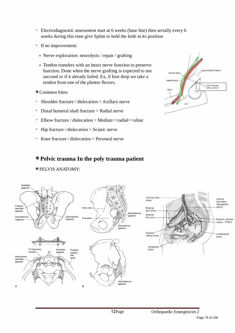
12Page Orthopaedic Emergencies 2
Page 75 of 236
- Electrodiagnostic assessment start at 6 weeks (base line) then serially every 6
weeks during this time give Splint to hold the limb in its position
- If no improvement:
• Nerve exploration: neurolysis / repair / grafting
• Tendon transfers with an intact nerve function to preserve
function. Done when the nerve grafting is expected to not
succeed or if it already failed. Ex, if foot drop we take a
tendon from one of the planter flexors.
Common Sites:
- Shoulder fracture / dislocation > Axillary nerve
- Distal humeral shaft fracture > Radial nerve
- Elbow fracture / dislocation > Median>>radial>>ulnar
- Hip fracture / dislocation > Sciatic nerve
- Knee fracture / dislocation > Peroneal nerve
Pelvic trauma In the poly trauma patient
PELVIS ANATOMY:

13Page Orthopaedic Emergencies 2
Page 76 of 236
Pathology:
A- anterior force causing a little opening in the sacro-illiac joint.
B- anterior force causing opening in the sacro-illiac joint and instability
C- More force cause open book pelvis. In this kind of injury, the left and right halves
of the pelvis are separated at front and rear, the front opening more than the rear, i.e.
like opening a book.
D, E and F lateral compression. e.g. car accident
G- Vertical shears pelvic fracture. Extremely unstable.
- Pelvic fractures / instability may cause life threatening
bleeding. The bleeding will be from the internal iliac artery
and it will be retroperitoneal so it’s hard to see in US. When
given blood I.V the blood pressure will increase for a while
then drop again.
- Diagnosing pelvic instability can save lives
Diagnosis:
- History: High vs. Low eneregy trauma
- Mechanism of injury: Anterior vs. Lateral vs. Axial force
- Pelvic skin contusion, bruising

14Page Orthopaedic Emergencies 2
Page 77 of 236
- Short extremity
- Careful neurologic assessment
- Primary survey : part of “C”. Circulation. If the pt showing signs of shock
(tachycardic, hypotension).
- Assess stability by gentle compression on the ASIS
- Traction on the leg and assess pelvic instability. the pt in supine position.
• traction in the leg while your hand in the lilac crest if it moves>>sheer.
- If unstable or painful:( pelvic binder )
• Apply sheet around hips and close the pelvis gently
• This results in decreased intra-pelvic volume leading to tamponading the bleeding by increasing the pressure around the injured vessel.>>BP will rise
• Traction on the leg to stabilize vertical instability.
• This minimizes ongoing vasculature injury and bleeding
• When the pt is stable enough arrange with the radiologist for radiological studies.
- Rectal exam:
• Bone fragments ( be careful)
• High riding prostate
• bleeding
- Blood at the meatus
- Labial or scrotal echymosis
- Vaginal exam
Management:
- Stabilize pelvis with binder
- If vertically unstable apply traction
- IV resuscitation
- Look for other injuries
- Check response
- If partial response, may require angiography for embolization of bleeders
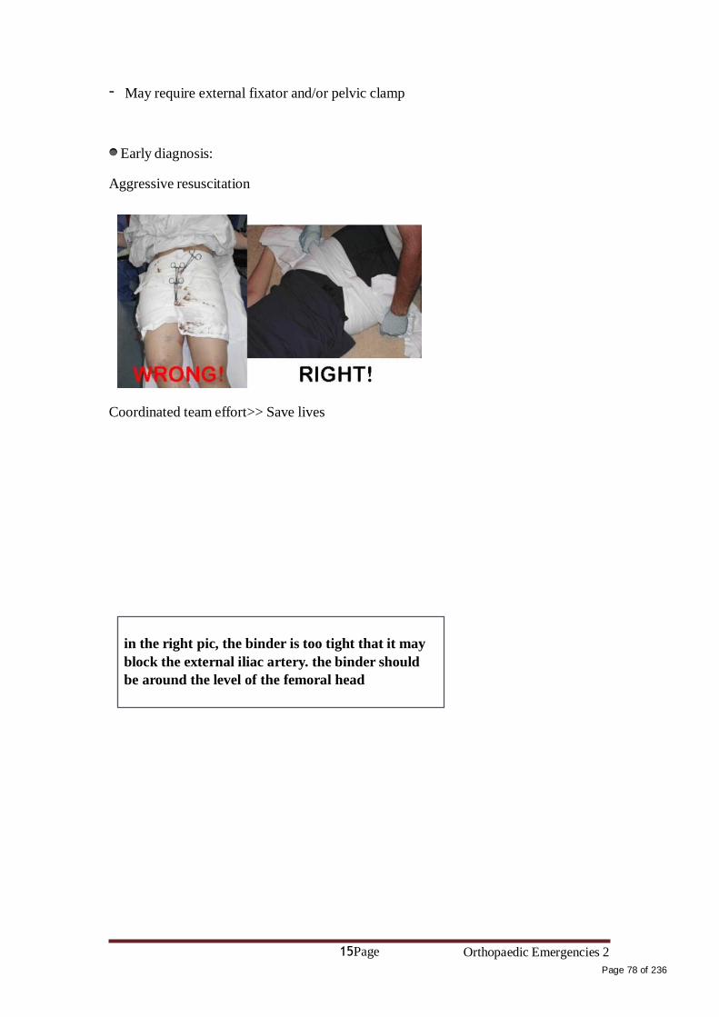
15Page Orthopaedic Emergencies 2
Page 78 of 236
- May require external fixator and/or pelvic clamp
Early diagnosis:
Aggressive resuscitation
Coordinated team effort>> Save lives
in the right pic, the binder is too tight that it may
block the external iliac artery. the binder should
be around the level of the femoral head
Isn't it funny how someone can say "I believe in Allah " but still follow the Satan who by the way also, " believes " in
Allah…
430 ORTHOPEDICS TEAM
Lecture: Compartment Syndrome/Acute Joint Dislocation.
Faten Al-‐Mohideb.
Team Members:
Hanan Al-‐Salman.
Aliya Al-‐Awaji.
Hadeel Al-‐Ghamdi.
Ghadeer Al-‐Wuhayd.
Hissa Al-‐Balla.
Lujain Al-‐Yousef.
Nouf Al-‐Hammad.
Jawaher Al-‐Faraydi.
Hiba Al-‐Rahiem.
Nour Al-‐Enezi.
Wejdan Al-‐Swayyid.
Arwa Abudawood.
Leena Al-‐Shaman.
Areej Al-‐Qunaitir.
Team Leader:
Ayedah Al-‐Ruhaimi
-‐‐The slides were provided by the doctor.
-‐‐429 team group A-‐1.
-‐‐Important notes in Red.
-‐‐Copied slides in Black.
-‐‐Doctor's notes in green.
Page1 Compartment Syndrome/ Acute Joint Dislocation
Page 79 of 236

Compartment Syndrome/Acute Joint Dislocation
• Objectives: 1. To explain the pathophysiology of CS 2. To Identify patients at risk of developing CS
3. To be able to diagnose and initially manage patients with CS 4. To be able to describe the possible complications of CS
• What is a compartment? - Area surrounded by fascia and contains muscles, bones, nerves and vessels.
• What is the tissue pressure normally? - Resting compartment pressure: 0-8 mmHg.
- Pressure difference between the tissues in the compartment
and capillary bed is what drives the gas exchange/diffusion. Therefore, when there is
an increase in the tissue in the compartment, an ischemia occur.
• Artery>arteriol>capillary bed (diffusion/exchange)>venule> vein
• Pathophysiology: • risk factor
1. Elevated tissue pressure
2. Absence of diffusion at the capillary bed 3. Cell damage and swelling
4. Further increase in tissue pressure 5. Lack of oxygenation
6. Vicious circle
• Threshold pressure: (IMP)
1. 30 mm Hg (rigid), (Intracompartment pressure)
2. Less than 30 mm Hg difference between compartment pressure and diastolic
pressure (clinically relevant)
Note: patient with hypotension they will have high compartment pressure and
more to get compartment syndrome.
• Risk factors (local):
Trauma, crush, fracture (open/closed)
Injection Bleeding
Prolonged vascular occlusion (reperfusion injury) Burns
Venomous bite
Page2 Compartment Syndrome/ Acute Joint Dislocation
Page 80 of 236

Intra-osseous fluid replacement
IV fluid extravasation Tight bandage
Post-surgery : most common factor.
Notes:
The risk with open fracture is higher than others, as it requires a high energy force.
Reperfusion injury is when the blood flow ceases transiently, the flow of the blood
the second time can cause leakage of the arterioles increases intracompartment
pressure.
Burns fluid collection in third space (interstitial) increases pressure of
the compartment
Post-surgery: bleeding, DVT (reperfusion injury)
Heavy weight lifters (body builders) might be a risk factor.
• Risk factors (general):
1. Head injury decreased consciousness!
2. Decreased concisenesslate diagnosis 3. Hypotension
Note : Unconscious patients can’t alert you when they feel pain (due to
compartment syndrome). So, check their compartment pressure every hour.
• Diagnosis:
• Early: >> all about pain Pain!!!
Pain increase with stretching the involved compartment Presence of risk factor
High index of suspicion Measurement of compartment pressure is high
The affected limb will be stiff
• Late: (IMP) Paresthesia Paralysis
Pallor
• Severely high pressure:
Pulselessness (RARE!)
Note: this happened when the compartment pressure is equal to the systolic
pressure.
• Tight, woody compartment
• Tender compartment
Page3 Compartment Syndrome/ Acute Joint Dislocation
Page 81 of 236

• Measurement: Rarely necessary
Must be done at area of highest expected pressure May give false low result
• Management: (IMP)
• Initial (undeveloped CS):
Maintain normal blood pressure Remove any constricting bandage
Keep limb at heart level Regular close monitoring (15-30 minute intervals)
Avoid nerve blocks, sedation and strong analgesia to obtain patients feed back
• Fully developed CS Maintain normal blood pressure
Remove any constricting bandage
Keep limb at heart level Diuresis to avoid kidney tubular injury if late.
Urgent surgical decompression (Fasciotomy). Muscle injury release of myoglobin toxic to kidney Acute Tubular
• Fasciotomy: (IMP) • Releasing the compartment fascia
• Allows swollen muscles to expand in volume
• Results in decreased compartment pressure
• Avoids further damage
• Does not reverse already occurred damage
• Ideally should be done as soon as diagnosis is made
• Should be done as long as there is still viable tissue
• Should not be done if there is no expected viable tissue, Otherwise infection is likely
•Debridement of all necrotic tissue is necessary
•Second and third look surgeries are often required
•Closure of skin is usually achieved after swelling has subsided
•Skin grafting is often required
• Indications: (IMP)
6 hours of total ischemia time (ex: arterial embolism) Significant tissue injury
Worsening initial clinical picture
Delayed presentation with a picture of developed CS
Absolute Compartment pressure >30 mmHg or <30 mm Hg difference from
diastolic pressure

Page4 Compartment Syndrome/ Acute Joint Dislocation
Page 82 of 236

Is a prophylactic procedure
Does not reverse injury to permanently damaged tissue
- So better to have a low threshold!
Notes:
The goal of fasciotomy is prevent further damage and not to regain the function of the
affected cells (reperfusion injury)
The ischemic muscles and nerves will be affected after one hour of the ischemia;
however permanent damage will occur after 6-8 hours.
• Complications: (IMP)
1. Myonecrosis>Myoglobenemia>Myoglobinuria>Kidney tubular damage
2. Loss of function of the involved compartment:
- Flexion contracture
- Paralysis
- Loss of sensation
Note:
The muscle will be replaced by a fibrous tissue (irreversibly damaged limb is not
amputated). However, it’s prone to infection if it was cut open.
3. Leg:
- Anterior compartment:
• Drop foot
- Deep posterior compartment:
• Clowed toes
• Loss of sensation in the sole
4. Forearm:
• Volar compartment
• Volkman contracture
Notes:
Volkmann's ischemic contracture is a permanent flexion contracture of the hand at the
wrist, resulting in a claw-like deformity of the hand and fingers.
"Acute Joint Dislocation"
• Objectives:
1. To describe mechanisms of joint stability 2. To be able diagnose patients with a possible acute joint dislocation
3. to be able to describe general principles of managing a patient with a dislocated joint
4. to describe possible complications of joint dislocations in general and in major joints such as the shoulder, hip and knee
• Joint stability: 1. Bony stability
Shape of the joint (ball and socket vs. round on flat)
Page5 Compartment Syndrome/ Acute Joint Dislocation
Page 83 of 236
2. Soft Tissue:
Dynamic stabilizer: Tendons/Muscles Static stabilizer: Ligaments ± meniscus/labrum
3. Complex synergy leading to a FUNCTIONAL and STABLE joint
4. It takes higher energy to dislocate a joint with bony stability than a joint with
mainly soft tissue stability
5. Connective tissue disorders may lead to increased joint instability due to
abnormal soft tissue stabilizers.
6. Dislocation of a major joint should lead to considering other injuries.
Notes:
Shoulder is round on flat (the glenoid surface persay is flat, however the labrum
on top of the surface renders it a round-like structure) prone to dislocation.
Connective tissue disorders: lax ligaments.
Some joints have different positions in which they can be easily dislocated. (i.e.
ball-throwing position risk for shoulder dislocation)
• At risk group: Major trauma victims
Athletes and sport enthusiasts Connective tissue disorder patients
• When a joint is subjected to sufficient force in certain directions it might sustain a fracture, a dislocation or a fracture dislocation
• Different joints have different force victors that may lead to a dislocation
• A joint might dislocate in different directions
• A joint dislocation is described by stating the location of the distal segment:
Anterior shoulder dislocation: anterior displacement of the humeral head
relative to the glenoid
Posterior hip dislocation: posterior displacement of the femoral head relative
to the acetabulum
• Dislocation: • Total loss of contact between the articular surfaces of the joint
• Sublaxation: • Partial loss of contact between the articular surfaces of the joint
• Acute joint dislocation
• Chronic joint dislocation: Example of chronic joint dislocation is DDH. Obviously, a recurrent joint
dislocation is NOT a chronic joint dislocation.
Page6 Compartment Syndrome/ Acute Joint Dislocation

Page 84
of 236

• Diagnosis:
• History of a traumatic event (major trauma or any trauma with the limb in high risk
position) • Pain and inability to use the limb
• Deformity
• Shortening • Malalignment
• Malrotation
• Should check for other injuries (distracting injury)
• Should always check the distal neurovascular status.
• Should check for compartment syndrome
• Shortening is a hint in diagnosing DDH.
• X-rays: 1. Should be done urgently without delay if dislocation is suspected.
2. Two perpendicular views of the involved joint .
3. Occasionally, special views are required such as the axillary view for shoulder
dislocation .
4. X-rays to the joint above and below.
• Management principles: (Very IMP)
• Must rule out other injuries
• Pain relief
• Urgent reduction
• Check stability and safety zone
• Check neurovascular status after reduction
• X-rays after reduction
• Protect the joint
• Rehabilitation • Follow for late complications
Notes :
Safety zone: The range of motion with stability (no danger of dislocation) For
example, 10 degrees flexion and 20 degrees extension. If the patient goes beyond that, the
joint will get unstable and might be dislocated.
Neurovascular status should be checked before and after the reduction. This goes for
the x-rays as well.
Page7 Compartment Syndrome/ Acute Joint Dislocation
Page 85 of 236

• Reduction:
• Monitor vitals
• IV analgesia (opiod) • IV sedation (to relax the muscles)
• Gradual traction to distract the joint
• Realignment and rotation to reduce the joint based on direction of
dislocation
• A palpable clunk well be felt
• Check ROM and stability of the joint
• Muscle relaxant to allow the traction and distracion.
• Once joint is felt to be reduced, check distal NV status . • If it was intact before but not after, farther urgent management is needed
• If it was not present before but intact after, check again later to confirm.
• Observe patients vitals until medications wear out.
• Stabilize joint and get X-rays.
• If irreducible or partial reduction only:
- Urgent closed reduction under general anesthesia and possible open reduction if
closed reduction fails
- Usually due to
Insufficient muscle relaxation Entrapment of soft tissue
- Fracture dislocation Open reduction (obviously!)
• Special considerations: - A fracture dislocation is usually reduced in an open fashion in the operating room
- Must confirm concentric reduction on the x-rays, otherwise an open reduction should be performed.
Page8 Compartment Syndrome/ Acute Joint Dislocation
Page 86 of 236
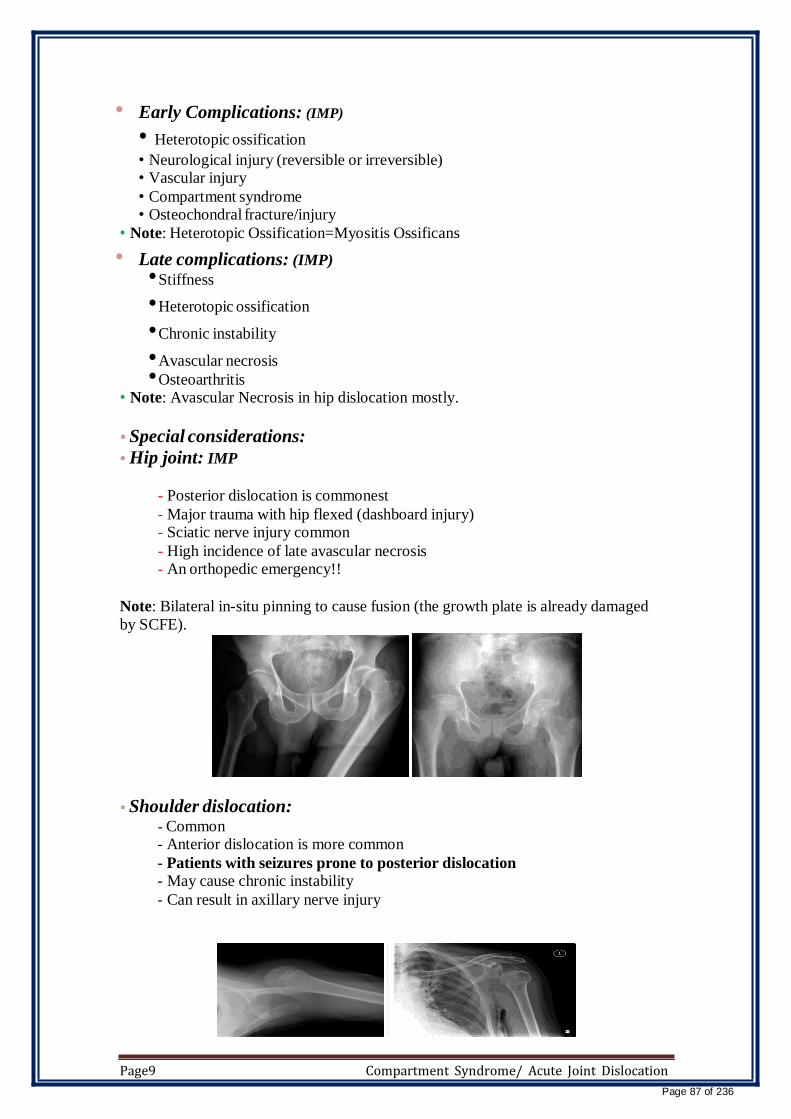
• Early Complications: (IMP)
• Heterotopic ossification
• Neurological injury (reversible or irreversible) • Vascular injury
• Compartment syndrome • Osteochondral fracture/injury
• Note: Heterotopic Ossification=Myositis Ossificans
• Late complications: (IMP)
• Stiffness
• Heterotopic ossification
• Chronic instability
• Avascular necrosis
• Osteoarthritis • Note: Avascular Necrosis in hip dislocation mostly.
• Special considerations:
• Hip joint: IMP
- Posterior dislocation is commonest
- Major trauma with hip flexed (dashboard injury) - Sciatic nerve injury common
- High incidence of late avascular necrosis - An orthopedic emergency!!
Note: Bilateral in-situ pinning to cause fusion (the growth plate is already damaged
by SCFE).
• Shoulder dislocation: - Common - Anterior dislocation is more common
- Patients with seizures prone to posterior dislocation - May cause chronic instability
- Can result in axillary nerve injury
Page9 Compartment Syndrome/ Acute Joint Dislocation
Page 87 of 236
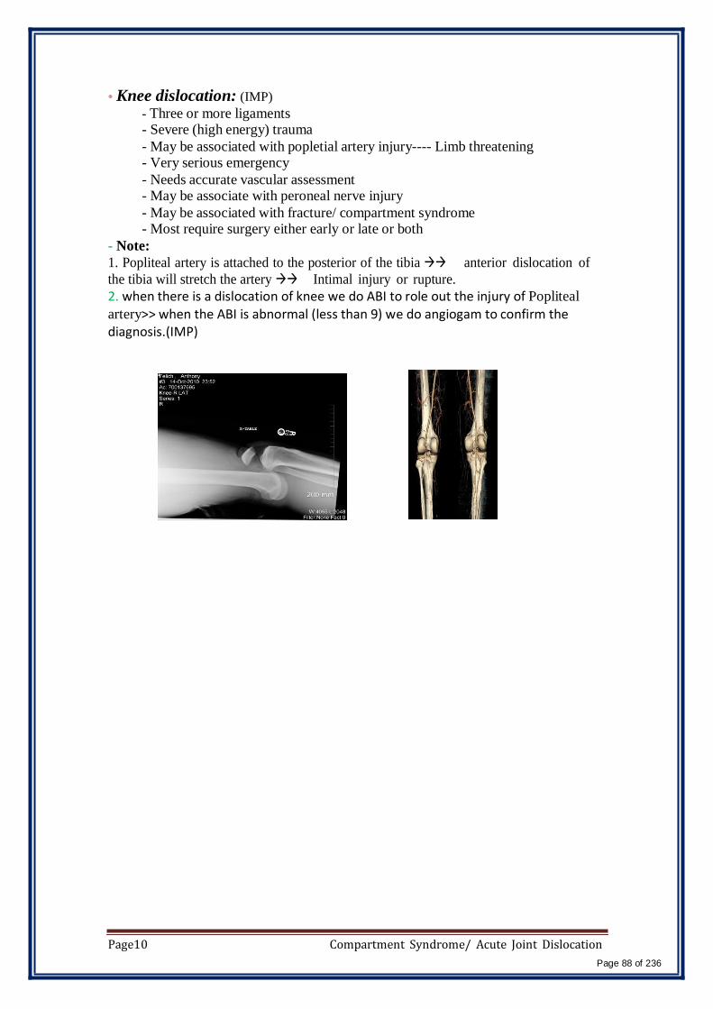
• Knee dislocation: (IMP)
- Three or more ligaments - Severe (high energy) trauma
- May be associated with popletial artery injury---- Limb threatening - Very serious emergency
- Needs accurate vascular assessment - May be associate with peroneal nerve injury
- May be associated with fracture/ compartment syndrome - Most require surgery either early or late or both
- Note:
1. Popliteal artery is attached to the posterior of the tibia anterior dislocation of
the tibia will stretch the artery Intimal injury or rupture.
2. when there is a dislocation of knee we do ABI to role out the injury of Popliteal
artery>> when the ABI is abnormal (less than 9) we do angiogam to confirm the diagnosis.(IMP)
Page10 Compartment Syndrome/ Acute Joint Dislocation
Page 88 of 236

Isn't it funny how someone can say, "I believe in Allah " but still follow the Satan who by the way also, " believes " in
Allah…
430 ORTHOPEDICS TEAM
Lecture: Common spinal disorders.
Team Members:
Faten Al-Mohideb. Hanan Al-Salman. Aliya Al-Awaji.
Hadeel Al-Ghamdi. Ghadeer Al-Wuhayd. Hissa Al-Balla. Lujain
Al-Yousef. Nouf Al-Hammad. Jawaher Al-Faraydi.
Hiba Al-Rahiem. Nour Al-Enezi. Wejdan Al-Swayyid.
Arwa Abudawood. Leena Al-Shaman. Areej Al-Qunaitir.
Team Leader:
- The slides were provided by the doctor.
- Important notes in Red
- Copied slides in Black.
-Doctor's notes in green.
Ayedah Al-Ruhaimi.
[Common Spinal Disorders] Page1
Page 89 of 236
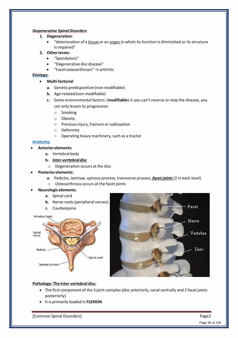
o Smoking
o Obesity
o Previous injury, fracture or subluxation
o Deformity
o Operating heavy machinery, such as a tractor
Anatomy:
Degenerative Spinal Disorders 1. Degeneration:
“deterioration of a tissue or an organ in which its function is diminished or its structure is impaired”
2. Other terms:
“Spondylosis”
“Degenerative disc disease” “Facet osteoarthrosis” → arthritis
Etiology:
Multi-factorial
a. Genetic predisposition (non-modifiable)
b. Age-related (non-modifiable)
c. Some environmental factors: (modifiable)→ you can’t reverse or stop the disease, you
can only lessen its progression.
Anterior elements:
a. Vertebral body
b. Inter-vertebral disc
o Degeneration occurs at the disc
Posterior elements:
a. Pedicles, laminae, spinous process, transverse process, facet joints (2 in each level)
o Osteoarthrosis occurs at the facet joints
Neurologic elements:
a. Spinal cord
b. Nerve roots (peripheral nerves)
c. Caudaequina
Pathology: The inter-vertebral disc:
The first component of the 3 joint complex (disc anteriorly, canal centrally and 2 facet joints
posteriorly)
It is primarily loaded in FLEXION
[Common Spinal Disorders] Page2
Page 90 of 236
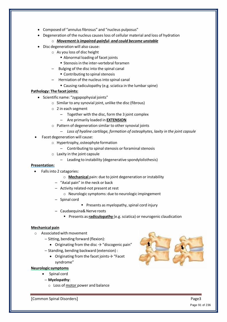
Composed of “annulus fibrosus” and “nucleus pulposus”
Degeneration of the nucleus causes loss of cellular material and loss of hydration
o Movement is impaired-painful- and could become unstable
Disc degeneration will also cause:
o As you loss of disc height
Abnormal loading of facet joints
Stenosis in the inter-vertebral foramen
– Bulging of the disc into the spinal canal
Contributing to spinal stenosis
– Herniation of the nucleus into spinal canal
Causing radiculopathy (e.g. sciatica in the lumbar spine)
Pathology: The facet joints:
Scientific name: “zygapophysial joints”
o Similar to any synovial joint, unlike the disc (fibrous)
o 2 in each segment
– Together with the disc, form the 3 joint complex
– Are primarily loaded in EXTENSION
o Pattern of degeneration similar to other synovial joints
– Loss of hyaline cartilage, formation of osteophytes, laxity in the joint capsule
Facet degeneration will cause:
o Hypertrophy, osteophyte formation
– Contributing to spinal stenosis or foraminal stenosis
o Laxity in the joint capsule
– Leading to instability (degenerative spondylolisthesis)
Presentation:
Falls into 2 catagories:
o Mechanical pain: due to joint degeneration or instability
– “Axial pain” in the neck or back
– Activity related-not present at rest
o Neurologic symptoms: due to neurologic impingement
– Spinal cord
Presents as myelopathy, spinal cord injury
– Caudaequina& Nerve roots
Presents as radiculopathy (e.g. sciatica) or neurogenic claudication
Mechanical pain
o Associated with movement
– Sitting, bending forward (flexion):
Originating from the disc → “discogenic pain”
– Standing, bending backward (extension) :
Originating from the facet joints→ “Facet
syndrome”
Neurologic symptoms
Spinal cord
– Myelopathy:
o Loss of motor power and balance
[Common Spinal Disorders] Page3
Page 91 of 236
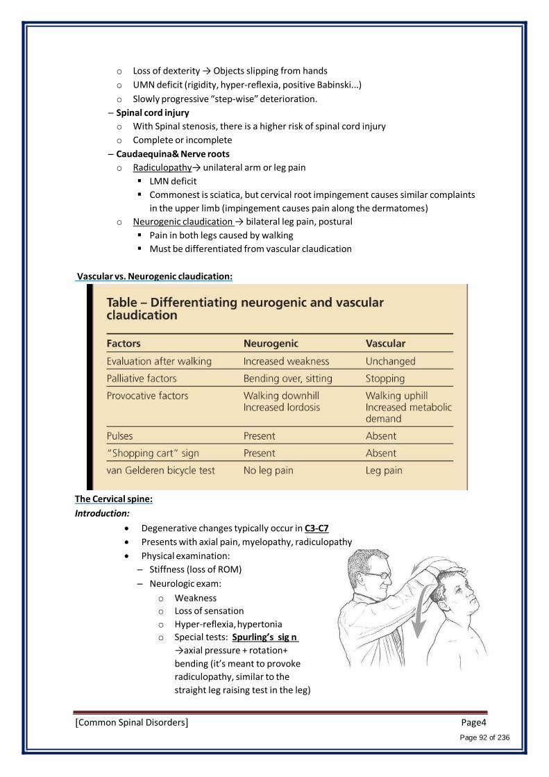
o Loss of dexterity → Objects slipping from hands
o UMN deficit (rigidity, hyper-reflexia, positive Babinski...)
o Slowly progressive “step-wise” deterioration.
– Spinal cord injury
o With Spinal stenosis, there is a higher risk of spinal cord injury
o Complete or incomplete
– Caudaequina& Nerve roots
o Radiculopathy→ unilateral arm or leg pain
LMN deficit
Commonest is sciatica, but cervical root impingement causes similar complaints
in the upper limb (impingement causes pain along the dermatomes)
o Neurogenic claudication → bilateral leg pain, postural
Pain in both legs caused by walking
Must be differentiated from vascular claudication
Vascular vs. Neurogenic claudication:
The Cervical spine:
Introduction:
Degenerative changes typically occur in C3-C7
Presents with axial pain, myelopathy, radiculopathy
Physical examination:
– Stiffness (loss of ROM)
– Neurologic exam:
o Weakness
o Loss of sensation
o Hyper-reflexia, hypertonia
o Special tests: Spurling’s sig n
→axial pressure + rotation+
bending (it’s meant to provoke
radiculopathy, similar to the
straight leg raising test in the leg)
[Common Spinal Disorders] Page4
Page 92 of 236

Management:
a. Conservative treatment
– First line of treatment for axial neck pain and mild neurologic symptoms (e.g. mild
radiculopathy without any motor deficit)
o Physiotherapy:
– Focus on ROM and muscle strengthening
o Non-steroidal anti-inflammatory medications (NSAID): forjoint (axial) pain
– E.g. Diclofenac, ibuprofen, naproxen
o Neuropathic medication: for radiculopathy pain
– E.g. Gabapentin or pregabalin
b. Surgical management(motor weakness is an indication for
surgery!)
– Indicated for:
o Spinal stenosis causing myelopathy
o Disc herniation causing severe radiculopathy and
weakness
o Failure of conservative treatment of axial neck pain or
mild radiculopathy
– Procedures:
o Anterior discectomy and fusion
o Posterior laminectomy
The Lumbar spine:
Introduction:
Degenerative changes typically occur in L3-S1
Anterior Discectomy + fusion
Presents with axial pain, Sciatica (radiculopathy) , neurogenic claudication
Physical examination:
– Stiffness (loss of ROM)
– Neurologic exam:
o Weakness
o Loss of sensation
o Hypo-reflexia, hypo-tonia
o Special tests: SLRT (straight leg raising test)
Management:
Axial low back pain:
– Conservative treatment if first-line and mainstay of treatment
o Physiotherapy: core muscle strengthening, posture training
o NSAID
– Surgical treatment indicated for:
o Instabilityor deformity(e.g. high-grade spondylolisthesis)
o Failure of conservative treatment
E.g. Lumbar Spondylosis:
[Common Spinal Disorders] Page5
Page 93 of 236

Spinal stenosis:(in the cervical spine it cause myelopathy)
– Conservative treatment is first line of treatment
o Activity modification, analgesics, epidural cortico-steroid injections
– Surgical treatment
o Indicated for:
Motor weaknesse.g. drop foot
failure of –minimum- 6 months of conservative treatment
o Spinal posterior decompression (laminectomy) is the commonest procedure
E.g. Spinal Stenosis:
Disc herniation:
– Conservative treatment is first line of treatment for mild sciatica without motor deficit
o Short (2-3 day) period of rest, NSAID, physiotherapy, epidural cortico-steroid
injection
o 95% of sciatica resolves within the first 3 months without surgery
– Surgical treatment:
o Indicated for cauda-equina syndrome, motor deficit, failure of 3 months of
conservative treatment
o Procedure: Discectomy (only the herniated part)+ spinal fusion
Spinal Fusion E.g. Disc Herniation:
[Common Spinal Disorders] Page6
Page 94 of 236

Osteoporotic Vertebral Fractures:
Pathologic fractures
Anterior column (±middle column) only compromised (Wedge/Burst Fracture)
Often missed
Repetitive fractures result in kyphoticdeformity (hunchback)
Treatment of underlying cause!!
Spinal Deformities:
Scoliosis
– deformity of the spine in the Coronal plane
Kyphosis
– deformity of the spine in the Sagittal plane
Spondylolisthesis
– Translation of one vertebra over another
Types of scoliosis:
Congenital
–Associated with anomalies of the bony vertebral column, e.ghemivertebra
Acquired (=secondary)
–Secondary to other pathology, e.gtumor, infection, spinal cord anomalies, degeneration
Idiopathic
–Most common is adolescent type
– Adolescent idiopathic scoliosis:
o Three dimensional deformity of the spine
o Vertebral Rotation is the hallmark
o Presents with deformity with little or no pain
o Usually noticed by parents/others, not the patient herself/himself
o Examination: neurologically normal, positive Adams test
o Management depends on degree of
deformity
Imaging shows truncal shift
[Common Spinal Disorders] Page7
Page 95 of 236
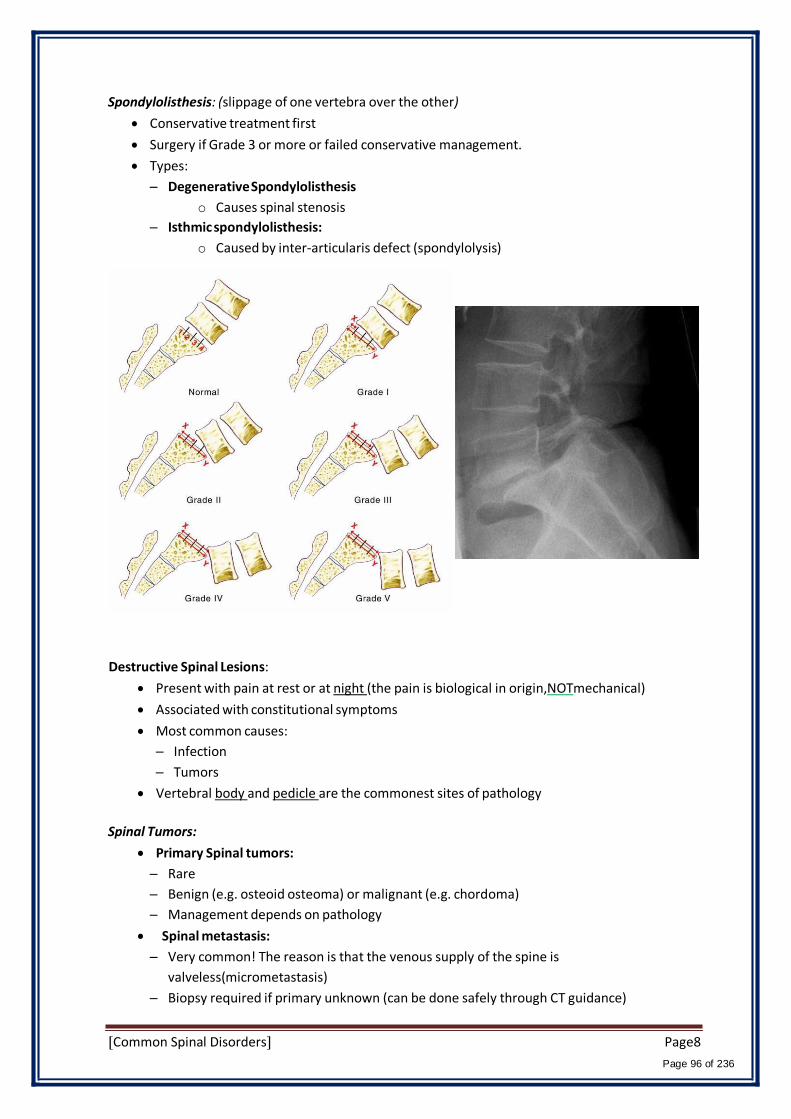
Spondylolisthesis: (slippage of one vertebra over the other)
Conservative treatment first
Surgery if Grade 3 or more or failed conservative management.
Types:
– Degenerative Spondylolisthesis
o Causes spinal stenosis
– Isthmic spondylolisthesis:
o Caused by inter-articularis defect (spondylolysis)
Destructive Spinal Lesions:
Present with pain at rest or at night (the pain is biological in origin,NOTmechanical)
Associated with constitutional symptoms
Most common causes:
– Infection
– Tumors
Vertebral body and pedicle are the commonest sites of pathology
Spinal Tumors:
Primary Spinal tumors:
– Rare
– Benign (e.g. osteoid osteoma) or malignant (e.g. chordoma)
– Management depends on pathology
Spinal metastasis:
– Very common! The reason is that the venous supply of the spine is
valveless(micrometastasis)
– Biopsy required if primary unknown (can be done safely through CT guidance)
[Common Spinal Disorders] Page8
Page 96 of 236

Spinal infections:
Most common is TBand Brucellosis
History of contact with TB patient, raw milk ingestion
Potentially treatable diseases once diagnosis is established and antimicrobials administered
Spinal Tuberculosis (with psoas abscess)
[Common Spinal Disorders] Page9
Page 97 of 236
Isn't it funny how someone can say "I believe in Allah” but still follow the Satan who by the way also, “believes” in
Allah…
430 ORTHOPEDICS TEAM
Lecture: Spinal Injuries.
Team Members:
Faten Al-‐Mohideb.
Hadeel Al-‐Ghamdi.
Lujain Al-‐Yousef.
Hiba Al-‐Rahiem.
Arwa Abudawood.
Hanan Al-‐Salman.
Ghadeer Al-‐Wuhayd.
Nouf Al-‐Hammad.
Nour Al-‐Enezi.
Leena Al-‐Shaman.
Aliya Al-‐Awaji.
Hissa Al-‐Balla.
Jawaher Al-‐Faraydi.
Wejdan Al-‐Swayyid.
Areej Al-‐Qunaitir.
Team Leader:
Ayedah Al-‐Ruhaimi.
_ The slides were provided by the doctor.
_ Important notes in Red.
_ 429 group A team in Orange.
_ Doctor's notes in Green.
_ Copied slides in Black.
Page1 [Spinal Injuries]
Page 98 of 236
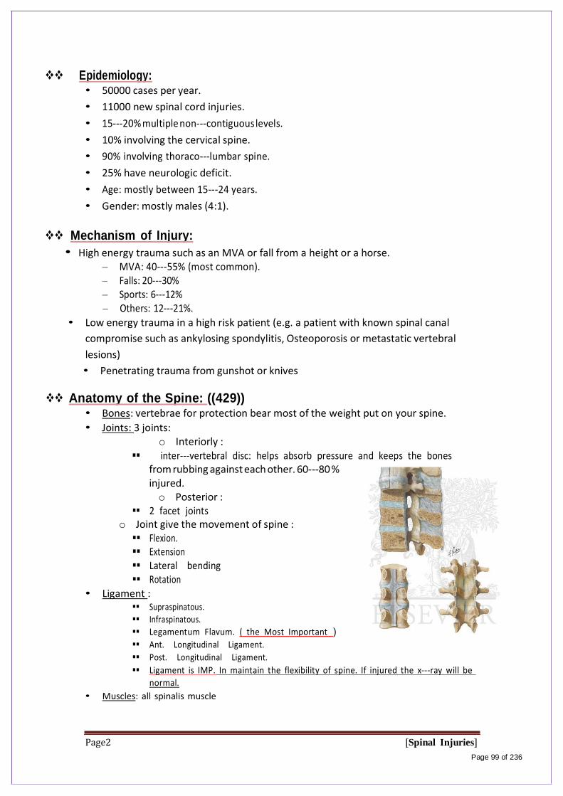
Epidemiology: • 50000 cases per year.
• 11000 new spinal cord injuries.
• 15-‐‐20% multiple non-‐‐contiguous levels.
• 10% involving the cervical spine.
• 90% involving thoraco-‐‐lumbar spine.
• 25% have neurologic deficit.
• Age: mostly between 15-‐‐24 years.
• Gender: mostly males (4:1).
Mechanism of Injury:
• High energy trauma such as an MVA or fall from a height or a horse. – MVA: 40-‐‐55% (most common). – Falls: 20-‐‐30% – Sports: 6-‐‐12% – Others: 12-‐‐21%.
• Low energy trauma in a high risk patient (e.g. a patient with known spinal canal
compromise such as ankylosing spondylitis, Osteoporosis or metastatic vertebral
lesions)
• Penetrating trauma from gunshot or knives Anatomy of the Spine: ((429))
• Bones: vertebrae for protection bear most of the weight put on your spine.
• Joints: 3 joints: o Interiorly :
inter-‐‐vertebral disc: helps absorb pressure and keeps the bones from rubbing against each other. 60-‐‐80 % injured. o Posterior :
2 facet joints o Joint give the movement of spine : Flexion. Extension Lateral bending Rotation
• Ligament : Supraspinatous.
Infraspinatous.
Legamentum Flavum. ( the Most Important )
Ant. Longitudinal Ligament.
Post. Longitudinal Ligament.
Ligament is IMP. In maintain the flexibility of spine. If injured the x-‐ray will be
normal.
• Muscles: all spinalis muscle
Page2 [Spinal Injuries]
Page 99 of 236

Cervical Anatomy: • C1 & C2 (50% of rotation occurs BTW C1 & C2 and then 5% at
each level). ( 50% of flexion and extension occurs BTW C1 and cranium)
o C1: No vertebra. Body Composed of Ant. & Post .arch.
o C2: Have Ant. Projection is: Odentoid process significant
in stability of C1 & C2. Open mouth x-‐‐ray (MCQ), we see the C1 &C2 and
their articulation. • C3-‐C7
C3-C7 is same. Have body +2 Lat. masses (facet joints). We differentiate it from the thoracic by Spinal
process.
Thoracic Spine • Articulate with ribs by body and spinal process (downward).
Lumbar Spine • More IMP.
• Lowest mobile segment.
• Most of ligament.
• More fracture.
• All joint here Ant +Post.
• Most of movement. o The Three columns (imp)
Ant. Column >Ant. part of body Middle column > Post. Part of body Post. Column > Pedicle + Lamina+ Spinal process.
• Stable injury : One column only (e.g. Wedge #in Ant. Column) Not required treatment or conservative only.
• Unstable injury: 2 or more column. Need intervention (surgery, etc.)
Page3 [Spinal Injuries]
Page 100 of 236

Spinal Column:
Page4 [Spinal Injuries]
Page 101 of 236
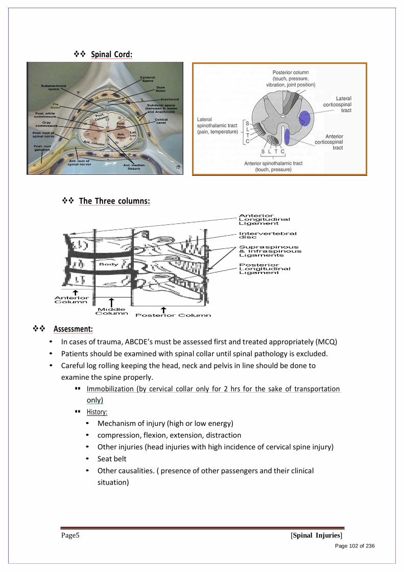
Spinal Cord:
The Three columns:
Assessment:
• In cases of trauma, ABCDE’s must be assessed first and treated appropriately (MCQ)
• Patients should be examined with spinal collar until spinal pathology is excluded.
• Careful log rolling keeping the head, neck and pelvis in line should be done to
examine the spine properly.
Immobilization (by cervical collar only for 2 hrs for the sake of transportation
only)
History:
• Mechanism of injury (high or low energy)
• compression, flexion, extension, distraction
• Other injuries (head injuries with high incidence of cervical spine injury)
• Seat belt
• Other causalities. ( presence of other passengers and their clinical
situation)
Page5 [Spinal Injuries]
Page 102 of 236

Physical examination:
• Inspection, palpation.
Neurologic examination (Muscle Test, Sensory exam, light touch, Sharp dull
discrimination, Vibration sense, Proprioception and two-‐point discrimination and
Reflexes).
Asia Score: Brief Trauma Neurologic Survey:
Page6 [Spinal Injuries]
Page 103 of 236

Level of Cord Injury determines level of function: (imp)
Notes: - You can palpate for Gaps and steps in the spine
C7 called: Cervical Prominence
- So, If there is a gap; you felt C1 then the other one below
it deep (not at the same level) then it is Steps
spondylotheiasis
- Gaps: you will feel one spine then below it space then
another Spine could be due to a rupture if the
ligaments .
- ASIA Scale: (American Spine injury association)
- The Closer the patient to normal (E) the better
prognosis.
(E excellent)
Prognosis for Recovery of spinal Cord Injury:
• Poor prognosis for recovery if:
o Pt arrives in shock.
o Pt cannot breath.
o Pt has a complete injury.
Severity of neurologic deficit
• Complete spinal cord injury:
o Flaccid paralysis below level of injury.
o May involve diaphragm if injury above C5.
o Sympathetic tone lost if fracture above T6.
• Incomplete
o Any sensation?
o Sacral sparing?
o Central cord syndrome:
Characterized by disproportionally (UL>LL).
Mechanism: hyper-‐‐extension.
Occur with or without fractures.
Recovery: 50% regaining function.
Prognosis is fair.
o Anterior cord syndrome:
Characterized by loss of corticospinal and
spinothalamic tract with preserved posterior column.
Mechanism: ischemia or infarction to spinal cord.
Common injury.
Recovery: 10%.
Page7 [Spinal Injuries]
Page 104 of 236
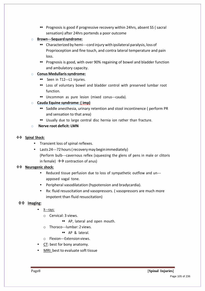
Prognosis is good if progressive recovery within 24hrs, absent SS ( sacral
sensation) after 24hrs portends a poor outcome
o Brown-‐Sequard syndrome:
Characterized by hemi-‐‐cord injury with ipsilateral paralysis, loss of
Proprioception and fine touch, and contra lateral temperature and pain
loss.
Prognosis is good, with over 90% regaining of bowel and bladder function
and ambulatory capacity.
o Conus Medullaris syndrome:
Seen in T12-‐L1 injuries.
Loss of voluntary bowel and bladder control with preserved lumbar root
function.
Uncommon as pure lesion (mixed conus-‐‐cauda).
o Cauda Equine syndrome: ( imp)
Saddle anesthesia, urinary retention and stool incontinence ( perform PR
and sensation to that area)
Usually due to large central disc hernia ion rather than fracture.
o Nerve root deficit: LMN
Spinal Shock:
• Transient loss of spinal reflexes.
• Lasts 24-‐‐72 hours ( recovery may begin immediately)
(Perform bulb-‐‐cavernous reflex (squeezing the glens of pens in male or clitoris
in female) contraction of anus)
Neurogenic shock:
• Reduced tissue perfusion due to loss of sympathetic outflow and un-‐‐
apposed vagal tone.
• Peripheral vasodilatation (hypotension and bradycardia).
• Rx: fluid resuscitation and vasopressors. ( vasopressors are much more
impotent than fluid resuscitation)
Imaging:
• X-‐‐rays:
o Cervical: 3 views.
AP, lateral and open mouth.
o Thoraco-‐‐lumbar: 2 views.
AP & lateral.
o Flexion-‐‐Extension views.
• CT: best for bony anatomy.
• MRI: best to evaluate soft tissue
Page8 [Spinal Injuries]
Page 105 of 236

Management of Spinal Injuries:
• Depends on: (MCQ)
o Level of injury.
o Degree and morphology of injury: STABILITY
o Presence of neurologic deficit.
o Other factors.
Some general rules:
• Stable injuries are usually treated conservatively.
• Unstable injuries usually require surgery.
• Neurologic compression requires decompression
Cervical spine fractures
Specific Injuries:
• Descriptive: depends on mechanism of injury.
o Flexion/extension.
o Compression/distraction.
o Shear.
• Presence of Sublaxation/dislocation
• SCI:
o High fracture results in quadriplegia.
o Low fracture results in paraplegia. ( C6 , C7 or C7, T1 )
Page9 [Spinal Injuries]
Page 106 of 236
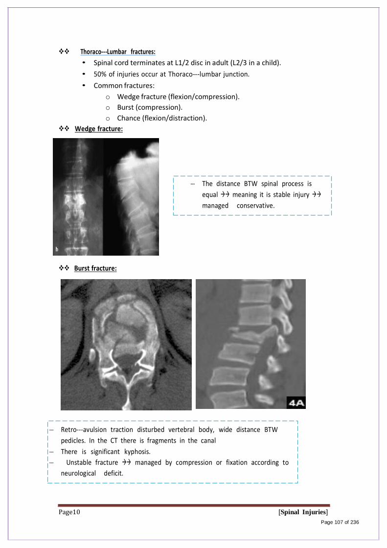
Thoraco-‐Lumbar fractures:
• Spinal cord terminates at L1/2 disc in adult (L2/3 in a child).
• 50% of injuries occur at Thoraco-‐‐lumbar junction.
• Common fractures:
o Wedge fracture (flexion/compression).
o Burst (compression).
o Chance (flexion/distraction).
Wedge fracture:
-‐‐ The distance BTW spinal process is
equal meaning it is stable injury
managed conservative.
Burst fracture:
-‐‐ Retro-‐‐avulsion traction disturbed vertebral body, wide distance BTW
pedicles. In the CT there is fragments in the canal
-‐‐ There is significant kyphosis.
-‐‐ Unstable fracture managed by compression or fixation according to
neurological deficit.
Page10 [Spinal Injuries]
Page 107 of 236
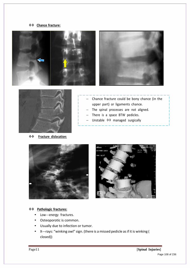
Chance fracture:
Fracture dislocation:
-‐‐ Chance fracture could be bony chance (in the
upper part) or ligaments chance.
-‐‐ The spinal processes are not aligned.
-‐‐ There is a space BTW pedicles.
-‐‐ Unstable managed surgically
Pathologic fractures:
• Low-‐‐energy fractures.
• Osteoporotic is common.
• Usually due to infection or tumor.
• X-‐‐rays: “winking owl” sign. (there is a missed pedicle as if it is winking (
closed))
Page11 [Spinal Injuries]
Page 108 of 236
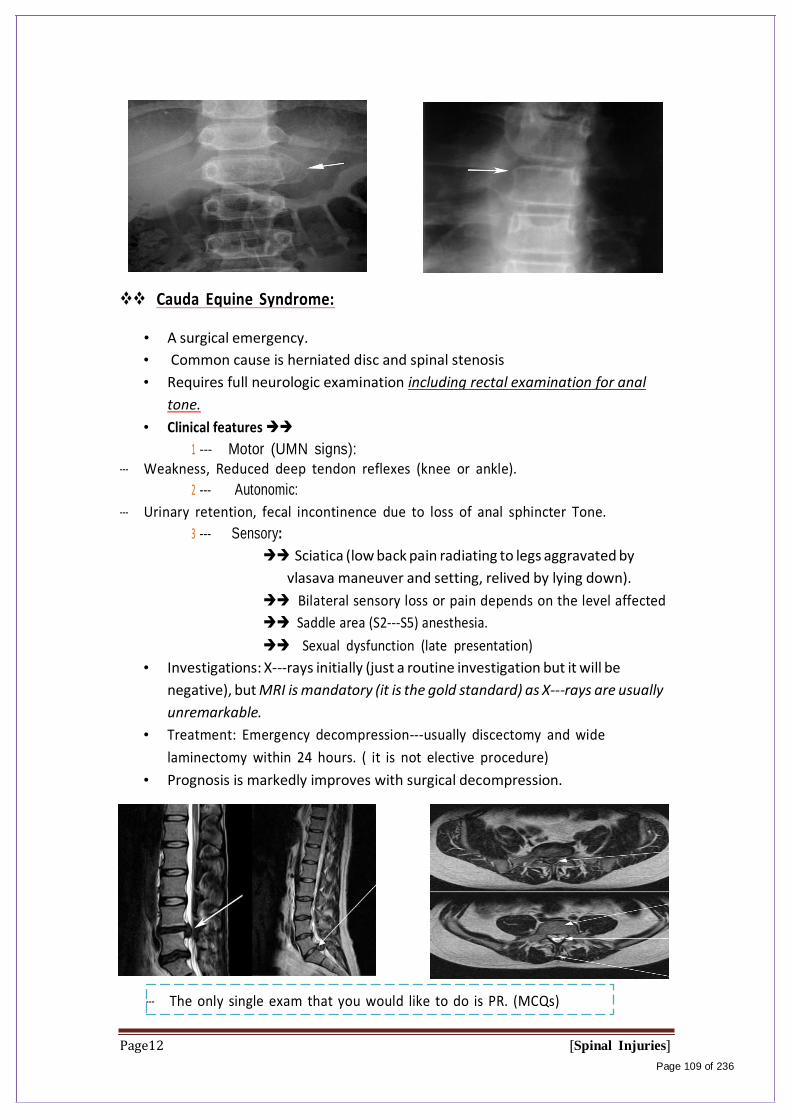
Cauda Equine Syndrome:
• A surgical emergency.
• Common cause is herniated disc and spinal stenosis
• Requires full neurologic examination including rectal examination for anal
tone.
• Clinical features
1 -‐‐ Motor (UMN signs):
-‐‐ Weakness, Reduced deep tendon reflexes (knee or ankle).
2 -‐‐ Autonomic:
-‐‐ Urinary retention, fecal incontinence due to loss of anal sphincter Tone.
3 -‐‐ Sensory:
Sciatica (low back pain radiating to legs aggravated by
vlasava maneuver and setting, relived by lying down).
Bilateral sensory loss or pain depends on the level affected
Saddle area (S2-‐‐S5) anesthesia.
Sexual dysfunction (late presentation)
• Investigations: X-‐‐rays initially (just a routine investigation but it will be
negative), but MRI is mandatory (it is the gold standard) as X-‐rays are usually
unremarkable.
• Treatment: Emergency decompression-‐‐usually discectomy and wide
laminectomy within 24 hours. ( it is not elective procedure)
• Prognosis is markedly improves with surgical decompression.
-‐‐ The only single exam that you would like to do is PR. (MCQs)
Page12 [Spinal Injuries]
Page 109 of 236
Notes //
-‐‐ Spinal red flags are Cauda Equine, spinal tumors, and Fractures with
instability.
-‐‐ Pediatric pt with cervical injury, the most important complication is
scoliosis.
-‐‐ 90% of injuries occur in thoraco-‐‐lumbar spine and 50% of these
fractures occur at the thoraco-‐‐lumbar junction.
Page13 [Spinal Injuries]
Page 110 of 236
Objectives: at the end of the lecture you should be able to:
Common Pediatric Fractures and Trauma
Isn't it funny how someone can say "I believe in Allah " but still follow the Satan who by the way also, " believes " in
Allah…
430 ORTHOPEDICS TEAM
Lecture:((Common Pediatric Fractures))
Team Members:
Faten Al-‐Mohideb. Hanan Al-‐Salman. Aliya Al-‐Awaji.
Hadeel Al-‐Ghamdi. Ghadeer Al-‐Wuhayd. Hissa Al-‐Balla.
Lujain Al-‐Yousef. Nouf Al-‐Hammad. Jawaher Al-‐Faraydi.
Hiba Al-‐Rahiem. Nour Al-‐Enezi. Wejdan Al-‐Swayyid.
Arwa Abudawood. Leena Al-‐Shaman. Areej Al-‐Qunaitir.
Team Leader:
Ayedah Al-‐Ruhaimi.
Special thanks to Norah Al-‐Turki
-‐The slides were provided by the doctor .
-‐Important notes in Red.
-‐Doctor's notes in green/blue.
-‐Copied slides in Black.
-‐MCQ in highlight.
Page 1 Common pediatric fractures
Page 111 of 236

Common Pediatric Fractures and Trauma
Objectives:
know most of the mechanism of injury
make the diagnosis of common pediatric fractures
request and interpret the appropriate x-rays
initiate the proper management of fractures
Know which fractures can be treated by conservative or operative methods
and the ways of fixation.
Know the possible complications of different fractures and how to avoid
them.
The different between adult and pediatric bones
• Pediatric bone has a higher water content and lower mineral content per unit
volume than adult bone so less brittle than adult bone. (weak bones)
• The physis (growth plate) is a unique cartilaginous structure is frequently
weaker than bone in torsion, shear, and bending, predisposing the child to
injury through it. (That’s why the most common pediatrics fractures occur in
growth plate).
• The physis is traditionally divided into four zones that the injury through it can
cause shortening, angular deformities. (In the Future).
• The periosteum in a child is a thick fibrous structure and Rich in blood supply
than adult bone so there is high remodeling rate. (Thick periosteeum fast
remodeling)
• Ligaments in children are functionally stronger than bone. Therefore, a
higher proportion of injuries that produce sprains in adults result in fractures in
children.
• The Growth Plate Diffuses at the age of 16-18 years old, it depends mainly at
the location of the growth plate.
• Twisted injury in adults will give you spiral Fracture but in Pediatric will give
you injury to the growth plate. Immature bone (Pediatric bone)
Page 2 Common pediatric fractures
Page 112 of 236

Common Pediatric Fractures and Trauma
Page 3 Common pediatric fractures
Page 113 of 236
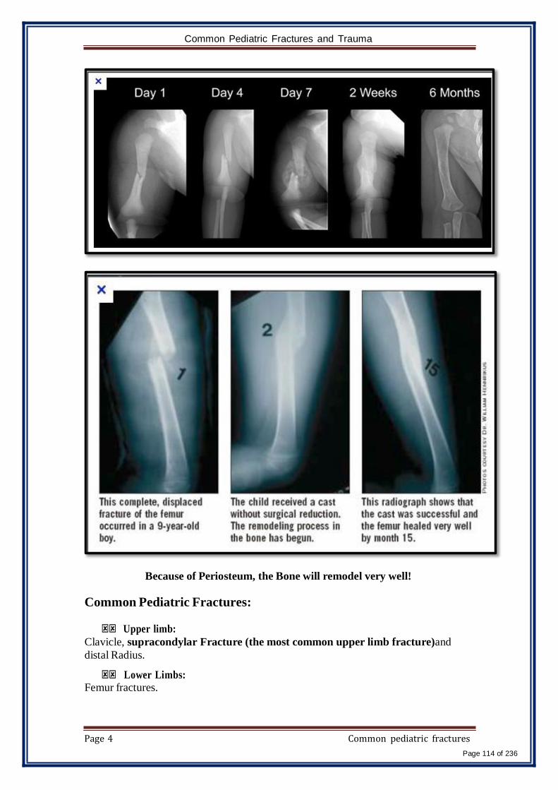
Common Pediatric Fractures and Trauma
Page 4 Common pediatric fractures
Page 114 of 236
Because of Periosteum, the Bone will remodel very well!
Common Pediatric Fractures:
Upper limb:
Clavicle, supracondylar Fracture (the most common upper limb fracture)and
distal Radius.
Lower Limbs: Femur fractures.

Common Pediatric Fractures and Trauma
Page 5 Common pediatric fractures
Page 115 of 236
A. CLAVICLE FRACTURES
• 8% to 15% of all pediatric fractures.
• 0.5% of normal deliveries and in 1.6% of breech deliveries. (Birth
Fracture).
• 90% of obstetric fractures. (During Delivery the baby will get shoulder
fracture)
• 80% of clavicle fractures occur in the shaft.(the MIDDLE)NOT the
Proximal or the Distal!
• The periosteal sleeve always remains in the anatomic position. Therefore,
remodeling is ensured.
Mechanism of Injury:
Indirect Direct Birth injury
Fall onto an outstretched
hand.
This is the most
common mechanism;
it carries the highest
incidence of injury to
the underlying
neurovascular and
pulmonary structures.
Patient will present with
pseudo-paralysis of the limbs
Clinical Evaluation:
• In Birth fractures: an asymmetric in the Shoulder, palpable mass overlying
the fractured clavicle.
• typically present with a painful ( ACUTE) , palpable mass along the
clavicle, Tenderness, Decrease Range of Motion (ROM), there may be
tenting of the skin, crepitus, and ecchymosis.
• Ecchymosis Subcutaneous Purpura > 1 cm or a Hematoma,
commonly called a bruise.
• Neurovascular status, the brachial plexus and upper extremity vasculature
may result.
• Pulmonary status must be assessed. Why? May Penetrate the Lung and
Cause Pneumothorax.

Common Pediatric Fractures and Trauma
Page 6 Common pediatric fractures
Page 116 of 236
Radiographic Evaluation:
1. AP view (The most COMMON and important view, it will show the location
of the fracture). No lateral view!
2. Cephalic tilt view (cephalic tilt of 35 to 40 degrees).
3. Apical oblique view (injured side rotated 45 degrees toward tube with a
cephalic tilt of 20 degrees).
AP view
Classification (Descriptive):
Location, open vs. closed, displacement, and angulation.
Fracture type: segmental, comminuted, greenstick.

Common Pediatric Fractures and Trauma
Page 7 Common pediatric fractures
Page 117 of 236
Allman classification: (Another way to Classify):
Type I Type II Type III
• Middle third
(most
common).
• Distal to the
coracoclavicul
ar ligaments
(lateral third).
• Proximal
(medial) third.
Treatment of clavicle fractures:
Newborn to Age 2 Years Age 2 to 12 Years
-Clavicle fracture in a newborn will
unite in approximately 1 week.
-Infants may be treated
symptomatically with a simple sling or
(figure-of-eight bandage) applied for 2
to 3 weeks. No Need For Reduction
Due to
presence of
periosteum
and the same
teqnuiqe
may apply to
adults.
The cullas
Formation will
be removed
from the body
After Healing!
So we don’t worry about them not
like adults.
A figure-of-eight bandage or sling is
indicated for 2 to 4 weeks.
Indirect Direct
The most common mechanism. Most commonly a
result of a fall onto an
outstretched (FOOSH) upper
extremity.
(Extension type >95%).
A fall onto a flexed elbow or from an object striking the
elbow (e.g., baseball bat,
automobile).
(Flexion type < 3%).
Common Pediatric Fractures and Trauma
Indication of operative treatment:
1- Open fractures
2- Neurovascular compromise
• 99% of The Close Fractures
Treated by the closed Method or
sling!
Example of X-ray Mid clavicle fracture:
• Post conservative treatment
• Healed completely
• With no complications ( No Cellus
Formation or Angulation )
Complications (Rare):
• Neurovascular compromise.
• Malunion.
• Nonunion.
• Pulmonary injury.
B. SUPRACONDYLAR FRACTURE
(The Commonest Fracture in Pediatric Age group)
• Comprise 55% to 75% of all elbow fractures.
• The male-to-female ratio is 3:2.
• The peak incidence is from 5 to 8 years, after which dislocations
become more frequent.
• The left, or nondominant side, is most frequently injured.
• Supracondylar fracture is a fracture in the metaphysis.
Mechanism of injury:
Page 8 Common pediatric fractures
Page 118 of 236

Page 9 Common pediatric fractures
Page 119 of 236
Clinical Evaluation:
o A swollen, tender elbow with painful range of motion.
o S-shaped angulation at the elbow.
o Pucker sign (dimpling of the skin anteriorly )
o Neurovascular examination: The median, radial, and ulnar nerves as well as their terminal branches. Capillary refill and distal pulses should be documented and the Radial Artery.
o The commonest Nerve to be injured is: Anterior
Interosseous Nerve which is branch from the Median
nerve and is associated more with Extension
(indirect)Type. The Way to assess: Ask the Patient TO
Do (OK) sign(medial nerve)!
o Muscles involvled with median nerve injury Flexor
Polices Longs, Flexor digitorm longs!
o E.g. if there is radial nerve injury, there will be wrist drop and loss of sensation in the lateral side.
o Ok sign Median nerve and loss of sensation
o Abduction of the thumb ulnar
o You can examine 3 nerves by the thumb!
Complete Fracture of
Supracondylar.
Gartland Classification:

Common Pediatric Fractures and Trauma
Type 1: Undisplaced -‐‐ Anterior humral line
pass to Cubetelum, minimally displaced.
Type 1 Diagnosis
Type 2: Anterior Cortex Disrupted, Posterior
cortex intact
Lateral elbow x-‐ray AP view
Page 10 Common pediatric fractures
Page 120 of 236

Common Pediatric Fractures and Trauma
Type 3A: Extension type-‐‐ posterior medial.
Type 3B: Flexion type -‐posterior
lateral (rare)
Page 11
AP view elbow
Lateral view
(To differentiate between
extension and flexion type)
Common pediatric fractures
Page 121 of 236
Common Pediatric Fractures and Trauma
Treatment of SUPRACONDYLAR fractures:
1- Initial approachin the ER ABCDE
"Close fracture"apply splint with flexion of 20-30 ◦ X-rays Dx Tx
according to the type.(Apply back-slab if there is swelling, if not complete cast)
"Open fracture"
control hemorrhage and correct coagulopathy
• e.g. direct pressure, tourniquets if in extremis
• fluid resuscitation, transfusion of packed cells and other blood products
as required
give analgesia and antibiotics early
• e.g. morphine or fentanyl +/- ketamine for analgesia
• see Q10 for antibiotics
Assess the limb and seek/treat complications
• e.g. neurovascular compromise, compartment syndrome, crush injury
and rhabomyolysis
Following the provision of adequate analgesia:
• remove gross contaminants from the wound
• if there will be more than a 1 to 2 hour delay in going to the operating
theatre for washout and debridement then clean by profusely irrigating
with saline and cover the wound with a sterile dressing (e.g. saline
soaked pads).
• reduce gross deformities using gentle traction and splint the injured
limb — this is a top priority if there is neurovascular compromise
• immobilise the limb with a POP back-slab and elevate the limb
• give tetanus toxoid/ tetanus immunoglobulin if indicated
Remember to look at the x-ray and consult orthopedics.
This protocol is applied for any open fracture (pediatric & adults)
Page 12 Common pediatric fractures
Page 122 of 236

Common Pediatric Fractures and Trauma
2- According to the type (definitive Tx)
Long arm cast (to mid-‐shaft od humerus)should be applied after all types of treatment. If there is swelling, do back-‐‐slab.
Page 13 Common pediatric fractures
Page 123 of 236

Common Pediatric Fractures and Trauma
X-rays of extension type:
Intra Op fluoroscope
AP view Lateral view
AP & Lateral views Post close reduction + pinning (k-‐‐wire)
Page 14 Common pediatric fractures
Page 124 of 236

Common Pediatric Fractures and Trauma
Complications of Supracondylar fracture:
A. Neurologic injury (7% to 10%)
• Most are neurapraxias requiring no treatment
• Median nerve/anterior interosseous nerve (most common),
ulnar and radial nerves.
• Extension type is the most common.
B. Vascular injury (0.5%)
• Direct injury to the brachial artery, or secondary to swelling.
C. Loss of motion. Cubitus varus deformity
D. Myositis ossificans: soft Bone Formation in the Muscle, during
manipulation, which will cause decrease ROM.
E. Angular deformity (Cubitus varus more frequently than Cubitus
valgus) (10 to 20 %).(the most common complication in
supracondylar fracture)
F. Compartment syndrome (<1%) Associated with Vascular Injury
leads to Volkmann’s Ischemic Contracture.
• Type of Fracture Depends on The Force and The
Mechanism! Extension > Flexion.
Compartment syndrome:
Volkmann's ischemic contracture
Page 15 Common pediatric fractures
Page 125 of 236

Common Pediatric Fractures and Trauma
C. DISTAL RADIUS FRACTURES
It is important to know the site of fracture, is itin thephysis (growth
plate) or metaphysis. It differs in prognosis and management.
1. Physeal Injuries.
• Salter-Harris Types I and II
Page 16 Common pediatric fractures
Page 126 of 236
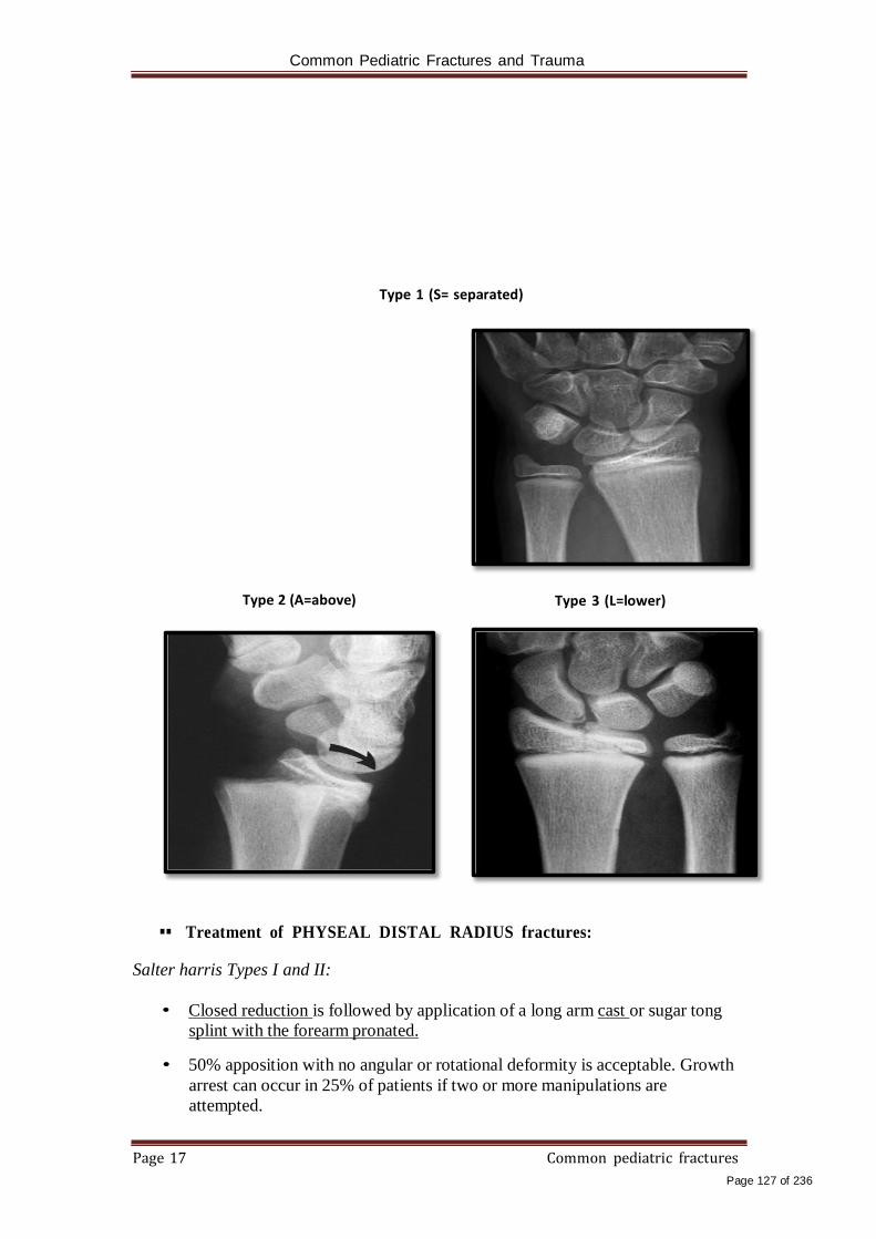
Common Pediatric Fractures and Trauma
Type 1 (S= separated)
Type 2 (A=above) Type 3 (L=lower)
Treatment of PHYSEAL DISTAL RADIUS fractures:
Salter harris Types I and II:
• Closed reduction is followed by application of a long arm cast or sugar tong
splint with the forearm pronated.
• 50% apposition with no angular or rotational deformity is acceptable. Growth
arrest can occur in 25% of patients if two or more manipulations are
attempted.
Page 17 Common pediatric fractures
Page 127 of 236
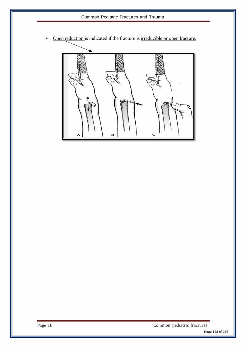
Common Pediatric Fractures and Trauma
• Open reduction is indicated if the fracture is irreducible or open fracture.
Page 18 Common pediatric fractures
Page 128 of 236

Common Pediatric Fractures and Trauma
Pre-‐op Salter harris 2 AP & lateral X-‐‐ray Post-‐‐op
Page 19 Common pediatric fractures
Page 129 of 236
Common Pediatric Fractures and Trauma
Salter-Harris Type III:
• Anatomic reduction is necessary
• Open reduction and internal fixation with smooth pins or screws parallel to
the physis is recommended if the fracture is inadequately reduced.
Salter-Harris Types IV and V:
• (Rare) need ORIF. Complications of Physealinjuries:
• Physeal arrest lead to shortening, an
angular deformity.
• Ulnar styloid nonunion.
• Carpal tunnel syndrome.
Page 20 Common pediatric fractures
Page 130 of 236
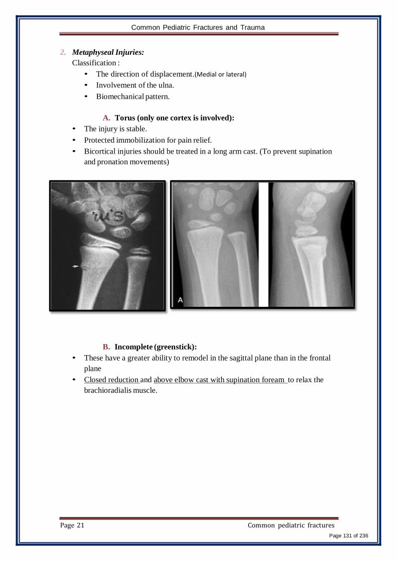
Common Pediatric Fractures and Trauma
2. Metaphyseal Injuries:
Classification :
• The direction of displacement.(Medial or lateral)
• Involvement of the ulna.
• Biomechanical pattern.
A. Torus (only one cortex is involved):
• The injury is stable.
• Protected immobilization for pain relief.
• Bicortical injuries should be treated in a long arm cast. (To prevent supination
and pronation movements)
B. Incomplete (greenstick):
• These have a greater ability to remodel in the sagittal plane than in the frontal
plane
• Closed reduction and above elbow cast with supination foream to relax the
brachioradialis muscle.
Page 21 Common pediatric fractures
Page 131 of 236

Common Pediatric Fractures and Trauma
The younger the
patient the more
amount of
angulation you
can accept!
The Younger the
Patient The more
Remodeling.
C. Complete :
• Closed reduction (by increase the deformity first, then after Reduction use the
cast).
• A well molded long arm cast for 3 to 4 weeks.
Page 22 Common pediatric fractures
Page 132 of 236

Common Pediatric Fractures and Trauma
Indications for percutaneous pinning without open reduction
• Loss of reduction, excessive local swelling, floating elbow and
multiple manipulations.
Page 23 Common pediatric fractures
Page 133 of 236

Common Pediatric Fractures and Trauma
Indication for ORIF
• Irreducible, open fracture and fracture with compartment syndrome.
Complications :
• Malunion Residual malangulation of more than 20% may
result in loss of forearm rotation.
• Nonunion – rare
• Refracture An early return to activity (before 6 weeks).
• Growth disturbance (overgrowth or undergrowth) 3mm/9-12Y
• Neurovascular injuries avoid extreme positions of
immobilization.
Page 24 Common pediatric fractures
Page 134 of 236

Common Pediatric Fractures and Trauma
D. FEMORAL SHAFT FRACTURES
• 1.6% of all pediatric fractures.
• Boys > girls
• Bimodal distribution of incidence 2 to 4 years of age, mid-adolescence.
• In children younger than walking age, 80% of these injuries are caused
by child abuse; this decreases to 30% in toddlers.
• In adolescence, >90% due to RTA (most common)
Mechanism of injury:
• Direct trauma: Motor vehicle accident, pedestrian injury, fall, and child abuse
• Indirect trauma: Rotational injury
• Pathologic fractures: osteogenesis imperfecta, nonossifying fibroma, bone cysts, and tumors.
How to know if it’s direct or indirect injury from the x-ray?
Transverse and comminuted fractures are direct
The spiral is indirect
Clinical evaluation:
• A history of high-energy injury should undergo full trauma evaluation as
indicated.
• An inability to ambulate, with extreme pain, variable swelling, and
variable gross deformity.
• A careful neurovascular examination is essential.
• A careful examination of the overlying soft tissues to rule out the
possibility of an open fracture.
Radiographic evaluation:
• Anteroposterior and lateral views
• X-ray most includes hip, knee joints.
A mass is showing in the x-‐ray
consider OPEN fracture.
Page 25 Common pediatric fractures
Page 135 of 236

Common Pediatric Fractures and Trauma
Classification
a) Descriptive:
• Open versus closed
• Level of fracture: proximal, middle, distal third • Fracture pattern: transverse, spiral, oblique, butterfly fragment
• Comminution
• Displacement
• Angulation
b) Anatomical:
• Subtrochanteric.
• Shaft.
• Supracondylar.
Treatment:
Age <6 Months • Pavlic harness or a posterior splint.
• Traction and spica casting.
Ages 6 Months to 6 Years
• Immediate spica casting is the treatment of choice (>95%).
• Skeletal traction followed by spica casting if there is difficulty to
maintain length and acceptable alignment.
Ages 6 to 12 Years
a) Flexible intramedullary nails It is contraindicated to put nails in children less than 16 years old, use elastic
b) Bridge plating
c) External fixation: When? C multiple injuries open fracture
comminuted B
A Unstable patient
Page 26 Common pediatric fractures
Page 136 of 236

Common Pediatric Fractures and Trauma
Ages 12 to Maturity
• Intramedullary fixation with either flexible or interlocked nails(age>16y)
is the treatment of choice.
Page 27 Common pediatric fractures
Page 137 of 236

Common Pediatric Fractures and Trauma
Operative Indications
• Multiple traumas, including head trauma.
• Open fracture.
• Vascular injury.
• Pathologic fracture.
• Uncooperative patient.
( Body habitus "OBESE PATIENT" not amenable to spica casting)
Complications:
1. Malunion: Remodeling will not correct rotational deformities (The
Important Thing is to restore the ALIGNMENT)
2. Nonunion –rare.
3. Muscle weakness.
4. Leg length discrepancy: Secondary to shortening or overgrowth,
Overgrowth of 1.5 to 2.0 cm is common in the 2- to 10-year age.
5. Osteonecrosis with antegrade IM nail<16year. (So try to avoid
Intramedullary Nail <16).
Page 28 Common pediatric fractures
Page 138 of 236

Common Pediatric Fractures and Trauma
Some extra information regarding X-ray
reading.
• Anteroposterior (AP) and lateral views:
The following angular relationships may be determined:
a. Baumann angle: 15 to 20 degrees
b. Humeral-ulnar angle: reflects the true carrying angle
(M=7.f=15)
c. Metaphyseal-diaphyseal angle: reverse of Baumann
Angle!
• True lateral radiograph of the elbow flexed to
90 degrees: (If any of them disrupted -
Fracture!)
•Teardrop.
•Diaphyseal-condylar angle: (30 to 45 degrees anteriorly)
•Anterior humeral line.
•Coronoid line.
The contralateral elbow should be obtained for
comparison as well as identification of ossification centers
• Lateral X-ray:
o Fat pad signs, black Presentation in front --
usually Fracture in X-ray!
o Anterior (coronoid) fat pad---joint effusion
o Posterior (The deep olecranon fossa) the deep
olecranon fossa normally completely contains the
posterior fat pad. large effusions cause posterior
displacement fracture, it is present >70% .
The End
Page 29 Common pediatric fractures
Page 139 of 236

O R T H O P E D I C S 4 2 9 T E A M
G r o u p A 1
Page 140 of 236 128
Nomenclature:
Common Pediatric Hip Problem
DDH : Developmental dislocation of the hip CDH : Congenital Dislocation of the Hip
DDH : Developmental Dysplasia of the Hip
SCFE : Slipped capital femoral epiphysis
Perthes
Normal pelvis
Triradiate cartilage
Acetabulum
ADULT
Lesser Trochanter
Pelvic Brim
CHILD
Ischium Pubic Bone
Grater Trochanter
Notes:
Triradiate cartilage (growth plate) connects three bones: acetabulum + ischium + pubic bone
Head of femur: in children it’s not called head of femur (because not fully developed) only in
adult
2arterialsuply of the femur anterior and posterior circumflex artery that supply the head of femur
Ligamentumteres is attached to the inside of the head of the femur, the ligament also contains
artery. So arteries inside and outside the bone
"DDH"
Normal Hip Dislocated Hip
Notes:
The head of femur is not articulating/attached to the acetabulum. Notice the under developed
epiphysis
The acetabulum is shallow (dysplastic acetabulum) not curved (cannot hold the head)
DDH is not due to injury during delivery (not congenital)
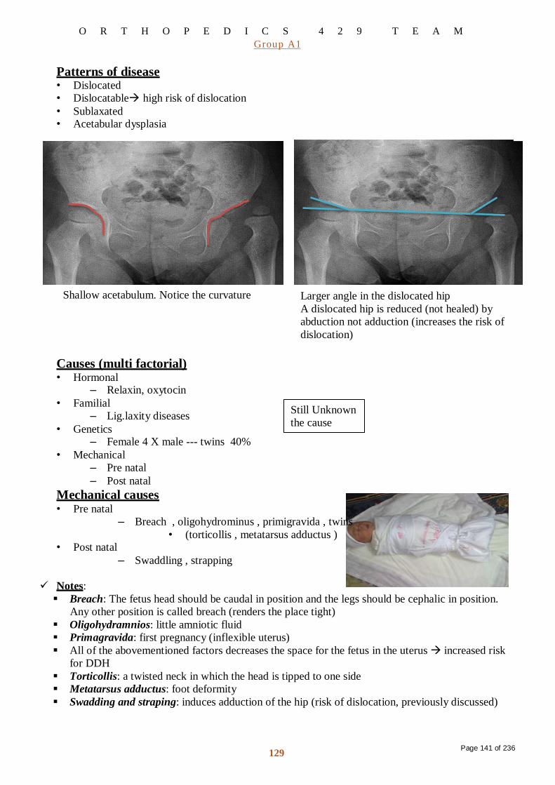
O R T H O P E D I C S 4 2 9 T E A M
G r o u p A 1
Page 141 of 236 129
Patterns of disease • Dislocated
• Dislocatablehigh risk of dislocation
• Sublaxated • Acetabular dysplasia
Shallow acetabulum. Notice the curvature Larger angle in the dislocated hip
A dislocated hip is reduced (not healed) by
abduction not adduction (increases the risk of
dislocation)
Causes (multi factorial) • Hormonal
– Relaxin, oxytocin
• Familial
– Lig.laxity diseases
• Genetics – Female 4 X male --- twins 40%
• Mechanical
– Pre natal
– Post natal
Mechanical causes • Pre natal
Still Unknown
the cause
– Breach , oligohydrominus , primigravida , twins
• (torticollis , metatarsus adductus ) • Post natal
– Swaddling , strapping
Notes:
Breach: The fetus head should be caudal in position and the legs should be cephalic in position.
Any other position is called breach (renders the place tight)
Oligohydramnios: little amniotic fluid
Primagravida: first pregnancy (inflexible uterus)
All of the abovementioned factors decreases the space for the fetus in the uterus increased risk
for DDH
Torticollis: a twisted neck in which the head is tipped to one side
Metatarsus adductus: foot deformity
Swadding and straping: induces adduction of the hip (risk of dislocation, previously discussed)
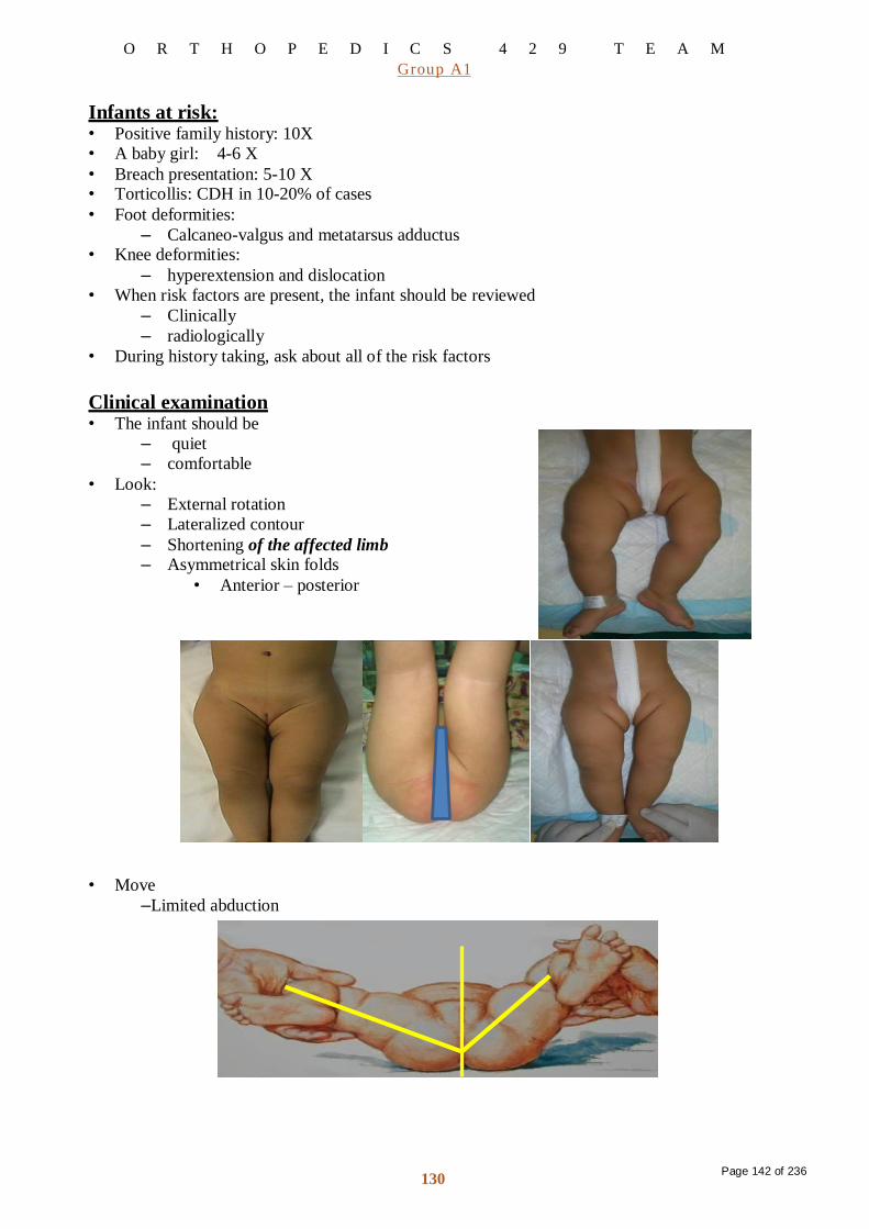
O R T H O P E D I C S 4 2 9 T E A M
G r o u p A 1
Page 142 of 236 130
Infants at risk: • Positive family history: 10X
• A baby girl: 4-6 X
• Breach presentation: 5-10 X • Torticollis: CDH in 10-20% of cases
• Foot deformities:
– Calcaneo-valgus and metatarsus adductus • Knee deformities:
– hyperextension and dislocation
• When risk factors are present, the infant should be reviewed
– Clinically
– radiologically
• During history taking, ask about all of the risk factors
Clinical examination • The infant should be
– quiet
– comfortable
• Look:
– External rotation
– Lateralized contour
– Shortening of the affected limb – Asymmetrical skin folds
• Anterior – posterior • Move
–Limited abduction

O R T H O P E D I C S 4 2 9 T E A M
G r o u p A 1
Page 143 of 236 131
• Special test
– Galiazzi
– Ortolani , Barlow test
– Trendelenburgh sign
– Limping ( waddling gait if bilateral)
• Only ortolani and Barlow tests are important
Galiazzi test
Ortolani test
• Pull and abduct
• A clunk will be felt after reduction. The hip will soon be dislocated again (not a treatment, just to
test for DDH)
• Cannot be done on dislocatable DDH Do barlow test
Barlow test
• Pull and adduct
Trendelenburgh sign

O R T H O P E D I C S 4 2 9 T E A M
G r o u p A 1
Page 144 of 236 132
Investigations
• -3 months U/S: most of the head and neck are cartilage (not
clear on x-ray)
• > 3months X-ray pelvis AP + abduction
Radiology • After 6 months: reliable
Notes
Red: Shenton line (disrupted in DDH)
Blue: Hilgenreiner’s line (through the triradiate cartilage). Notice the angle marking the acetabulum
Orange: Perkin's line: it drowns vertically and perpendicular to Hilgeneiner’s line starts at the
lateral acetabulum
Hilgenriner’s line + Perkin’s line form four quadrants. In the normal limb, the head of the femur
is found in the bottom inner quadrant, unlike the affected limb, where the head is in the top outer
quadrant.
Five signs of DDH on x-ray: MCQ
1. Disrupted shenton’sline
2. Wide acetabulum angle on Hilgenriner’s line
3. Head of femur located on the top outer quadrant when Perkin’s line is drawn
4. Shallow acetabulum
5. Ossification center/head of femur is smaller than the one in the other limb
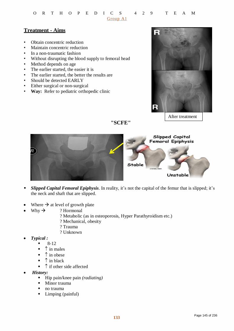
O R T H O P E D I C S 4 2 9 T E A M
G r o u p A 1
Page 145 of 236 133
Treatment - Aims
• Obtain concentric reduction
• Maintain concentric reduction
• In a non-traumatic fashion • Without disrupting the blood supply to femoral head
• Method depends on age • The earlier started, the easier it is
• The earlier started, the better the results are
• Should be detected EARLY • Either surgical or non-surgical
• Way: Refer to pediatric orthopedic clinic
"SCFE"
After treatment
Slipped Capital Femoral Epiphysis. In reality, it’s not the capital of the femur that is slipped; it’s
the neck and shaft that are slipped.
Where at level of growth plate
Why ? Hormonal
? Metabolic (as in osteoporosis, Hyper Parathyroidism etc.)
? Mechanical, obesity
? Trauma
? Unknown
Typical : 8-12
in males
in obese
in black
if other side affected
History: Hip pain/knee pain (radiating)
Minor trauma
no trauma
Limping (painful)

O R T H O P E D I C S 4 2 9 T E A M
G r o u p A 1
Page 146 of 236 134
On Examination: Hip in ER (external rotation)
IR (internal rotation)
Abduction
Usually painful ROM (Range of motion)
Limping (painful)
Investigation:
X-ray
- Pelvis – slippage positive or
Growth plate space
[Pre slip phase]
- Knee
If not clear but still doubtful MRI can help
Treatment:
Refer to orthopedic as emergency case
What they will do?
In situ pinning – to prevent further damage to the vascularity
Protected weight bearing for 3-4 weeks then full weight bearing
No sport for 6 months
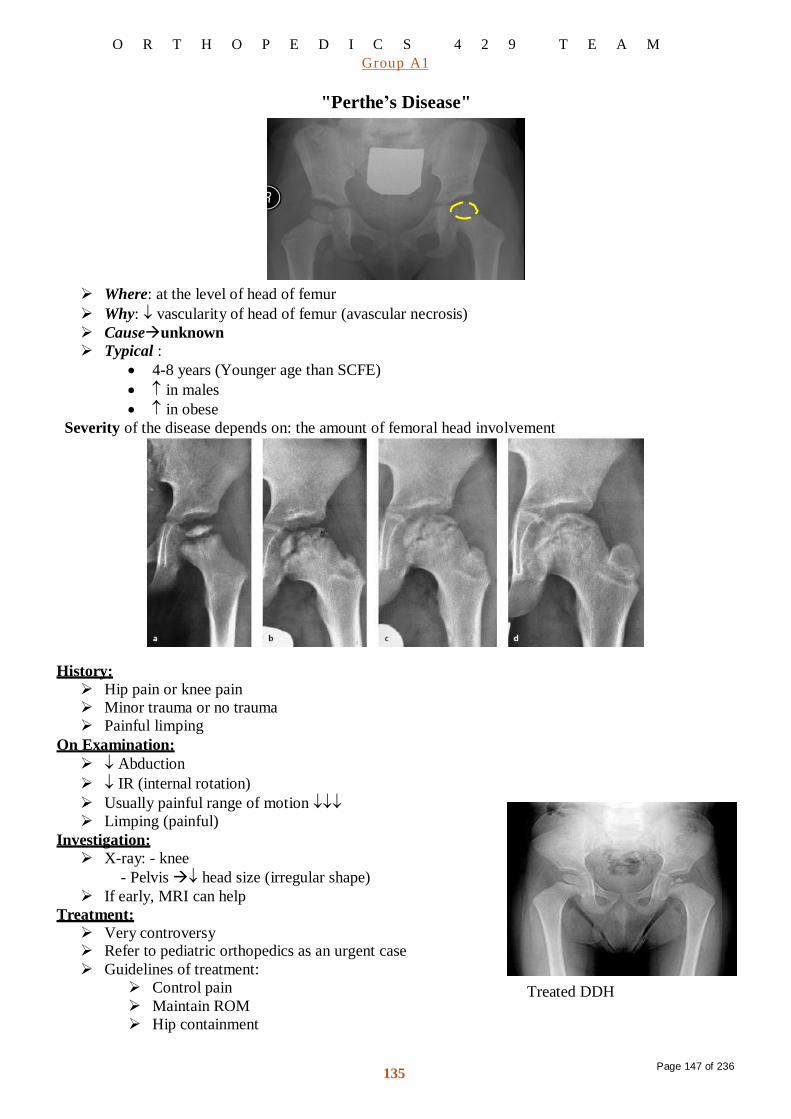
O R T H O P E D I C S 4 2 9 T E A M
G r o u p A 1
Page 147 of 236 135
"Perthe’s Disease"
Where: at the level of head of femur
Why: vascularity of head of femur (avascular necrosis)
Causeunknown
Typical :
4-8 years (Younger age than SCFE)
in males
in obese
Severity of the disease depends on: the amount of femoral head involvement
History:
Hip pain or knee pain
Minor trauma or no trauma
Painful limping
On Examination:
Abduction
IR (internal rotation)
Usually painful range of motion
Limping (painful)
Investigation:
X-ray: - knee
- Pelvis head size (irregular shape)
If early, MRI can help
Treatment:
Very controversy Refer to pediatric orthopedics as an urgent case
Guidelines of treatment:
Control pain
Maintain ROM
Hip containment
Treated DDH

Common Pediatrics Lower Limb Deformities
430 ORTHOPEDICS
Lecture: Common Pediatrics Lower Limb Deformities.
Faten Al-Mohideb.
Team Members:
Hanan Al-Salman.
Aliya Al-Awaji.
Hadeel Al-Ghamdi. Lujain Al-Yousef.
Ghadeer Al-Wuhayd.
Nouf Al-Hammad.
Hissa Al-Balla.
Jawaher Al-Faraydi.
Hiba Al-Rahiem. ArwaAbudawood.
Nour Al-Enezi.
Leena Al-Shaman.
Wejdan Al-Swayyid.
Areej Al-Qunaitir.
Team Leader:
Ayedah Al-Ruhaimi.
Black: Slides (by Dr. Zamzam)
Green: My notes (recording)
Red: Important
Blue: Extra notes (429 – webmd)
MC/MCC: Most common cause
ST: Soft tissue - IR: Internal Rotation – ER: External Rot.
Page 148 of 2361

2
Torsional Deformities
1 COMMON PRESENTATIONS
1.1 LEG ACHES
• History o It’s a growing pain o With no functional
disability o Benign o It resolves
spontaneously
o Unknown cause • Screening Examination
• Tenderness Joint Motion
• Diagnosis by exclusion
• Differential Diagnosis from serious problems mainly tumor
o Osteoid osteoma o Osteosarcoma o Ewing sarcoma
• Management o Symptomatic o Reassurance
1.2 LIMPING
• Abnormal gait due to pain, weakness or deformity
• Evaluation o History (Mainly age of onset) o Observation
• Evaluate the limp by studying the child’s gaitwhile the child walks in the clinic hallway
• Types of Limp o Painful gait: Antalgic gait o Waddling gait = abductors weak in both legs o Toe to heel gait (plantar flexion) = Equinus gait
• Management o Generalization regarding
management cannot be made
o Treatment of the cause
Page 149 of 2362

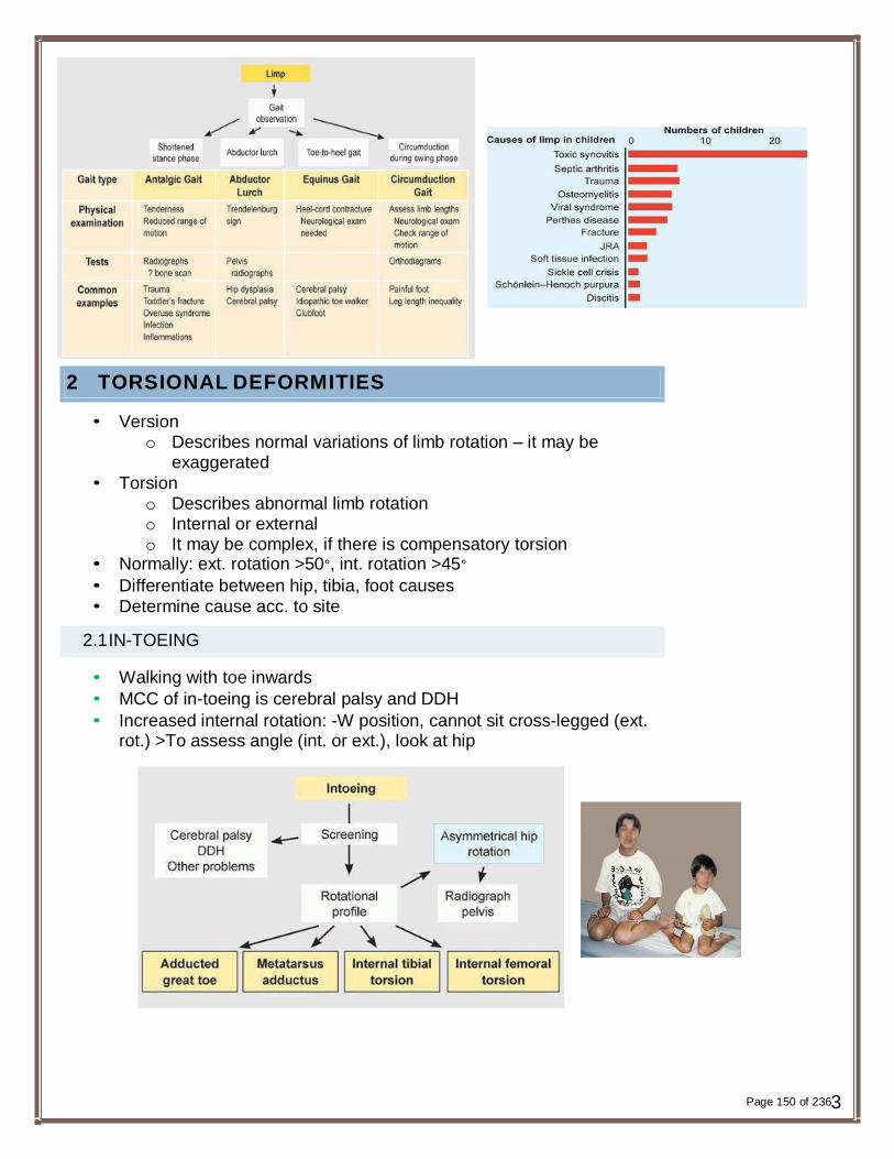
2 TORSIONAL DEFORMITIES
• Version o Describes normal variations of limb rotation – it may be
exaggerated
• Torsion o Describes abnormal limb rotation o Internal or external o It may be complex, if there is compensatory torsion
• Normally: ext. rotation >50°, int. rotation >45°
• Differentiate between hip, tibia, foot causes
• Determine cause acc. to site
2.1 IN-TOEING
• Walking with toe inwards
• MCC of in-toeing is cerebral palsy and DDH
• Increased internal rotation: -W position, cannot sit cross-legged (ext. rot.) >To assess angle (int. or ext.), look at hip
Page 150 of 2363
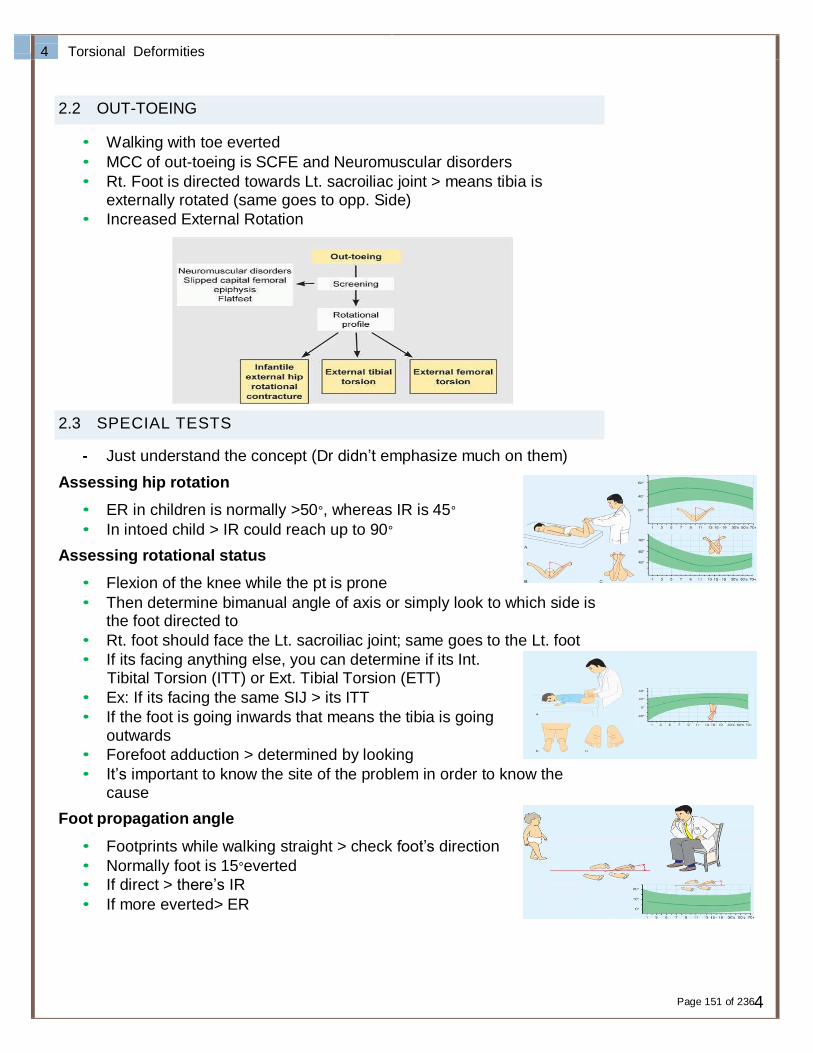
4
Torsional Deformities
2.2 OUT-TOEING
• Walking with toe everted
• MCC of out-toeing is SCFE and Neuromuscular disorders
• Rt. Foot is directed towards Lt. sacroiliac joint > means tibia is externally rotated (same goes to opp. Side)
• Increased External Rotation
2.3 SPECIAL TESTS
- Just understand the concept (Dr didn’t emphasize much on them)
Assessing hip rotation
• ER in children is normally >50°, whereas IR is 45°
• In intoed child > IR could reach up to 90°
Assessing rotational status
• Flexion of the knee while the pt is prone
• Then determine bimanual angle of axis or simply look to which side is the foot directed to
• Rt. foot should face the Lt. sacroiliac joint; same goes to the Lt. foot
• If its facing anything else, you can determine if its Int. Tibital Torsion (ITT) or Ext. Tibial Torsion (ETT)
• Ex: If its facing the same SIJ > its ITT
• If the foot is going inwards that means the tibia is going outwards
• Forefoot adduction > determined by looking
• It’s important to know the site of the problem in order to know the cause
Foot propagation angle
• Footprints while walking straight > check foot’s direction
• Normally foot is 15°everted
• If direct > there’s IR
• If more everted> ER
Page 151 of 2364

2.4 MANAGEMENT
• Determine site to determine the cause
• Define severity to determine Rx accordingly
• Allow for spontaneous correction (observational management) until 8 years of age
• Controlling a child’s walking, sitting or sleeping is extremely difficult and frustrating
• Shoe wedges or inserts are ineffective
• Bracing with twister cables limits child’s activities
• Night splints have no long term benefit
• If mild > wait for spont. correction
• If severe/past the age of spont. Corr. > surgical: o Rotate the bone after doing osteotomy and fix it in
place with K-wires for 6-7 wks then remove them
• But if v. severe > can be done earlier than 8 years of age
• Operative correction o Indicated for children above the age of 8 years with
significant cosmetic and functional deformity o Foot: correction of muscle imbalance that has caused the inward
or outward deviation of foot
o Break the bone & rotate it the other direction
o Ex: 40°intoeing (in hip or femur) > osteotomy of the bone under trochanteric area & then rotate the femur 40°& fixate it using k- wires for 6-7 weeks
3 LIMB LENGTH INEQUALITY
• True: Medial malleolus to superior anterior iliac spine
• Apparent: Medial malleolus to umbilicus
• Etiology o Congenital
- Proximal focal femoral discrepancy
- Congenital short femur
o Developmental - DDH - SCFE
o Traumatic - Trauma will cause overlap or
fracture near epiphysis > initiation of bone growth > lengthening > discrepancy
o Infection - By irritation/suppression of epiphysis or initiation to
more bone growth
Page 152 of 2365
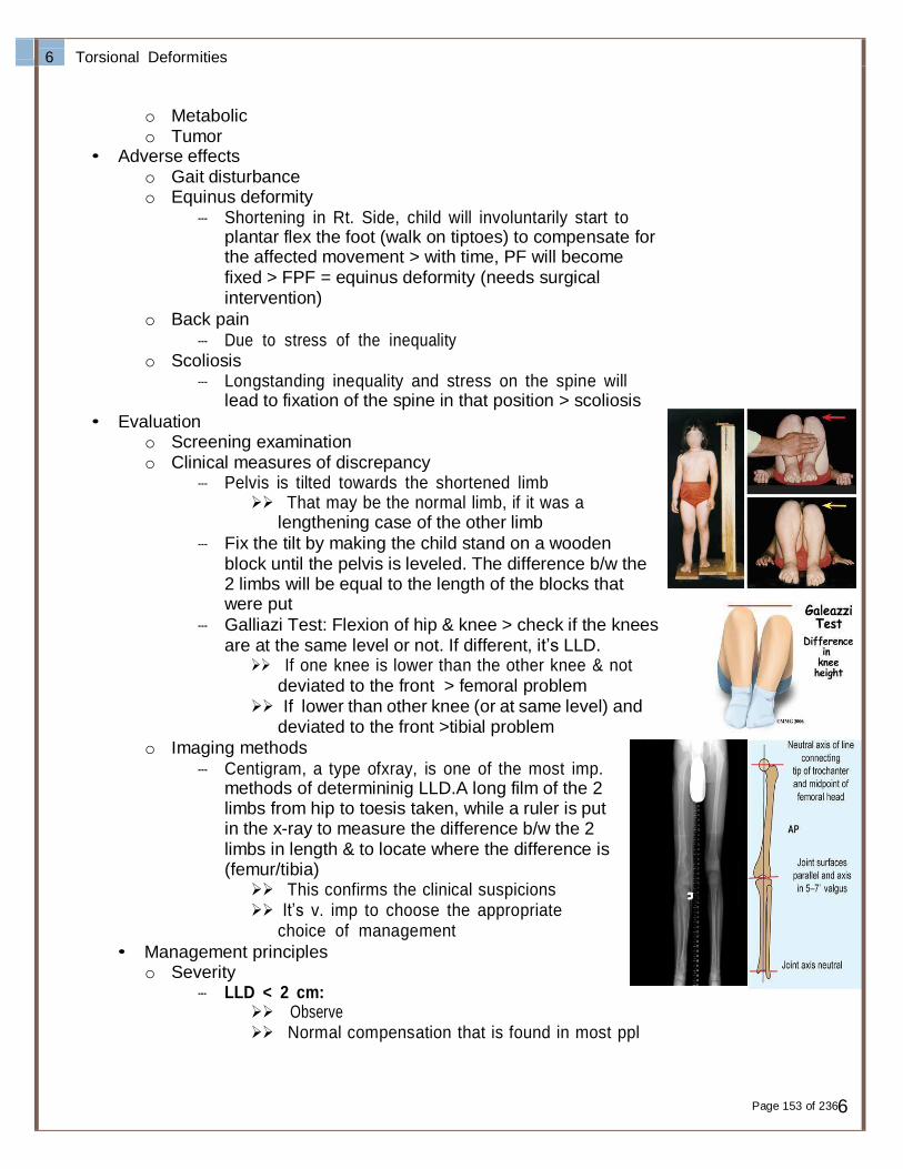
6
Torsional Deformities
o Metabolic o Tumor
• Adverse effects o Gait disturbance o Equinus deformity
- Shortening in Rt. Side, child will involuntarily start to plantar flex the foot (walk on tiptoes) to compensate for the affected movement > with time, PF will become fixed > FPF = equinus deformity (needs surgical intervention)
o Back pain
- Due to stress of the inequality o Scoliosis
- Longstanding inequality and stress on the spine will lead to fixation of the spine in that position > scoliosis
• Evaluation o Screening examination o Clinical measures of discrepancy
- Pelvis is tilted towards the shortened limb That may be the normal limb, if it was a
lengthening case of the other limb
- Fix the tilt by making the child stand on a wooden block until the pelvis is leveled. The difference b/w the 2 limbs will be equal to the length of the blocks that were put
- Galliazi Test: Flexion of hip & knee > check if the knees are at the same level or not. If different, it’s LLD.
If one knee is lower than the other knee & not deviated to the front > femoral problem
If lower than other knee (or at same level) and deviated to the front >tibial problem
o Imaging methods
- Centigram, a type ofxray, is one of the most imp. methods of determininig LLD.A long film of the 2 limbs from hip to toesis taken, while a ruler is put in the x-ray to measure the difference b/w the 2 limbs in length & to locate where the difference is (femur/tibia)
This confirms the clinical suspicions It’s v. imp to choose the appropriate
choice of management
• Management principles o Severity
- LLD < 2 cm: Observe Normal compensation that is found in most ppl
Page 153 of 2366

Not really apparent clinically
- LLD 2-5 cm: Shoe lift (for the whole foot NOT only heel)
- LLD > 5 cm: Consider surgery or active treatment
o Lifts(as we said, used in MILD cases) o Shortening
- Osteotomy then fixation (remove part of bone according to length required)
- Ex: In neurofibromatosis, there is overgrowth of the whole limb >Debulking of soft tissue + shortening to equalize both limbs
o Epiphysiodesis - Temporary Epiphys. is done when you want to do
shortening in young children that still have growth potential
- Clips are inserted both medially and laterally of the
bone for a certain period until desired equalization of the limb is achieved. Then have them removed so both limbs can continue to grow normally together at the same pace
- Permanent method is done for children that are close to skeletal maturity (12-13 y/o). They’ll have around 4-5 cms of growth potential left to reach, which equals the length desired to shorten the limb
Insertion of screw will destroy the epiphysis > bone stops growing
o Lengthening (just understand the concept)
- More common - We use circular frames or do partial osteotomies& then
insert a device that will start to lengthen bit by bit 1 mm per day (or so), by having the patient manipulate a screw of the external apparatus > callous formation b/w the 2 fragments of bone that are lengthening
- Could reach up to 8 cm in tibia and 15 cm in femur
- Takes up to 2-3 months. Success rate is high. - Can be performed in any age group, easier in children
though
4 GENU VARUS & GENU VALGUS
4.1 DEFINITION
• Valgus: Away from center (distal in relation to proximal) = knock knees
• Varus: Towards center = bow legs
Page 154 of 2367
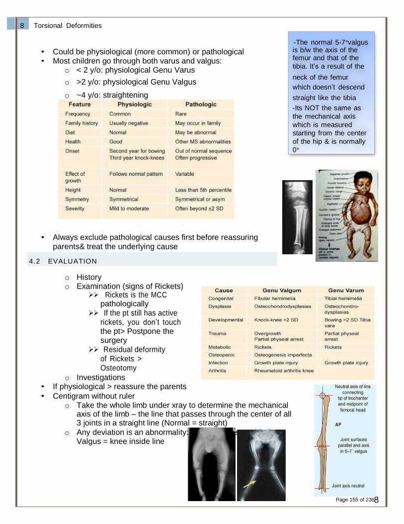
8
Torsional Deformities
-The normal 5-7°valgus
• Could be physiological (more common) or pathological is b/w the axis of the
• Most children go through both varus and valgus: femur and that of the
o < 2 y/o: physiological Genu Varus tibia. It’s a result of the
o >2 y/o: physiological Genu Valgus neck of the femur
which doesn’t descend
o ~4 y/o: straightening straight like the tibia
-Its NOT the same as
the mechanical axis
which is measured
starting from the center
of the hip & is normally
0°
• Always exclude pathological causes first before reassuring parents& treat the underlying cause
4.2 EVALUATION
o History o Examination (signs of Rickets)
Rickets is the MCC pathologically
If the pt still has active rickets, you don’t touch the pt> Postpone the surgery
Residual deformity of Rickets > Osteotomy
o Investigations • If physiological > reassure the parents
• Centigram without ruler o Take the whole limb under xray to determine the mechanical
axis of the limb – the line that passes through the center of all 3 joints in a straight line (Normal = straight)
o Any deviation is an abnormality: Varus = knee outside line , Valgus = knee inside line
Page 155 of 2368
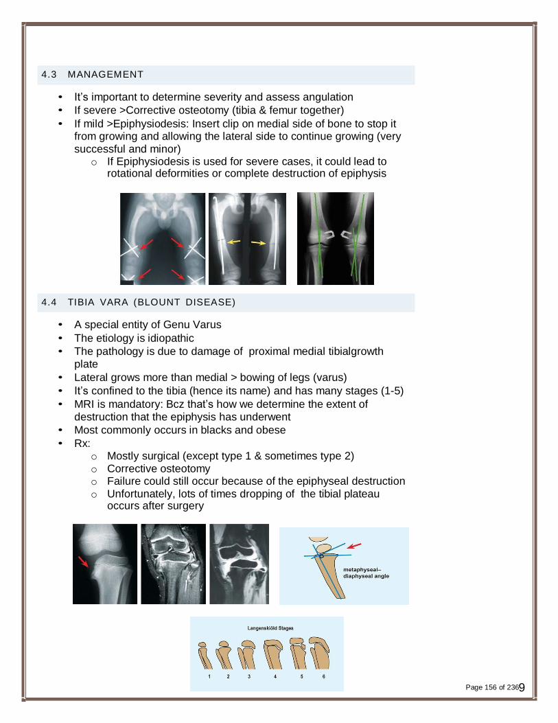
4.3 MANAGEMENT
• It’s important to determine severity and assess angulation
• If severe >Corrective osteotomy (tibia & femur together)
• If mild >Epiphysiodesis: Insert clip on medial side of bone to stop it from growing and allowing the lateral side to continue growing (very successful and minor)
o If Epiphysiodesis is used for severe cases, it could lead to rotational deformities or complete destruction of epiphysis
4.4 TIBIA VARA (BLOUNT DISEASE)
• A special entity of Genu Varus
• The etiology is idiopathic
• The pathology is due to damage of proximal medial tibialgrowth plate
• Lateral grows more than medial > bowing of legs (varus)
• It’s confined to the tibia (hence its name) and has many stages (1-5)
• MRI is mandatory: Bcz that’s how we determine the extent of destruction that the epiphysis has underwent
• Most commonly occurs in blacks and obese
• Rx: o Mostly surgical (except type 1 & sometimes type 2) o Corrective osteotomy o Failure could still occur because of the epiphyseal destruction o Unfortunately, lots of times dropping of the tibial plateau
occurs after surgery
Page 156 of 2369

10
Torsional Deformities
5 FOOT DEFORMITIES
• Let’s look at the characteristics of a normal foot first
o Stable: for supporting the body weight in standing o Resilient:for walking and running o Mobile: to accommodate variations of surface o Cosmetic
5.1 CLUBFOOT(IMP)
5.1.1 ETIOLOGY
• Postural
o Abnormal posture of uterus (IUGR) causing this deformity
>assessed after delivery
• Idiopathic (CTEV)
o Congenital TalipesEquinoVarus
• Secondary
o (Spina bifida [MCC],myelodysplasia, arthrogryposis)
5.1.2 DIAGNOSIS
• Diagnosis by exclusion o If deformity is identified after delivery, try to do correction
manually If corrected successfully > Postural If not > Idiopathic or secondary
- If there’s no syndrome or underlying pathology > Idiopathic
• Differentiate by exclusion (DDx): o Neurological lesion that can cause the deformity “Spina
Bifida”(excluded by spine x-rays) o Other abnormalities that can explain the deformity:
“Arthrogryposis, Myelodysplasia” o Presence of concomitant congenital anomalies: “Proximal
femoral focal deficiency” o Syndromatic clubfoot: “Larsen’s syndrome, Amniotic band
Syndrome”
Page 157 of 23610

5.1.3 CLINICAL EXAMINATIONOF IDIOPATHIC
• Characteristic deformity (a MUST in every case)
o Hind foot: - Equinus(fixed plantar flexion of ankle joint - FPF)
- Varus(inversion of subtalar joint)
o Fore foot - Forefoot Adduction(of talus) - Cavus(high-arched foot)
• Additional findings o Short Achilles tendon(due to FPF) o High and small heel (due to FPF) o No creases behind Heel(due to FPF) o Abnormal crease in middle of the foot(due to forefoot add.) o Affected foot is smaller (obvious if unilateral) o Callosities at abnormal pressure areas o Internal torsion of the leg: Varusforefoot add. > internal tibial
torsion
o Calf muscles wasting o Deformities don’t prevent walking
5.1.4 MANAGEMENT
• Aim of treatment: o To reach an antigrade, mobile, normal,
painless foot which looks cosmetically acceptable and fits normal shoes
• Ponseti technique: o Age limit is up to 12 months (controversial,
won’t be asked about it)
• Manipulation is maintained by a plaster cast
o It’s changed periodically, weekly at 1st, then every 2 wks> until full correction is obtained (4-6 casts)
• Then maintain correction by Dennis Brown splints until pt starts to walk
• Success rate is very high
• Indications for Surgery o If pt presents late after 12 months of age o Failure of conservativemanagement o Complementary to conservative treatment o Recurrence of deformity
For the 3 above, it could be soft tissue only or ST +/- bone surgery > To regain function
o Complementary surgery (salvage) > To regain appearance Longstanding varus>osteotomy: wedge removed
ofcalcaneous
Page 158 of 23611

12
Torsional Deformities
Short Achilles tendon > Lengthening Before and after correction slide Triple osteotomy (talus – calcaneous – navicular)
Most common salvage procedure Loss of inversion or eversion. Pt will feel pain
walking on uneven ground, otherwise, they will walk normally
6 CEREBRAL PALSY
Cerebral palsy (CP) is a broad term used to describe a group of chronic "palsies" -- disorders that impair control of movement -- due to damage to the developing brain.
6.1 PHYSIOLOGICAL CLASSIFICATION
• Spastic
o Spastic is the MC that goes to surgery • Athetosis
• Ataxia o In ataxia and athetosis> surgery is contraindicated
• Rigidity
• Mixed
6.2 TOPOGRAPHIC CLASSIFICATION
• Monoplegia>one limb affected
• Paraplegia>both legs and trunk affected
• Hemiplegia>one side of the body (arm&leg) affected
• Triplegia>three limbs affected
• Quadriplegia or tetraplegia>all four limbs (+/- trunk, tongue and windpipe) affected
• Bilateral hemiplegia
• Diplegia>both legs affected
6.3 ASSESSM ENT
• Hip: o Flexion
Do Thomas test to assess fixed flexed deformity of hip
o Adduction: Scissoring gait Hip ROM
o IR: Intoeing Hip ROM
• Knee: o Flexion
Page 159 of 23612
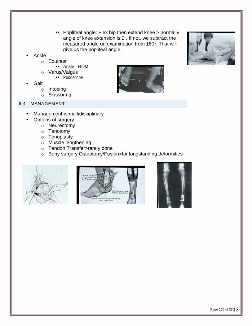
• Ankle
Popliteal angle: Flex hip then extend knee > normally angle of knee extension is 0°. If not, we subtract the measured angle on examination from 180°. That will give us the popliteal angle.
o Equinus Ankle ROM
o Varus/Valgus Podoscope
• Gait o Intoeing o Scissoring
6.4 MANAGEMENT
• Management is multidisciplinary
• Options of surgery o Neurectomy o Tenotomy o Tenoplasty o Muscle lengthening o Tendon Transfer>rarely done o Bony surgery Osteotomy/Fusion>for longstanding deformities
Page 160 of 23613
Definition: A non-inflammatory (DEGENERATIVE) disease affecting articular cartilage of joints.
TYPES:
Primary
Intrinsic defect (mechanical, vascular, cartilage, hereditary-generalized O.A)
-usually familial , hereditary.
- Generalized and affects the upper limbs more than lower limbs.
Secondary
Secondary to local or systemic disease
Etiology:
Increased load: obesity (hips and knees take 3-4 body weight with each step) (the most common movements which can affect the knee is jogging & squatting).
Trauma: osteochondral, malunion, sport injury
Congenital/developmental: CDH, multiple epiphyseal dysplasia
Infection
Necrosis: Perth’s disease, osteonecrosis, steroids
Hematologic: SCD, hemophelia ( mostly by oseonecrosis )
Endocrine: DM (due to neuropathy, it can lead to unorganized remodeling of destroyed painless joint called "Charcot's Joint" which in turns lead to severe secondary OA), acromegaly
Metabolic: crystaline deposition disease (gout mainly affect small peripheral joint "Feet", CPPD "crystalline pyrophosphate deposition which is psudo gout, mainly affect the knee "), Paget’s disease
Inflammatory: RA, SLE, Reiter’s syndrome
Neuropathic: DM, tabes dorsalis.
Occupation (indirect cause of OA).
-The first part affected by the normal aging degeneration process is the cervical spine.
Page2 [osteoarthrosis]
Page 162 of 236
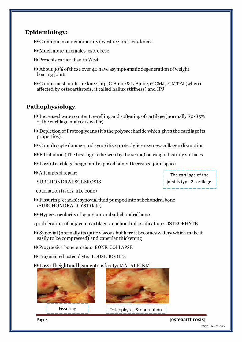
Epidemiology:
Common in our community ( west region ) esp. knees
Much more in females ;esp. obese
Presents earlier than in West
About 90% of those over 40 have asymptomatic degeneration of weight bearing joints
Commonest joints are knee, hip, C-Spine & L-Spine,1st CMJ,1st MTPJ (when it affected by osteoarthrosis, it called hallux stiffness) and IPJ
Pathophysiology:
Increased water content: swelling and softening of cartilage (normally 80-85% of the cartilage matrix is water).
Depletion of Proteoglycans (it's the polysaccharide which gives the cartilage its properties).
Chondrocyte damage and synovitis › proteolytic enzymes› collagen disruption
Fibrillation (The first sign to be seen by the scope) on weight bearing surfaces
Loss of cartilage height and exposed bone› Decreased joint space
Attempts of repair:
SUBCHONDRAL SCLEROSIS
eburnation (ivory-like bone)
The cartilage of the
joint is type 2 cartilage.
Fissuring (cracks): synovial fluid pumped into subchondral bone ›SUBCHONDRAL CYST (late).
Hypervascularity of synovium and subchondral bone
›proliferation of adjacent cartilage › enchondral ossification› OSTEOPHYTE
Synovial (normally its quite viscous but here it becomes watery which make it easily to be compressed) and capsular thickening
Progressive bone erosion› BONE COLLAPSE
Fragmented osteophyte› LOOSE BODIES
Loss of height and ligamentous laxity› MALALIGNM
Page3
Fissuring Osteophytes & eburnation [osteoarthrosis]
Page 163 of 236
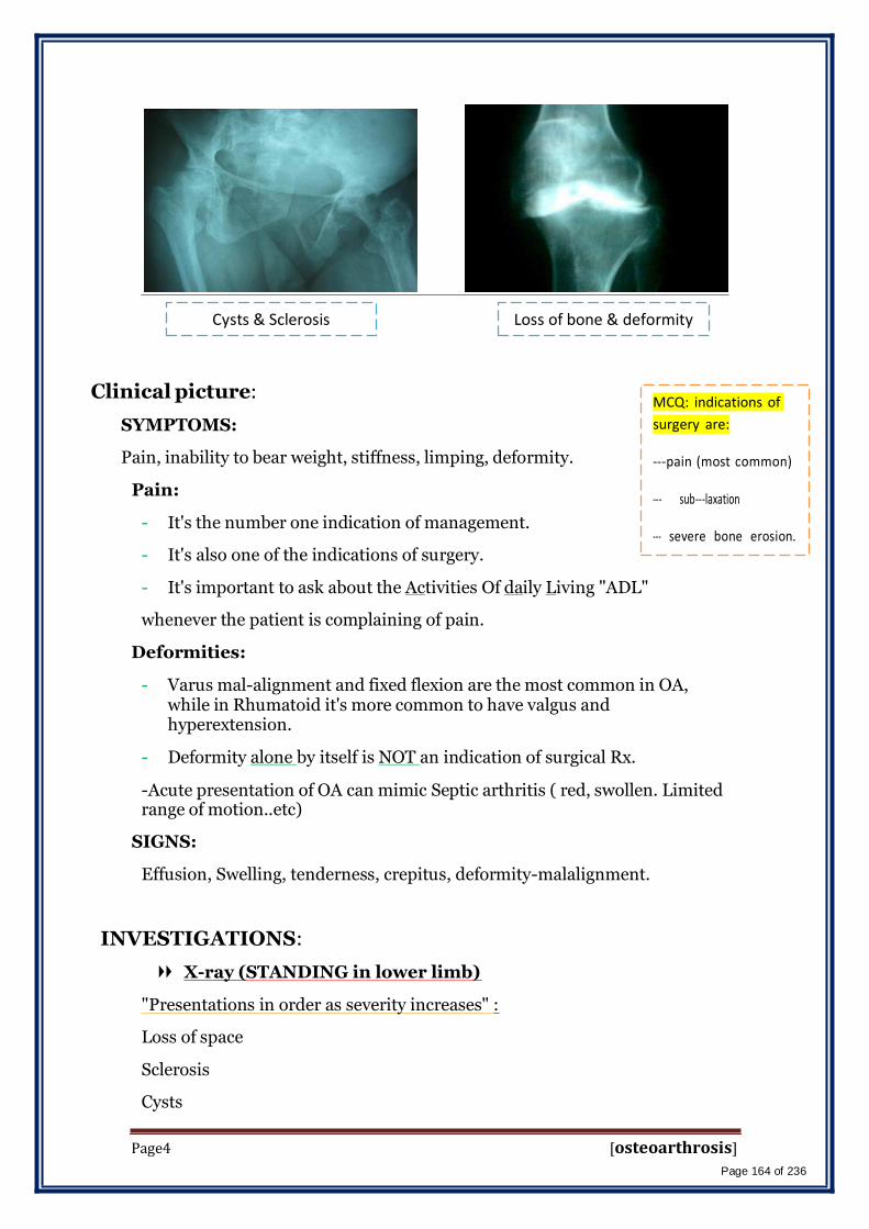
Cysts & Sclerosis Loss of bone & deformity
Clinical picture:
SYMPTOMS:
Pain, inability to bear weight, stiffness, limping, deformity.
Pain:
- It's the number one indication of management.
- It's also one of the indications of surgery.
- It's important to ask about the Activities Of daily Living "ADL"
whenever the patient is complaining of pain.
Deformities:
MCQ: indications of
surgery are:
-pain (most common) - sub-laxation - severe bone erosion.
- Varus mal-alignment and fixed flexion are the most common in OA, while in Rhumatoid it's more common to have valgus and hyperextension.
- Deformity alone by itself is NOT an indication of surgical Rx.
-Acute presentation of OA can mimic Septic arthritis ( red, swollen. Limited range of motion..etc)
SIGNS:
Effusion, Swelling, tenderness, crepitus, deformity-malalignment.
INVESTIGATIONS:
X-ray (STANDING in lower limb)
"Presentations in order as severity increases" :
Loss of space
Sclerosis
Cysts
Page4 [osteoarthrosis]
Page 164 of 236

Osteophytes (at the peripheral lines of the joint).
Loose bodies
Malalignment.
Subluxation (in advanced cases)
sulux.
Erosion.
synovial analysis (in differential diagnosis)
• Synovial fluid analysis should NOT be performed always, unless there is a massive effusion, otherwise don't do it all the time.
Septic Arthritis (>20,ooo-30,ooo polymorph cell count).
Inflammatory Arthritis
Protein Level Glucose Level
High Low
High Normal
Osteoarthrosis Normal Normal
Diagnosis:
- OA is mainly diagnosed clinically, there is no much need to do many several investigations. X-ray is the best modality for investigation with AP & lateral standing views + Skyline "Axial" view in case of hip or knee OA to show the patella-femoral joint.
- Whenever a patient is complaining of knee joint, you should automatically examine the hip. Because hip disease can transfer to the knee and vise versa.
Page5 [osteoarthrosis]
Page 165 of 236
Management:
Case scenarios:
1-a 42 years old female complaining of right leg pain since 6 months . She has the pain after 1 hour of
walking & praying. She doesn't have it when she use the stairs up & down. Her weight= 80, Height=165.
On examination the patient has mild tenderness at the medial side of the joint, there is no redness. X-ray
shows 3 degrees of valgus instead of 7 degrees. Her mother & father have osteoarthrosis. She is mildly
diabetic on regulatory treatment. No Hx of trauma, previous surgeries or infection. She is on glucophage.
How to manage this patient?
-Exercise, reduse weight & modification of the activities. – give her paracetamol.
2-a 47 years old female complaining of left knee pain since 2 years . The pain increase with praying , using
of stairs & walking. She has a Hx of twisting injury over knee when she was 25 years old. She had meniscal
tear & she has arthroscopy for meniscal tear. She has been seen by other doctors & she took NSAID. She
did exercise & she is considered obese & she failed to lose weight. Height=165, weight= 95. X-ray shows
subchondral sclerosis, decrease medial joint space osteophytes on the medial side of the joint. Also, she
has varus deformity of 10 degrees. How to manage?
- Hyaluronic acid. -Arthroscopic lavage as a second choice if there is no hyluronic acid available.
3-a 55 years old male, porter complaining of knee pain since 5 years . Height =180, weight=100 He loves exercise. X-ray shows 15 degrees of varus , sclerosis & redused medial .بنةیی جسھھم عضلھھییjoint line. He wants to complete his work & he tried everything. What modalities can be used?
-Arthroplasty (if patient accept to change his job & be carful about exercise).
-Osteotomy "closed wedge osteotomy" (reallighnment of the joint).
Points to remember from the scenarios: IMP
- If the x-ray is only showing mild varus exercise and weight reduction would be advisable.
- Paracetamole can be used in case of mild pain. NSAID are usually not beneficial in case of OA.
- Activity modification is advisable if the pain gets worse with specific movement or activity.
- Arthroscopic lavage "debridement" used as a temporary Rx and in case of moderate OA.
- Steroid injections has NO effect on OA as a main method of treatment, it's only beneficial in case of acute presentation of OA because its duration of effect = 3 weeks.
- Hyaluronic acid injection has almost similar results of arhtrosope, so it's preferred as it
doesn't require anesthesia.
- If patient had arthroplasty, he should restrict his activity after this procedure especially sports & exercise.
- Osteotomy used to over correct the valgus "overcorrection". Normal Anterior cruciate
ligaments + knee stability & range of motion are one of conditions that should be considered before performing this type of surgery.
-
Page6 [osteoarthrosis]
Page 166 of 236
History
Examination
Investigations Conservative
treatment:
Decrease load (stick, brace, reduce weight)
Modify activity
Physiotherapy:
prevent contractures
muscle strengthening
range of motion
Medications
systemic
Local
Surgical treatment:
1- Joint Debridement if there are lose body ( bone ) in the joint and it block the movement
2- Corrective Osteotomy ( there are 2 pictures in the slid no. 22 if you want
to see them cus I couldn't copy them) :
What? varus/valgus, abd./add.
Why ? to realign axis and redistribute weight
Which joint? knee/hip
What joint? mobile, stable, minimally deformed
Which patient? young, thin, active
3- Arthrodesis: ( fusion of the joint )
Why? Transfer painful stiff into painless stiff joint.
Which joint? -wrist, ankle, C-Spine, L-Spine, hand (Small joints "hand & foot").
- hips and knees (LESS COMMON)
Page7 [osteoarthrosis]
Page 167 of 236
When?
- failed TKR ( Chronic infection & acute infection on post-hip replacement).
- Neuropathic joint ( the best treatment for it).
- paralytic (flail).
- Loss of quad.
- Stiff in young.
When NOT to use it? - Ipsilateral disease.
- Contralateral hip disease.
- bilateral joint disease.
TRANSFER LOAD TO DISTAL and CONTRALATERAL JOINTS
4-Arthroplasty:
Excision Arthroplasty ( remove part of the joint to allow movement )
Disadvantage:
-weakness.
–shortening.
-walking aid
Which joint?
-Hip; post infection (girdle stone)
-1st MTPJ.
-1st.MPJ
Excisional arthroplasty is indicated for small joints in hand & foot. It's not used for big joints
EXCEPT in case of chronic infections "TB or Brucella'' or acute infection in post-total hip
replacement" because joint replacement is contraindicated in case of infection" so we use
the excisional instead of it.
-patient with sever varus (25 degrees varus), sclerosis, osteophytes , lose of height , lose of bone = Arthroplasty.
Page8 [osteoarthrosis]
Page 168 of 236

Partial Replacement: one side of the joint
Which joint?
- hip (fracture).
- knee
- shoulder(SCD, RA)
When?
When NOT?
- necrosis
- degenerative
- trauma
- Inflammatory (ONLY SHOULDER)
- infection
- young
- inflammatory
-Partial or hemi-arthroplasty is indicated usually for trauma (e.g. femur neck fracture, hip fracture, knee fracture…). It is CONTRAINDICATED in Rheumatoid Arthritis
5- TOTAL REPLACEMENT (pictures in slide 33 & 34 which
can't be copied also):
Which?
- Knees (mainly).
- Hips.
- Shoulders.
- Ankles.
- elbow
When?
- painful, deformed stiff joint, old patient!!
Page9 [osteoarthrosis]
Page 169 of 236
When NOT?
- Neuropathic
- Infection
- Paralysis (e.g. polio)
- young, active (RELATIVE)
Because young and active pt will consume the artificial joint very fast.
Page10 [osteoarthrosis]
Page 170 of 236

Isn't it funny how someone can say "I believe in Allah " but still follow the Satan who by the way also, " believes " in
Allah…
430 ORTHOPEDICS TEAM
Lecture: Peripheral Nerve Injury.
Team Members:
Faten Al-Mohideb. Hanan Al-Salman. Aliya Al-Awaji.
Hadeel Al-Ghamdi.
Ghadeer Al-Wuhayd.
Hissa Al-Balla.
Lujain Al-Yousef.
Nouf Al-Hammad.
Jawaher Al-Faraydi.
Hiba Al-Rahiem.
Nour Al-Enezi.
Wejdan Al-Swayyid.
Arwa Abudawood.
Leena Al-Shaman.
Areej Al-‐Qunaitir.
Team Leader:
Ayedah Al-Ruhaimi
-‐‐The slides were provided by the doctor.
-429 team group A-1.
-‐‐Important notes in Red.
-‐‐Copied slides in Black.
-Doctor's notes in green.
Page1 Peripheral Nerve Injury
Page 171 of 236

OUTLINE
• DEFINITION.
• TYPES OF NERVE INJURIES.
• FATE (pathophysiology) AND REHABILITATION.
• ETIOLOGY.
• PRESENTATION.
• DIAGNOSIS.
• CLINICAL EXAMPLES:
(ERB’ S,CARPAL TUNNEL,RADIAL,ULNAR,SCIATIC AND
PERONEAL)
What is peripheral nerve?
It is a nerve which connects between CNS & periphery, with EXCLUSION of cranial
nerves. It is composed of: cell body, axon & nerve end.
If the Cell Body is Motor: it is located in anterior Horn cell in spinal cord (motor end
plate).
If axon is sensory: it is located in posterior Horn cell.
Always mixed (contain both sensory & motor fibers) No pure sensory or motor.
It has the capability of regeneration unlike of CNS.
Rehabilitation is very important in peripheral nerve injury.
WHAT ARE THE FEATURES OF A PERIPHERAL NERVE?
• RELATES PERIPHERY AND SPINAL CORD.
• MIXED (SENSORY AND MOTOR).
• REGENERATES.
Definition of nerve injury:
It is Partial OR Complete interruption of normal physiology of the nerves (nerve
conduction is affected).
Types:
1. Neuropraxia:
E.g .: when you set for long time you feel numbness.
• Commonest & easiest in recovery
• Reversible failure propagation of electrical impulses across the affected nerve
segment.
• No anatomical change of the nerve structure (anatomy is intact).
Page2 Peripheral Nerve Injury
Page 172 of 236

• Duration: usually: seconds – minutes. Rare: hours - days.
e.g.:
• Saturday night palsy:alcoholics→radial nerve palsy
• Honeymooner's syndrome.
• Wheel chair bound persons.
2. Axontemesis:
• Complete absence of sensory & motor activity of that nerve
• Associated with accident & trauma:
Fracture of humeral shaft →radial nerve injury
Fracture of medial epicondyle → ulnar nerve injury.
Fracture of proximal fibula → peronealnerve injury.
Fracture of acetabulum &posteriordislocation of femoral head→sciatic
nerve injury.
• No sensation or motion (loss of nerve function).
• Axon+myline sheath are damaged (histological changes).
• The cell body losses its continuity with nerve.
• Endo, epi, perineum are intact (fascia is intact).
• Anatomy is intact.
• There is wallerian degeneration.
• Good prognosis.
3. Neurotemesis:
• Anatomical damaged in the nerve (nerve ends are not in continuity)
→complete disturbance of activity of that nerve + loss of supporting tissues.
• There is wallerian degeneration.
• Prognosis is poor& worst without surgical repair.
Wallerian degeneration:
• It is a process of proximal part regeneration &distal part degeneration of the
nerve regarding to the damage site.
• Length of regeneration: 1mm/day. e.g.: if the length of sciatic nerve injury is
50cm→it needs 500 days to recover.
Page3 Peripheral Nerve Injury
Page 173 of 236

• During the period of regeneration, if the distal part is not stimulated from
outside→ lead to atrophy of motor end plate & loss its function. So, you have
to stimulate motor end plate by rehab to prevent degeneration of it (it is not
reversible).
• If the anatomy is intact (axontemesis) → no need to repair.
• If the anatomy is disturbed (neurotemesis) → you have to repair. Then wait for
regeneration.
• N.B: if the tunnel is not in continuity that is mean there is no stimulus for
regeneration which may cause neuroma (group of N. tissue which doesn't have
any function).
Rehabilitation:
• Pain control by simple analgesia.
• Nerve& muscle stimulation.
• Dynamic splints to avoid stiffness. (We have to prevent pressure
sores).*NECROSIS STARTS AFTER 20 MINS
• Nearby joints range of motion by doing passive movement to prevent muscle
stiffness.
• It takes months-years (long time) to recover.
Remember:
• Pressure sores develops due to the pressure of splint in case of complete loss
of sensation of the splinted limb.
• You can prevent pressure sores by padding the splint with cotton between
splint & limb.
Etiology:
■ Acute:
• Fractures (the commonest) →axontmesis.
• Wrong position (Saturday night syndrome, handicap) {neuropraxia}.
• Surgery.{neurotmesis}
Page4 Peripheral Nerve Injury
Page 174 of 236

• Electrical burn (the worst) because it damages everything.USUALLY
IRREVERSABLE
■ Chronic:
• Tight N. passage (e.g.: carpal tunnel syndrome, tarsal tunnel syndrome).
• Tumors which compress the nerve.
Presentation:
• Pain
• History of trauma.
• Loss of sensation.
• Loss of motion
• Loss of power.
• Loss of reflexes.
• Muscle wasting.
• Contractures (deformity).
• Tropic changes: loss of normal well-being of limb due to disuse (shiny skin,
hair disturbance…..).
Diagnosis:
Confirmed by:
• X-ray: in trauma.
• EMG: study of muscle electrical activity.
• NCS (neural conduction study): study of activity of electricity in the N.
itself.IT IS VERY IMPORTANT TO COMPARE THE PROGRESS FROM
ONE VISIT TO ANOTHER AND TO DECIDE WHAT TO DO NEXT
• MRI. (Like in brachial plexus injury so, we use it in special cases).
Erb’s palsy:
• Birth injury (difficult labor e.g.: shoulder dystocia & breech
presentation).
• Traction often on N. roots C5-C6 but may occur on C4-C7-C8.
• Stretch or rupture or avulsion ← the worst.
• Upper limb in extension + internal rotation ← waiter phenomena.
Page5 Peripheral Nerve Injury
Page 175 of 236
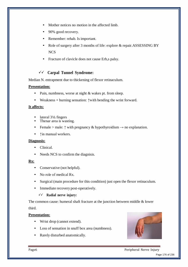
• Mother notices no motion in the affected limb.
• 90% good recovery.
• Remember: rehab. Is important.
• Role of surgery after 3 months of life: explore & repair.ASSESSING BY
NCS
• Fracture of clavicle does not cause Erb,s palsy.
Carpal Tunnel Syndrome:
Median N. entrapment due to thickening of flexor retinaculum.
Presentation:
• Pain, numbness, worse at night & wakes pt. from sleep.
• Weakness + burning sensation: ↑with bending the wrist forward.
It affects:
• lateral 3⅕ fingers • Thenar area is wasting.
• Female > male: ↑ with pregnancy & hypothyroidism → no explanation.
• ↑in manual workers.
Diagnosis:
• Clinical.
• Needs NCS to confirm the diagnisis.
Rx:
• Conservative (not helpful).
• No role of medical Rx.
• Surgical (main procedure for this condition) just open the flexor retinaculum.
• Immediate recovery post-operatively.
Radial nerve injury:
The common cause: humeral shaft fracture at the junction between middle & lower
third.
Presentation:
• Wrist drop (cannot extend).
• Loss of sensation in snuff box area (numbness).
• Rarely disturbed anatomically.
Page6 Peripheral Nerve Injury
Page 176 of 236

Rx:
• Conservative (dynamic splint).
• Do NCS, if no improvement within 3 months → surgical intervention.
• N.B: dynamic splint= splint with joint movement but static splint= splint with
no joint movement.
Ulnar N. injury:
Associated with elbow injury + tight compartment.
Usually in children with supracondylar fracture.
Presentation:
• Numbness in medial 1⅕ fingers. • Hypothener muscle wasting.
• In late stage: CLAW hand (extension of MCP +flexion of IPJ) due to loss of
lumbrical&interosseous action.
Rx: as in radial nerve.
Sciatic nerve injury:
(no loss of hip flexion + Knee Extension)
With trauma or posterior dislocation of the hip + distal injury to its branches
(posterior Tibial + common peroneal).
Presentation:
• Loss of sensation & motion below the knee level.
• Loss of all activities (flexion, extension …) below the knee level.
• Extension of the knee is not lost.
• Long period of recovery.
Peroneal nerve injury:
Presentation:
Foot drop → no heel strike= inability to extend the foot.
Loss of sensation. - Leg weakness.
Causes:
• Direct injury is rare.
• Tight splint without proper cotton padding.
• Skeletal traction.
Rx:
Page7 Peripheral Nerve Injury
Page 177 of 236

• Skeletal traction.
• Dynamic splint.
Quiz:
1) Axon degeneration occurs from mild compression injury. X
2) The prognosis for Neuropraxia is poor. X
3) Axonotmesis is generally caused from separation of the cell body from the
neuron. XWallerian Degeneration typically does not occur in Neuropraxic
injury. √
4) Surgical reconstruction is necessary in Neurotmesis. √
5) Wallerian Degeneration does not occur in Neurotmesis. X
6) A ligamentous structure can cause Neuropraxia. √
Page8 Peripheral Nerve Injury
Page 178 of 236

Isn't it funny how someone can say "I believe in Allah " but still follow the Satan who by the way also, " believes " in
Allah…
430 ORTHOPEDICS TEAM
Lecture: Common Shoulder Disorders
Team Members:
Faten Al-Mohideb. Hanan Al-Salman. Aliya Al-Awaji.
Hadeel Al-Ghamdi.
Ghadeer Al-Wuhayd.
Hissa Al-Balla.
Lujain Al-Yousef.
Nouf Al-Hammad.
Jawaher Al-‐Faraydi.
Hiba Al-Rahiem.
Nour Al-Enezi.
Wejdan Al-Swayyid.
Arwa Abudawood.
Leena Al-Shaman.
Areej Al-Qunaitir.
Team Leader:
Ayedah Al-Ruhaimi.
-‐‐ The slides were provided by the doctor.
-‐‐ Important notes in Red
-‐‐ Copied slides in Black.
-Doctor's notes in green.
Special thanks to hend Al-Qhtani.
Page1 [Common Shoulder Disorders]
Page 179 of 236
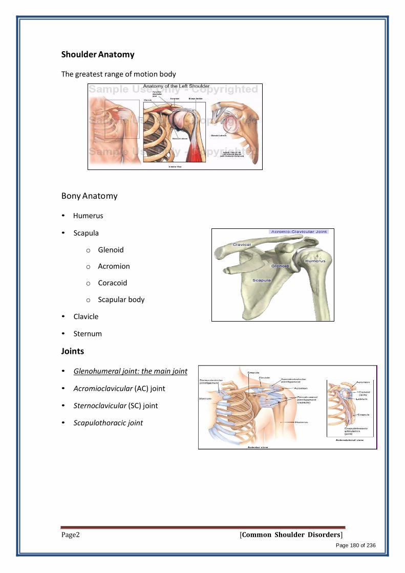
Shoulder Anatomy
The greatest range of motion body
Bony Anatomy • Humerus
• Scapula
o Glenoid
o Acromion
o Coracoid
o Scapular body
• Clavicle
• Sternum
Joints • Glenohumeral joint: the main joint
• Acromioclavicular (AC) joint
• Sternoclavicular (SC) joint
• Scapulothoracic joint
Page2 [Common Shoulder Disorders]
Page 180 of 236

Rotator Cuff Muscles (SITS)
Depress humeral head against glenoid
• Supraspinatus:
o Initiating of Abduction.(there
can be cases of complete tears
of the ms. And the Pt. can still
abduct his shoulder)
• Infraspinatus:
o External rotation
• Teres Minor:
o External rotation
• Subscapularis:
o Internal rotation
Teres minro is not important clinically –there is no specific test for Teres minor
Muscles
*Deltoid:
Largest, strongest muscle of the shoulder. Most important muscle
is the deltoid: 3 origins(scapula spine –acromion-clavicle) and one
attachment(proximal third of humerus ) supplied by axillary nerve
*Biceps : 2 heads . long head is deep – the only tendon in the body that goes into a
joint . short head attach to cricoid process
Page3 [Common Shoulder Disorders]
Page 181 of 236
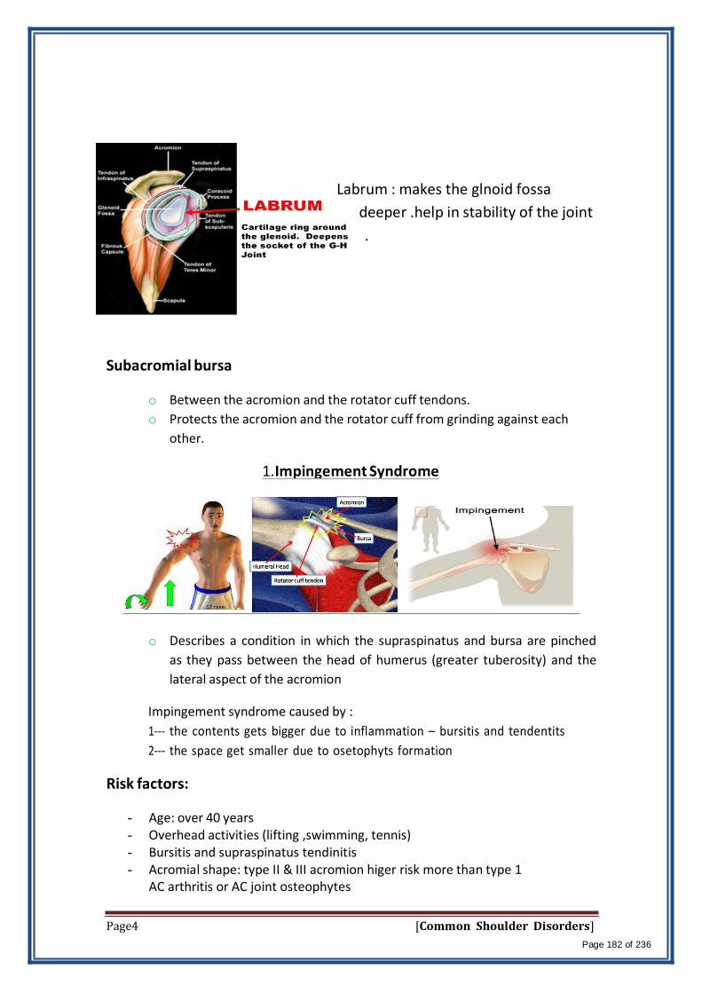
Labrum : makes the glnoid fossa
deeper .help in stability of the joint
.
Subacromial bursa
o Between the acromion and the rotator cuff tendons.
o Protects the acromion and the rotator cuff from grinding against each
other.
1. Impingement Syndrome
o Describes a condition in which the supraspinatus and bursa are pinched
as they pass between the head of humerus (greater tuberosity) and the
lateral aspect of the acromion
Impingement syndrome caused by :
1- the contents gets bigger due to inflammation – bursitis and tendentits
2- the space get smaller due to osetophyts formation
Risk factors:
- Age: over 40 years - Overhead activities (lifting ,swimming, tennis) - Bursitis and supraspinatus tendinitis - Acromial shape: type II & III acromion higer risk more than type 1
AC arthritis or AC joint osteophytes
Page4 [Common Shoulder Disorders]
Page 182 of 236

� Symptoms
- 3 Types of acromion: - I : flat - II : curved - III: hooked
• Pain in the acromial area when the arm is flexed and internally rotated
Inability to use the overhead position.
• The pain may result from subacromial bursitis or rotator cuff tendinitis
• Pain when sleeping on the affected side..
• Pain will often become worse at night, as the subacromial bursa becomes
hyperemic after a day of activity
• Decreased range of motion especially abduction
• Weakness
sever impingement may lead to rotator cuff tear
Differential diagnosis
- Rotator cuff tears - Calcific tendinitis - Biceps tendinitis - Cervical radiculopathy - Acromioclavicular arthritis - Glenohumeral instability - Glenohumeral osteoarthritis.
Physical examination
- Atrophy of rotator cuff muscles. - Decreased range of motion (esp. internal rotation & abduction) - Weakness in flexion and external rotation. - Pain on resisted abduction and external rotation. - Pain on “impingement tests”.
Page5 [Common Shoulder Disorders]
Page 183 of 236
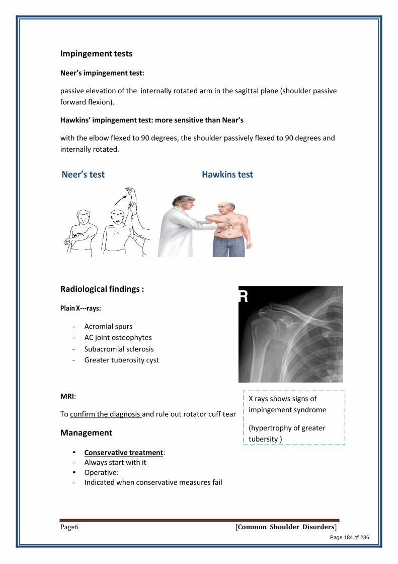
Impingement tests
Neer’s impingement test:
passive elevation of the internally rotated arm in the sagittal plane (shoulder passive
forward flexion).
Hawkins’ impingement test: more sensitive than Near’s
with the elbow flexed to 90 degrees, the shoulder passively flexed to 90 degrees and
internally rotated.
Radiological findings :
Plain X-‐rays:
- Acromial spurs
- AC joint osteophytes
- Subacromial sclerosis
- Greater tuberosity cyst
MRI:
To confirm the diagnosis and rule out rotator cuff tear
Management
• Conservative treatment: - Always start with it
• Operative: - Indicated when conservative measures fail
X rays shows signs of
impingement syndrome
(hypertrophy of greater
tubersity )
Page6 [Common Shoulder Disorders]
Page 184 of 236
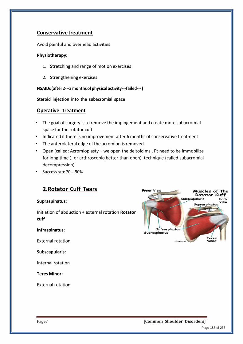
Conservative treatment
Avoid painful and overhead activities
Physiotherapy:
1. Stretching and range of motion exercises
2. Strengthening exercises
NSAIDs (after 2-‐3 months of physical activity-‐failed-‐‐ )
Steroid injection into the subacromial space
Operative treatment
• The goal of surgery is to remove the impingement and create more subacromial
space for the rotator cuff
• Indicated if there is no improvement after 6 months of conservative treatment
• The anterolateral edge of the acromion is removed
• Open (called: Acromioplasty – we open the deltoid ms , Pt need to be immobilize
for long time ), or arthroscopic(better than open) technique (called subacromial
decompression)
• Success rate 70-90%
2.Rotator Cuff Tears
Supraspinatus:
Initiation of abduction + external rotation Rotator
cuff
Infraspinatus:
External rotation
Subscapularis:
Internal rotation
Teres Minor:
External rotation
Page7 [Common Shoulder Disorders]
Page 185 of 236

Function of rotator cuff muscles
• Keep the humeral head centered on the glenoid regardless of the arm’s
position in space.
• Generally work to depress the humeral head while powerful deltoid contracts
to pull the humeral head up
Causes of rotator cuff tears
• Intrinsic factors:
- Vascular = in the greater tubresity is very poor>risk of avasacular necrosis
due to any small injury , maluniuon
- Degenerative ( age-related)
• Extrinsic factors:
- Impingement ( in acromion, or sub acromioclavicular joint)
- Acromial spurs
- AC joint osteophytes
- Repetitive use
• Traumatic (e.g. a fall or trying to catch or lift a heavy object)
Diagnosis
- History
- Physical examination(empty can, lift off test)
- X-rays( not showing any abnormalities )
- MRI (best )
Symptoms
• Pain(can't lift his arm up ) radiating to deltoid insertion or biceps( Pt. said I
had pain in the upper part of my arm or in my neck)
• Insidious progression of pain
• Night pain
• Popping noises
• Weakness
• Could be asymptomatic
Page8 [Common Shoulder Disorders]
Page 186 of 236
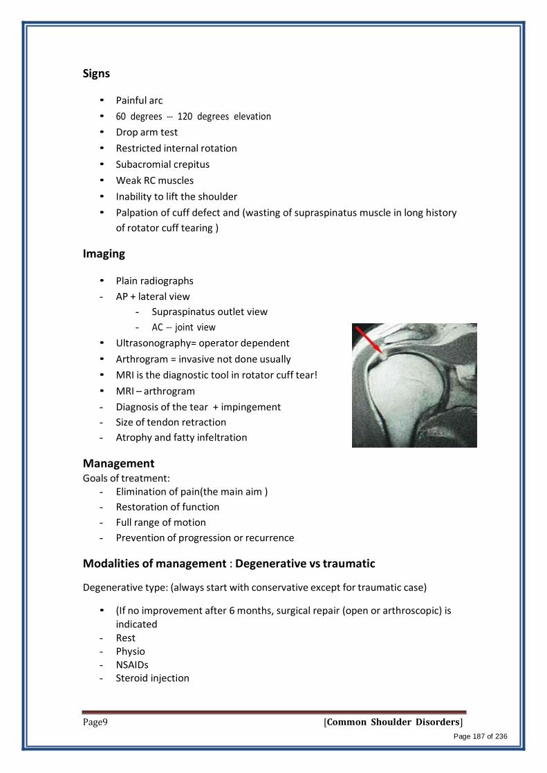
Signs
• Painful arc
• 60 degrees - 120 degrees elevation
• Drop arm test
• Restricted internal rotation
• Subacromial crepitus
• Weak RC muscles
• Inability to lift the shoulder
• Palpation of cuff defect and (wasting of supraspinatus muscle in long history
of rotator cuff tearing )
Imaging
• Plain radiographs
- AP + lateral view
- Supraspinatus outlet view
- AC - joint view
• Ultrasonography= operator dependent
• Arthrogram = invasive not done usually
• MRI is the diagnostic tool in rotator cuff tear!
• MRI – arthrogram
- Diagnosis of the tear + impingement
- Size of tendon retraction
- Atrophy and fatty infeltration
Management Goals of treatment:
- Elimination of pain(the main aim )
- Restoration of function
- Full range of motion
- Prevention of progression or recurrence
Modalities of management : Degenerative vs traumatic
Degenerative type: (always start with conservative except for traumatic case)
• (If no improvement after 6 months, surgical repair (open or arthroscopic) is indicated
- Rest - Physio - NSAIDs - Steroid injection
Page9 [Common Shoulder Disorders]
Page 187 of 236

Traumatic type: (acute surgical repair)
- Can’t treat it with conservtive ! Muscle may retract .
• Operative treatment
Depends on:
Patient factors:
- Activity level - Expectations - Needs
Pain or weakness:
- Interferes with work, sports, activities of daily living. - Unresponsive to appropriate non-operative treatment
Wide spectrum of tears:
• Partial ( just shaving the partial tear )
• Complete ( need repair the tendon )
- Small
- Large
- Massive (irreparable) : muscle atrophy and contracts– cant do repair – too
late and need joint replacement
Surgical Modalities :
If conservative failed after 6 month of trials
1. Rotator cuff tendon debridement
2. Subacromial decompression
- open
- arthroscopic = much easier
3. Rotator cuff tendon repair = most immobilize the shoulder to help the tendon bone fixation
- completely surgically open repair
- arthroscopic assisted mini-open repair
- completely arthroscopic repair
4. Reverse shoulder arthroplasty = in massive tear
Page10 [Common Shoulder Disorders]
Page 188 of 236

Complications of RC tears
• Complete tears: if not treated chronic pain and loss of motion and with
time becomes irreparable deltoid take of the role leading to shoulder
elevation and Glenohumeral –rotator cuff arthropathy-
• Complications of surgery: not improving, stiffness, infection, RSD
4-6 wk of immobilization = risk of stiffness in longer duration
3. Adhesive Capsulitis
- Also called “frozen shoulder”
- It is characterized by pain and restriction of all movements of the shoulder
(global stiffness)
- ( in adhesive capsulitis all movements of shoulder are affected in contrast to
impingement syndrome where limitation of abduction only) –
- Usually self limiting (typically begins gradually, worsens over time and then
resolves but may take >2 years to resolve)
- Adhesive Capsulitis
Risk factors:
- DM (esp. insulin dependent) the most imp risk factor - Following injury or surgery to the shoulder - Hypo and Hyperthyroidism - High cholesterol
Diagnosis:
- Mainly clinical = global movement limitation - X-rays and MRI to rule out other pathologies
Stages: each stage takes 2 years
1 -Pain (freezing stage)= pain
2 -Stiffness (frozen stage)= pain with movement limitation
3-Resolution (thawing stage)= start healing
Treatment
- Resolves if untreated over 2-4 years
- Start with conservative after 6 month if no improvement go for surgery
- Aggressive Physiotherapy
- Pain and anti-inflammatory medications used all together
- Steroid injections
- Manipulation under anesthesia = risk of fracture due to disuse osteopenia
Page11 [Common Shoulder Disorders]
Page 189 of 236

- Arthroscopic capsular release = best way of management and safer
4. Glenohumeral osteoarthritis
- Mainly wear and tear
- Could be a result of RA, AVN or malunited fractures
- Glenohumeral osteoarthritis
- The management starts with conservative measures.
- If it fails to relieve the pain, the best management for
elderly patient is arthroplasty (hemi or total shoulder
replacement)
- If associated with irreparable rotator cuff tear for
reverse shoulder replacement
- Note there is 2 types of glenohumeral osteoarthritis:
1) Primary osteoarthorosis like the knee > Rx with arthroplasty
2) 2ry to irreparable rotator cuff tear >Rx with reverse shoulder replacement
Reverse shoulder
replacement
Hemi arthroplasty
4. AC arthritis
Across the chest adduction test( positive if there is pain)
Page12 [Common Shoulder Disorders]
Page 190 of 236

Causes of AC Arthritis
- Degenerative osteoarthritis.( wear and tear in old aged people)
- Rheumatoid Arthritis .
- Gouty Arthritis.
- Septic Arthritis.
- Atraumatic distal claivcle osteolysis in weight lifters.
Signs and Symptoms
- Pain , which worsens with movement and progressively worsens.( the patient
may suffer a night pain which is a sign of arthritis)
- It is commonly associated with impingement syndrome
- Positive across the chest adduction
Diagnosis: Clinical
and by x-rays
Treatment :
Non-surgical Treatment
- Rest , avoid weightlifting and push-ups(triggering activities )
- Pain medications and NSAID to reduce pain and inflammation
Surgical treatment
-it involves removing of the AC joint applying an artificial joint.
-the bone spurs must also be removed .
5. Dislocation of the Shoulder
Basic anatomy about Glenohumeral Joint :
- Most common dislocated joint
- Lacks bony stability
- Composed of:
Fibrous capsule
Ligaments
Surrounding muscles
Glenoid labrum
Page13 [Common Shoulder Disorders]
Page 191 of 236
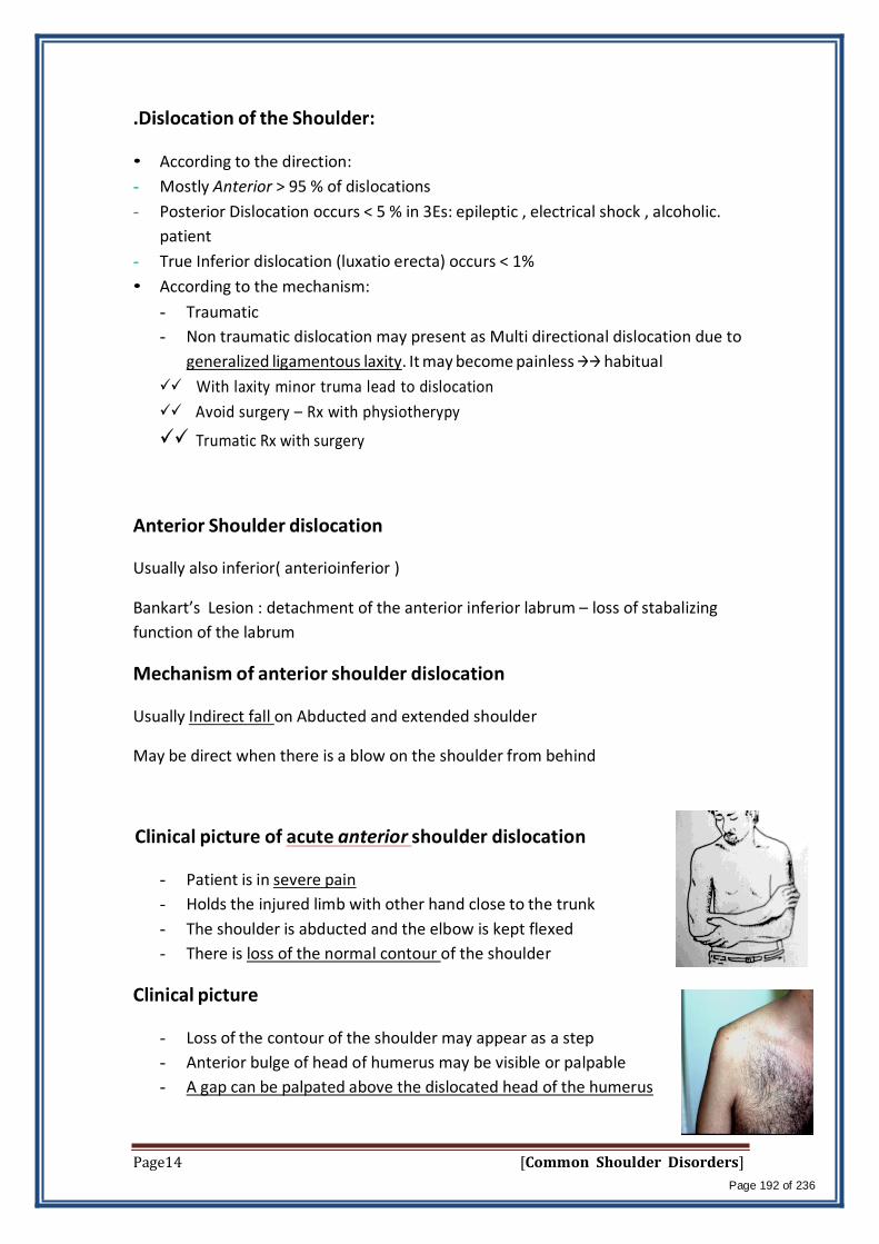
.Dislocation of the Shoulder: • According to the direction:
- Mostly Anterior > 95 % of dislocations
- Posterior Dislocation occurs < 5 % in 3Es: epileptic , electrical shock , alcoholic.
patient
- True Inferior dislocation (luxatio erecta) occurs < 1%
• According to the mechanism:
- Traumatic
- Non traumatic dislocation may present as Multi directional dislocation due to
generalized ligamentous laxity. It may become painless habitual
With laxity minor truma lead to dislocation
Avoid surgery – Rx with physiotherypy
Trumatic Rx with surgery
Anterior Shoulder dislocation
Usually also inferior( anterioinferior )
Bankart’s Lesion : detachment of the anterior inferior labrum – loss of stabalizing
function of the labrum
Mechanism of anterior shoulder dislocation
Usually Indirect fall on Abducted and extended shoulder
May be direct when there is a blow on the shoulder from behind
Clinical picture of acute anterior shoulder dislocation
- Patient is in severe pain
- Holds the injured limb with other hand close to the trunk
- The shoulder is abducted and the elbow is kept flexed
- There is loss of the normal contour of the shoulder
Clinical picture
- Loss of the contour of the shoulder may appear as a step
- Anterior bulge of head of humerus may be visible or palpable
- A gap can be palpated above the dislocated head of the humerus
Page14 [Common Shoulder Disorders]
Page 192 of 236
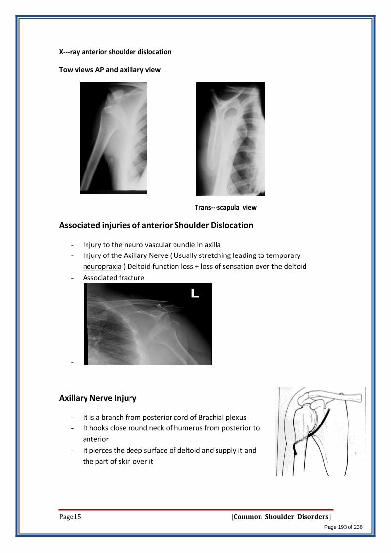
X-‐‐ray anterior shoulder dislocation
Tow views AP and axillary view
Trans-‐scapula view
Associated injuries of anterior Shoulder Dislocation
- Injury to the neuro vascular bundle in axilla
- Injury of the Axillary Nerve ( Usually stretching leading to temporary
neuropraxia ) Deltoid function loss + loss of sensation over the deltoid
- Associated fracture
-
Axillary Nerve Injury
- It is a branch from posterior cord of Brachial plexus
- It hooks close round neck of humerus from posterior to
anterior
- It pierces the deep surface of deltoid and supply it and
the part of skin over it
Page15 [Common Shoulder Disorders]
Page 193 of 236
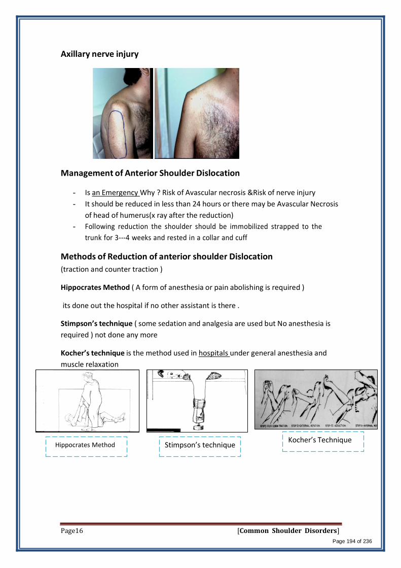
Axillary nerve injury
Management of Anterior Shoulder Dislocation
- Is an Emergency Why ? Risk of Avascular necrosis &Risk of nerve injury
- It should be reduced in less than 24 hours or there may be Avascular Necrosis
of head of humerus(x ray after the reduction)
- Following reduction the shoulder should be immobilized strapped to the
trunk for 3-4 weeks and rested in a collar and cuff
Methods of Reduction of anterior shoulder Dislocation
(traction and counter traction )
Hippocrates Method ( A form of anesthesia or pain abolishing is required )
its done out the hospital if no other assistant is there .
Stimpson’s technique ( some sedation and analgesia are used but No anesthesia is
required ) not done any more
Kocher’s technique is the method used in hospitals under general anesthesia and
muscle relaxation
Hippocrates Method Stimpson’s technique Kocher’s Technique
Page16 [Common Shoulder Disorders]
Page 194 of 236

Complications of anterior Shoulder Dislocation : Early
- Neurovascular injury ( rare )
- Axillary nerve injury
- Associated Fracture of neck of humerus or greater or lesser tuberosities
Complications of anterior shoulder Dislocation : Late
- Avascular necrosis of the head of the Humerus (high risk with delayed
reduction)
- Recurrent shoulder dislocations (the commonest complication) , treated by
Bankart repair (either arthroscopic or open)re-attachemtn of the labrum
Bankart lesion Arthroscopic Bankart Bankart lesion
6. Acromioclavicular separation
Acromioclavicular Joint
The AC joint is different from joints like the knee or ankle, because it doesn't need
to move very much. The AC joint only needs to be flexible enough for the shoulder to
move freely. The AC joint just shifts a bit as the shoulder moves.
The AcromioClavicular joint is stabilized by three ligaments
- 2 Coracoclavicular ligaments
- Conoid
- trapezoid
- AC ligament
Acromioclavicular separation
Mechanisms of Injury:
- Fall on the tip of the unprotected shoulder.
- Fall on the outstretched hand.
- Downward force on the acromion from above.
-
Page17 [Common Shoulder Disorders]
Page 195 of 236
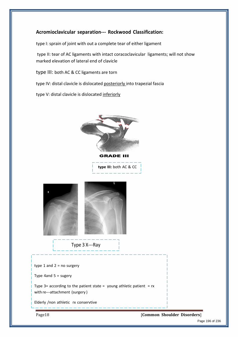
Acromioclavicular separation-‐‐ Rockwood Classification:
type I: sprain of joint with out a complete tear of either ligament
type II: tear of AC ligaments with intact coracoclavicular ligaments; will not show
marked elevation of lateral end of clavicle
type III: both AC & CC ligaments are torn
type IV: distal clavicle is dislocated posteriorly into trapezial fascia
type V: distal clavicle is dislocated inferiorly
type III: both AC & CC
Type 3 X-Ray
type 1 and 2 = no surgery
Type 4and 5 = sugery
Type 3= according to the patient state = young athletic patient = rx
with re-attachment (surgery )
Elderly /non athletic rx conservtive
Page18 [Common Shoulder Disorders]
Page 196 of 236

Isn't it funny how someone can say "I believe in Allah " but still follow the Satan who by the way also, " believes " in
Allah…
430 ORTHOPEDICS TEAM
Lecture: Metabolic Bone Disorders
Team Members:
Faten Al-Mohideb. Hanan Al-Salman. Aliya Al-Awaji.
Hadeel Al-Ghamdi.
Ghadeer Al-Wuhayd.
Hissa Al-Balla.
Lujain Al-Yousef.
Nouf Al-‐Hammad.
Jawaher Al-Faraydi.
Hiba Al-Rahiem.
Nour Al-Enezi.
Wejdan Al-Swayyid.
Arwa Abudawood.
Leena Al-Shaman.
Areej Al-Qunaitir.
Team Leader:
Ayedah Al-Ruhaimi.
-‐‐The slides were provided by the doctor.
-‐‐Important notes in Red
-‐‐Copied slides in Black.
-Doctor's notes in green.
Page1 [Metabolic Bone Disorders]
Page 197 of 236

Orthopedic Surgeons and Bone:
Orthopedic surgeons have to deal with all types of bone: healthy or
diseased; and that’s why they have to know about bone metabolism.
Bones in the body protect vital organs eg: skull protects brain, ribs protect
lungs and heart.
Bones give support to muscles and tendons.
Bone may become weak in certain conditions.
Bone is a living structure:
There is a continuous activity in bone during all stages of life
There is continuous bone resorption and bone formation as well as
remodeling.
While osteoblasts are forming a new bone, osteoclasts are removing the
dead aged ones. This process accelerates with aging and when estrogen
levels drop (menopause) with decreased rate of formation and increased rate
of bone loss. Opposite to that is happening in childhood where bone is
formed in higher rates than loss, the age period from 18-21 years is the
period of equilibrium.
That means bone is not only for protection and support but its contents
play an important part in blood homeostasis
Many factors are involved in this process
Bone Metabolism:
Bone metabolism is controlled by many factors:
Calcium
Phosphorus
regulated by:
Parathyroid gland
Thyroid gland
Estrogen
Glucocorticoid hormones
Intestinal absorption
Renal excretion
Patients with malabsorption condition like celiac disease or sprue or having a
kidney disease like tubular necrosis or glomerular nephropathy will not
Page2 [Metabolic Bone Disorders]
Page 198 of 236

benefit from the dietary or supplemental calcium and vitamin D intake till this
condition is corrected.
Diet
Vitamin D
Sun
exposure
Exercise
Bone Structure:
Bone is formed by
Bone matrix : which consists of
40% organic : collagen type1 (responsible for tensile strength)
60% Minerals: mainly Calcium hydroxyapatite, Phosphorus,
and traces of other minerals like zinc
Cells in bone : osteoblasts, osteoclasts, osteocytes
Plasma levels:
Calcium : 2.2-2.6 mmol/l
Phosphorus : 0.9-1.3 mmol/l
you should remember those values only if you are intern or resident
Both absorbed by intestine and secreted by kidney in urine
Alkaline phosphatase : 30-180 units/l shows the activity of bone metabolism
Is elevated in bone increased activity like during growth or in metabolic bone
disease or destruction
in adult it should not exceed 180 units/l, but in children it is considered
normal up to 400 units/l because of growth
Vitamin D level : 70-75 nmol/l
this value is controversial, because here in Saudi Arabia all women are less
than 75, they still don’t know if it is normal to have less than that in our
population or that all Saudi females are truly vitamin D deficient. Most
probably it shouldn’t be less than 75 even in our population :/
Vitamin D is not only important for human skeleton, it also gives immunity
against cancer and several diseases. Recently they discovered that all
Alzheimer patients are vitamin D deficient.
Parathyroid Hormone (PTH):
Production levels are related to serum calcium levels:
It increases calcium levels in the blood by increasing its release from
bone, this is a fast action that increases ca levels in blood secondary
to disease as a compensatory mechanism.
increase absorption from the intestine
Page3 [Metabolic Bone Disorders]
Page 199 of 236

and increase reabsorption from the kidney (also increase secretion of
phosphorus)
Hyperparathyroidism:
This part and the last part of the lecture which also about hyperparathyroidism are
not included in the syllabus and you are not going to ask about, it is only to help you in
DDx.
Primary: Adenoma of the gland very rarely carcinoma.
Secondary: as a result of low calcium "Chronic" malabsorption, low
calcium diet, kidney excrete more Ca .
Tertiary: as a result of prolonged or sustained stimulation =
hyperactive nodule or hyperplasia
Calcitonin:
Is secreted by C cells of thyroid gland
Its secretion is regulated by serum calcium
Its action is to cause inhibition of bone resorption and increasing
calcium excretion by this it causes lowering of serum calcium
Bone Strength:
Bone strength is affected by mechanical stress which means exercise and
weight bearing, those who were exercising since childhood or farmers have
very strength bone.
Bone strength gets reduced with menopause and advancing age
Reduced bone density on X rays is called Osteopenia
Osteopenia is also a term used to describe a degree of reduced bone density,
which if advanced becomes Osteoporosis
Bone Density:
Bone density is diagnosed at current time by a test done at radiology
department called : DEXA scan which tests 3 areas: vertebrae, hip and wrists.
DEXA is ( Dual Energy X ray Absorbtionometry )
Increased bone density does not always mean increased bone strength, as
sometimes what is called Brittle bone which is a dense bone is not a strong
bone but fragile bone which may break easily
Dexa Scan: for example, if bone is dense and we send 100 rays, 20 will go back and thin
means that 80 are absorebed. But if bone is osteopenic 50 will go back so
only half is absorbed which means less density.
Page4 [Metabolic Bone Disorders]
Page 200 of 236

Normal,
- 0.9
Osteopenia,
less than - 1
Osteoprosis,
less than - 2.5
Disorders to be discussed:
Rickets Osteomalacia Osteoporosis Hyperparathyroidism (not included)
Rickets & Osteomalacia:
Different expressions of the same disease
Inadequate mineralization
Rickets: Affects areas of endochondral growth in children (wrist, knee, shoulder, hip) We can diagnose rickets by looking at knees (inspection) or wrist x-ray
Osteomalacia: Affects all skeleton is incompletely calcified in adults
Causes Calcium deficiency Hypophosphataemia Defect in Vitamin D metabolism
o Nutritional (poor, war zoon, …) o underexposure to sunlight o intestinal malabsorption ( ex. Celiac disease) o liver & kidney diseases, ( renal tubular acidosis , Glomerular
nephropathy) collect 24 hour urine Ca ( normal : 500 mg of Ca )
Page5 [Metabolic Bone Disorders]
Page 201 of 236

Rickets: Symptoms and Signs:
o Child is restless, babies cry without obvious reason o Failure to thrive o Muscle weakness
Knees are the
first one to be
affected then
wrists then
costochondral
Skull enlarge
=Delayed
sutures closure
o In severe cases with very low calcium: tetany or convulsions o Joint thickening (hypertrophy ) especially around wrists and knees o Deformity of limbs, mostly Genu varum (bowl legs) or Genu Valgum
in late presentation o Pigeon chest deformity, Rickety Rosary (beads like), craniotabes
Between costochondral junction in rib cage
Chondral
hypertrophy
especially in
growing bone
X Ray Findings in Rickets:
Growth Plate& Metaphysial Changes: Long Bones Deformities:
Epihyseal Widening
Metaphysieal
Cupping
Page6 [Metabolic Bone Disorders]
Page 202 of 236
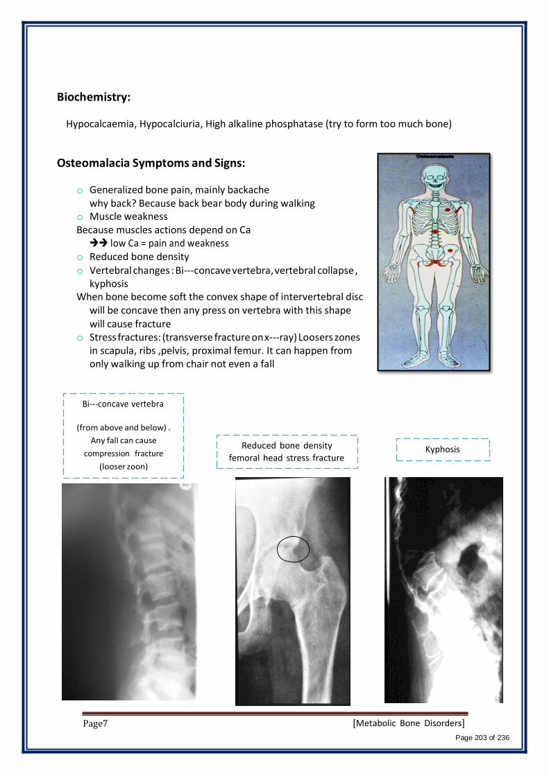
Biochemistry:
Hypocalcaemia, Hypocalciuria, High alkaline phosphatase (try to form too much bone)
Osteomalacia Symptoms and Signs:
o Generalized bone pain, mainly backache why back? Because back bear body during walking
o Muscle weakness Because muscles actions depend on Ca low Ca = pain and weakness
o Reduced bone density o Vertebral changes : Bi-concave vertebra, vertebral collapse ,
kyphosis When bone become soft the convex shape of intervertebral disc
will be concave then any press on vertebra with this shape will cause fracture
o Stress fractures: (transverse fracture on x-ray) Loosers zones in scapula, ribs ,pelvis, proximal femur. It can happen from only walking up from chair not even a fall
Bi-concave vertebra
(from above and below) .
Any fall can cause
compression fracture
(looser zoon)
Page7
Reduced bone density
femoral head stress fracture
Kyphosis
[Metabolic Bone Disorders]
Page 203 of 236

Treatment: easily treated unless the patient is having intestinal or kidney disease. ( should be treated first. because there is no point of the treatment without treat the primary cause ).
Vitamin D deficiency
- Rickets
- Osteomalacia
• adequate Vitamin D replacement
• sun exposure
• correct residual deformities
• Vitamin D + Ca (milk, eggs, fish)
• fracture management • correct deformity if needed
Osteoporosis: loss of bone unit unlike osteomalacia which affects bone mineralization
Decreased bone mass: decreased amount of bone per unit volume (and this causes reduced density) osteoclast activity
Mineralization is not affected
Mainly post-menopausal (drop Estrogen level) and age related
Normally we lose 0.3% of bone but in osteoporosis will be about 3% per year
Osteoporosis: Primary and Secondary
Primary Osteoporosis:
o Post menopausal
o Senile process happen in every men and women
Post menopausal Osteoporosis: Due to rapid decline in estrogen level This results in increased osteoclastic activity Normal bone loss usually 0.3% per year Post menopausal bone loss 3% per year
Risk Factors in Post menopausal Osteoporosis:
o Race: Caucasian (fair skin)
o Hereditary o Body build (thin people) o Early menopause o Smoking/ alcohol intake/ drug abuse o ? Calcium intake (low Ca)
-Osteoporosis is painless disease
unless it cause fracture (neck of
femur).
- Hx. Of F# (high risk group)
Page8 [Metabolic Bone Disorders]
Page 204 of 236

Senile Osteoporosis: Usually by 7th to 8th decades there is steady loss of at least 0.5% per year
female after 50 years and male after 70 years It is part of physiological manifestation of aging
Risk factors in Senile Osteoporosis: o Male menopause
o Dietary : less calcium and vitamin D and protein o Muscle weakness o reduced activity
Clinical Features of Osteoporosis:
Osteoporosis is a Silent disease
Osteoporosis is Serious due to possible complications :mainly fractures
Osteoporosis does not cause pain usually pain comes with fracture especially femoral neck which can cause death, or vertebral fracture which starts with gradual back pain as a result of micro-fracture due to vertebral changes. Osteoporotic patients are 50% more prone to recurrent fractures.
Osteoporosis causes gradual increase in dorsal kyphosis Osteoporosis leads to loss of height Osteoporosis is not osteoarthritis; but the two conditions may co-
exist
How does kyphosis and loss of height occurs:
With osteoporosis the anterior part of
vertebra narrows which leads to kyphosis
and loss of height.
Can't be prevented
Page9 [Metabolic Bone Disorders]
Page 205 of 236

Osteoporotic Fractures:
They are Pathological fractures (malignancies, cyst)
Most common is osteoporotic compression fracture ( OVC #s )
Vertebral micro fractures occur unnoticed (dull ache)
Most serious is hip fractures (increased vascularity bleeding)
Also common in wrist fractures (Colles fracture)
colles fractures are distal radius fractures, characterized by dorsal
angulation, radial deviation and impaction.
Secondary Osteoporosis:
Drug induced :steroids (change the dose after one year to minimize side
effects), alcohol, smoking, phenytoin (for Epilepsy Pt.),heparin
Hyperparathyroidism, hyperthyroidism, Cushing's syndrome,
gonadal disorders, malabsorption, mal nutrition
Chronic diseases : RA, renal failure, tuberculosis
Malignancy : multiple myeloma, leukemia, metastasis
Disuse Osteoporosis:
Occurs locally adjacent to immobilized bone or joint
May be generalized in bed ridden patients
Awareness of and attempts for prevention (by moving the immobilized
limb from time to time) are helpful
Osteomalacia vs. Osteoporosis
Osteomalacia Osteoprosis
Any age Post-menopause, old age Pt. ill Not ill
General ache Asymptomatic till
Weak muscles Normal
Looser zones Nill
Alkaline ph increase Normal
PO4 decrease Normal
Prevention of Osteoporosis:
Prevention of osteoporosis should start from childhood
Healthy diet, adequate sunshine, regular exercise, avoidance of smoking
or alcohol, caution in steroid use
Page10 [Metabolic Bone Disorders]
Page 206 of 236

At some time in the past there was a recommendation of HRT (Hormone
replacement Therapy) for post menopausal women ? And men; but now
this is discontinued because of HRT side effects
Management of Osteoporosis:
Drugs
Exercise
Management of fractures
Drug Therapy in Osteoporosis:
o Estrogen has a definite therapeutic effect and was used extensively as
HRT but cannot be recommended now due to serious possible side
effects
o Adequate intake of calcium and vitamin D is mandatory
o Drugs which inhibit osteoclast activities : e.g. Bisphosphonates like
sodium alendronate (FOSAMAX , BONVIVA)
o Drugs which enhance osteoblast activities: bone stimulating agents like
PROTELOS, FORTEO. The problem in this type of medications is the risk of
malignancies.
Exercise in Osteoporosis: Resistive exercises Weight bearing exercises Exercise should be intelligent to avoid injury which may lead to fracture
Management of Fractures in Osteoporosis:
Use of load shearing implants in fracture internal fixation instead of plating. In osteoporosis the fractures can heal but not like normal bones.
Vertebral Osteoporotic Compression Fracture:
Page11 [Metabolic Bone Disorders]
Page 207 of 236

Management of OVC Fractures:
Pain relief
Prevention of further fractures
Prevention of instability
Vertebroplasty
Kyphoplasty
Vertebroplasty:
o Is the injection of bone cement into the collapsed vertebra
o The injection is done under X ray control ( image intensifier ) by
experienced orthopedist or interventional radiologist
o It results in immediate pain relief
o It helps to prevent further OVF
o Possible complication is leakage of cement into spinal canal (nerve injury )
or venous blood (cement PE )
Page12 [Metabolic Bone Disorders]
Page 208 of 236

Kyphoplasty:
o Is the injection of bone cement into the collapsed vertebra AFTER
inflating a balloon in it to correct collapse and make a void (empty space)
into which cement is injected
o It is possible that some correction of kyphosis is achieved
o It is safer because cement is injected into a safe void
Balloon Kyphoplasty
Hyperparathyroidism: (not included)
Excessive PTH secretion : primary, secondary or tertiary
Leads to increased bone resorption , sub periosteal erosions,
osteitis manifested by fibrous replacement of bone
Significant feature is hypercalcemia
In severe cases : osteitis fibrosa cystica and formation of Brown tumours
Radiological changes in Hyperparathyroidism:
o Generalised decrease in bone density
o Sub-periosteal bone resorption ( scalloping of metacarpals and phalanges
)
o Brown tumors. Too much bone reuptake causing areas of empty bone
with bleeding, this blood will accumulate like paste forming what calls
brown tumors.
DDx: multiple myeloma, metastasized cancer.
o Chondrocalcinosis ( wrist, knee, shoulder )
Page13 [Metabolic Bone Disorders]
Page 209 of 236
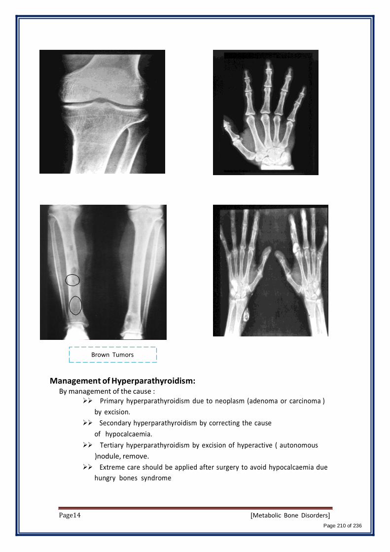
Brown Tumors
Management of Hyperparathyroidism: By management of the cause :
Primary hyperparathyroidism due to neoplasm (adenoma or carcinoma )
by excision.
Secondary hyperparathyroidism by correcting the cause
of hypocalcaemia.
Tertiary hyperparathyroidism by excision of hyperactive ( autonomous
)nodule, remove.
Extreme care should be applied after surgery to avoid hypocalcaemia due
hungry bones syndrome
Page14 [Metabolic Bone Disorders]
Page 210 of 236

Isn't it funny how someone can say "I believe in Allah " but still follow the Satan who by the way also, " believes " in
Allah…
430 ORTHOPEDICS TEAM
Lecture : Sports and soft tissue Injuries.
Team Members:
Faten Al-Mohideb. Hanan Al-‐Salman. Aliya Al-Awaji.
Hadeel Al-Ghamdi.
Ghadeer Al-Wuhayd.
Hissa Al-Balla.
Lujain Al-Yousef.
Nouf Al-Hammad.
Jawaher Al-Faraydi.
Hiba Al-Rahiem.
Nour Al-Enezi.
Wejdan Al-Swayyid.
Arwa Abudawood.
Leena Al-Shaman.
Areej Al-Qunaitir.
Team Leader:
Ayedah Al-Ruhaimi.
-‐The slides were provided by the doctor.
-429 team group A-1 in blue.
-‐Important notes in Red.
-‐Copied slides in Black.
Doctor's notes in green.
Page1 Sports & Soft Tissue Injuries
Page 211 of 236

By the end of this teaching session the students should be able to
• Specify the symptoms, signs and potential immediate complications of common sport and soft tissues
injuries involving muscles, tendons, and ligaments for commonly injured joints; like shoulder, knee, and
ankle.
• Outline the assessment and appropriate investigation and to outline the immediate and long term
management of patients with muscles, tendons, ligaments and meniscus
• Demonstrate knowledge of indications for non-operative and operative treatment and to know the most
common non-operative and operative measurements used for sport/soft tissue injuries.
Soft tissues injuries • Muscle
• Tendon
• Ligament
• Meniscus
Muscle injury
• Knee
• Shoulder
• Ankle
• The muscles most at risk are those in which the origin and the insertion cross two
jointse.g. quadriceps, hamstring muscles.
• Frequently injured muscles act in an eccentric fashion (i.e., lengthening as they
contract). (eg. Squats: contracted quads while knee is flexed).
• Frequently injured muscles have a relatively high percentage of type II (fast-
twitch) fibers.
• Muscle strain
• Muscle Contusion
• Muscle Laceration
• Delayed-‐onset soreness
1: Muscle Strainمتززقق عضلي
Most of muscle injury
management is conservative,
rarely need surgery.
– The most common muscle injury suffered in sports.
– Immediate pain associated with diminished function.
– Both complete and incomplete muscle tears can occur
by passive stretch of an activated muscle.
– Muscle tears also typically occur at or near to the
myotendinous junction.
– Treatment
– RICE,immediate measurement(to decrease edema).
– NSAID
– physical therapy
2: Muscle Contusion
• Caused by a nonpenetrating blunt injury (direct blow) to the muscle resulting in
hematoma and inflammation.
• Quadriceps and Brachialis muscles are common involved regions.
• Clinical features:
– Pain with active and passive motion +/_
swelling.
– Decreased range of motion of joints
Page2 Sports & Soft Tissue Injuries
Page 212 of 236
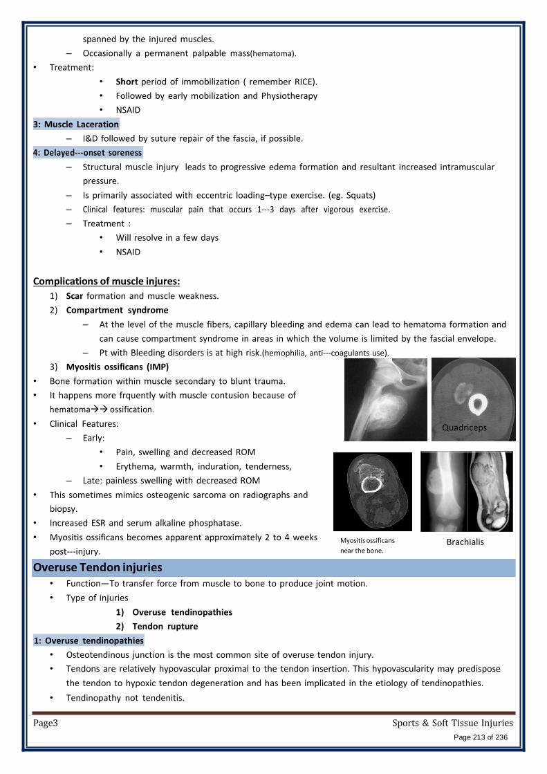
spanned by the injured muscles.
– Occasionally a permanent palpable mass(hematoma).
• Treatment:
• Short period of immobilization ( remember RICE).
• Followed by early mobilization and Physiotherapy
• NSAID
3: Muscle Laceration
– I&D followed by suture repair of the fascia, if possible.
4: Delayed-‐onset soreness
– Structural muscle injury leads to progressive edema formation and resultant increased intramuscular
pressure.
– Is primarily associated with eccentric loading–type exercise. (eg. Squats)
– Clinical features: muscular pain that occurs 1-3 days after vigorous exercise.
– Treatment :
• Will resolve in a few days
• NSAID
Complications of muscle injures:
1) Scar formation and muscle weakness.
2) Compartment syndrome
– At the level of the muscle fibers, capillary bleeding and edema can lead to hematoma formation and
can cause compartment syndrome in areas in which the volume is limited by the fascial envelope.
– Pt with Bleeding disorders is at high risk.(hemophilia, anti-coagulants use).
3) Myositis ossificans (IMP)
• Bone formation within muscle secondary to blunt trauma.
• It happens more frquently with muscle contusion because of
hematomaossification.
• Clinical Features:
– Early:
• Pain, swelling and decreased ROM
• Erythema, warmth, induration, tenderness,
– Late: painless swelling with decreased ROM
• This sometimes mimics osteogenic sarcoma on radiographs and
biopsy.
• Increased ESR and serum alkaline phosphatase.
• Myositis ossificans becomes apparent approximately 2 to 4 weeks
post-injury.
Overuse Tendon injuries
Myositis ossificans
near the bone.
Quadriceps
Brachialis
• Function—To transfer force from muscle to bone to produce joint motion.
• Type of injuries
1) Overuse tendinopathies
2) Tendon rupture
1: Overuse tendinopathies
• Osteotendinous junction is the most common site of overuse tendon injury.
• Tendons are relatively hypovascular proximal to the tendon insertion. This hypovascularity may predispose
the tendon to hypoxic tendon degeneration and has been implicated in the etiology of tendinopathies.
• Tendinopathy not tendenitis.
Page3 Sports & Soft Tissue Injuries
Page 213 of 236
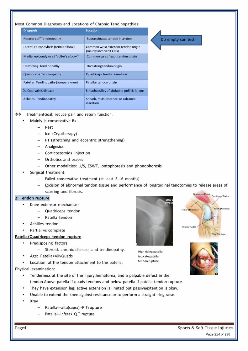
Most Common Diagnoses and Locations of Chronic Tendinopathies:
Do empty can test.
TreatmentGoal: reduce pain and return function.
• Mainly is conservative Rx
– Rest
– Ice (Cryotherapy)
– PT (stretching and eccentric strengthening)
– Analgesics
– Corticosteroids injection
– Orthotics and braces
– Other modalities: U/S, ESWT, iontophoresis and phonophoresis.
• Surgical treatment:
– Failed conservative treatment (at least 3-6 months)
– Excision of abnormal tendon tissue and performance of longitudinal tenotomies to release areas of
scarring and fibrosis.
2: Tendon rupture
• Knee extensor mechanism
– Quadriceps tendon
– Patella tendon
• Achilles tendon
• Partial vs complete
Patella/Quadriceps tendon rupture
• Predisposing factors:
– Steroid, chronic disease, and tendinopathy.
• Age: Patella<40>Quads
• Location: at the tendon attachment to the patella.
Physical examination:
High riding patella
indicate patella
tendon rupture.
• Tenderness at the site of the injury,hematoma, and a palpable defect in the
tendon.Above patella if quads tendons and below patella if patella tendon rupture.
• They have extension lag: active extension is limited but passiveextention is okay.
• Unable to extend the knee against resistance or to perform a straight-leg raise.
• Xray
– Patella-alta(supra)> P.T rupture
– Patella-infera> Q.T rupture
Page4 Sports & Soft Tissue Injuries
Page 214 of 236

• RX: usually surgical.
Achilles tendon rupture(remember David Beckham)
• Most ruptures (75%) occur during sporting activities.
• Usually due to eccentric load, rupture occurs while
performing actions requiring acceleration e.g. jumping,
pushing off.
• Injuries happen during dorsiflexionwith contracted
gastrocnemius muscle.
• History:
– The patient reports a “pop” or the sensation
of being kicked in the heelduring the injury.
– weakness and difficulty walking.
• Examination:
injured
– Increased resting dorsiflexion with the knees flexed, a palpable gap,
weak plantar flexion, and an abnormal Thompson test
(lack of plantar flexion when squeezing the calf).
• Diagnosis is clinical, but MRI or ultrasound can confirm.
• Rx: usually surgically.
injured because
unopposed
dorsiflexion .
Knee ligaments injuries
mid- substance tear of ligaments usually occurs in adult.
proximal avulsion usually occurs in pediatric age group in
tibia because ligament is stronger than bone.
Anterior cruciate ligament injuries:common in soccer players
Anatomy
originates from between the tibial eminencespine to
the medial surface of the lateral epicondyle.Needed
when you change direction immediately or
performing rough movements.
Mechanism of Injury
• Noncontact (70%) most common.twist knee (valgus and external rotation)or jump and land in wrong position.
– Cutting or Pivoting (zig-zag movement)
– Contact = MCL
• Sports-Related (80%) soccer sport.
• “Pop” sound (70%)
• Female: 2-4x > Male
Diagnosis
• Symptoms:
– Acute: “pop” sound &swelling (Hemarthrosis) is noted within a 1-2 days
of the injury.
– Chronic: Instability “giving way episodes”– توخوني ررجلي
– Pain if associated with meniscus tear
– If the patient is walking straight on flat floor s/he will have no complain even with injured ACL. The
problem when suddenly changing direction or walking on uneven floorinsatbility.
• Physical examination
• The patient need to be relaxed and comfortable.
Page5 Sports & Soft Tissue Injuries
Page 215 of 236

• Must be compared with those of the normal knee.
• A moderate to severe effusion is usually present
• ROM: in acute injury the range of motion may limited by:
– Pain
– Effusion
– Hamstring spasm,
– ACL stump impingement,
– Meniscal pathology.
• Special tests:
– Lachman’s test
– Anterior drawer test.
– Pivot shift test : is pathognomonic for
ACL injury (best in the chronic setting).
• Investigations:
– In acute stage: X-ray will be normal, it is done to
rule out fractures.
Tibial spine avulsion in
pediatric(wedge
shaped) , ACL is intact
but the tibial
attachment is avulsed ,
same symptoms and
signs of torn ACL.
Segond fracture
Avulsion of
antrolateralcapsculea
ttachemt,pathognoic
of ACL injury but it’s
a rare finding in x-ray.
most common finding
in ACL injury is
– MRI ( ACL injury is a clinical diagnosis, no need for MRI except if suspecting other injuries).
• In the skeletally mature patient, the femoral insertion or midsubstance is usually the site of disruption.
• In the skeletally immature patient, the tibial attachment may be avulsed with or without a piece of bone.
Normal ACL Torn ACL
Injuries associated with ACL disruption
performed in acute stage and shows: bone
bruise, posterior part of tibia hits
thelateralfemoral condyle(kissing lesion).
• Injuries of the ACL rarely occur in isolation. The effects of other injuries, including:
– Other ligament sprains (MCL)
– Meniscal tears
– Articular cartilage injuries
– Bone bruises ,
• Complicate the treatment and eventual outcomes of ACL disruptions.
• Long term complications with meniscal damage results in early osteoarthritis (meniscal injury in young
patients is major cause for ACL reconstruction in all young people).
Treatment
Nonsurgical treatment
• Appropriate for asymptomatic or old patients with partial injuries to the ACL.
• Older or less physically active may elect to modify their activities and proceed with nonsurgical treatment.
• Nonsurgical treatment involves rehabilitation to strengthen hamstrings and quadriceps, as well as
proprioceptive training.
• Activity modification is also an important part of nonsurgical management, as patients who avoid cutting
and pivoting sports are at lower risk for knee instability.
Page6 Sports & Soft Tissue Injuries
Page 216 of 236
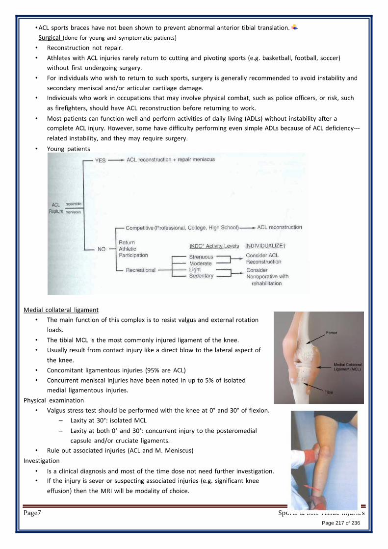
orts & Soft Tissue Injurie
• ACL sports braces have not been shown to prevent abnormal anterior tibial translation.
Surgical (done for young and symptomatic patients)
• Reconstruction not repair.
• Athletes with ACL injuries rarely return to cutting and pivoting sports (e.g. basketball, football, soccer)
without first undergoing surgery.
• For individuals who wish to return to such sports, surgery is generally recommended to avoid instability and
secondary meniscal and/or articular cartilage damage.
• Individuals who work in occupations that may involve physical combat, such as police officers, or risk, such
as firefighters, should have ACL reconstruction before returning to work.
• Most patients can function well and perform activities of daily living (ADLs) without instability after a
complete ACL injury. However, some have difficulty performing even simple ADLs because of ACL deficiency-
related instability, and they may require surgery.
• Young patients
Medial collateral ligament
• The main function of this complex is to resist valgus and external rotation
loads.
• The tibial MCL is the most commonly injured ligament of the knee.
• Usually result from contact injury like a direct blow to the lateral aspect of
the knee.
• Concomitant ligamentous injuries (95% are ACL)
• Concurrent meniscal injuries have been noted in up to 5% of isolated
medial ligamentous injuries.
Physical examination
• Valgus stress test should be performed with the knee at 0° and 30° of flexion.
– Laxity at 30°: isolated MCL
– Laxity at both 0° and 30°: concurrent injury to the posteromedial
capsule and/or cruciate ligaments.
• Rule out associated injuries (ACL and M. Meniscus)
Investigation
• Is a clinical diagnosis and most of the time dose not need further investigation.
• If the injury is sever or suspecting associated injuries (e.g. significant knee
effusion) then the MRI will be modality of choice.
Page7 Sp s
Page 217 of 236

• Xray: to R/O fracture (lateral tibia plateau fracture)
Treatment
• Conservative Rx
– Is the mainstay of treatment for the isolated MCL injuries
– Crutches, RICE, and anti-inflammatory/pain medication.
– No brace is usually required for partial tear
– A knee brace is recommended for complete tear.
• Surgical Rx:
– if failed conservative Rx + complete tear + associated with other ligaments injury
Lateral Collateral Ligament
• The LCL is the primary restraint to varus stress at 5° and 25° of knee flexion.
• Less commonly injuries than MCL
• Injuries to the lateral ligament of the knee most frequently result from motor vehicle accidents and athletic
injuries.
• Rx:
– Isolated injury: non operative
– Combined injury: surgical
Posterior Cruciate Ligament
o Originates from lateral surface of medial epicondyle to posterior aspect
of tibia.
• The PCL is the primary restraint to posterior tibial translation in the
intact knee.
• Mechanism of injury
– A direct blow to the proximal aspect of the tibia is the most common
cause of PCL injury.
– Dashboard injury
– In athletes >a fall onto the flexed knee with the foot in plantarflexion,
which places a posterior forces on the tibia and leads to rupture of the
PCL.
• PCL insuffiency significantly increased the risk of developing medial femoral
condyle and patellar cartilage degeneration over time.
• Rx
• Non operative
• Surgical if combined ligamnet injury
Knee dislocation Multiligament Knee Injuries
Knee dislocation (IMP because it’s a limp threatening condition)
• Multiligament knee injuries are usually caused by high-energy trauma and are
often considered knee dislocations.
• Less frequently, low-energy trauma or ultra-low-velocity trauma in obese
patients can also result in this injury pattern.
• Most patient will have spontaneous reduction.
• A bicruciate (ACL+PCL) injury or a multiligament knee injury involving three or
more ligaments should be considered a spontaneously reduced knee
Page8
Sports & Soft Tissue Injuries
Page 218 of 236

dislocation.
• A knee dislocation should be considered a limb-threatening injury, and careful monitoring of vascular status
after the injury is imperative.
• Popliteal artery (estimated at 32%) or peroneal nerve injury (20% to 40%) also can
occur.
• Vascular examination is critical in an acutely dislocated knee.
• Pulse and ankle-brachial index (ABI) should be carefully assessed. An ABI of less
than 0.90, and most certainly less than 0.80, should be considered abnormal.
• If there is any concern about an abnormal vascular examination, there should
be a low threshold for ordering an angiogram.
• If pulses are still abnormal or absent following reduction of the dislocation,
immediate vascular surgery consultation with intraoperative exploration
should be the next step in management.
• A vascular injury in a knee dislocation is a limb-threatening injury and needs to
be corrected within 6 to 8 hours. If not corrected, amputation may be
required.
• Neurologic examination is also critical, as peroneal nerve injury can occur with
multiligament injuries, particularly in
concomitant lateral/posterolateral corner
injuries.
• emergent closed reduction and splinting or
bracing should be performed immediately.
Postreduction
radiographs should be taken to confirm knee
reduction.
Meniscus Tear Anatomy
• The menisci are crescent-shaped, with a triangular appearance on cross-section.
• The lateral meniscus covers 84% of the condyle surface; it is 12 to 13 mm wide and 3 to 5 mm thick.
• The medial meniscus is wider in diameter than the lateral meniscus; it covers 64% of the condyle surface and
is 10 mm wide and 3 to 5 mm thick.
• Meniscus function
– The meniscus provides stability, absorbs shock,
increases articular congruity, aids in lubrication,
prevents synovial impingement, and limits
flexion/extension extremes.
– The most important function of the meniscus is load-
sharing across the knee joint, which it accomplishes
by increasing contact area and decreasing contact
stress.
• Epidemiology of meniscus injuries
– Meniscus injuries are among the most common injuries seen
in orthopaedic practices.
– Arthroscopic partial meniscectomy is one of the most
common orthopaedic procedures.
Page9 normal
Sports & Soft Tissue Injuries
Page 219 of 236
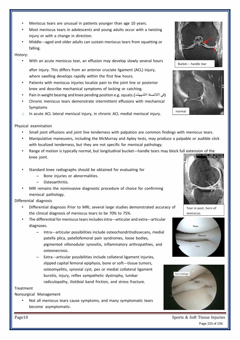
• Meniscus tears are unusual in patients younger than age 10 years.
• Most meniscus tears in adolescents and young adults occur with a twisting
injury or with a change in direction.
• Middle-aged and older adults can sustain meniscus tears from squatting or
falling.
History:
• With an acute meniscus tear, an effusion may develop slowly several hours
after injury. This differs from an anterior cruciate ligament (ACL) injury,
where swelling develops rapidly within the first few hours.
• Patients with meniscus injuries localize pain to the joint line or posterior
knee and describe mechanical symptoms of locking or catching.
• Pain in weight bearing and knee pending position e.g. squats.(في االالصةة ااتلدھھشد)
• Chronic meniscus tears demonstrate intermittent effusions with mechanical
Symptoms
o In acute ACL lateral meniscal injury, in chronic ACL medial meniscal injury.
Bucket- handle tear
normal
Physical examination
• Small joint effusions and joint line tenderness with palpation are common findings with meniscus tears.
• Manipulative maneuvers, including the McMurray and Apley tests, may produce a palpable or audible click
with localized tenderness, but they are not specific for meniscal pathology.
• Range of motion is typically normal, but longitudinal bucket-handle tears may block full extension of the
knee joint.
• Standard knee radiographs should be obtained for evaluating for
– Bone injuries or abnormalities.
– Osteoarthritis.
• MRI remains the noninvasive diagnostic procedure of choice for confirming
meniscal pathology.
Differential diagnosis
• Differential diagnosis Prior to MRI, several large studies demonstrated accuracy of
the clinical diagnosis of meniscus tears to be 70% to 75%.
• The differential for meniscus tears includes intra-articular and extra-articular
diagnoses.
– Intra-articular possibilities include osteochondritisdissecans, medial
patella plica, patellofemoral pain syndromes, loose bodies,
pigmented villonodular synovitis, inflammatory arthropathies, and
osteonecrosis.
– Extra-articular possibilities include collateral ligament injuries,
slipped capital femoral epiphysis, bone or soft-tissue tumors,
osteomyelitis, synovial cyst, pes or medial collateral ligament
bursitis, injury, reflex sympathetic dystrophy, lumbar
radiculopathy, iliotibial band friction, and stress fracture.
Tear in post. horn of
meniscus.
Treatment
Nonsurgical Management
• Not all meniscus tears cause symptoms, and many symptomatic tears
become asymptomatic.
Page10 Sports & Soft Tissue Injuries
Page 220 of 236
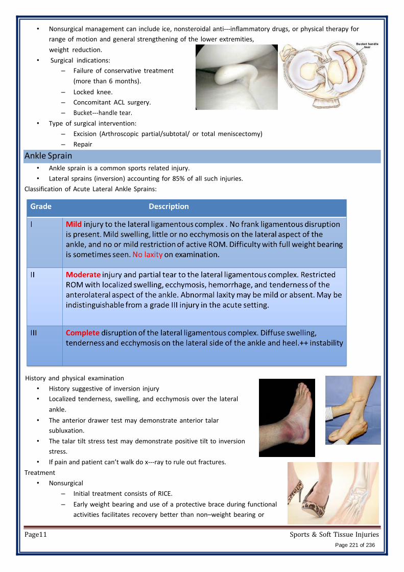
• Nonsurgical management can include ice, nonsteroidal anti-inflammatory drugs, or physical therapy for
range of motion and general strengthening of the lower extremities,
weight reduction.
• Surgical indications:
– Failure of conservative treatment
(more than 6 months).
– Locked knee.
– Concomitant ACL surgery.
– Bucket-handle tear.
• Type of surgical intervention:
– Excision (Arthroscopic partial/subtotal/ or total meniscectomy)
– Repair
Ankle Sprain • Ankle sprain is a common sports related injury.
• Lateral sprains (inversion) accounting for 85% of all such injuries.
Classification of Acute Lateral Ankle Sprains:
History and physical examination
• History suggestive of inversion injury
• Localized tenderness, swelling, and ecchymosis over the lateral
ankle.
• The anterior drawer test may demonstrate anterior talar
subluxation.
• The talar tilt stress test may demonstrate positive tilt to inversion
stress.
• If pain and patient can’t walk do x-ray to rule out fractures.
Treatment
• Nonsurgical
– Initial treatment consists of RICE.
– Early weight bearing and use of a protective brace during functional
activities facilitates recovery better than non–weight bearing or
Page11 Sports & Soft Tissue Injuries
Page 221 of 236
immobilization.
– Functional instability may result and should be treated with a course of physical therapy and
proprioceptive training.
– Residual mechanical instability may be managed effectively with bracing or taping.
– Patients may return to unrestricted activity when cutting, running, and hopping on the affected leg
are no longer painful.
– Ninety percent of acute ankle sprains resolve with RICE and early functional rehabilitation.
• Surgical––Surgery is a reasonable option when an adequate trial of nonsurgical treatment fails to control
symptoms for grade III.
Page12 Sports & Soft Tissue Injuries
Page 222 of 236

O R T H O P E D I C S 4 2 9 T E A M
G r o u p A 1
Musculoskeletal Tumors
Bone tumors
Benign Malignant
Primary Secondary
(Benign transformation
of) benign process
Metastatic
Most common bone Tumors → BENIGN.
Most common benign tumors → OSTEO CHONDROMA.
Majority of Malignant tumors → METASTATIC of other tumors elsewhere in the body.
Most common primary malignant tumors → M. MYELOMA THEN OSTEOSARCOMA.
Malignant Bone tumors are called SARCOMA
Primary Malignant bone tumors is minority
Benign bone tumors do not invade the soft tissue nor do they affect the cortex
Certain tumors have predilection for some bones (favorite places) it can recognized by X-ray.
E.g.: Ewing's sarcoma most commonly in the DIAPHYSIS. Giant cell tumors are almost always
found in a subarticular position, i.e. METAPHYSIC location.
Classification according to the cell origin: Tissue of origin Benign Malignant
Bone (osteogenic)
Ostoidosteoma
Osteoblastoma
Osteosarcoma
(most common in
epiphysis)
Cartilage (chondrogenic) 1- Enchodroma 2- Chondroblastoma
Chondrosarcoma
Bone & cartilage (both) Osteochondroma
Fibrous (fibrogenic) Fibrous cortical defect
Bone marrow (hematopoitic)
Gaint cell tumor
1- Multiple myeloma 2- Ewing's sarcoma (most
common in diaphysis)
Unknown (lesion like tumor)
1- Simple cyst.
2- Aneurismal bone cyst
(ABC).
Metastasize Metastasis
161 Page 223 of 236

O R T H O P E D I C S 4 2 9 T E A M
G r o u p A 1
How to describe bone tumors?
1- Site: Epiphysis, Diaphysis or Metaphysic whish most of the tumor arises from metaphysic.
2- Centric (away from the border) or eccentric (in the bone border).
3- Border of the tumor:
a. Well define & sharp border → usually in benign tumors.
b. Ill-defined →usually in malignant.
c. Sclerotic margin: sign of benign tumors.
4- Matrix of the tumor :
a. Sclerotic: it means forming bone .so on x-ray →opaque.
b. Lytic: it means forming tissue other than bone (it may be cartilage, fibrous tissue or cyst) on
X-ray →translucent.
N.B. this calcification (lytic and sclerotic) helps in the differentiation b/w the tumors .i.e. you
can't say from this calcification that this tumor is benign or malignant.
5- Periosteal reaction :
-occur in some bone tumors:
A. Characteristic periosteal reaction: mostly occur with malignant tumors (teeth like)
B. Smooth periosteal reaction: with benign tumor.
6- Geographic appearance :
Describe the tumor: e.g. oval shape, surface tumor, multiple tumor, etc. 7- Soft tissue extension: occurs mostly with malignant tumors or aggressive benign.
162 Page 224 of 236
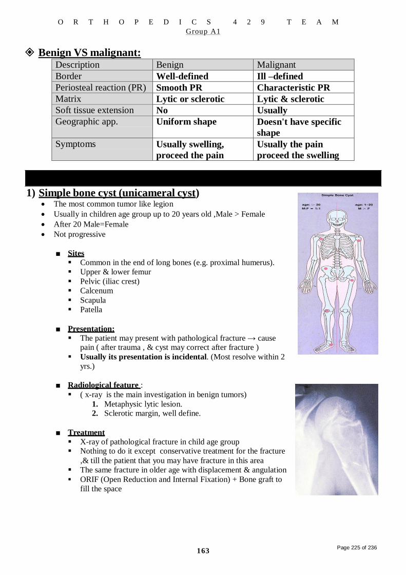
Description Benign Malignant
Border Well-defined Ill –defined
Periosteal reaction (PR) Smooth PR Characteristic PR
Matrix Lytic or sclerotic Lytic & sclerotic
Soft tissue extension No Usually
Geographic app. Uniform shape Doesn't have specific
shape
Symptoms Usually swelling,
proceed the pain
Usually the pain
proceed the swelling
O R T H O P E D I C S 4 2 9 T E A M
G r o u p A 1
Benign VS malignant:
Tumor like lesions 1) Simple bone cyst (unicameral cyst)
The most common tumor like legion
Usually in children age group up to 20 years old ,Male > Female
After 20 Male=Female
Not progressive
■ Sites Common in the end of long bones (e.g. proximal humerus).
Upper & lower femur
Pelvic (iliac crest)
Calcenum
Scapula
Patella
■ Presentation:
The patient may present with pathological fracture → cause pain ( after trauma , & cyst may correct after fracture )
Usually its presentation is incidental. (Most resolve within 2
yrs.)
■ Radiological feature :
( x-ray is the main investigation in benign tumors)
1. Metaphysic lytic lesion. 2. Sclerotic margin, well define.
■ Treatment
X-ray of pathological fracture in child age group Nothing to do it except conservative treatment for the fracture
,& till the patient that you may have fracture in this area
The same fracture in older age with displacement & angulation
ORIF (Open Reduction and Internal Fixation) + Bone graft to
fill the space
163
Page 225 of 236

O R T H O P E D I C S 4 2 9 T E A M
G r o u p A 1
2) Aneurysmal bone cyst Aggressive and balloon-like.
Progressive tumor like legion.
Arteriovenous formation.
Recurrence rate is high.
Fusiform cyst.
Aneurismal cysts have bloody content, while simple cysts have straw
colored fluid.
■ Site (common site ): Upper humerus.
Upper femur.
Spine (neural arch: lamina &pedicle) may give pressure
effect.
Proximal tibia.
Scapula.
■ Presentation : The usual presentation is swelling.
Patient may present with pathological fracture → pain.
■ Radiological feature: X-ray shows different content inside aneurysmal cyst unlike simple cysts.
Metaphyseal lytic lesion.
Well- defined, sclerotic margin.
Expansile →thin cortex (like egg shell)
■ Treatment ORIF + BONE graft ( from fibula as support BCZ lesion is very big) Sequestration = divide it to small champers
Aspiration with alcohol ,phenol & steroid
164 Page 226 of 236
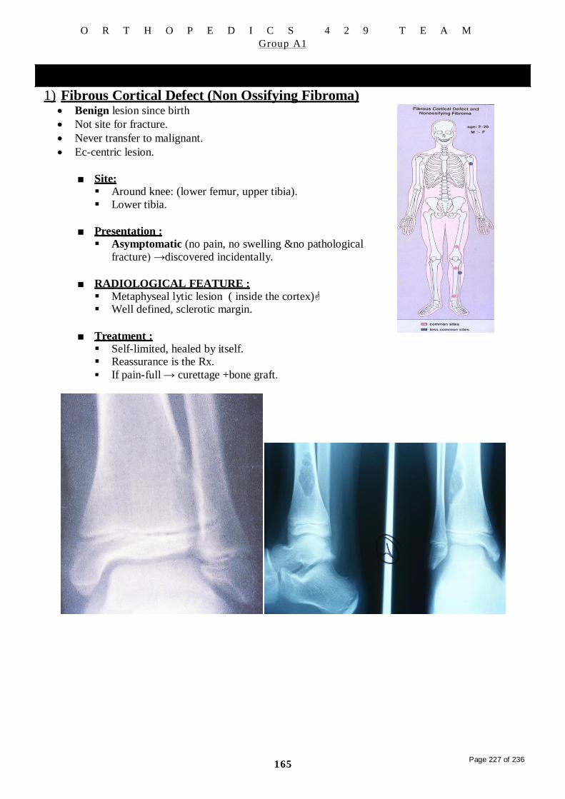
O R T H O P E D I C S 4 2 9 T E A M
G r o u p A 1
Benign tumors 1) Fibrous Cortical Defect (Non Ossifying Fibroma)
Benign lesion since birth
Not site for fracture.
Never transfer to malignant.
Ec-centric lesion.
■ Site: Around knee: (lower femur, upper tibia).
Lower tibia.
■ Presentation : Asymptomatic (no pain, no swelling &no pathological
fracture) →discovered incidentally.
■ RADIOLOGICAL FEATURE : Metaphyseal lytic lesion ( inside the cortex) Well defined, sclerotic margin.
■ Treatment : Self-limited, healed by itself. Reassurance is the Rx.
If pain-full → curettage +bone graft.
165 Page 227 of 236

O R T H O P E D I C S 4 2 9 T E A M
G r o u p A 1
2) Osteoid Osteoma: Benign tumor which has different behavior → main presentation is pain.
Usually affects young patients 10-35 and more common in males.
May arise in the cortex of long bones, or occasionally in the cancellous
bone of the spine and less commonly talus
■ Presentation
Usually well localized pain that is worse at night and prevents patients from sleep.
Character of pain :
1. Pain at the site of tumor.
2. Aggravated by activity.
3. Relived by aspirin & NSAID.
■ Radiological features : Metaphyseal or diaphyseal lesion.
Lytic lesion inside patch of sclerotsis.
The lytic lesion called (( nidus )) , nidus is the part which
surrounded by a reactive zone of dense sclerotic new bone
formation, therefore in the treatment →we remove nidus only.
■ Investigation: 1. X-ray→ a nidus surrounded by a thick cortex (sclerosis).
2. CT scan →nidus
3. Bone scan used sometimes.→↑uptake.
■ Treatment
NSAIDs for 3-6 weeks
If not relived A CT-guided needle can be inserted in to the nidus and the lesion ablated
with radiofrequency coagulation (also called radiofrequency ablation).
If it easy to access > excise it &lable it then send it to histopathologist to make sure all
the lesion was excised
1 66 Page 228 of 236
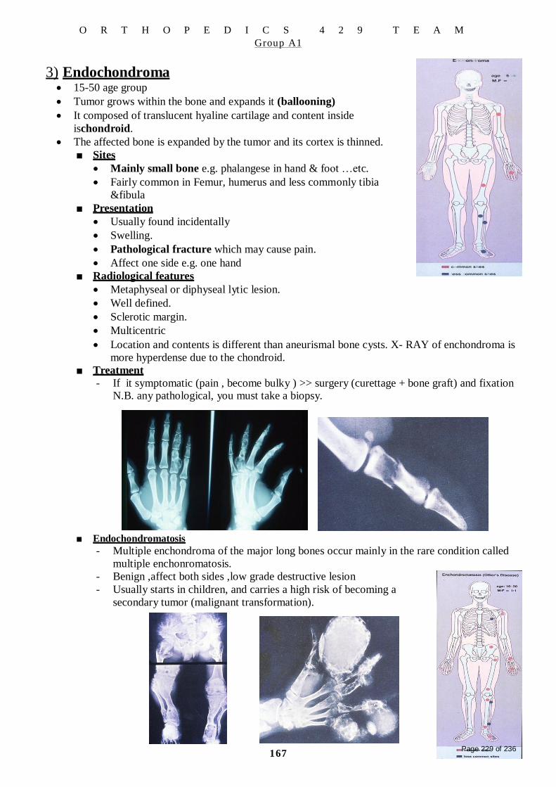
O R T H O P E D I C S 4 2 9 T E A M
G r o u p A 1
3) Endochondroma 15-50 age group
Tumor grows within the bone and expands it (ballooning)
It composed of translucent hyaline cartilage and content inside
ischondroid.
The affected bone is expanded by the tumor and its cortex is thinned.
■ Sites
Mainly small bone e.g. phalangese in hand & foot …etc.
Fairly common in Femur, humerus and less commonly tibia
&fibula
■ Presentation
Usually found incidentally
Swelling.
Pathological fracture which may cause pain.
Affect one side e.g. one hand
■ Radiological features
Metaphyseal or diphyseal lytic lesion.
Well defined.
Sclerotic margin.
Multicentric
Location and contents is different than aneurismal bone cysts. X- RAY of enchondroma is
more hyperdense due to the chondroid.
■ Treatment
- If it symptomatic (pain , become bulky ) >> surgery (curettage + bone graft) and fixation N.B. any pathological, you must take a biopsy.
■ Endochondromatosis
- Multiple enchondroma of the major long bones occur mainly in the rare condition called
multiple enchonromatosis.
- Benign ,affect both sides ,low grade destructive lesion
- Usually starts in children, and carries a high risk of becoming a
secondary tumor (malignant transformation).
167 Page 229 of 236

O R T H O P E D I C S 4 2 9 T E A M
G r o u p A 1
4) Osteo chondroma Commonest benign tumor of bone.
The only surface tumor, (outside the bone) .therefore, we call it
exostosis or pedunculated.
It is acombination of both bone & cartilage.
Usually in 10-20 age group and Male > Female.
Painful in children due to growth plate compression and pressure
effects on adjacent nerve or vascular structures.
■ Sites - The commonest are( around knee) distal femur + proximal
tibia
- Scapula, neck of femur.
■ Presentation: a. Swelling : it can reach huge size .(keeps on growing )
b. Symptom of complication :
1- Pressure symptom:
- Pseudo-aneurysm.→ artery.
- Hypothesis or parasethesia →nerve.
- Rendering the movement → tendon.
- Restrict the movement of the movement nearby the joint
- Adventitia bursea.
2- Fracture especially with pedunculated type.
3- If transfer to malignant
■ Radiological feature:
- Metaphyseal lesion. - Mushroom-like stalk of the bony tumor. (connected to the bone)
- On x-ray the cartilaginous cap: which is the cartilaginous part of the tumor not seen
(translucent).it is located around the bony part.
- Direction of the tumor →away from the bone.
Start from the growth plate & in the direction of Ms. Tendon.
Stop growing when patient stop to growth usually at 18 yrs.
- According to the shape of the neck of the tumor , we divide it into :
1- Pedunculated type: has long & thin neck.
2- Sessile type: has short & thick neck.
■ Treatment - Usually we do nothing, just reassure the patient.
- Surgery ( just excision , no need for bone graft b/c it is surface tumor) & the surgery is
indicated in :
a. When there is complication.
b. Cosmetic.
168 Page 230 of 236

■ Sites
- -
-
Most common distal epiphysis of radius Less common are distal tibia ,sacrum
Very bad one in sacrum because it will affect nerve roots
O R T H O P E D I C S 4 2 9 T E A M
G r o u p A 1
5) Giant cell tumor From bone marrow
Occurs most commonly in young adults, 20-40 age groups.
Benign aggressive tumor.
It's only the benign bone tumors that can metastases to the lung. So it's
important to get chest x-ray.
Origin: osteoclast, therefore it is sometimes called osteoclastoma.
Very destructive tumor
■ Presentation
- Mostly patient present first with: pain, then swelling & later on pathological fracture. - Not clear
- Osreopenia.
■ Radiological feature :
- Epiphyseal lytic lesion. - No new bone formation b/c this new bone will be eaten by the osteoclast.
- Ill define margin.
- Radiograph shows lucent regions, lytic destruction of the bone with expansion of the
cortex, without a sclerotic rim.
- Ill-defined borders when aggressive or pre-malignant
- Usually looks like a bone cyst, definite diagnosis made by biopsy.
- No marginal sclerosis.
- Soft tissue extension.
■ Investigation :
- The assessment depends on : a. Soft tissue extension: by MRI.
b. Joint extension: the articular cartilage is a very resistant
against progression of the tumor from going to the joint.
c. Bone extension: by CT scan.
d. Further investigation :
- Bone scan: for metastasis.
So, the tumor will not destruct the joint. But the
surgeon during treatment
will destruct the joint b/c
he has to remove the entire
region around the joint.
■ Treatment
- CBC: FOR ASSESMENT of general condition of patient.
- Biopsy: for confirmation it doesn't convert to malignant tumor.
- No place for conservative treatment. - In general we do : excision followed by either : (depend on the site of the tumor)
1- Bone graft.
2- Prosthesis.
a. If the tumor in non-weight bearing area ( e.g. lower radius ) → radical
excision + bone graft + arthrodesis for nearby joint
b. If the tumor in weight bearing area (e.g. lower
femur) →excision + prosthesis (N.B. no bone
graft here).
■ Radiotherapy &chemotherapy are indicated in : 1- Frank malignancy.
2- Recurrence of the tumor.
- Recurrence rate is 50% ,it will be more aggressive with
metastasis
169 Page 231 of 236

O R T H O P E D I C S 4 2 9 T E A M
G r o u p A 1
1- Ewing's sarcoma
Malignant tumors
Origin: from the endothelial lining of the bone marrow canal.
Most of the Ewing's sarcoma is miss-diagnosed firstly as acute osteomyelitis.
(Mimic the OA) because of systemic symptoms and elevated ESR.
Young age group 5-25
Pulmonary metastasis can occur.
■ Sites:
- It is the only bone tumor which takes it origin from diaphysis → so; we
will find a diaphyseal lesion.
- The diaphyses of the femur are the most common sites, followed by
the tibia and the humerus.
■ Presentation : Very characteristic:
1- Febrile patient.
2- High WBC's.
3- Local Pain & redness like the presentation of infection
4- Ulceration of skin.
5- swelling
+ Scapula, less commonly clavicle.
■ Radiological feature: X-ray: peal onion reaction.
--- You think it is AOM→YOU do aspiration for drainage of pus →there will be no pus
&you will find tumor tissue →biopsy →Ewing's sarcoma.
- So, it is a diaphyseal lytic lesion (not sclerotic).
- Ill- defined.
- Sometimes →soft tissue extension.
- N.B: you can't differ it from osteosarcoma unless you do biopsy.
■ Investigation :
- Definite diagnosis made by MRI and biopsy.
■ Management o In history usually asymptomatic unless it cause pressure in vital structures or in late
stage
o Blood investigations , ESR
o Radiology (X-ray ,bone scan ,CT,MRI)
o Biopsy –same surgeon who will do surgery should take the biopsy- To avoid contamination then spreading the tumor
■ Treatment
It is sensitive to chemotherapy So start with chemotherapy –to decrease the tumor in size so it will be easy to excise it &
to control metastasis (microcell) everywhere in the body as in bacteremia.
Tumor treated by operative excision and disarticulation and You may need amputation
170 Page 232 of 236

O R T H O P E D I C S 4 2 9 T E A M
G r o u p A 1
2- Osteo sarcoma Most common primary malignant tumor .{
remember the most common malignant tumor is
metastatic }
More common than Ewing's sarcoma
10- 25 year old and Male > Female
■ Sites - Arises from primitive bone-forming cells - Around the knee.
- Common in the lower femur, upper tibia, and upper humerus.
- Less commonly clavicle
■ Presentation :
Patient present firstly with pain, then swelling,
lastly pathological fracture (typical presentation
of malignant tumor). Overlying skin is warm due
to high vascularity.
May present as cachectic (in lated& advanced
case).
■ Radiological presentation
Very dense
Irregular medullary and cortical destruction of the metaphysis.
Sun rise periosteal reaction (surrounded by low dense)
Could be lytic or sclerotic."mixed"
Ill defines no sclerotic margin.
Metaphyseal lesion.
Definite diagnosis made by biopsy
■ Investigation :
1- CT scan = bone extension.
2- MRI = soft tissue extension.
3- Bone scan = metastasis.
4- Searching for metastasis
■ Treatment
- Surgical intervention : 1- Limb salvage procedure: which is radical
excision of the affected bone & the
affected compartements (e.g. flexor
compartement of thigh , adductor
compartment of the thigh … etc )around
the bone + followed by reconstruction
(reconstruction may be :prosthesis , bone
graft , or bone cement ) + lastly we do
radiotherapy &chemotherapy .
2- Amputation : is indicated:
a. The whole compartment around the bone is
involved (e.g. the flexor, adductor & extensor
compartment of the thigh). If there the Vessels
involved you can do bypass graft
b. Pathological fracture.
3- in all stages: chemotherapy surgery chemotherapy
4- survival rate is 80 %
N.B. CHEMOTHERAPY TO AT LEAST ↓ED SIZE OF THE MASS.
171
Page 233 of 236

O R T H O P E D I C S 4 2 9 T E A M
G r o u p A 1
3- Multiple myeloma - Arise from plasma cells in the bone marrow. Plasma cells are responsible
for antibodies production (gamma globulins).
- The most common primary malignant tumor.
- Arises from the plasma cells of the bone marrow
- Tumor of bone marrow, occurring in older adults > 50 and predominantly
Males.
- Bence Jones proteins test found in 24-hour urine collection.
- Disseminates too many parts of the skeleton through the blood stream, thus
usually multiple.
■ Site: - Central bones (axial skeleton): skull, ribs, pelvic girdle. & spine. - It may be solitary, multiple (commonest) or generalized osteoporosis (mylometosis:
multiple myeloma + only generalized osteoporosis).
■ Presentation :
- Pt ill (decreased immunity) - Sclerotic (no more elasticity) so more prone to fractures
- In skull there will be pepper (lytic) &salt (sclerotic)... {if sclerotic →un flexible →easy
to fracture) mottled appearance).
- P.t came with bone ach (backache) + osteopenia.
- More common in periphery than center
- The only one definitive to diagnose it, is Bone marrow aspiration."Biopsy".
■ Treatment
o Bone marrow transplant Success rate is 30% Cost million $ all 3 done by oncologist.
o Radiotherapy.
o Chemotherapy.
o The rule of orthopedic surgeon is only when you have pathological fracture →do internal fixation.
172 Page 234 of 236

O R T H O P E D I C S 4 2 9 T E A M
G r o u p A 1
4- Metastatic lesions Tumor outside the bone. The most common tumors are: prostate, thyroid,
breast, lung and kidney.
More than 45 in age, F<M.
More common than primary tumors in later adult life.
Affect SAME SITE which affected by multiple myloma.
Take biopsy to know where is the primary
If there is fracture do curettage first then fixation.
■ Presentation :
- Patient present & primary tumor is known & the patient is treating
from it.
- May present with secondary metastasis. So, we have to search for the
primary and try to treat it.
- Usually metastasis occurs in the highly vascular bones e.g. vertebral
body, ribs, pelvis, upper end of femur, and humerus.
■ Radiological feature : - It may be solitary or multiple (common) or just osteoporosis (called
carcinomatosis).
- It differs from multiple myeloma because it may present with lytic or sclerotic
lesion while the M.M only present with lytic lesion.
- If the primary tumor is from the prostate →the lesion will be sclerotic.
- If the primary tumor is from the breast → the lesion will be lytic.
■ Investigation :
Same as Multiple myeloma
X-ray shows decreased bone density (bone eaten away) with thinning of the cortex.
Resembles bone cysts but the age group directs the diagnosis.
Metastasis usually of the lung sarcoma
CT- scan of the chest performed for definite diagnosis.
■ TREATMENT : Same as Multiple myeloma
173 Page 235 of 236
O R T H O P E D I C S 4 2 9 T E A M
G r o u p A 1
Some IMP Notes From 427 All bone tumors all metaphyseal except:
1- Osteoid osteoma: metaphyseal + diaphyseal.
2- Enchondroma: metaphyseal + diaphysis.
3- Chondroblastoma + giant cell tumor: epiphysis.
4- Ewing's sarcoma: diaphyseal tumor.
Most of the tumor occurring in children or growing age (>25) except:
1- Osteoid osteoma: 10-35.
2- Osteoblatoma: 10-35.
3- Osteochondroma: 10-35.
4- Enchondroma: 15-40.
5- Giant cell tumor: 20-40. 6- Multible myeloma & metastasis: old age <50.
Most of the tumor occurs more common in male except:
- Giant cell tumor: F<M.
- MUTIPLE myeloma +metastasis: f <m
- Simple bone cyst +ABC+ enchondroma: F=M.
Benign tumor that may transfer to malignant:
1- Osteoblastoma.
2- Osteochondroma.
3- Giant cell tumor.
4- Chondromatosis.
- Metastatic tumor more common than primary tumor.
- Most common primary benign tumor: osteochondroma.
- Most common primary malignant tumor: multiple myeloma then osteosarcoma.
Important tumors (study them very well)
1- Simple cyst.
2- Know the difference between osteblastoma& osteoid osteoma.
3- Enchondroma&condroblastoma : اطالير ىلع
4- Osteosarcoma: مهأ يش
5- Ewing sarcoma: pay attention to its presentation.
6- Multiple myeloma: pay attention about it investigation.
174 Page 236 of 236