2-Bone pathology.pdf - KSUMSC
17
-
Upload
khangminh22 -
Category
Documents
-
view
2 -
download
0
Transcript of 2-Bone pathology.pdf - KSUMSC

Bone pathology
Musculoskeletal block Page 1
Objectives:
Be aware of some important congenital
and developmental bone diseases and
their principal pathological features.
Be familiar with the terminology used in
some important developmental and
congenital disorders.
Understand the etiology, pathogenesis
and clinical features of osteoporosis.
Videos to Watch:
• Bone Modelling, Remodelling, and Peak Bone Mass
http://www.youtube.com/watch?v=GenafQna0J4
• Developmental Abnormalities in Bone Cells, Matrix, and Structure http://www.youtube.com/watch?v=p8UYRhs76F4&list=PLfefVARFaHqUsQuEHGx8iVcHhjwvMPFFK
• Fractures - Robbins Pathology Audiobook http://www.youtube.com/watch?v=daEqlRtbFkc&list=PLfefVARFaHqUsQuEHGx8iVcHhjwvMPFFK

Bone pathology
Musculoskeletal block Page 2
Bone pathology
Musculoskeletal block Page 3
Formation of Bone
Directly:
Intramembranous
ossification
Pre-existing cartilage:
Endochondral
ossification
Bone:
206 bones
Organic matrix (35%) and inorganic elements (65%): calcium
hydroxyapatite [Ca10(PO4)6(OH)2]
The bone-forming cells include osteoblasts and osteocytes, while
cells of the bone-digesting lineage are osteoclasts
In bone development, action of osteoblast predominates. When the
skeleton reaches maturity, actions of osteoblast and osteoclast are
equal (remodeling). By the third decade, the action of osteoclast
predominates.
Development and Growth of the skeleton:
Is very dynamic and subject to constant
breakdown and renewal: Remodeling

Bone pathology
Musculoskeletal block Page 4
First: Intramembranous ossification:
Bone is laid down as woven bone that matures into lamellar
bone, e.g. (skull and clavicles)
Second: Endochondral ossification:
The cartiladinous template undergoes ossification at
ossification centers. As in long bones, the cartilages at the
epiphysis ossify after puberty and the area is called growth
plate.
IMPORTANT SCHEDULE HERE!!!!!!
sease
Achondroplasia &Thanatophoric
(Dwarfism القزامة)
Osteogenesis Imperfecta
(brittle bone disease)
(OI)
Osteopetrosis
Types
Congenital disease. Many of its types transmitted as an autosomal dominant.
Achondroplasia ( most common form of
dwarfism )
Thanatophoric ( lethal variant of dwarfism)
It is Congenital
disease and it is also inherited autosomal dominant disease.
Type I OI & Type II OI. Type III OI’s , type IV
OI
There are several
forms of it.
It is a Congenital and rare diseases
Bone pathology
Musculoskeletal block Page 5
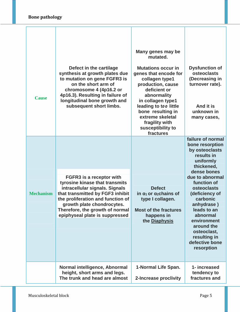
Cause
Defect in the cartilage synthesis at growth plates due to mutation on gene FGFR3 is
on the short arm of chromosome 4 (4p16.2 or
4p16.3). Resulting in failure of longitudinal bone growth and
subsequent short limbs.
Many genes may be mutated.
Mutations occur in genes that encode for
collagen type1 production, cause
deficient or abnormality
in collagen type1
leading to too little bone resulting in extreme skeletal
fragility with susceptibility to
fractures
Dysfunction of osteoclasts
(Decreasing in turnover rate).
And it is unknown in many cases,
Mechanism
FGFR3 is a receptor with
tyrosine kinase that transmits intracellular signals. Signals
that transmitted by FGF3 inhibit the proliferation and function of
growth plate chondrocytes. Therefore, the growth of normal epiphyseal plate is suppressed
Defect in ɑ1 or ɑ2chains of
type I collagen.
Most of the fractures happens in
the Diaphysis
failure of normal bone resorption by osteoclasts
results in uniformly thickened,
dense bones due to abnormal
function of osteoclasts
(deficiency of carbonic
anhydrase ) leads to an abnormal
environment around the osteoclast, resulting in
defective bone resorption
Normal intelligence, Abnormal height, short arms and legs.
The trunk and head are almost
1-Normal Life Span.
2-Increase proclivity
1- increased tendency to
fractures and

Bone pathology
Musculoskeletal block Page 6
Features
of normal size (the head may look larger but it is not large it only looks large because his
limbs are short) with depression of the nasal bridge. General health is not affected, and life expectancy is normal
Thanatophoric dwarfism Features: Thanatophoric means
(death-loving).
1-Lethal.
2-Extreme shortening of the limbs.
3-Extreme frontal bossing.
4-Extreme small thorax, which will be the cause of fatal
respiratory failure.
for fractures during childhood (decrease after puberty) from any minor trauma.
3-Blue Sclera )مسحة في بياض العين( زرقاء
4-Small misshapen teeth.
5-Hearing loss (sometimes).
6-In addition, other tissues contain
collagen type I are affected such as:
tendons, ligaments, skin and dentine.
Type II OI’s Features:
Uniformly fatal in utero or immediately postpartum because of multiple fractures during pregnancy.
Type 1 Features: blue sclera in both eye,
deformed teeth and hearing loss
osteomyelitis
2-Recurrent infections
because of the reduced bone
marrow size and activity.
3-anemia and extramedullary hematopoiesis
Notes: Achondroplasia can occurs as a sporadic
mutation in approximately 80% of cases
(associated with advanced paternal age)
People with Achondroplasia, their
Membranous ossification are not affected, so
that the skull, facial bones, and axial skeleton
develop normally.
The Four main types of
Osteogenesis Imperfecta occur with different
clinical manifestations classified according to
the severity of bone fragility, the presence or
absence of blue scleras, hearing loss,
abnormal dentition, and the mode of
inheritance.
FGFR3 : Fibroblast growth factor receptor 3
Sclera appears blue because it is thin that the
underlying uveal pigment becomes visible.

Bone pathology
Musculoskeletal block Page 7
HYPERPARATHYROIDISM PAGET'S DISEASE OF BONE OSTEOMALACIA AND RICKETS,
Excessive secretion of PTH increased produces
. osteoclastic activity
There is excessive destruction of cortical and
trabecular bone, with inadequate compensatory
osteoblastic activity.
Excessive uncontrolled destruction of bone by abnormally large and active osteoclasts, with concurrent inadequate attempts at haphazard new bone formation by osteoblasts, producing physically weak woven bone.
It may result from a paramyxovirus infection in genetically susceptible persons.
Osteomalacia and rickets are the same, but osteomalacia occur in adult and rickets occur in children . Bone biopsy taking from osteomalacia patient shows: Black area: calcified bone
Red area: osteoid (Uncalcified bone) Space: bone marrow with progenitor cells The main pathology of osteomalacia is: 1. Abnormal calcification 2. Mineralization due to deficiency vitamin D
Causes: 1.Malnutrition 2.Malabsorbtion 3.Renal diseases Symptoms of rickets are: big head, frontal bone is fosselated, opened gap in skull suture. One of fractures of osteomalacia is pseudofracture (rupture of cortical bone)
Metabolic bone disease
Comprises four fairly common conditions in which
there is an imbalance between osteoblastic (bone
forming) and osteoclastic (bone destroying) activity
Osteomalacia
Hyperparathy
roidism
Paget's
disease of
bone
Osteoporosis

Bone pathology
Musculoskeletal block Page 8
OSTEOMALACIA AND RICKETS:
They are characterized by defective mineralization of osteoid matrix with lack of vitamin D
which maintains the serum calcium and phosphorous levels. In osteomalacia, lack of vitamin
D impairs normal mineralization of osteoid laid down in the remodeling of bone. In children,
lack of vitamin D leads to inadequate mineralization of the epiphyseal cartilage and the
osteoid (rickets).
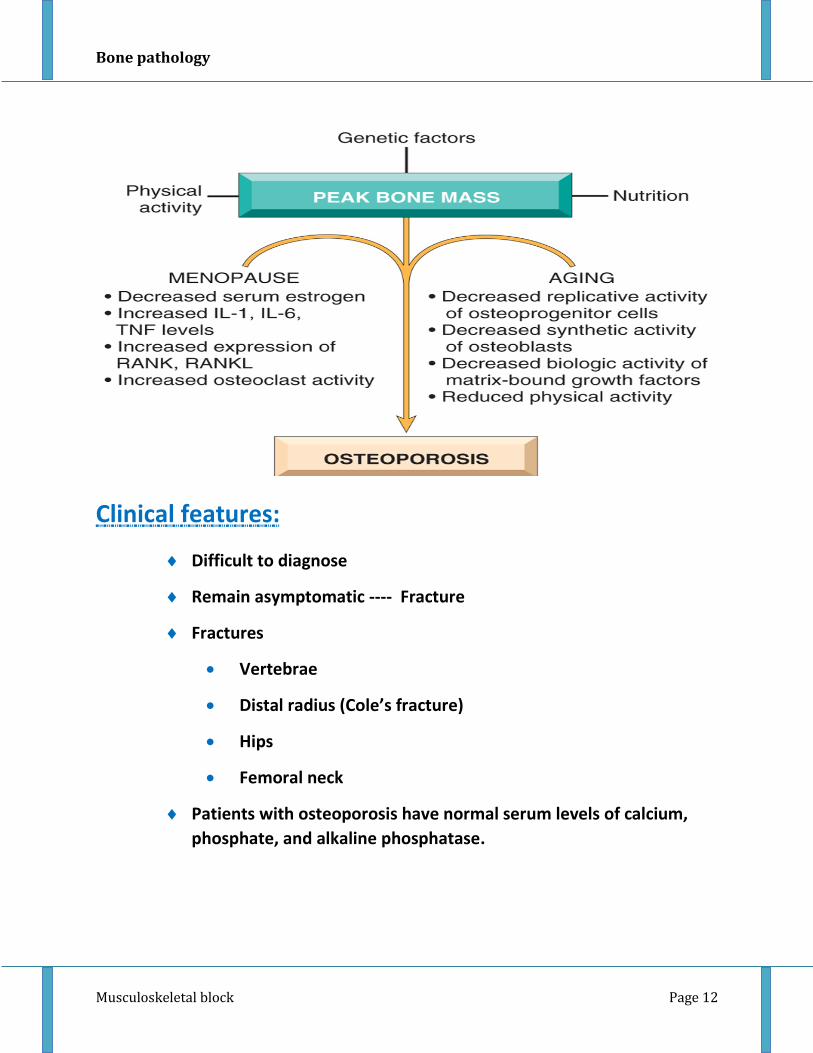
Osteoporosis:
Osteoporosis is an acquired condition characterized by reduced bone mass, leading to bone
fragility and susceptibility to fractures.
-Trabecular bone ("large amount" in vertebral bodies and pelvis) is affected before cortical
bone ("large amount" in long bones).
There is a slowly progressive increase in bone erosion
It may be:
localized generalized
cause disuse osteoporosis of a limb
a metabolic bone disease
types ---------- primary or secondary
(Primary forms of osteoporosis are most common and may be associated with aging (senile
osteoporosis) or the postmenopausal state in women. The drop in estrogen following
menopause tends to exacerbate (يفاقم) the loss of bone that occurs with aging, which means
women are more likely to get this disease more than men.
The risk of osteoporosis with aging is related to the maximum of bone mass earlier in life, the
greater the peak bone mass, the greater the delay in onset of osteoporosis.)
More info about Osteoporosis:
The main pathology of Osteoporosis is slim and slender Trabeculae (Woven).
Osteoporosis shows normal calcification. Alkaline Phosphatase Isoenzymes: can be in the
liver, placenta and bone. Alkaline Phosphatase of the bone is really high in Paget’s disease
of the bone.
Bone pathology
Musculoskeletal block Page 9
Osteoporosis fractures are neck of femur, wrist (Coles fracture),
vertebrae. Kyphosis is due to Osteoporosis and shows compression fracture in
vertebrae, in Kyphosis )التحدب( the elbow becomes in the level of hip. Scliosis )االلتواء(
shows deviation in vertebrae taking S shape.
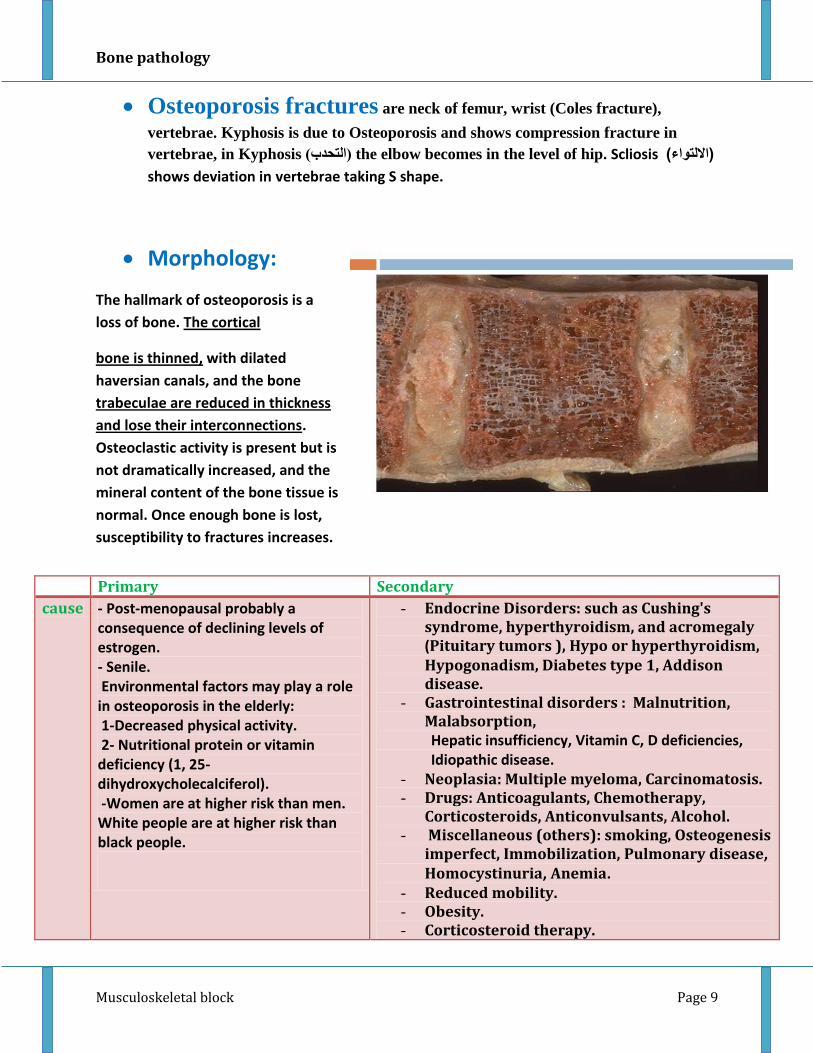
Morphology:
The hallmark of osteoporosis is a
loss of bone. The cortical
bone is thinned, with dilated
haversian canals, and the bone
trabeculae are reduced in thickness
and lose their interconnections.
Osteoclastic activity is present but is
not dramatically increased, and the
mineral content of the bone tissue is
normal. Once enough bone is lost,
susceptibility to fractures increases.
Primary Secondary
cause - Post-menopausal probably a consequence of declining levels of estrogen. - Senile. Environmental factors may play a role in osteoporosis in the elderly: 1-Decreased physical activity. 2- Nutritional protein or vitamin deficiency (1, 25-dihydroxycholecalciferol). -Women are at higher risk than men. White people are at higher risk than black people.
- Endocrine Disorders: such as Cushing's syndrome, hyperthyroidism, and acromegaly (Pituitary tumors ), Hypo or hyperthyroidism, Hypogonadism, Diabetes type 1, Addison disease.
- Gastrointestinal disorders : Malnutrition, Malabsorption,
Hepatic insufficiency, Vitamin C, D deficiencies, Idiopathic disease.
- Neoplasia: Multiple myeloma, Carcinomatosis. - Drugs: Anticoagulants, Chemotherapy,
Corticosteroids, Anticonvulsants, Alcohol. - Miscellaneous (others): smoking, Osteogenesis
imperfect, Immobilization, Pulmonary disease, Homocystinuria, Anemia.
- Reduced mobility. - Obesity. - Corticosteroid therapy.

Bone pathology
Musculoskeletal block Page 10
Note:
-Post- menopausal women lose up to 2% of cortical bone per year and up to
9% of trabecular bone per year for 8-10 years with declining to the normal loss
after that.
Pathophysiology:
Osteoporosis occurs when the dynamic balance between bone formation by
osteoblasts and bone resorption by osteoclasts tilts in favor of resorption.
يميل

Bone pathology
Musculoskeletal block Page 11
Bone pathology
Musculoskeletal block Page 12
Clinical features:
Difficult to diagnose
Remain asymptomatic ---- Fracture
Fractures
Vertebrae
Distal radius (Cole’s fracture)
Hips
Femoral neck
Patients with osteoporosis have normal serum levels of calcium,
phosphate, and alkaline phosphatase.

Bone pathology
Musculoskeletal block Page 13
OSTEOPOROSIS
Diagnosis Prognosis Prevention strategies
1. Plain X ray: cannot detect osteoporosis until 30% to 40% of bone mass has already disappeared.
2. Dual-emission X-ray absorptiometry (DXA scan): is used primarily to evaluate bone mineral density, to diagnose and follow up pt. with osteoporosis.
Osteoporosis is rarely lethal.
Patients have an increased mortality rate due to the complications of fracture. e.g.
-- hip fractures can
lead to decreased
mobility and an
additional risk of
numerous
complications: deep
vein thrombosis,
pulmonary embolism
and pneumonia
--vertebral bodies
fractures may lead to
progressive loss of height
and pain or may cause
deformity of the
spine(kyphosis)
The best long-term approach to osteoporosis is prevention.
Children and young adults, particularly women, with a good diet (with enough calcium and vitamin D) and get plenty of exercise, will build up and maintain bone mass.
This will provide a good reserve against bone loss later in life. Exercise places stress on bones that builds up bone mass
Taking hormonal replacement therapy.

Bone pathology
Musculoskeletal block Page 14

Bone pathology
Musculoskeletal block Page 15
MCQs
1- Which of following strategies can provide the best overall long-term reduction in risk of fracture from osteoporosis in women? Page 278 Q 27
(A) Supplement the diet with calcium and vitamin D after menopause.
(B) Begin estrogen replacement therapy after a fracture.
(C) Increase bone mass with exercise in childhood and young adulthood.
(D) Limit alcohol use and avoid use of tobacco
2- After a minor fall, a 63 year old woman sustains a complete right femoral neck fracture. Of the following conditions, the most significant contributing factor for this fracture is Page 276 Q 13
(A) Multiple myeloma
(B) Vitamin D deficiency
(C) Chronic osteomyelitis
(D) Postmenopausal bone loss
3- In a 75 year old male, which of the following processes contributes to the occurrence of osteoporosis?
(A) Decreased production of osteoid by osteoblasts.
(B) Increased resorption of bone by osteoclasts.
(C) Synthesis of chemically abnormal osteoid.
4- A 2-year-old child has a history of multiple bone fractures with minor trauma. Radiographs reveal diffusely and symmetrically sclerotic bones with poorly formed metaphysis. He is treated with bone marrow stem cell transplantation. Which of the following cells in his bones was most likely functionally deficient and replaced following transplantation?
(A) Chondroblast
(B) Chondrocyte
(C) Osteoblast
(D) Osteoclast
(E) Osteocyte

Bone pathology
Musculoskeletal block Page 16
5- A 5 year old child has a GI tract problem that causes malabsorption of a certain substance. The same disease seen in adults is known as osteomalacia. What condition is also associated with the deficiency?
A. Osteoporosis
B. Osteopetrosis
C. Scurvy
D. Cushing's syndrome
E. Pneumonia of the Lung
6- A 15 year old boy has shortened limbs and ribs, frontal bossing of the forehead, bowing of the limbs, his IQ however is normal. The cells that are affected in his disorder are:
A. Osteoclasts
B. Chondrocytes
C. Osteoblasts
D. Macrophages
E. Osteocytes
7- A patient presents with multiple fractures and blue sclera of the eye. The same disease in infants would result in:
A. Death
B. Tumor of the bone
C. Fractures
D. Blue sclera
E. A C and D
1 C, 2 D, 3 A, 4 D, 5 A, 6 B, 7 E


![[5] HEMA - Megaloblastic Anemia.pdf - KSUMSC](https://static.fdokumen.com/doc/165x107/631deac95ff22fc7450674ca/5-hema-megaloblastic-anemiapdf-ksumsc.jpg)