Basic ECG interpretation - KSUMSC
38
1 Basic ECG interpretation NOTE: This ECG interpretation approach to help to diagnose some most common disorders. It is important to note that there are many other helpful approaches to interpret ECG and there are many disordered not covered in this approach. -------------------- Recall the approach 1. Take a deep breath 2. . Analyze rate 3. Analyze rhythm 4. . Look at axis 5. . Look for injury/strain/ischemic patterns 6. . Look for conduction deficits (RBBB, LBBB) 7. . Hypertrophy, meds, toxic effects 8. . Make your measurements (PR, QT/QTc, QRS) Another approach (which I prefer for Basic level) Look at: 1. Rhythm 2. Rate 3. Axis 4. P wave, P-R interval, Q wave, QT interval, QRS complex, ST segment, T-wave. 5. LBBB, RBBB or LVH
-
Upload
khangminh22 -
Category
Documents
-
view
12 -
download
0
Transcript of Basic ECG interpretation - KSUMSC

1
Basic ECG interpretation
NOTE:This ECG interpretation approach to help to diagnose some most common disorders. It is important to note that there are many other helpful approaches to interpret ECG and there are many disordered not coveredin this approach.
--------------------
Recall the approach
1. Take a deep breath 2. . Analyze rate 3. Analyze rhythm 4. . Look at axis 5. . Look for injury/strain/ischemic patterns 6. . Look for conduction deficits (RBBB, LBBB) 7. . Hypertrophy, meds, toxic effects 8. . Make your measurements (PR, QT/QTc, QRS)
Another approach (which I prefer for Basic level)
Look at:
1. Rhythm
2. Rate
3. Axis
4. P wave, P-R interval, Q wave, QT interval, QRS complex, ST segment, T-wave.
5. LBBB, RBBB or LVH

2
===========================================================================

3
Irregular rhythm z there are many causes but the most important and common are:
1. Atrial Fibrillation
2. Atrial flutter
3. Second degree heart block Type1 (mobitzl )
4. Second degree heart block T2 (mobitzll )
5. Sinus arrhythmia
Rate: • if regular rhythm:
1. calculate big square between R-R:
if > 5 big square (bradycardia)
if < 3 big square (tachycardia)
if between 3 and 5 big squares (normal heart rate)
2. calculate the big squares between R-R
Number of squares
1 square 2 squares 3 squares 4 squares 5 squares 6 squares
Heart rate 300 150 100 75 60 40
3. in standard ECG: calculate the number of QRS complex in ECG and multiply by 6 = HR

4
4. Method working for regular and irregular rhythm:
measure 30 big square on ECG strip, then calculate how many QRS complex within this 30 big square, then multiply the number of QRS complex by 10 then you will get the heart rate.
Axis trick
Positive in I and II = normal
Positive in I and Negative in II = LAD
Negative in IPositive in II = RAD

5
P-Wave x P wave: better seen in lead ll. x absent P wave can be seen in many disorders, the most important and common causes
are: 1. Atrial Fibrillation: (if absent P wave + irregular rhythm = consider it as Atrial Fibrillation) 2. SVT (supraventricular tachycardia): regular narrow complex tachycardia with absent P wave. 3. VT (V tach) (Ventricular tachycardia): any wide QRS complex tachycardia is considered VT
until proven otherwise. 4. VF (V fib) (ventricular Fibrillation): An ECG finding of a rapid grossly irregular ventricular
rhythm with marked variability in QRS cycle length, morphology, and amplitude.
P-R interval x Prolong PR interval >0.2 sec ( > 200 ms) (>5 small boxes) = AVB ( 1st , 2nd type1, 2nd type2
or 3rd degree heart block) or hyperkalemia x Short PR interval <0.120 sec (120 ms) (< 3 small squares) = most important cause is WPW
which is associated with delta wave. Remember 3rd degree heart block causing Variable P-R interval length, So it will cause short and prolong P-R interval.
Q-Wave z Q waves: >1 small square in width and >25% height of R wave in >2 contiguous leads suggest
old MI
Q-T interval z It is the time between the start of the Q wave and the end of the T wave
z normal value for the QTc in men is ≤0.44 sec (440 ms) and in women is ≤0.45 (450 ms)
z if QT interval > half the RR interval; then consider prolonged QT interval.
z Q-T interval is a marker for the potential of ventricular tachyarrhythmias like Torsadesdepointes and a risk factor for sudden death.
QRS complex z Wide QRS if more than 0.12 sec (120 ms) (more than 3 small squares)
z Most important causes of wide QRS complex:
1. Ventricular tachycardia
2. Hyperkalemia
3. Bundle branch block (Rt or Lt)
4. Some drug toxicity like TCA
5. WPW (not always wide QRS complex)
6. 3rd degree heart block (not always wide QRS complex)

6
S-T segment z Either elevated or depressed.
z Better determined by J point.
z The best isoelectric line to measure the ST segment elevation or depression is TP.
z Most important and common causes of ST-elevation:
1. Acute STEMI
2. Acute pericarditis (Widespread ST elevation and PR depression in most leads, expect lead AVR will be ST depression and PR elevation)
3. LBBB
4. Benign early repolarization
z If you find a notch in ST-segment, then very less likely to be ischemia
z Most important cause of ST segment depression:
1. Ischemia (either as part of non STEMI or as a reciprocal changes)
2. LVH with repolarization abnormality
T-wave z Peaked, Inverted, biphasic or flattened.
z may be ischemia / injury but NONSPECIFIC
notch

7
Bundle Branch Block z - LBBB: terminal deflection in lead I (+); bunny ears in V5-V6 (WiLLiaM)
z - RBBB: terminal deflection in lead I (-); bunny ears in V1-V2 (MaRRoW)
i.e., with LBBB, there is a W in lead V1 and an M in lead V6, whereas, with RBBB, there is an M in V1 and a W in V6.
Left Ventricular Hypertrophy z Hypertrophy (more muscle -> more voltage)
LVH = SV1 + (RV5 or RV6)>35 small squares (>7 big boxes)
Viewpoints of the heart:

8
Let’s interpret the following ECGs:
Interpretation:
x Important findings:
Flutter waves (“saw-tooth” pattern)
x Diagnosis:
Atrial Flutter

9
ECG
z Important findings:
� Irregular rhythm � Absent P-wave
z Diagnosis:
� Atrial Fibrillation

10
ECG
x Important findings:
� Irregular rhythm � Present P-wave with normal PR interval
x Diagnosis:
� Sinus arrhythmia

12
ECG
Important findings:
� Regular tachycardia � Absent P- wave � Wide QRS complex
Diagnosis:
x Wide QRS complex tachycardia most likely: Ventricular Tachycardia
14
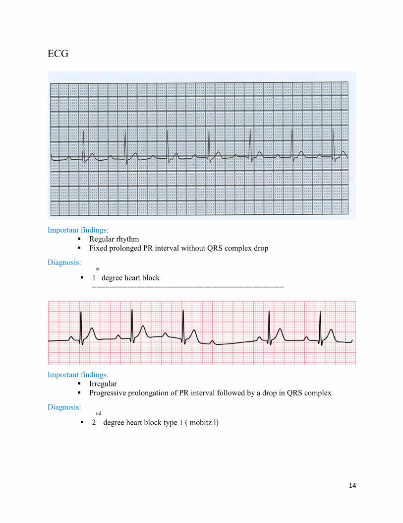
ECG
Important findings:
� Regular rhythm � Fixed prolonged PR interval without QRS complex drop
Diagnosis:
� 1st
degree heart block ===========================================
Important findings:
� Irregular � Progressive prolongation of PR interval followed by a drop in QRS complex
Diagnosis:
� 2nd
degree heart block type 1 ( mobitz l)

15
ECG
Important findings: � Regular rhythm � bradycardia � The P wave with a regular P-to-P interval � The QRS complex with a regular R-to-R interval. � The PR interval will be variable � As the hallmark of complete heart block is lack of any apparent relationship
between P waves and QRS complexes.
Diagnosis:3rd
degree heart block (complete heart block)

16
ECG
Important findings:
� Short PR interval
� Delta wave
Diagnosis:
z WPW
17
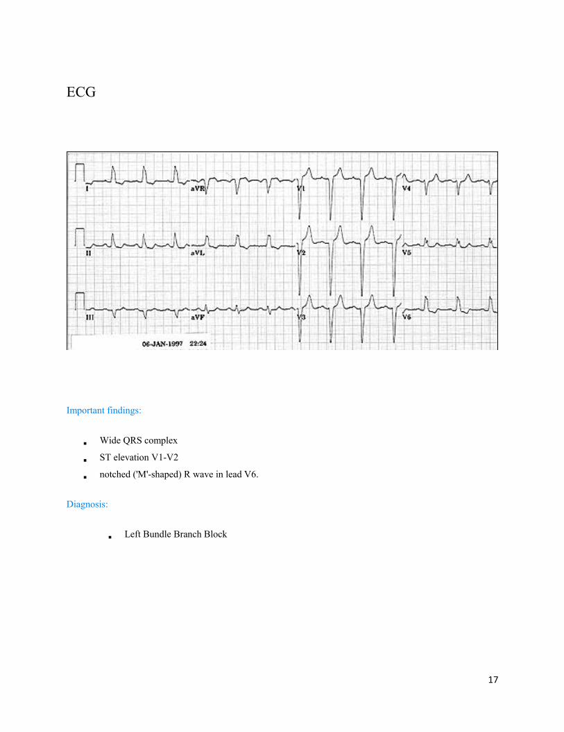
ECG
Important findings:
� Wide QRS complex
� ST elevation V1-V2
� notched ('M'-shaped) R wave in lead V6.
Diagnosis:
� Left Bundle Branch Block

18
ECG
Important findings: � Wide QRS complex � notched ('M'-shaped) RSR wave in lead V1.
Diagnosis: � Right Bundle Branch Block

19
LBBB
RBBB
00

20
ECG
Important findings:
� SV1 + RV5 or 6 >35 small squares (>7 big squares)
� ST segment depression with T wave inversion in V5-V6 (not always present)
Diagnosis:
z Left Ventricular Hypertrophy (LVH)

21
ECG
z Left Ventricular Hypertrophy (LVH)


25
ECG
Important findings:
� ST segment elevation in V1-V6
� Flattened T wave in inferior Lead
Diagnosis:
z Most likely anterior MI (based on the Hx and complete picture)

26
ECG
.Important findings:
� Widespread ST elevation
� There is a notch in ST segment which make it less likely to be ischemia.
Diagnosis:
z Benign early repolarization

28
ST depression
===============================================================
hyper acute T waves
adapted from Medscape.

29
ECG
Important findings:
� There is T wave inversion in inferior leads
Diagnosis:
z Ischemia (most likely NON STEMI which depend on Hx and complete picture)
30
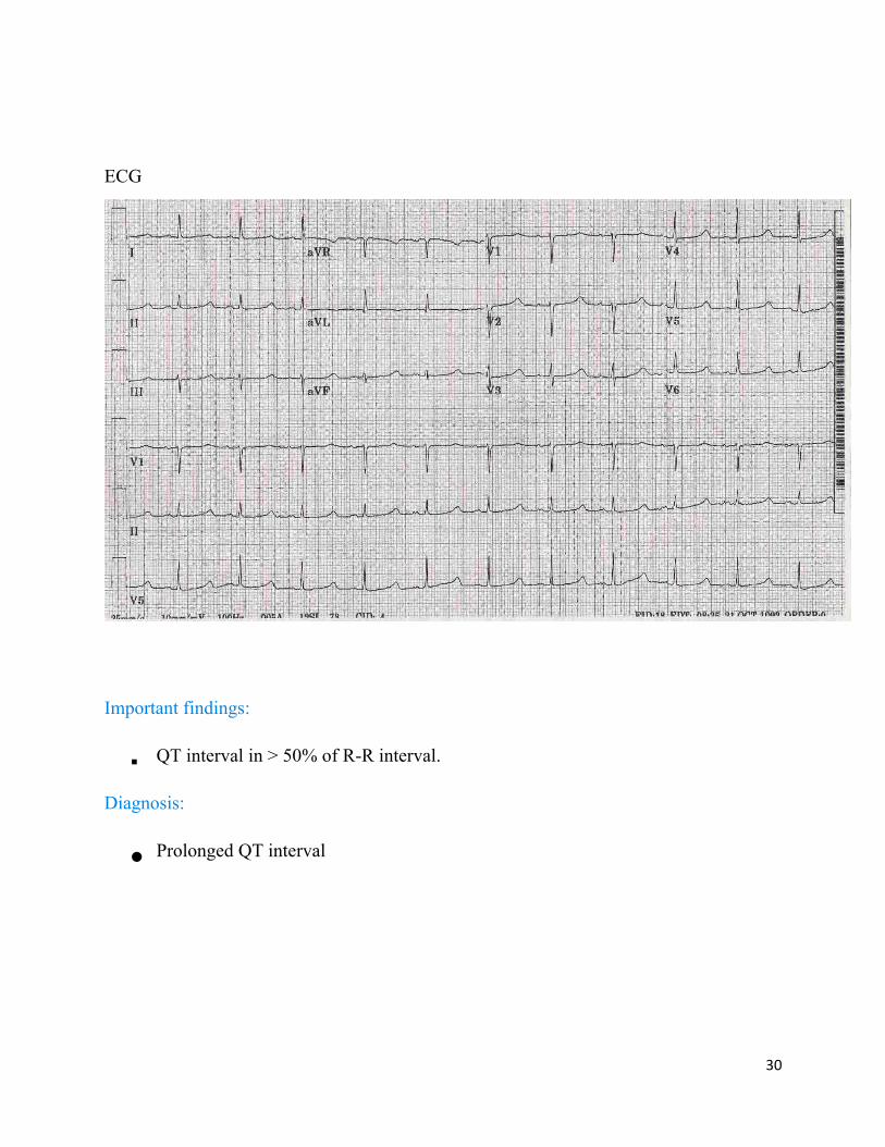
ECG
Important findings:
� QT interval in > 50% of R-R interval.
Diagnosis:
z Prolonged QT interval

31
we done with ECG approach and most important ECG diagnosis, now try to interpret the
following ECGs:
Important findings: � Regular rhythm � Fixed prolonged PR interval without QRS complex drop
Diagnosis:
z 1st
degree heart block


34
ECG
Important findings:
� Regular rhythm
� Bradycardia
� Wide QRS complex
� The P wave with a regular P-to-P interval
� The QRS complex with a regular R-to-R interval.
� The PR interval will be variable
� As the hallmark of complete heart block is lack of any apparent relationship between P waves and
QRS complexes.
Diagnosis:
z 3
rd degree heart block (complete heart block)


37
ECG
Important findings:
� Irregular rhythm
� Present P-wave with normal PR interval
Diagnosis:
z Sinus arrhythmia

38
ECG
z Important findings:
flutter waves “Saw tooth appearance”
z Diagnosis:
Atrial Flutter
40
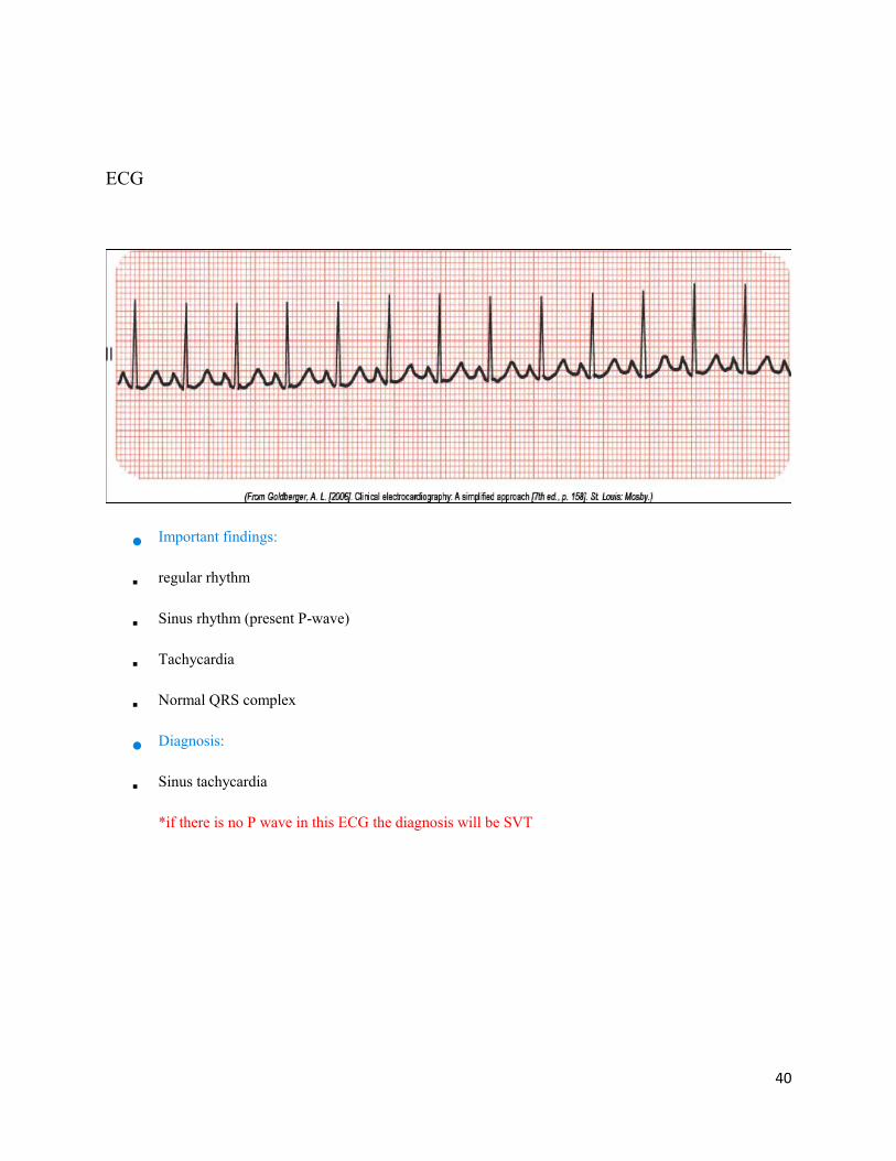
ECG
z Important findings:
� regular rhythm
� Sinus rhythm (present P-wave)
� Tachycardia
� Normal QRS complex
z Diagnosis:
� Sinus tachycardia
*if there is no P wave in this ECG the diagnosis will be SVT

41
55 Years old with heavy retrosternal chest pain for 3 hours
Findings:
1) progressive prolongation of PR interval with a drop of QRS complex.
2) Inferior ST segment elevation MI (leads II, III, and aVF) with reciprocal ST
depression (leads I and aVL)
Diagnosis:
Acute inferior STEMI with 2nd
degree type 1 heart block.


43
48 years old with Hx of palpitation and SOB for 3 hours
z Important findings:
� Irregular rhythm
� Absent P-wave
� there is PVC
z Diagnosis:
� Atrial Fibrillation
� PVC

44
22 years old pre-op ECG
Important findings:
� Irregular rhythm.
� Present P-wave before each QRS complex and there is QRS complex after every P-wave.
� normal PR interval.
Diagnosis:
z Sinus arrhythmia.

45
32 years old male with Hx of SOB + Palpitation for 1 hour
Important findings:
� Regular tachycardia
� Absent P wave
� Normal QRS complex
Diagnosis:
z SVT

47
47 years old with Hx of palpitation and dizziness
z Short PR interval and delta waves consistent with Wolff-Parkinson-White (WPW) syndrome
Good Luck-


![[5] HEMA - Megaloblastic Anemia.pdf - KSUMSC](https://static.fdokumen.com/doc/165x107/631deac95ff22fc7450674ca/5-hema-megaloblastic-anemiapdf-ksumsc.jpg)