Cerebrospinal fluid (CSF) - KSUMSC
13
Cerebrospinal fluid (CSF) [email protected] OUTLINES: • CSF definition, functions and methods • of sampling • Indications and contraindications of CSF Sampling. • Examination of CSF. • Abnormalities in CSF examination.
-
Upload
khangminh22 -
Category
Documents
-
view
1 -
download
0
Transcript of Cerebrospinal fluid (CSF) - KSUMSC

Cerebrospinal fluid (CSF)
OUTLINES:
• CSF definition, functions and methods
• of sampling • Indications and contraindications of CSF
Sampling. • Examination of CSF.
• Abnormalities in CSF examination.

q Physical support & protec8on (shock absorber)
q Provides controlled chemical environment
(nutrient supply and waste removal) q Intra and extra cerebral
transport (Neuroendocrine func8on)
q The liquid surrounding the brain and spinal cord.
q It flows in the
subarachnoid space (Between arachnoid and pia maHer)
EXCRETION • Formed at the choroid plexuses by the
cells lining the ventricles. • Rate of formaAon: 500 ml/day • Mechanism of formaAon: 1) SelecAve ultrafiltraAon of plasma 2) AcAve secreAon by epithelial membranes.
ABSORPTIO
N Excre8on volume =
produc8on volume => constant CSF volume
occurs at the arachnoid villi à protrude through the dura to the venous sinuses of the brain. à bloodstream
CIRCULATIO
N

Obtained by lumbar puncture
(At the interspace L3-‐4, or lower)1 Using asep8c techniques ⇒ Then, separated into 3 aliquots: ü For chemistry & serology ü For microbiology ü For cell count. Immediate analysis is required to
avoid exposure to microbes Because it is a precious sample,
preserving any remaining sample is highly suggested.
CONTRAINDIC
ATIONS
OF LP (lumbar pun
cture) q Bleeding diathesis (tendency)
q Increased intracranial pressure
q Infec8on at site of needle inser8on
q CNS infec8on q Demyelina8ng
diseases q CNS Malignancy q Hemorrhage in CNS
1: Because spinal cord ends at L2


Blood & Hemoglobin pigments in CSF Due to:
TRAUMATIC TAP Subarachnoid hemorrhage (SAH)
Xanthochromia Hemoglobin pigment breakdown = RBCs lysis and metabolism previously occurred (2 hours earlier)
u Bright red color u RBCS decrease in number as
the fluid is sampled. It indicates hemorrhage if we exclude:
1) Prior trauma8c tap 2) Hyperbilirubinemia (bilirubin > 20 mg/dL)
o Normal CSF is: 1) Colorless 2) Clear 3) Free of clots 4) Free of blood
o If CSF is turbid => perform microscopic examina8on. Usually due to leucocytes or may be due to micro-‐organisms 1-‐
Physical
exam
inaA
on of
CSF

Tests of interest: o Glucose1 o Protein1: total and Specific e.g. Albumin, Immunoglobulin, others e.g. myelin basic protein o Lactate o Glutamine :(replaced by measuring plasma [ammonia] Bi
oche
mical
analysis of
CSF
1. The most reliable diagnos8cally and accessible analy8cally 2. A plasma sample must be obtained 2-‐4 hours before CSF sample
q Enters CSF via facilita8ve transporter (GLUT).
q CSF [glucose] is ~ 2/3 that of PLASMA
Which equals 50 -‐ 80 mg/dl2 v In hypoglycemia: => CSF glucose may be very low v In hyperglycemia: => CSF glucose is raised. q Analysis should be done
Immediately or preserve the specimen with anIglycolyIc e.g. fluoride ion.
• Not clinically informative • Provides only confirmation of hyperglycemia ↑ CSF [glucose]
• Disorder in carrier-mediated transport e.g. TB meningitis, sarcoidosis
• Active metabolism of glucose by cells or organisms e.g:
acute purulent, amebic, & fungal meningitis • Increased metabolism by the CNS
e.g. by CNS neoplasm
↓CSF [glucose] (hypoglycorrhachia):
N.B. In viral meningi8s, CSF [glucose] is usually normal

q Proteins, mostly albumin are found in the CSF (0.15-‐0.45 g/L)
q Source of CSF proteins: 80% from plasma by ultrafiltra8on (Albumin mainly) 20% from intrathecal synthesis (Immunoglobulin mainly)
q ↑ CSF [total protein]: q Must be compared to the serum [protein]
Useful nonspecific indicator of pathological
states:
Lysis of contaminant
blood (traumatic
tap)
↑ permeability of the epithelial
membrane due to:
↑ production by CNS tissue in:
Obstruction Like in:
• Bacterial or fungal infec8on • Cerebral hemorrhage
• Multiple sclerosis (MS) • Subacute Sclerosing Panencephalitis
(SSPE)
• Tumors • Abscess
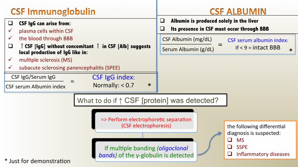
q CSF IgG can arise from: ü plasma cells within CSF ü the blood through BBB q ↑CSF [IgG] without concomitant ↑ in CSF [Alb] suggests
local production of IgG like in: ü mul8ple sclerosis (MS) ü subacute sclerosing panencephali8s (SPEE)
Serum Albumin (g/dL) =
* CSF Albumin (mg/dL) CSF serum albumin index:
If < 9 = intact BBB
q Albumin is produced solely in the liver q Its presence in CSF must occur through BBB
* CSF IgG/Serum IgG
CSF serum Albumin index =
CSF IgG index: Normally: < 0.7
What to do if ↑ CSF [protein] was detected?
* Just for demonstra8on
=> Perform electrophore8c separa8on (CSF electrophoresis)
If mul8ple banding (oligoclonal bands) of the γ-‐globulin is detected
the following differen8al diagnosis is suspected: q MS q SSPE q Inflammatory diseases

prealbumin
albumin
α1 α2
β1 β2 γ
prealbumin α1
α2
β1
β2
albumin

o CSF [Calcium], [Potassium] & [Phosphates] are lower than their levels in the blood. o CSF [Chloride] & [Magnesium] are higher than their levels in the blood
o Abnormal CSF [Chloride] o marked ↓↓ in acute bacterial meningi8s o slight ↓ in viral meningi8s & brain tumors O
ther Che
mical
Compo
nents o
f CSF
Clear ,Colorless Appearance
<5/mm3 Lymphocytes
Nil Polymorphs
7.4 pH
100 -‐ 150 ml Total Volume
450 -‐ 500 ml Daily Secre8on
1.006 -‐ 1.007 Specific Gravity
0.15 – 0.45 g/L Protein
50 -‐ 80 mg/dL (2.8-‐4.2 mmol/L)
(>50% plasma level)
Glucose
115 -‐ 130 mmol /L Chloride
1.0 -‐ 1.40 mmol/L Calcium
0.4 -‐ 0.7 mmol/L Phosphorus
1.2 -‐ 1.5 mmol/L Magnesium
2.6 -‐ 3.0 mmol/L Potassium
1: No need to memorize them. References rates will be given in the exam

Condi8on Parameter
Viral Meningi8s
Tuberculous Meningi8s
Bacterial Meningi8s (pyogenic)
Usually clear Oten fibrin web Oten turbid Appearance
Mononuclear Mononuclear Polymorphs Predominant cell
50-‐1000 10-‐1000 90-‐1000+ Cell count/mm3
None seen or cultured Oten none in smear In smear & culture Bacteria
<1 (Normal) 1-‐5 (↑ ↑) >1.5 (↑ ↑) Protein (0.15-‐0.45 g/L)
>1/2 plasma (Normal or slightly ↓)
<1/2 plasma (↓ ↓) <1/2 plasma (↓ ↓) Glucose (2.8-‐4.2 mmol/L)
Normal or ↓ ↓ ↓ ↓ ↓ Chlorides (115 -‐ 130 mmol/l)
Otorrhea: leakage of CSF from the ear Rhinorrhea: leakage of CSF into the nose Q: How to iden8fy it as CSF? =>Measure β-‐transferrin (a protein unique to the CSF)

Answers: 1) C 2) A 3) B 4)C 5) E 6)A 7) D 8) C 1) CSF sampling is contraindicated in which ONE of the following ? A- CSF infections B- Vertigo C- Increased intracranial pressure D- Immunocompromised patients 2) While sampling CSF from a patient, the nurse noticed that a bright red color of blood has appeared and it was decreasing in color as he continue sampling. This hemorrhage is most-likely due to: A- Traumatic tap. B- Subarachnoid hemorrhage. C- Infection at site of needle insertion. D- The sample is normal. 3) Increased CSF (GLUCOSE) is a confirmation of : A- Diabetes mellitus B- Not clinically informative C- Presence of micro organisms in CSF D- Hypoglycemia 4) CSF (GLUCOSE) in a patient with viral meningitis is expected to be : A- Very high B- Very low C- Normal D- Depend on the cause. 5) Abnormal CSF total protein can be an indicator of which of the following pathological states A- Tumors B- MS C- Bacterial or fungal infection D- Hemorrhage E- All of them
6) A CSF sample was taken from a 55-years old female for her annual check up, the biochemistry lab noted the presence of oligoclonal band of of γ-globulin In electrophoresis, this CAN indicate which ONE of the following: A- SSPE B- Hemorrhage C-Tumors D- Autoimmune diseases 7) Which ONE of the following components is present in higher levels in CSF than in the blood A- Ca B-Zn C- Phosphate D- Cl 8) The following schedule illustrate the result of some parameters of CSF sample. Based on the results shown above, the most-likely diagnosis is: A-Bacterial meningitis B- Viral meningitis C- Tuberculous meningitis D- SSPE
Parameter Result Reference rate
Appearance Fibrin web clear
Predominant cell Mononuclear -‐-‐
Protein 4 g/dl 0.15-‐0.45 g/L
Bacteria None appear in smear None

Done by: Mohammed Alnafisah
Sara Aldokhyl Ahmed alqahtani
Thank You!
If you have any ques8ons or comments, don’t hesitate to contact us