Refractive Errors - KSUMSC
12
Refractive Errors [ Color index: Important | Notes: F1 , F2 | Extra ] EDITING FILE Objectives: ➢ Not given. Done by: Lamya Alsaghan, Ruba Alselaimy, Munira Alhussaini. Edited & revised by: Lamya Alsaghan, Munerah AlOmari. Resources: Slides + Notes + Lecture Notes of Ophthalmology + 434 / 435 Team. Last 2 pages are extras from Group A!
-
Upload
khangminh22 -
Category
Documents
-
view
7 -
download
0
Transcript of Refractive Errors - KSUMSC
Refractive Errors
[ Color index: Important | Notes: F1, F2 | Extra ] EDITING FILE
Objectives: ➢ Not given.
Done by: Lamya Alsaghan, Ruba Alselaimy, Munira Alhussaini. Edited & revised by: Lamya Alsaghan, Munerah AlOmari. Resources: Slides + Notes + Lecture Notes of Ophthalmology + 434 / 435 Team.
Last 2 pages are extras from Group A!
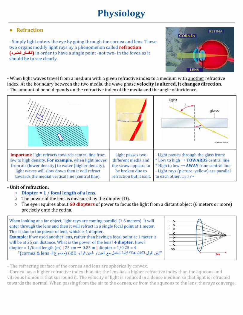
Physiology
● Refraction - Simply light enters the eye by going through the cornea and lens. These two organs modify light rays by a phenomenon called refraction in order to have a single point -not two- in the fovea as it (انكسار الضوء)should be to see clearly.
- When light waves travel from a medium with a given refractive index to a medium with another refractive index. At the boundary between the two media, the wave phase velocity is altered, it changes direction. - The amount of bend depends on the refractive index of the media and the angle of incidence.
Important: light refracts towards central line from low to high density. For example, when light moves from air (lower density) to water (higher density),
light waves will slow down then it will refract towards the medial vertical line (central line).
Light passes two different media and the straw appears to
be broken due to refraction but it isn’t.
- Light passes through the glass from: * Low to high → TOWARDS central line * High to low → AWAY from central line - Light rays (picture: yellow) are parallel to each other. متوازیین
- Unit of refraction: ○ Diopter = 1 / focal length of a lens. ○ The power of the lens is measured by the diopter (D). ○ The eye requires about 60 diopters of power to focus the light from a distant object (6 meters or more)
precisely onto the retina.
When looking at a far object, light rays are coming parallel (≥ 6 meters). It will enter through the lens and then it will refract in a single focal point at 1 meter. This is due to the power of lens, which is 1 diopter.
Example: If we used another lens, rather than having a focal point at 1 meter it will be at 25 cm distance. What is the power of the lens? 4 diopter. How? diopter = 1/focal length (m) | 25 cm → 0.25 m | diopter = 1/0.25 = 4
"(cornea & lens مجموع الـ) 60D لیش نقول الكالم هذا؟ ألننا نتعامل مع العین و العین قوتها"
- The refracting surface of the cornea and lens are spherically convex. - Cornea has a higher refractive index than air; the lens has a higher refractive index than the aqueous and vitreous humours that surround it. The velocity of light is reduced in a dense medium so that light is refracted towards the normal. When passing from the air to the cornea, or from the aqueous to the lens, the rays converge.
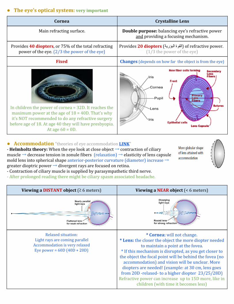
● The eye’s optical system: very important
Cornea Crystalline Lens
Main refracting surface. Double purpose: balancing eye’s refractive power and providing a focusing mechanism.
Provides 40 diopters, or 75% of the total refracting power of the eye. (2/3 the power of the eye)
Provides 20 diopters (القوة البؤریة) of refractive power. (1/3 the power of the eye)
Fixed Changes (depends on how far the object is from the eye)
In children the power of cornea = 32D. It reaches the maximum power at the age of 18 = 40D. That’s why it’s NOT recommended to do any refractive surgery
before age of 18. At age 40 they will have presbyopia. At age 60 = 0D.
● Accommodation “theories of eye accommodation LINK” - Helmholtz theory: When the eye look at close object → contraction of ciliary muscle → decrease tension in zonule fibers (relaxation) → elasticity of lens capsule mold lens into spherical shape anterior-posterior curvature (diameter) increase → greater dioptric power → divergent rays are focused on retina.
- Contraction of ciliary muscle is supplied by parasympathetic third nerve.
- After prolonged reading there might be ciliary spasm associated headache.
Viewing a NEAR object (< 6 meters) Viewing a DISTANT object (≥ 6 meters)
* Cornea: will not change. * Lens: the closer the object the more diopter needed
to maintain a point at the fovea. * If this mechanism is disrupted, as you get closer to
the object the focal point will be behind the fovea (no accommodation) and vision will be unclear. More
diopters are needed! (example: at 30 cm, lens goes from 20D -relaxed- to a higher diopter 23/25/28D)
Refractive power can increase up to 15D more, like in children (with time it becomes less)
Relaxed situation: Light rays are coming parallel
Accommodation is very relaxed Eye power = 60D (40D + 20D)

Accommodation
Looking at a far object.
No accommodation.
1. Change of lens shape. Notice how the shape of the lens changes in relation to different distances (in near object the rays are not parallel, so we need a more powerful lens to maintain the point in the fovea so the lens becomes more thick) 2. Miosis: to allow as much light rays as possible to enter. 3. Convergence: contraction of medial recti.
● Axial length (AL):
○ Normal axial length is 22.5 mml (it’s measured from the tip of the cornea to the surface of the retina). ○ if the axial length is longer = the picture will be in front of the retina “Myopia”. ○ If the axial length is shorter = the picture will be behind the retina “Hyperopia”.
Refractive Errors
- Facts: 75% of avoidable blindness is due to: uncorrected refractive error, cataract, trachoma. - A mismatch between the refractive power (RP) & the focusing distance of the eye. AL & RP should match! - Inability to see clearly is often caused by refractive errors.
- Emmetropia (normal) ما یلبس النظارات وال یعرفها - Ametropia = refractive error, three types of refractive errors:
○ Myopia (nearsightedness) ○ Hyperopia (farsightedness) ○ Astigmatism.
Point at the retina Matching between axial length (AL) &
refractive power (RP)
Point in front of the retina * high RP & normal AL OR
* normal RP & long AL
Point behind the retina * low RP & normal AL OR
* normal RP & short AL

Emmetropia
- Adequate correlation OR matching between axial length (distance from cornea to the fovea) and refractive power of the eye (cornea and the lens). - Rays of light from a distant object are brought to a pinpoint sharp focus on the retina (without accommodation). fovea لما یكون قوة العین وطولها متطابقین معناته النقطة بتسقط على الـ - All refractive errors are some deviation from emmetropia.
Ametropia
Myopia
- More common than hyperopia and astigmatism. - Most prevalent among Asians (80-90%), African Americans (25%), and Caucasians (13%) varies according to race. - Etiology: not clear, genetic (family history is important), acquired (excessive accommodation, near objects, aging) - Average age of onset: 6/8/10 (preschool/ school age) to 20 years (normally stops at 20)
- Rays of light from a distant objects converge in front of the retina, causing a blurred image on the retina. - The myopes can see close objects clearly, commonly known as “short-sightedness” قصر النظر: القریب أوضح من البعید - Myopia can be: essential (↑ RP, ↑ AL) OR secondary (other causes)
Causes of myopia: refractive & axial
1. Refractive myopia كبیرة ولكن حجم العین طبیعي
a) Change in lens nucleus or shape: - Cataract (ex. senile cataract: high density/thick/hard, low elasticity → high refractive power = induced myopia)
"senile cataract جدیدة بسبب الـ myopia طول عمره ما یعرف النظارة لكن لما یوصل عمره 60/70 یبدأ تصیر عنده أرقام“ - Spherophakia: congenital anomaly where the lens is spherical. - Diabetes. How it affects the lens? Through a process called “osmosis” due to variabilities in blood sugar. * Diabetics have both myopia and hyperopia depending on the level of the blood sugar. * High blood sugar → high sugar in aqueous humour → shrink of lens → HYPEROPIA. * Low blood sugar → fluid shift into lens → globular shape → MYOPIA * Undiagnosed diabetics might end up with a brain injury. Uncontrolled (fluctuations in blood sugar) lead to blurry vision.
"لما تتابعي patient ویقولك أنا عندي زغللة نظر مستمرة تعرفي أنه مو controlled الـ blood sugar ألن فیه fluctuation عالي"b) Lens repositioning: - Ciliary muscle shift: treatments like miotics. Contraction of ciliary muscles persistently → induced myopia - Lens movement e.g. anterior lens dislocation. Trauma → lens moves forward → image will be in front → myopia c) Ciliary muscle tone: Excessive accommodation, e.g. medical students. Reading a lot at a near distance.
d) Increase corneal power: Keratoconus (cone shaped cornea), Congenital glaucoma (big eye + protrusion of cornea)

2. Axial myopia
- Excessive long globe. القوة طبیعیة ولكن حجم العین كبیر - More common ★ - Causes: (what causes big eye) a) Congenital glaucoma, b) Posterior staphyloma (bulging of posterior part of eye).
Myopia Forms
Benign myopia (school age myopia) Progressive OR Malignant myopia
- Commonest type ★ - Onset 8-12 years, myopia increases until the child stops growing in height. - Generally tapers off at about 18 to 20 years of age.
- Also called degenerative or pathological myopia. - Myopia increases rapidly each year and is associated with, fluidity of vitreous and chorioretinal change.
"األرقام مستمرة في النزول"
Symptoms
Headache (due to eyestrain), blurred distance vision, squint in an attempt to improve uncorrected visual acuity when gazing into the distance. | Children: strabismus & amblyopia.
Morphologic eye changes (axial myopia)
Changes occur with axial myopia specifically. The following changes occur with increase in length (from front to back): 1. Deep anterior chamber. | 2. Atrophy of ciliary muscle. due to excessive stretching 3. Vitreous may collapse prematurely leading to opacification. The eye is bigger (more space) and the vitreous (gel-like substance) will stay the same. The space will be partially filled with vitreous and the rest with aqueous humour (watery) causing them to mix in a process called “liquefaction” (less density) [important] 4. Fundus changes: loss of pigment in RPE , large disc and white crescent-shaped area on temporal side , RPE atrophy in macular area , posterior staphyloma , retinal degeneration → hole → increase risk of retinal detachment & vision loss.
“لو قلنا أن أرضیة الغرفة هي الـ retina وجینا بعدنا الجدار، وش حیصیر في األرضیة؟ فیه أماكن ما راح یكون فیها coverage في الـ retina، في البدایة لو كان موكیت ممكن یجي معاك بس بعدین حینقطع بیصیر حاجة اسمها retinal break الـ retina ما عاد تقدر أنها تغطي حجم العین الكبیرة فیصیر فیه breaks والجل
liquified فیدخل مع الـ breaks هذي، لما یدخل وش یسوي في الـ retina ؟ یدفها/یفصلها، فیصیر عندنا حاجة اسمها retinal detachment أو انفصال الشبكیة"
One of the most important risk factors of retinal detachment is high myopia. وممكن توصل للعمى
Posterior Staphyloma (bulging)
Yellow arrow: optic nerve with distorted margins because of vitreous | Blue arrow: retinal margins. | White area: The retina (which is transparent ) and choroid (red in color ) are very thin and the sclera (white) is showing (behind the retina). | US: shows bulging at
the back of the eye because the pressure of the eye pushes the weak structures. (eye is getting bigger and the walls weaker)
Correction of myopia
Concave lenses (negative)

Hyperopia
- Parallel rays converge at a focal point posterior to the retina. myopia نعكس كل األشیاء اللي كانت بالـ - Etiology: not clear, inherited. trauma may cause dislocation of the lens.
- Rays of light from a distant object now focus behind the retina. - Must accommodate when gazing into distance to bring focal point on to the retina. However, this reduces their accommodative reserve when they want to view close objects. So, their distance vision is generally better than near vision, hence the term “long- sightedness” "األشیاء البعیدة أوضح من القریبة لكن حتى البعیدة مو واضح مره" - Can be: essential (↓ RP, ↓ AL) OR secondary (other causes)
Causes of hyperopia: refractive & axial
1. Refractive hyperopia
- Decreased refractive power of the eye (insufficient): a) Absent (aphakia: absent lens) or posteriorly repositioned lens (image at the back). can be congenital or due to trauma. b) Weak accommodation: trauma (it affects muscles or zonules), marijuana. (causes weak accommodation)
2. Axial hyperopia
- Decreased effective axial length (excessive short globe) - More common (retina is pushed forward) - Causes: Tumor, orbital mass.
retina العین ماهي صغیرة فعلًیا ولكن في حاجة ثانیة دفت العین والـ
Symptoms
Eye pain / strain, headache in frontal region (especially after reading, they require more accommodation), visual acuity at near tends to blur (blurry vision) relatively early ‘’inability to read fine print’’ | Pediatrics: strabismus & amblyopia.
Correction of hyperopia
Convex lenses (positive)
Myopia Hyperopia
Other name Nearsightedness / Short-sightedness Farsightedness / Long-sightedness
Focal point Single point in front of the retina Single point behind the retina
Refractive power Increased Decreased
Axial length Increased (long eye) Decreased (short eye)
Correction Concave lens Convex lens
Picture

Astigmatism
- الالُبَؤَرّیة: ما عندها بؤرة وحدة /االنحراف (misleading ألن االنحراف ممكن یعني حول وهذا مو حول)- Cornea is usually shaped like half a football. In these eyes there will be no astigmatism. you may describe it to the patient as “ your eye is shaped as a rugby ball instead of a football” - In astigmatism parallel rays come to focus in ≥ 2 focal lines rather than a single point. - Etiology: hereditary. - Cause: refractive media is not spherical → refract differently along one meridian than along meridian perpendicular to it → focal.
No astigmatism طبیعي ← single point focus ← الكرة أقطارها متساویة
astigmatism غیر طبیعي ← two focal points ← وجزء حاد flat الكرة أقطارها غیر متساویة جزء
2 Meridians:
Blue: flat Red: perpendicular
= 2 focal points
Classification
Regular astigmatism 2 focal points. Irregular astigmatism ≥ 2 focal lines
"الزم أتعامل مع two points، كیف أعالجها؟ (1) أشكلهم كـ one بعدین (2)
أحطها على الـ retina، أما الـ myopia و hyperopia بس مجرد أحطها على الـ
"retina
(eye injury) بسكین مضروبة cornea الـ الیمین بس منتظم شكلها الیسار على "الليالـ وباقي نقطة) من أكثر في األضواء تشتت اللي (هي متعوجة المنطقة باقي لكن وخیطناها
مع أتعامل أنا هنا ،clear تكون cornea عالجها! فأصعب اثنین) من (أكثر نقاط عشرةأجیب ،treatment للـ ثانیة لطریقة فنجيمنتظم شكلها فأخلي القرنیة في وأضعها حاجةینفع وال glasses معها ینفع ما علیها، غصب
معها شيء"
Types فیه افتراضات كثیرة
1. Simple Myopic Astigmatism: one before the retina, and one on the retina. 2. Simple Hyperopic Astigmatism: one on the retina and another behind the retina. 3. Compound Myopic Astigmatism: both are before the retina but at two different locations. 4. Compound Hyperopic Astigmatism : both behind the retina but at different virtual locations. 5. Mixed Astigmatism: one is before the retina and the other is behind the retina.
Causes
Corneal causes (majority) Lenticular causes
- Simple corneal astigmatism. بس جزء بسیط متضرر الباقي منتظم - Keratoconus (causes myopic astigmatism) very common in our community → Disease of the collagen of the cornea → astigmatism عالي الدرجة - Masses e.g. lid tumor یضغط من فوق ویتغیر شكلها
cornea وترجع الـ lid بیروح الوزن ویترفع الـ tumor شیل الـ للوضع الطبیعي
- Ptosis: یضغطها cornea لما یكون الجفن مرتخي على الـ شوي فـ induce astigmatism، و مهم عند األطفال، طفل عنده
congenital ptosis یصیر عنده astigmatism ویسوي له.amblyopia
"lens واحد جاه بوكس وصار اختالل في شكل الـ" - Lens dislocation. part of the zonules are cut.
جزء من العدسة مرتخي فتصیر غیر متساویة- Lenticonus. ↓
Symptoms
Asthenopic symptoms (headache, eye pain), blurred vision, distortion of vision مع العالي الدرجة، یا عین أو عینین, head tilting یحاول یبعد عن المیالن اللي یزعجه and turning, amblyopia (with uncorrected astigmatism > 1.5 Diopters)

Presbyopia
- Physiological loss of accommodation in advancing age - Deposit of insoluble proteins in the lens with advancing age → elasticity of lens progressively decrease → decrease accommodation. - Around 40 years of age, accommodation become less than 3D → reading is possible at 40-50 cm → difficulty reading fine print, headache, visual fatigue. - Lens is not as flexible because of decreased elasticity (gets dry) + zonules relaxes. - Correction of Presbyopia: convex lenses for reading.
"المریض یستخدم النظارة للقراءة لكن یناظرك بدونها، تلقى طول عمره ما یعرف النظارة، ولما وصل األربعین بدأ یالحظ األشیاء القریبة موب واضحة فیبدأ یبّعد عشان توضح لكن بعدین مهما یبّعد ما توضح، فیضطر یستخدم نظارة عشان یقدر یقرأ لكن البعید كویس"
Testing Vision
● Visual acuity حدة البصر “refraction ال تلخبطون بینها وبین الـ” - Vital sign of the eye and first thing to do in the clinic with Intraocular pressure (IOP) - Central visual acuity: display of different sized targets shown at a standard distance from the eye: Allen’s & Snellen chart. “أشكال أو حروف، تتدرج بالحجم من كبیر إلى صغیر بقیاس معین ”ومسافة معینة- Patient should be sitting & examine each eye alone (cover the other eye). Always start by showing large letters (assuming everyone is blind) and go smaller till normal. - VA: 20/20 (American), 6/6 (British) | 20/20 ft = 6/6 m. What does 20/20 mean? The patient can see at 20 feet what the normal population can see at 20 feet. Just like vital signs we have ranges from the normal population. Examples: 20/200: the patient can see at 20 ft what the normal population can see at 200 ft (far away from the normal). 20/40: the patient can see at 20 ft what the normal population can see at 40 ft (not that far away from the normal)
● Testing poor vision First: If the patient is unable to read the largest letter < 20/200 → Move the patient closer e.g. 5/200. at 10/200 الحرف الكبیر he can see 10 وأجي أقول واللهيft 20 أخلیهاft حتى الحرف الكبیر ما یشوفه فأول شيء أسویه أقرب المریض، یعني مثًال بدل ما أخلیها"
فـ( he can see at 10 ft what the normal can see at 200 ft) بعد كذا ممكن حتى الحروف كلها ما یشوفها مهما قربت، نجي إلى ↓"Second: If patient cannot read:
1. Count fingers (CF) examine each eye alone and write down the distance ex. CF at 1 foot or 2 feet. Not ↓ 2. Hand motion (HM) Not ↓ 3. Light perception (LP) with a torch examine all quadrants: up, down, right, left (sometimes all quadrants
are affected except one). You have to document that. 4. No light perception (NLP) complete blindness ما یشوف تماًما
● Pinhole testing simple but very important. Routinely done to all patients with decreased VA. "مثال: مریض زار العیادة، أول ما جاء قسنا الـ VA حقته، لقینا حدة البصر 20/100 معناته أنه decreased، و (he is far away from normal)، طیب لیش؟
"tool نجي هنا نستخدم هذي الـ we don't know ؟optic nerve وال retinal وال cataract زي الـ lens disease وال refractive error هل عنده
"زي البالستیك یغطي عین والعین الثانیة فیها فتحات صغیرة، نحط قدامه الـ Snellen chart ونقوله ركز على.(pinhole will cause muscle spasm) مرة ثانیة visual acuity وحده من الفتحات الصغیرة و نقیس الـ
corrects for about three) بس ما ندري وش نوعه refractive error إذا تحسن لـ 20/20 فعنده diopters) وإذا ما تحسن فهو ماهو refractive error (شيء ثاني). كیف یتحسن النظر من فتحة صغیرة
وأوصل 20/20؟ الحظوا في الصورة الـ المریض عنده myopia لما أجي أحط له الـ pinhole بتسمح بدخول الـwe فـ confusion هي اللي مسببة مشاكل و rays أما بقیة الـ fovea فقط وتروح على الـ central ray
eliminate them. الفتحة هذي 1.2mm فـ (it has to be 1.2) لو زادت أو نقصت بتغیر القیاسات وما بتوصل 20/20، زي اللي یصغرون فتحة عینهم لما یناظرون التلفزیون یعملون pinholing عشان یتحسن
نظرهم"

● Measuring refraction video (1:05 to 1:31)
"اآلن لقینا بالفعل المریض عنده refractive error، ما ندري عنده myopia وال hyperopia وال astigmatism، الزم نفرق هو جاي عندي ویبي أعطیه prescription، نبي نعرف النوع والدرجة in diopter، عشان نفصل له نظارة. فاستخدم الـ
" retinoscopy
Correction of Refractive Errors
● Spectacle lenses “Glasses”: most common ○ Monofocal lenses لها قوة وحدة: spherical lenses for myopia and hyperopia, cylindrical lenses for astigmatism ○ Multifocal lenses لها قوتین: for patients with: presbyopia + myopia/hyperopia.
First picture: Hyperopia Biconvex إما حجم العین صغیر أو القوة ضعیفة، نحتاج عدسة قویة (+) | شكلها
Second picture: Myopia إما حجم العین كبیر أو القوة قویة، نحتاج عدسة (-) نصعب علیها األمور ونبعد الـ
Biconcave زیادة ألنها قویة | شكلها rays Third picture: Regular astigmatism corrected with cylindrical lens: has both (plus +, minus -)
optometrists یدرسونها الـ advanced باقي التفاصیل العمیقة و الـ
Multifocal lenses Has 2 lenses (powers) One
for far vision (up) and the other for near vision (down).
To summarize the Tx: (1) Cylindrical lens → astigmatism +/- myopia or hyperopia (2) Biconcave lens → myopia (-) (3) Biconvex lens → to correct hyperopia (+)
● Contact lenses. common
● Keratorefractive surgery: last option,options: cornea (change shape) or intraocular to get rid of glasses
1. Cornea نقلل من سماكتها بطریقة محسوبة: LASIK لیزك (Laser-Assisted In-Situ Keratomileusis), INTRALASIK, LASEK We remove part of the stroma (central part) using laser by evaporating .(Laser Epithelial Keratomileusis) الزیكtissue from corneal surface. Video1 | Video2 (not the same as presented by the doctor) 2. Intraocular: PRK (Photorefractive Keratectomy): another lens نشیل العدسة الطبیعیة ونزرع بدالها"
أو نزرع عدسة أمام العدسة الطبیعیة (نربط العدسة بالـ iris) بدل ما أحط العدسة (نظارة أو عدسة الصقة) أمام العین أحطها في العین"
Clinical Scenario
Scenario: 14 year-old boy complaining of difficulty seeing the blackboard (common presentation) - Visual acuity (VA): 20/200 (first thing to do) - PH (Pin hole):
○ Improve: (1) to 20/20 → Refractive errors | (2) to 20/80 → both. (RE & other cause) ○ No improvement → Other causes: cataract, optic nerve disease..etc

Extra from 435 team
- The refractive index of a medium is defined as the ratio of the phase velocity of a wave light in a reference medium to its velocity in the medium itself. - To have a clear picture in the retina & to be seen in the brain, there should be a clear cornea, clear anterior chamber, and clear lens, clear vitreous cavity then the picture should be focused on the retina with normal refractive index. - The retina is responsible for the perception of light. It converts light rays into impulses; sent through the optic nerve to your brain, where they are recognized as images. - Power of accommodation is {(15 - age)/4} of the lens. accommodation is strong in children. - All three types of ametropia can be corrected by spectacle lenses . These diverge the rays in myopia, converge the rays in hypermetropia, and correct for the non - spherical shape of the cornea in astigmatism. - Japanese tend to have myopia more due to their crowded narrow surroundings which requires excessive accommodation. - Symptoms of hyperopia: Accommodative esotropia: because accommodation is linked to convergence leading to easotropia.. - It should be noted that in hypermetropia, accommodative effort will bring distant objects into focus by increasing the power of the lens. This will use up the accommodative reserve for near objects. - Astigmatism is a common and generally treatable imperfection in the curvature (mismatched curves) of the eye that causes blurred distance and near vision. It’s the worst in the quality of vision. - In astigmatism, surface of cornea is not homogenous. Usually it is congenital. - Regular astigmatism: (2 meridians) power and orientation of principal meridians are constant. The principal meridians are 90 degrees apart (perpendicular to each other). With the rule astigmatism, Against the rule astigmatism, Oblique astigmatism. - Irregular astigmatism: (different meridians > 2) power and orientation of principal meridians change across the pupil. The principal meridians are not perpendicular. - Anisometropia: (not mentioned by the doctor or slides) * Anisometropia is the condition in which the two eyes have unequal refractive power. Generally a difference in power of two diopters or more is the accepted threshold to label the condition anisometropia. the image of an object in one eye differs in size or shape from the image of the same object in the other eye. * More than 3 diopters difference if not detected in pediatrics and corrected it can cause unilateral amblyopia “in the weaker eye”. * Individuals can tolerate up to 2-3 Diopters of anisometropia before becoming symptomatic. * If the difference between 2 eyes: (D=diopter), > 3D → glasses | > 3 but < 7 → contact lenses | > 7D → surgery. Causes: Correction of a refractive error Anisometropia, Antimetropia (being myopic (nearsighted) in one eye and hyperopic (farsighted) in the other.), Meridional aniseikonia occurs when these refractive differences only occur in one meridian (see astigmatism), Refractive surgery.
Allen chart (age 3 – 6 years) Snellen chart (more than 6 years)
- Notes for testing vision & VA: * In the first 2 months of life: do light objection test (if the baby objecting or closing the eye in response to light it means he/she is seeing) * From 2 months – 3 years: do follow and fixate test. At this age, babies will start to follow the objects, so bring a toy in front of them and do the test. (If following the toy → good vision). OR you can do (central= seeing centrally. Steady= no nystagmus. Maintained= baby is following object & after blinking he/she continues following the same object) * The vision maturation is acquired skill for the brain, so babies when they’re first born they will be legally blind.

* The axial length of the eye will grow quickly in the first 6 months. So if anything stops the growing they will have amblyopia (lazy eye) E.g: vitreous hemorrhage, congenital cataract. * Legal blindness: if the vision in the best eye is w/ best correction and providing less than 20/200, this is considered legal blindness. (patient needs assistance). The criteria used to determine eligibility for government disability benefits and which do not necessarily indicate a person's ability to function. In the US, the criteria for legal blindness are: * Visual acuity of 20/200 or worse in the better eye with corrective lenses. * Visual field restriction to 20 degrees diameter or less (tunnel vision) in the better eye. Note that the definition of legal blindness differs from country to country. * Testing near visual acuity: It is done at a standard working distance ~ 30-40 cm. A variety of charts are available:
* - To assess the effect of pathology on VA. You must eliminate the effect of refractive error. This is achieved by measuring: the patient’s best spectacle correction or viewing the test chart through a pinhole. - Contact lenses: * Higher quality of optical image and less influence on the size of retinal image than spectacle lenses * Indication:cosmetic,athletic activities,occupational,irregular corneal astigmatism,high anisometropia & corneal disease * Disadvantages: careful daily cleaning and disinfection. * Complications: infectious keratitis, giant papillary conjunctivitis, corneal vascularization, and severe chronic conjunctivitis. - Refractive surgery – flattens corneal surface (more successful because it’s easier to flatten than to make it more convex) for myopia or increases its curvature in Hyperopia. - Improves unaided visual acuity but may have complications. - Intraocular surgery: for high power للي نقصهم مرة عالي, نشیل العدسة بكبرها ونحط جدیدة أو نزرع عدسة قدام العدسة الطبیعیة ○ Give best optical correction for aphakia; avoid significant magnification and distortion caused by spectacle lenses. ○ Clear lens extraction(with or without IOL) ○ Phakic IOL ( intraocular lenses): lenses made of plastic or silicone that are implanted into the eye permanently to
reduce a person's need for glasses or contact lenses. ○ One of the side effects of intraocular lens procedure => loss of accommodation.
Photorefractive keratectomy (PRK) Laser-assisted-in-situ Keratomileusis (LASIK)
Flap No flap. We just remove the epithelium apply laser then the epithelium will grow.
Thin flap.
Advantage Safer on the long run. Immediate 20/20 vision, no pain, good visual rehabilitation, can correct high numbers (up
to-80)
Disadvantage Severe pain for 1 week, blurred vision for 2-3 weeks
Severe trauma, the flap can fall down