10-Common Adult Fractures - KSUMSC
21
Objectives : 1. Clavicle fracture 2. Humerus (proximal & shaft) 3. Both 'bone' forearm 'fracture' 4. Distal 'radius' fracture 5. Hip fracture 6. Femur 'shaft' fracture' 7. Tibial 'shaft' fracture' 8. Ankle' fracture [ Color index : Important | Notes | Extra ] Editing file link Feisal Mussa , Salem Basamad , Ahmed Badahdah Team members: Mohammed Baqais Team leader: Abdulaziz ALmohammed Revised by: 435 Team, Toronto note, Slide, Note References: 10-Common Adult Fractures
-
Upload
khangminh22 -
Category
Documents
-
view
7 -
download
0
Transcript of 10-Common Adult Fractures - KSUMSC

Objectives : 1. Clavicle fracture
2. Humerus (proximal & shaft)
3. Both 'bone' forearm 'fracture'
4. Distal 'radius' fracture
5. Hip fracture
6. Femur 'shaft' fracture'
7. Tibial 'shaft' fracture'
8. Ankle' fracture
[ Color index : Important | Notes | Extra ] Editing file link
Feisal Mussa, Salem Basamad ,Ahmed Badahdah Team members:
Mohammed Baqais Team leader:
Abdulaziz ALmohammed Revised by:
435 Team, Toronto note, Slide, Note References:
10-Common Adult Fractures

2
Toronto notes
It is a common fracture in both children (unites rapidly without complications) and adults (much more troublesome in jury), you see it in young active people, who are engaged in contact sports and sometimes in RTA too.
Anatomy:
❏ Clavicle is S shape bone
❏ It is anchored to scapula via ACJ (Acromioclavicular joint)
❏ It is anchored to trunk via SCJ (Sternoclavicular Joint)
Mechanism: ❏ Most of fracture occurs as result from fall onto shoulder
❏ Direct trauma to clavicle or or FOOSH (Fall on An Outer Stretched Hand)
Classification:
❏ Fracture is classified into: proximal, middle and lateral third fractures
❏ Most of fractures are of middle third. Why do we classify things? To determine management, prognosis and it is helpful for communication, like if someone calls you at midnight and tells you that there is a clavicle fracture, I would like to hear the classification. However, for this classification it does not describe any of the prognostic indicators such as comminution, shortening or displacement.
Clinical findings:
❏ Arm is clasped to chest to splint shoulder and prevent movement
❏ Check the skin: looking for any skin tenting, because it will affect your management. The skin is tented by the bony fragments, sometimes it is sharp like a knife which threatens the skin so you have to make sure that the skin is intact.
❏ Injury to brachial plexus and subclavian artery/vein may be present (uncommon) you have to examine N.V, so you have to keep in mind to evaluate the neurovascular status distal to the clavicle (entire upper
limb)
❏ Rarely, Pneumothorax can occur.
Imaging: to exclude pneumothorax
❏ X-Ray: AP chest and Clavicle special view (focused on clavicle) (30°cephalic tilt), This X-Ray shows Middle third clavicle fracture with minimal displacement.
Clavicle fracture
3
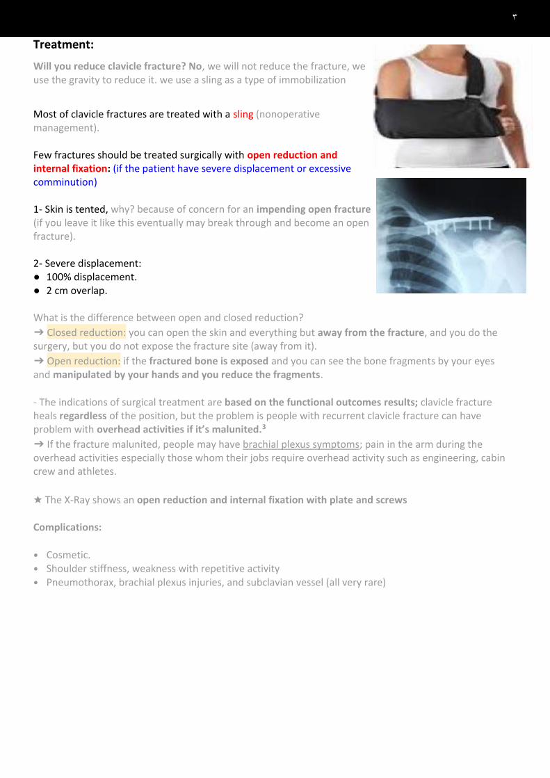
Treatment:
Will you reduce clavicle fracture? No, we will not reduce the fracture, we use the gravity to reduce it. we use a sling as a type of immobilization
Most of clavicle fractures are treated with a sling (nonoperative management). Few fractures should be treated surgically with open reduction and internal fixation: (if the patient have severe displacement or excessive comminution) 1- Skin is tented, why? because of concern for an impending open fracture (if you leave it like this eventually may break through and become an open fracture). 2- Severe displacement: ● 100% displacement. ● 2 cm overlap. What is the difference between open and closed reduction?
➔ Closed reduction: you can open the skin and everything but away from the fracture, and you do the surgery, but you do not expose the fracture site (away from it).
➔ Open reduction: if the fractured bone is exposed and you can see the bone fragments by your eyes and manipulated by your hands and you reduce the fragments.
- The indications of surgical treatment are based on the functional outcomes results; clavicle fracture heals regardless of the position, but the problem is people with recurrent clavicle fracture can have problem with overhead activities if it’s malunited.3
➔ If the fracture malunited, people may have brachial plexus symptoms; pain in the arm during the overhead activities especially those whom their jobs require overhead activity such as engineering, cabin crew and athletes.
★ The X-Ray shows an open reduction and internal fixation with plate and screws Complications: • Cosmetic. • Shoulder stiffness, weakness with repetitive activity
• Pneumothorax, brachial plexus injuries, and subclavian vessel (all very rare)

4
Anatomy: Proximal humerus has four anatomic parts:
● Head ● Greater tuberosity ● Lesser tuberosity ● Shaft ● Anatomical neck vs. surgical neck anatomical neck is between the tuberosities and the head while surgical neck is between the tuberosities and the shaft. -Why is it called surgical neck? because this is the location of many fractures that require surgery -Surgical neck fractures are more common and carry good outcomes. -Anatomical neck fractures: rare and carry bad outcomes because of the blood supply, the healing will be affected, and the patient may have AVN (avascular necrosis)
Fracture: These fractures happen in a bimodal fashion so you can see it in: ● Younger patients: violent trauma such as RTA. ● Older patients: minor trauma Most fractures are minimally displaced, and treated it conservative like what is seen on the X-Ray.
Physical exam: Start with ATLS history (You need to know what was happened and the
mechanism of the injury) then proceed to the physical examination: ● Expose the shoulder very well. ● Look for fracture signs (swelling, tenderness, inability to move,
ecchymosis) ● Check the skin, you have to examine the axilla. to know if it is an open or
closed fracture (unlikely to have an open fracture of proximal humerus bc it’s a deep joint).
● Peripheral N/V exam ● Axillary nerve: lateral skin patch Check if the patient has sensation (fine touch) over the lateral side of the shoulder which is supplied by
terminal branches. ● Examine cervical spine, you have to examine joint above (cervical spine) and joint below (the elbow)
X-Ray: ● AP and lateral views: you need 2 perpendicular (Orthogonal) views, why? to
have 3D image of the fracture ● Axillary view (special X-ray): the patient is laying down, and the beam will go
through the axilla to allow you see this view (the whole joint), it can show you if there is a fracture dislocation (it has different management)
Humerus fracture

5
Classification: ● Imagine that you cracked the anatomical neck, the surgical neck and you have a crack between
the GT and LT→ you will end by having 4 pieces. If you have 1 ● fracture line → you will get 2 pieces. 2 fracture lines → 3 pieces. 3 fracture lines → 4 pieces ● If we have all the fractures but not displaced, we call this nondisplaced humerus fracture (one-part fracture) ● If not displaced, we don’t count the fragments
● If there is a fracture with displacement more than 1 cm between the fragments, then we count the fragments. If there is 2 fragments, we call it 2 fragments fracture
● “two-part fracture”, if there is 3 fragments, we call it 3 fragments fracture “three-part fracture” and so on
● if all the major parts are displaced (the head of the humerus, the lesser tuberosity, the greater tuberosity and the shaft), it is a four-part fracture.
● Fracture is defined by the fragments displaced: Neer’s classification ● Displacement: more than 1 cm and/or angulation >45°
Normal AP Shoulder
Extra Figures

6
Treatment: ❖ If fracture is not displaced:
● Treatment with sling immobilization and NWB of upper extremity for 6-8 weeks, why 6-8 weeks? based on the healing process of the fracture Early ROM exercises after 2-4 weeks.
● Normal function can be resumed after 3-4 months. What type of healing for this fracture? Primary (direct) or secondary (indirect)? secondary healing
❖ If the fracture is displaced:
● Surgery is indicated. ● ORIF is indicated (plate and screws). ● Shoulder hemi-arthroplasty is indicated in some cases such as fracture dislocation (the humerus is
out of the joint in the axillary view) Hemi-arthroplasty: when we change the humeral part of the joint and it’s indicated in old patient with comminuted fracture
A patient with proximal humerus fracture, AP and lateral X-Ray were done, what is your next step in the management? A) CT
B) MRI
Humerus shaft fracture: Toronto notes Classification: It can be classified based on location of fracture. (proximal, middle and distal) Symptoms: pain, swelling, weakness ± shortening, motion/crepitus at fracture site
Physical exam: ● Skin ● Compartment ● N/V (neurovascular): watch for radial nerve palsy.
How to examine the radial nerve? Motor: extension of the wrist. Sensory over the dorsum of the first webspace.
X-Ray: 2 orthogonal views. It shows spiral fracture in the middle third of the humerus.
Treatment: Almost all humerus shaft fracture can be treated non-surgically.
● Close reduction ● Functional brace x 4-6 weeks + NWB ● Early ROM of elbow and shoulder to avoid stiffness
What is the difference between brace and cast? the brace is removable, plastic with velcro tape, clamshell. There is no significant difference compared to the cast but it’s easier to the patient. Surgery is indicated for specific conditions like:
● Segmental fracture, big fragment in the middle.
● Open fracture
● Obese patient, why? because of body built which will push the humerus and displace it, and also, they have a lot of fat which push the arm into varus
● Bilateral fracture, why? patient can’t function with 2 casts (inhumane). ● Floating elbow (Fracture of forearm and humerus); difficult to control.
Surgery: ORIF with plate and screws

7
Anatomy:
Forearm is complex with two mobile parallel bones, we consider the forearm as a one joint (quadrilateral joint). Radius and ulna articulate proximally and distally, by the proximal and distal radioulnar joint (DRUJ) to allow forearm rotation.
Fracture: fractures are often from fall or direct blow. It very unlikely to fracture only one bone without disruption of their articulation: A-Both bone fracture
Clinical: Symptoms and signs of fracture: deformity, pain, swelling loss of function in hand and forearm
● Check the skin ● Check the compartments of forearm, you have to check because
forearm and leg fractures have a higher risk for compartment syndrome especially leg fractures.
● Check Ulnar, median and radial nerve (PIN, AIN)
How to examine AIN and PIN? AIN: ask the patient to do opposition, it gives branches to the flexor pollicis longus (FPL) and flexor digitorum profundus (FDP) muscles. If it get injured the patient will be unable to flex the distal interphalangeal joint (DIP), Cannot make a perfect “O” sign. PIN: ask the patient to put his thumb up, it gives branche to Extensor pollicis longus (EPL).
● Check vascularity: color, temperature, capillary refill and pulse.
Monteggia fracture:
Toronto notes
Means proximal or middle third ulna shaft fracture
with dislocation of radius proximally (at elbow)
Galeazzi fracture:
Toronto notes
Means distal or middle third shaft radius fracture
with disruption of DRUJ.
Ulna is the fractured big bone, radius is the dislocated one. -Mechanism: direct blow on the posterior aspect of the forearm, hyper-pronation or fall on the hyperextended elbow. -Clinical Features: decreased rotation of forearm ± palpable lump at the radial head.
radius is fractured, ulna is dislocated from DRUJ (Distal radioulnar joint) -Mechanism: hand FOOSH (Fall on An Outer Stretched
Hand) with axial loading of pronated forearm or direct wrist
trauma
Both bones forearm fracture: (Means radius and ulna are broken)

8
Images:
2 orthogonal views perpendicular on each other with joint
above and below
CT scan if fracture extends into joint.
Treatment:
Both bone fracture:
● Reduce and splint at ER/clinic (temporary)
● Are treated almost always with ORIF: (plate and screws)
Monteggia fracture:
● ORIF ulna and close reduction of radial head Galeazzi fracture:
● ORIF radius and close reduction of DRUJ.
If the close reduction fails, then we do an open reduction of the joint
ما نفتحون نرجعه بدر ما نقدر وإذا لمكسوالعظم انثبت < علمخلوامفصل لانرجع و نفتح

9
● Most common fracture of upper extremity. ● Most frequently are seen in older women. ● Young adults fractures are most commonly secondary to high energy trauma
Extra-articular
Colles’ Fracture: Toronto notes Dorsal angulation displacement is more accurate, shortening and radial deviation
Smith’s fracture: Toronto notes Volar angulation(displacement) and shortening.
(reverse Colles’)
A dinner fork deformity, also known as a bayonet deformity
Dorsal angulation with apex directed vollary
Volar displacement , we always describe distal to proximal
Volar angulation with apex directed dorsally, always look at the thumb to know if it is volar = palmar side of the hand
Distal radius fracture

10
Intra-articular
Barton’s fracture: volar or dorsal
Treatment:
❖ Extra-articular fractures:
❏ Closed reduction and cast application.
❏ After fracture reduction we do X-Ray to decide the definitive treatment, if the fracture is in accepted position then continue in the cast, if the fracture is not in accepted position > do surgery for the patient. I will not tell you about the accepted position (too much information for you).
❏ Immobilization for 6-8 weeks.
❏ ROM exercises after cast removal. (usually we in adult we put the cast below
elbow to prevent elbow stiffness)
❏ Surgery: if reduction is not accepted
❖ Intra-articular fracture:
❏ A step more than 2 mm displacement is an indication for surgery.
❏ ORIF with plate and screws.

11
Toronto notes
The usual story of this fracture: a geriatric patient falls down in the bathroom and it is usually managed by
surgery.
● It is the most common fracture of LL. ● It is associated with osteoporosis. ● Most common mechanism is a fall from standing height. ● Other causes of fall (stroke, MI) should be rolled out during clinical evaluation, you should ask the patient
about the cause of falling down, because this can be the only manifestation of MI or stroke.
● It is a life changing event it’s not about the fracture itself, but bc it represents a systemic failure of the
patient “the patient starts to be senile”. most people will walk but they will not be the same. Mortality:
20% of these people will die 1 year after the fracture. Not bc of the fracture, it just tells you how it’s
linked to systemic failure.
Fractures can be classified into:
Joint capsule attaches proximally to the acetabulum and distally to the neck of the femur anteriorly at the greater trochanter.
-Intra-capsular:
● Subcapital (below the head) (Femoral head and neck junction)
● Trans-cervical (mid portion of femoral neck)
-Extra-capsular:
● Basicervical (base of femoral neck)
● Intertrochanteric
● subtrochanteric
AVN risk is higher with intra-capsular fracture.
Why to bother about the capsule? because the blood supply is related to the capsule itself, and we know
patient who has fracture in this area has a higher chance of avascular necrosis, while if the fracture is
outside the capsule, he has a lesser chance of AVN.
Displaced vs nondisplaced
Clinical:
Keep in mind it is not an adult hip it is a geriatric hip, so the patients age usually ranges between 60-80s, they may have osteoporosis, and you will see it more in the future because the number of old people is increasing in SA.
● Full detailed history of mechanism of injury. ● Rule out syncope, chest pain, weakness etc. ● A detailed systematic review. ● Deformity (old MCQs): Abduction, External rotation and shortening. ● Assess distal N/V status ● Avoid ROM if fracture is expected
X-Ray: ● 3 views are needed
● AP pelvis
● AP hip
● Lateral hip, how can we get lateral hip X-Ray? cross table lateral, the patient rises the normal leg, and the image is taken from down
Hip fracture (Old patients)

12
Treatment:
● No close reduction is needed, why? a study showed that there is no difference if you put a traction or
not, not cost effective, and no benefit for the patient.
● No traction is needed. ● Patient needs surgery ideally within 48 hrs, why? a study showed that mortality is higher after 48 H. ● The goal is to ambulate patient as soon as possible. ● Be sure that DVT prophylaxis is started. ● Be sure that patient will be evaluated for osteoporosis after discharge.
60 years old lady, seen in the ER, she has an external rotation and abduction deformity of the leg, X-Ray
shown below, which of the following is the appropriate management?
a. Reduce the fracture and place skin traction
b. Reduce the fracture and place skeletal fracture
c. Do not reduce the fracture
d. Reduce the fracture and place above the knee cast If fracture is intra-capsular:
Hemiarthroplasty: percutaneous in situ Screws fixation.
● Displaced: Hemiarthroplasty, I do not want to do ORIF because although theoretically it works in 65%, 35% will have AVN and they will need another surgery
● Nondisplaced: (if not displaced the treatment is percutaneous in-situ screw fixation)
Examples
What is the type of this fracture? A) Subcapital
B) Trans-cervical C) Basicervical D) Intertrochanteric
this is high intertrochanteric and yes this is maybe
basicervical (doctor said it is more likely to be
basicervical than intertrochanteric (extracapsular)
Percutaneous in-situ screw fixation

13
If fracture is Extra-capsular: the chance of AVN is minimum less than 5%
● Stable: Close reduction and DHS ● Unstable: Intra-medullary device
Fracture instabilities signs:
1. Large LT (lesser trochanter) fragment 2. Extension to subtrochanteric region 3. 4 parts fracture
Remember that they are old patients and if you have one shot in your gun, you want it be accurate (you do want to take the patient multiple times to the OR)
if young patient always fix even if displaced (if you done hemiarthroplasty he will live for long time and will need to repeat for multiple time and eventually total hip replacement)
Complications: Nonunion
● 2% (IT fractures) ● 5% (non displaced neck fracture) ● 30% (displaced neck fracture)
AVN (femoral neck fracture):
● 10% (non displaced) ● 30% (displaced)
Death:
● early 4%. ● At 1 year: 20-40%
VTE (Venous thromboembolism)
Femoral neck fracture:
● It is a completely different entity from similar fractures in elderly (>60 years). ● High energy mechanism, we do not expect to see hip fracture in young patients. ● Patient should be taken to operative room for ORIF within 6 hours. ● MCQ: A 30 years old male, presented to the ER, he missed a step on the stairs, the X-Ray will
show you a hip fracture, what do you think? It is a pathological fracture (most likely a tumor)
● ATLS protocol. ● 2.5% associated femoral shaft fracture. (long femur X-ray) ● Nonunion: 30% (most common complication) ● AVN: 25-30%
They have the same chances for AVN, but I can take them to the OR again
Hip fracture (young patients)

14
Management
Intracapsular fractures Extracapsular fractures
Displaced Nondisplaced Close reduction and DHS or IM nail fixation
closed reduction open reduction and fixation
with cannulated screws. the same as
nondisplaced No hemiarthroplasty for
young patients
closed reduction and Screw fixation
(cannulated screws).
Toronto notes
Most common:
● high energy mechanisms (MVC, fall from a height, gunshot wound) ● Young patients (male, < 30 years). ● ATLS protocol,
Less common:
● low energy mechanism (torsional forces) ● Old patients. ● Spiral type fracture
Associate musculoskeletal injuries:
● Ipsilateral femoral neck fracture (10%. Missed in 30-50%) ● Knee ligaments injuries: 50% ● Meniscal tear 30% ● Floating knee injury: less common ● Vascular/nerve injuries: rare ● Contralateral femur shaft fracture (worse prognosis among above)
Associated non-MS injuries:
● Fat embolism ● ARDS ● Head injuries. ● Abdominal injuries
● Clinical: ● ATLS ● Fracture symptoms and signs ● Skin integrity ● N/V exam. ● Compartment assessment ● Knee swelling or ecchymosis.
Femoral shaft fracture
15
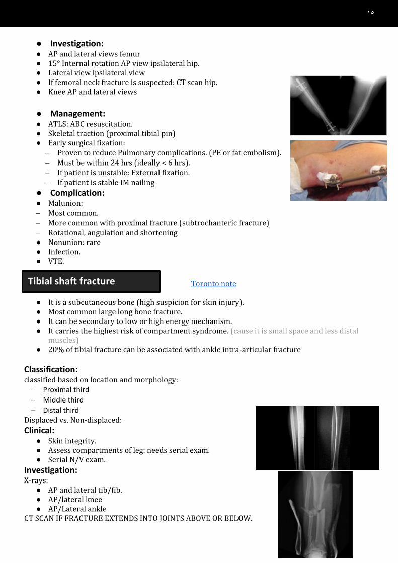
Tibial shaft fracture
● Investigation: ● AP and lateral views femur ● 15° Internal rotation AP view ipsilateral hip. ● Lateral view ipsilateral view ● If femoral neck fracture is suspected: CT scan hip. ● Knee AP and lateral views
● Management: ● ATLS: ABC resuscitation. ● Skeletal traction (proximal tibial pin) ● Early surgical fixation: − Proven to reduce Pulmonary complications. (PE or fat embolism). − Must be within 24 hrs (ideally < 6 hrs). − If patient is unstable: External fixation. − If patient is stable IM nailing
● Complication: ● Malunion: − Most common. − More common with proximal fracture (subtrochanteric fracture) − Rotational, angulation and shortening ● Nonunion: rare ● Infection. ● VTE.
Toronto note
● It is a subcutaneous bone (high suspicion for skin injury). ● Most common large long bone fracture. ● It can be secondary to low or high energy mechanism. ● It carries the highest risk of compartment syndrome. (cause it is small space and less distal
muscles) ● 20% of tibial fracture can be associated with ankle intra-articular fracture
Classification: classified based on location and morphology: − Proximal third
− Middle third
− Distal third Displaced vs. Non-displaced:
Clinical: ● Skin integrity. ● Assess compartments of leg: needs serial exam. ● Serial N/V exam.
Investigation: X-rays:
● AP and lateral tib/fib. ● AP/lateral knee ● AP/Lateral ankle
CT SCAN IF FRACTURE EXTENDS INTO JOINTS ABOVE OR BELOW.

16
Ankle fracture
Management: Indications for non-surgical treatment:
● NO displacement: < 10° angulation on AP/lateral x rays. ● < 1 cm shortening. ● Not comminuted.
C/I: ● Displacement. ● Open fracture. ● Compartment syndrome. ● Floating knee.
Close reduction and cast immobilization:
● Above knee back slab and U slab if surgical treatment is chosen. ● Above knee full cast if non-surgical treatment is chosen: it must be bivalved to minimized
compartment syndrome. ● Always provide patient with Compartment Syndrome checklist if patient is discharged home
with cast. ● NWB for 8 weeks with cast immobilization.
Surgical treatment:
● Most common modality of treatment
● Most commonly IM nail fixation.
Complications: ● Non-union: most common complication ● Delayed union ● Infection: open fracture ● DVT/PE
Toronto note Low energy (torsional): malleoli fracture.
Anatomy:
● Medial and lateral malleoli, distal tibia and talus.
● Highly congruent joint
● Fibula is held to distal tibia by syndosmotic ligament.
● Medial malleolus is held to talus by deltoid ligament.
● Lateral malleolus is held to talus by LCLI(lateral collateral ligment)
● The ankle Joint: consists of a deep socket formed by the lower ends of
the tibia and fibula, into which is fitted the upper part of the
body of the talus. The shape of the bones and the strength
of the ligaments (deltoid ligament attach the medial
malleolus to the talus, If I remove the fibula completely the
talus will stay in position because of the deltoid ligament
attachment and syndesmosis. ) and tendons make this joint
strong and stable.
● The syndesmosis is the ligament that connects two bones of the leg (tibia and fibula), you do
not need to know about its tears.

17
● In order to move the talus out, I have to crack the fibula and
cut the deltoid and syndesmosis, the talus will go with the
fibula toward the fracture. If the syndesmosis and deltoid
ligaments are intact the talus will not move.
● Equivalent of deltoid rupture: fracture of the medial
malleolus but the ligament is still intact so both will move
together.
● Make sure that there is no lateral translation (1 mm) of the
talus, because 100% the patient will get osteoarthritis after 1
year.
● Usually it is a twisting injury either RTA or falling down.
Classification:
● Stable v.s. Unstable fracture: Lateral displacement of talus
● Medial, lateral or bimalleolar fracture
● Lateral malleolus: Weber A, B, C
The Danis–Weber classification is based on the level of the fibular fracture: ● Type A: a fibular fracture below the syndesmosis, and an oblique fracture
of the medial malleolus. ● Type B: a fracture at the syndesmosis often associated with disruption of the anterior fibers of the
tibiofibular ligament and fracture of the posterior and/ or medial malleolus, or disruption of the medial ligament
● Type C: a fibular fracture above the syndesmosis; the tibiofibular ligament must be torn, or else ● Bimalleolar fracture: when both medial and lateral malleolus are broken.
Clinical:
● Look for Fracture symptoms and signs.
● Assess medial joint ecchymosis or tenderness to assess medial malleolus and deltoid ligament
integrity.
● Assess N/V status (before and after reduction).

18
Imaging: X-Ray:
● AP
● Lateral
● Mortise view
● Long leg x-rays: if only medial malleolus is broken.
CT SCAN IF FRACTURE EXTENDS TO ARTICULAR DISTAL TIBIA SURFACE. Unstable: > 4mm lateral translation Stable
Management:
Intact medial malleolus:
● Weber A: No surgery
•Splint + NWB X 6 weeks.
•Early ROM.
● Weber B/C: Plate -/+ syndesmotic screw
•If medial joint line widen (unstable): ORIF , if the tibia and fibula are displaced I will put the
syndesmotic screw between them, it is called syndesmotic screw because it acts as a
syndesmotic ligament (hold the bones together until syndesmosis heal).
•If not: Call Orthopedic for stress film x-rays.
● If both malleoli are broken: ORIF (ORIF both bones -/+ syndesmotic screw)
when do we have to put the syndesmotic screw? if there is lateral translation of the talus
intraoperative → if the talus is still moving with stress after fixation → syndesmosis is open, and
we put screws. so, after fixation in weber B or C/ bimalleolar fracture, we do stress test and accordingly we put syndesmotic screws or not

19
1- 6-year-old came through the ER after he sustained a 3-meter fall from a building, he was cleared
except for a solitary left femur injury (X-ray shows mid femoral shaft fracture)
how would you manage this injury?
A-External fixation.
B-Screws and plate.
C-Rigid IM
D-Flexible IM
Ans: D
2- A picture of a fracture of the radius, what is the Diagnosis:
A-Gelazzi.
B-Distal radial
Ans: A
3- What is the type of the fracture shown in the picture below?
A-Intertrochanteric Fracture.
B-Subcapital neck fracture
C-Transcervical Fracture.
D-Fracture of the greater trochanter.
Ans: A
4- 20-year-old male, who fell down from 2 steps stairs and fractured his ankle. The parents report that
their son' personality has been changed since the past weeks,which one of the following describe
the patient situation?
A-Pathological fracture.
B-Psychological factors
Ans: A
5- Which one of the following is considered as unstable fracture?
A-Lateral displacement of the talus.
6- Patient has a fracture in the humerus as shown below in the picture, how would you manage him?
A-Screw and plates. B-ORIF.
C-Brace
Ans: A
MCQs

20
7- Patient has an isolated fracture of the femur, what is the management?
A-IMN 8- A distal humeral fracture will result in?
A-Wrist drop.
9- Which fracture is described as dislocation of the distal head of the radius and a fracture of the
ulnar?
A-Monteggia 10- What is the diagnosis (Can’t remember the case but they gave us a pic and asked)?
A-Barton’s fracture
B-Smith’s Fracture
C-Colle’s Fracture
D-Galeazzi
Ans: A
11- Case about an old patient with intracapsular fracture what is the
treatment?
A-IM nail
B-hemiarthroplasty
C-DHS
D-percutaneous in situ fixation
Ans: B
12- Isolated R clavicle fracture with skin tinting, N/V is normal. what is the management?
A. Immobilization and arm sling B. Immobilization and figure of 8 C. Closed reduction and k-wires fix D. ORIF and plate and screws Ans: D
13- A 66 years old male fell from standing height, he’s unable to bare weight on his right leg, upon
examination the right leg is shortened and rotated. Patient is stable. What’s the management of choice for this patient?
A. Closed reduction and in situ pinning B. Hemiarthroplasty C. Open reduction + DHS
Ans: B

21
14- a 17-year football player and had an injury to the shoulder. X ray shows dislocation of the humerus.
What concomitant sign can you see clinically?
A. Deltoid atrophy B. Wrist drop. C. claw hand Ans: A
15- A scenario of a 17/o male w/ Hx of “Fall on Outstretched Hand” x-ray showing:
A. Monteggia. B. Colles. C. Distal radius fracture. D. Galeazzi. Ans: A