Cell adaption & Cell Injury - KSUMSC
32
PATHOLOGY: CELL INJURY FOUNDATION BLOCK TEAM 432 Cell Injury Notes on Dr. Ammar C. Al-Rikabi's handout First year Medicine-Foundation Block Pathology Team September 2012 Please note: This paper does not replace the main sources,it's only a facilitator. 1
-
Upload
khangminh22 -
Category
Documents
-
view
2 -
download
0
Transcript of Cell adaption & Cell Injury - KSUMSC

PATHOLOGY: CELL INJURY FOUNDATION BLOCK TEAM 432
Cell Injury Notes on Dr. Ammar C. Al-Rikabi's handout
First year Medicine-Foundation Block Pathology Team
September 2012
Please note: This paper does not replace the main sources,it's only a facilitator.
!1

PATHOLOGY: CELL INJURY FOUNDATION BLOCK TEAM 432
Acknowledgement
Dear colleague, this paper was a result of hours and days of hard work from both female & male pathology teams...
All what they want from you is "Dua'a"
!2

PATHOLOGY: CELL INJURY FOUNDATION BLOCK TEAM 432
OBJECTIVES
1. Understand the concept of cell and tissue adaptation: A. Hypertrophy (size) B. Hyperplasia (number) C. Atrophy (shrinkage) D. Metaplasia (shape of cell)
2. Know the concept of hypoxic cell injury and it's causes. 3. Understand mechanism and definition of free radical injury. 4. Know types of cell death , their definitions , types & clinical examples:
A. Apoptosis programmed death (physiologic & pathologic) B. Necrosis group cell death (pathologic)
5. Differentiate between apoptosis & necrosis. 6. understand causes of steatosis , accumulations of exogenous & endogenous pigaments. 7. Understand causes & differences between :
A. Dystrophic calcification: B. Metastatic calcification:
!3

PATHOLOGY: CELL INJURY FOUNDATION BLOCK TEAM 432
!4
PATHOLOGY: CELL INJURY FOUNDATION BLOCK TEAM 432
Definitions:
1. Cell injury : Cell injury is best defined as the cellular changes caused by stress مجهود .قدرة الخلية على التكيف the cell's adaptive capability يتعدى which exceed أو ضغط
2. Cell death : The end of the cell's life موت الخلية . There are two types of cell death:
A. Apoptosis : Programmed cell death.
B. Necrosis : The ultimate result أسوأ نتيجة ممكنة of irreversible cell injury
3. Cell adaptive capability : It's the range of stress the cell can manage .
4. Hyperplasia : The increase in number of cells. (Antonym: Hypoplasia)
5. Hypertrophy : The increase in the cell's size. (Antonym : Hypotrophy)
6. Metaplasia : The cell change from one form to another تغير نوع الخلية وشكلها
i.e. the change of pseudostratified columnar ciliated epithelium to stratified squamous epithelium .
7. Atrophy : Shrinkage in cell size or tissue size
8. Hypoxia : loss of blood supply in a tissue.
9. Ischemia : loss of blood supply in a tissue due to impeded arterial flow or reduced venous drainage
!5

PATHOLOGY: CELL INJURY FOUNDATION BLOCK TEAM 432
Cell Adaption
Intro : Cells are active participants in their environment , constantly adjusting their structure & function to accommodate تتكيف changing demands & extracellular stress. Cells maintain homeostasis , it's vital ضروري جدا وحيوي for cells' life and function.
Cell Adaption : When a cell undergoes physiological stresses or pathological affect, it encounters the stress by undergoing adaption , it achieves a new steady state preserving . قدرتها على العمل viability & function محافظةً على
This adaption is provided in several mechanisms:
1. Hyperplasia: It is an increase in the number of cells زيادة في عدد الخاليا
Cause: hyperplasia is usually a response to growth factors (i.e. hormones).
Note: Hyperplasia can be physiologic or pathologic.
# Clinical application :
A. Endometrial hyperplasia induced by estrogen.
B. Endocrine stimulation, e.g. Thyroid.
C. Focal nodular hyperplasia (liver).
D. The response of foot cells to uncomfortable shoes.
2. Hypertrophy : It is an increase in the size of cells resulting an increase in organ size( occurs in tissues incapable of cell division i.e. muscle tissues) زيادة في حجم الخاليا مما يؤدي إلى زيادة في حجم العضو
Note: Hypertrophy can be physiologic or pathologic
# Clinical applications:
A. (Picture) : hypertrophy of the uterus during
!6

PATHOLOGY: CELL INJURY FOUNDATION BLOCK TEAM 432
pregnancy. A, Gross appearance of a normal uterus (right) and a gravid uterus (left) that was removed for postpartum bleeding. B, Small spindle-shaped uterine smooth muscle cells from a normal uterus. Compare this with (C) large, plump hypertrophied smooth muscle cells from a gravid uterus (B and C, same magnification).
B. (Picture): hypertrophy of cardiac muscle,typically myocardial tissue. (a) you can see normal myocardial cells , (b) you can see large hypertrophied myocardial cells .
3. Atrophy : Shrinkage in the size of the cell by the loss of cell substance. Note: Even though atrophic cells have diminished function فعاليتها منخفضة they are not dead . الخلية ال تموت وإنما تنخفض فعاليتها
# Clinical applications :
A. Atrophy of the brain in Alzheimer disease
B. Thinning of the bones in Osteoporosis.
C. Picture:Atrophy. A, Normal brain of a young adult. B, Atrophy of the brain in an 82-year-old male with atherosclerotic disease. Atrophy of the brain is due to aging and reduced blood supply. Note that loss of brain substance narrows the gyri and widens the sulci. The meninges have been stripped from the right half of each specimen to reveal the surface of the brain.
!7

PATHOLOGY: CELL INJURY FOUNDATION BLOCK TEAM 432
4. Metaplasia : a change in which an adult cell type is replaced by another type. The cell is replaced by another cell more capable to withstand the adverse environment. تغير يصيب الخاليا ففتحول من نوع إلى آخرأكثر قدرة على تحمل الوضع الذي تواجهه الخلية األصلية
Clinical applications:
A. (Picture) : Bronchial mucosa replaced by squamous epithelium (smoking).
B. Cervical columnar epithelium replaced by squamous epithelium (cervicitis)
C. Metaplasia of the squamous epithelium of the esophagus into intestinal or gastric epithelium due to reflux of gastric juice العصارة . الهضمية
SUMMARY
Cellular Adaptations to Stress Hypertrophy: increased cell and organ size, often in response to increased workload; induced by mechanical stress and by growth factors; occurs in tissues incapable of cell division Hyperplasia: increased cell numbers in response to hormones and other growth factors; occurs in tissues whose cells are able to divide Atrophy: decreased cell and organ size, as a result of decreased nutrient supply or disuse; associated with decreased synthesis and increased proteolytic breakdown of cellular organelles Metaplasia: change in phenotype of differentiated cells, often a response to chronic irritation that makes cells better able to withstand the stress; usually induced by altered differentiation pathway of tissue stem cells; may result in reduced functions or increased propensity for malignant transformation.
!8

PATHOLOGY: CELL INJURY FOUNDATION BLOCK TEAM 432
Cell injury
cell injury results when cells are stressed so severely that they are no longer able to adapt, or when cells are exposed to inherently damaging agents, or suffer from intrinsic abnormalities.
In other words, if the cell adaptive capability is exceeded (overtaxed) then cell injury develops.
Types Of Cell Injury
1. Reversible: In early stages or mild forms of injury the functional and morphologic changes are reversible, if the damaging stimulus is removed. At this stage, although there may be significant structural and functional abnormalities, the injury has typically not progressed to severe membrane damage and nuclear dissolution Earliest changes associated with cell injury are :
A. decreased generation of ATP. B. loss of cell membrane integrity. C. defects in protein synthesis. D. cytoskeletal damage. E. DNA damage.
!9

PATHOLOGY: CELL INJURY FOUNDATION BLOCK TEAM 432
2. Irreversible injury is marked by : (VERY IMPORTANT) A. Severe mitochondrial vacuolization. B. extensive damage to plasma membranes. C. swelling of lysosomes. D. The appearance large, amorphous densities in mitochondria
Cell death(will be covered later on) :When the injury becomes irreversible, at that time the cell cannot recover , thus it dies.
Causes of cell injury
1. Oxygen Deprivation: hypoxia or oxygen deficiency, it’s the common cause of cell injury and death. Hypoxia is caused several mechanisms i.e.:
A. Ischemia : loss of blood supply in a tissue due to impeded arterial flow or reduced venous drainage . Note: ischemia is the main cause of hypoxia
B. Pneumonia
C. Blood loss anemia
D. CO poisoning : CO forms a stable complex with hemoglobin preventing oxygen binding.
2. Free Radicals: Free radicals are chemical species with a single unpaired electron in an outer orbital. Oxygen free radicals are the most common.
3. Chemical agents: E.g. Poisoning, alcohol and smoking
4. Physical agents: E.g. radiation, trauma, burn and excessive freezing
!10
PATHOLOGY: CELL INJURY FOUNDATION BLOCK TEAM 432
Mechanisms of cell injury:
A. Depletion نضوب of ATP Depletion of ATP causes
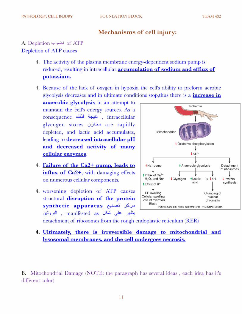
4. The activity of the plasma membrane energy-dependent sodium pump is reduced, resulting in intracellular accumulation of sodium and efflux of potassium.
4. Because of the lack of oxygen in hypoxia the cell's ability to preform aerobic glycolysis decreases and in ultimate conditions stop,thus there is a increase in anaerobic glycolysis in an attempt to maintain the cell's energy sources. As a consequence نتيجة لذلك , intracellular glycogen stores مخازن are rapidly depleted, and lactic acid accumulates, leading to decreased intracellular pH and decreased activity of many cellular enzymes.
4. Failure of the Ca2+ pump, leads to influx of Ca2+, with damaging effects on numerous cellular components.
4. worsening depletion of ATP causes structural disruption of the protein synthetic apparatus مركز تصنيع يظهر على شكل manifested as , البروتينdetachment of ribosomes from the rough endoplastic reticulum (RER)
4. Ultimately, there is irreversible damage to mitochondrial and lysosomal membranes, and the cell undergoes necrosis.
B. Mitochondrial Damage (NOTE: the paragraph has several ideas , each idea has it's different color)
!11

PATHOLOGY: CELL INJURY FOUNDATION BLOCK TEAM 432
Mitochondrial damage often results in the formation of a high-conductance channel in the mitochondrial membrane, called the mitochondrial permeability transition pore. The opening of this channel leads to the loss of mitochondrial membrane potential and pH changes, resulting in failure of oxidative phosphorylation and progressive depletion of ATP, culminating قد يصل in n e c r o s i s o f t h e c e l l . The mitochondria also contain several proteins that are capable of activating apoptotic pathways, including cytochrome c (the major protein involved in electron transport). Increased permeability of the mitochondrial membrane may result in leakage of these proteins into the cytosol and death by apoptosis. Thus, cytochrome c plays a key dual role in cell survival and death; in its normal location inside mitochondria, it is essential for energy generation and the life of the cell, but when mitochondria are damaged so severely that cytochrome c l e a k s o u t , i t s i g n a l s c e l l s t o d i e . Mitochondria can be damaged by : (IMP) a. Increases of cytosolic Ca2+ b. Oxidative stress c. Breakdown of phospholipids d. Lipid breakdown products.
C. Influx of Intracellular Calcium & loss of Calcium Homeostasis Ischemia causes an increase in cytosolic calcium concentration due to the damage of calcium pump
D. Accumulation of Oxygen derived free Radicals
!12

PATHOLOGY: CELL INJURY FOUNDATION BLOCK TEAM 432
Free radicals production
Free radicals are chemical species with a single unpaired electron in an outer orbital. These chemicals are extremely unstable, and rapidly react with organic & inorganic compound. Free radicals avidly strike nucleic acids as well as a بسرعة ونهمvariety of other compounds causing severe damages. The molecules that react with free molecules also turn into free radicals propaganding ًمضخمة the chain of damage .
(ROS)
Reactive oxygen species (ROS) are a type of oxygen-derived free radical whose role in cell injury is well establish, they are produced normally during the cell in the mitochondrial respiration & energy generation . The cell bets rid of these free radicals normally by cellular defenses. When there is an excessive production of (ROS) or when the cell is not able to get rid of theme , they damage the cell.
The sources of free radicals are:- • Normal metabolism
• Chemical toxicity
• Reperfusion injury: ischemia -----> blood supply returns -----> oxygen returns -----> free radicals form.
Reperfusion injury is the tissue damage caused when blood supply returns to the tissue after a period of ischemia or lack of oxygen. The absence of oxygen and nutrients from blood during the ischemic period creates a condition in which the restoration of circulation results in inflammation and oxidative damage through the induction of oxidative stress rather than restoration of normal function.
For more info: http://en.wikipedia.org/wiki/Reperfusion_injury
• Ionizing radiation
• O2 therapy : excessive intake of O2 is toxic and causes injury.
• Immune response\ inflammation
!13

PATHOLOGY: CELL INJURY FOUNDATION BLOCK TEAM 432
Ultra structural signs of cell injury:
1. Seen in both reversible and irreversible cell injury
A. Cellular swelling\ Diminished activity of the sodium pump in the cell membrane causes an influx of sodium .
B. Mitochondrial swelling\ results in reduced aerobic respiration
C. Dilatation and degranulation of the rough endoplasmic reticulum\ results incessation of protein synthesis
D. Autophagocytosis\ is the ingestion of damaged organelles by lysosomes
2. Seen only in irreversible cell injury
A. Cell membrane rupture
B. Nuclear changes
!14

PATHOLOGY: CELL INJURY FOUNDATION BLOCK TEAM 432
Cell Death
Recall: When the injury becomes irreversible, at that time the cell cannot recover , thus it dies, this is called (cell death).
1. Apoptosis: is programmed cell death (single cell death) which is commanded by genes inside the cell, and it happens due to physiologic or pathologic events.
In case of cancer, the cells don't undergo apoptosis due to a translocation or a change of the structure of the chromosomes which will create a new gene called bcl2 which is anti-apoptosis (switch off apoptosis) and it will enable the cell to multiply indefinitely.
A. Examples of physiologic (normal) apoptosis include the:
1- (picture) Programmed death of embryonic cells (in the embryonic life) in the limb buds leading to the formation of fingers and toes (this is useful)
2- Hormone-induced cell death of endometrial cells at the end of the menstrual cycle.
3- During lactation in the female, the breast becomes big and the glands become hyperplastic and big and they produce milk. To stop lactation it has to undergo apoptosis therefore the breast shrink (becomes small) and the glands disappear.
4- To eliminate immune cells after cytokine depletion and auto reactive T-cells in developing thymus.
B. Examples of pathologic apoptosis include:
1- (picture) Hepatitis virus-induced liver cell apoptosis (acidophilic bodies)
2- Immune injury-related skin kerationcytes (civatte
!15

PATHOLOGY: CELL INJURY FOUNDATION BLOCK TEAM 432
bodies)
3- Corticosteroid-induced atrophy of the neonatal thymus.
2. Necrosis: is a morphologic sign of cell death in a living tissue.
Types of necrosis:
1- Coagulative necrosis is caused by hypoxemia and ischemia and it causes myocardial infarction (coagulative necrosis affects mainly the heart, the lung, the spleen and the kidneys)
Character : Cells' shape is maintained even after it's death.
2- Liquifactive necrosis has two good examples (brain and abscess) and it caused by hypoxia to the brain.
Characterr:part of the tissue appear as liquid (no cells)
3- Caseous necrosis is typically seen in tuberculosis (happens mainly in the lung or the lymph node) Histologically, it consists of granular material surrounded by epithelioid and multinucleated giant cells.
Character: the tissue appears cheesy.
4- Fibrinoid necrosis is seen usually in the kidney
!16

PATHOLOGY: CELL INJURY FOUNDATION BLOCK TEAM 432
and the blood vessels. It is immunologically mediated and seen in auto-immune diseases. Blood vessels are impregnated by fibrin and other serum proteins and appear magenta-red in histologic sections.
Character: Blood vessels are impregnated by fibrin
5- Fat necrosis happens in the organs which are rich in fat.
In case of acute pancreatitis, the enzymes (lipolytic enzymes like amylase and lipase) in the necrotic pancreatic cells are released to the abdominal cavity and start to digest the fat. Free fatty acid released from fat cells bind with calcium to form white specks or streaks composed of calcium soaps.
Character: white specks نقاط or streaks شريط composed of calcium soaps.
Differences between apoptosis and necrosis:
!17

PATHOLOGY: CELL INJURY FOUNDATION BLOCK TEAM 432
Remember: There are some proteins that spread during certain injuries .
These proteins help us detect the type of injury .
SUMMARY
If there is any stress there will be adaptation.
If the stress is over exceeded and the cell can't adapt there will be injury.
If the injury is to severe , it causes cell death.
Proteins used in diagnosis of tissue damage by blood testing.
Cell damaged Enzyme elevated in blood
Cardiac muscle 1. cardiac troponin
2. Creatine kinase
Hepatocyte 1. alanine transaminase (ALT)
2. Aspartate tansaminase (AST)
Striated muscles Creatine kinase
Exocrine pancreas Amylase
!18

PATHOLOGY: CELL INJURY FOUNDATION BLOCK TEAM 432
Accumulation occurs when abnormal amounts of substances "such as: Fatty acids, pigments, calcium" accumulate !"#$% in the cell under physiological or pathological process.
It can be: HARMFUL OR ASSOCIATED WITH VARYING DEGREE OF INJURY.
"note: Accumulation occurs after cell injury, while the cell is still alive. It might lead to necrosis"
1- Cytoplasm 2- Nucleus 3- Organelles
A.Endogenous: occurs by synthesis of affected cells
B.Exogenous: produced elsewhere "Outside the cell"
1- Metabolism change. example: Fatty liver.
!19
Accumulation:
Accumulation can be found in:
Accumulation is classified:
Mechanisms that lead to accumulation:

PATHOLOGY: CELL INJURY FOUNDATION BLOCK TEAM 432
2-Protein deficiency example: folding in abnormal way leads to accumulation of abnormal proteins
3- Lack of certain enzymes that participate in metabolism.
Complex substance Insoluble material
Example: Lysosomes will store insoluble endogenous materials in the absence of a certain enzyme. "Storage diseases"
4-Accumulation of ingested indigested materials by the cells "Cannot be digested or exit
!20
NO ENZYME
Fatty change

PATHOLOGY: CELL INJURY FOUNDATION BLOCK TEAM 432
It's any abnormal accumulation of triglycerides within parenchymal cells. "Such as: Hepatocytes and Mayocytes" It's usually an early indicator of cell stress and reversible injury.
-Severe steatosis in liver ! Inflammation ! Fibrosis ! Necrosis
-Steatosis is reversible until it reaches to fibrosis stage; it becomes IRREVERSIBLE because it's hard to remove fibers.
It's most common in the liver because it has the central role of fat metabolism.
It might also occur in heart (resulting from Anemia or Starvation "Anorexia nervosa")
Other sites: Skeletal muscle, kidney and other organs.
1-Toxins "most importantly: Alcohol abuse"(chronic alcoholism).
2-Diabetes mellitus 3-Protein malnutrition "starvation"
4-Obesity 5-Anoxia
!21
Sites of Steatosis:
Causes of Steatosis:-
PATHOLOGY: CELL INJURY FOUNDATION BLOCK TEAM 432
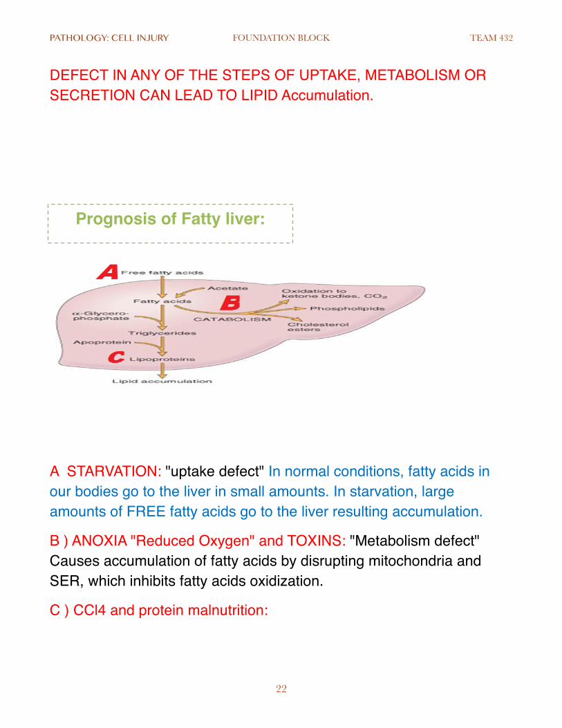
DEFECT IN ANY OF THE STEPS OF UPTAKE, METABOLISM OR SECRETION CAN LEAD TO LIPID Accumulation.
�
A STARVATION: "uptake defect" In normal conditions, fatty acids in our bodies go to the liver in small amounts. In starvation, large amounts of FREE fatty acids go to the liver resulting accumulation.
B ) ANOXIA "Reduced Oxygen" and TOXINS: "Metabolism defect" Causes accumulation of fatty acids by disrupting mitochondria and SER, which inhibits fatty acids oxidization.
C ) CCl4 and protein malnutrition:
!22
Prognosis of Fatty liver:

PATHOLOGY: CELL INJURY FOUNDATION BLOCK TEAM 432
Fatty acids change can be:- MILD: has no effect on cellular function
SEVERE: may transiently impair cellular function
Fatty acids accumulate together and push cells' nucleus peripherally &'(&) , occasionally they rupture together and form fatty cysts.
Sign of Early stages : small fat vacuoles in the cytoplasm around the nucleus.
Signs of Later stages: the vacuoles coalesce !"$#%و +,$% to create cleared spaces that displace the nucleus to the cell periphery
Occasionally: contiguous cells rupture (fatty cysts)
Is Fatty liver reversible? Fatty change is reversible except if some vital intracellular process is irreversibly impaired (e.g. in CCl4 poisoning)
•Mild: benign natural history (approximately 3% will develop cirrhosis
•Moderate to severe: inflammation, degeneration in hepatocytes, +/- fibrosis (30% develop cirrhosis)
•5 to 10 year survival:67% and 59%
!23
Stages of fatty change:
Other forms of accumulations:

PATHOLOGY: CELL INJURY FOUNDATION BLOCK TEAM 432
Cholesteryl esters: These give atherosclerotic plaques their characteristic yellow color and contribute to the pathogenesis of the lesion. This is called atherosclerosis
�
Pigments are colored substances that are either:
1.Exogenous coming from outside the body.
2.Endogenous synthesized within the body itself.
!24
Pigments:
1-Exogenous pigments:-

PATHOLOGY: CELL INJURY FOUNDATION BLOCK TEAM 432
They may be toxic and produce inflammatory tissue reactions or they may be relatively inert -./,05 %4$23! ا.
Examples:
A ) Indian ink pigments
produce effective tattoos because they are engulfed by dermal macrophages which become immobilized and permanently deposited.
B ) The most common exogenous pigment is CARBON
When inhaled, it is phagocytosed by alveolar macrophages and transported through lymphatic channels to the regional tracheobronchial lymph nodes. It causes (heavy accumulation that leads to fibrosis or anthracsis).
-Heavy accumulations may induce fibrosis. "This might happen to coal workers pneumoconiosis".
-Aggregates of the pigment blacken the draining lymph nodes and pulmonary parenchyma (anthracosis).
They're synthesized within the body itself.
Examples:
-Hemosidren
-Melanin
-Lipofuscin
!25
2-Endogenous pigments:-
PATHOLOGY: CELL INJURY FOUNDATION BLOCK TEAM 432
HEMOSIDREN:-
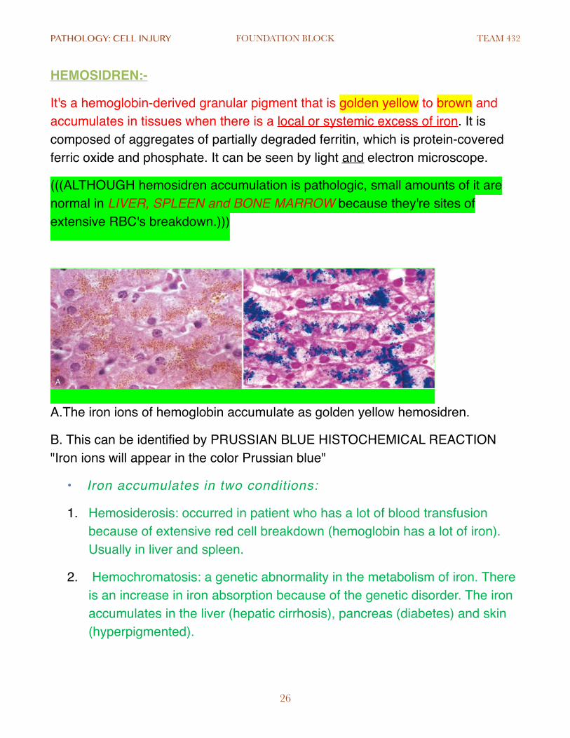
It's a hemoglobin-derived granular pigment that is golden yellow to brown and accumulates in tissues when there is a local or systemic excess of iron. It is composed of aggregates of partially degraded ferritin, which is protein-covered ferric oxide and phosphate. It can be seen by light and electron microscope.
(((ALTHOUGH hemosidren accumulation is pathologic, small amounts of it are normal in LIVER, SPLEEN and BONE MARROW because they're sites of extensive RBC's breakdown.)))
�
A.The iron ions of hemoglobin accumulate as golden yellow hemosidren.
B. This can be identified by PRUSSIAN BLUE HISTOCHEMICAL REACTION "Iron ions will appear in the color Prussian blue"
• Iron accumulates in two conditions:
1. Hemosiderosis: occurred in patient who has a lot of blood transfusion because of extensive red cell breakdown (hemoglobin has a lot of iron). Usually in liver and spleen.
2. Hemochromatosis: a genetic abnormality in the metabolism of iron. There is an increase in iron absorption because of the genetic disorder. The iron accumulates in the liver (hepatic cirrhosis), pancreas (diabetes) and skin (hyperpigmented).
!26

PATHOLOGY: CELL INJURY FOUNDATION BLOCK TEAM 432
Hemosidren Lipofuscin (wear-and-tear
pigment)
Melanin
A hemoglobin-derived granular pigment Golden yellow to brown (identified by the Prussian blue histochemical reaction ) accumulates in tissues when there is excess of iron. composed of ferritin ( protein -covered ferric oxide and phosphate) small amounts of this pigment are normal It's occur in the mononuclear phagocytes of the : bone marrow spleen & liver. Because of extensive red cell breakdown . Local excesses of iron and consequently of hemosiderin, result from hemorrhage (نزيف ( الدمBruise . systemic overload of iron . Hemosiderosis occurs in the setting of: increased absorption of dietary iron . impaired utilization of iron. hemolytic anemias . A lot of blood transfusions. In systemic hemosiderosis, parenchymal cells are not damaged . Extensive accumulations of iron are seen in hereditary hemochromatosis Including tissue injury like: liver fibrosis heart failure & diabetes mellitus. Spleen.
an insoluble brownish-yellow granular intracellular material seen in the heart liver & brain as a function of age or atrophy.(old people) (Brown atrophy) By electron microscopy >>> seen as perinuclear electron-dense .
brown/black pigment normally present in melanocytes. Function : to block harmful UV rays from the epidermal nuclei. May accumulate in : benign or malignant (حميدة أو خبيثة)melanocytic neoplasms ( أورام صبغيه ) Melanin presence is a useful diagnostic feature melanocytic lesions (لتشخيص الآلفات ( الصبغية Post-inflammatory pigmentation of the skin. It causes melanosis coli in small intestine.
!27

PATHOLOGY: CELL INJURY FOUNDATION BLOCK TEAM 432
It accumulates in the liver, muscles or kidneys in patients with inborn errors of glycogen metabolism or diabetes mellitus. Degeneration : describing pathological processes which result in deterioration of tissues and organs.
• PLEASE , MAKE SURE YOU SEE THE EXAMPLES IN THE LECTURE !
!28
Glycogen:

PATHOLOGY: CELL INJURY FOUNDATION BLOCK TEAM 432
Pathologic calcification
abnormal deposition of calcium salts with smaller amounts of :
• iron
• magnesium
• and other minerals.
Dystrophic calcification Metastatic calcification In normal tissues
normal serum levels of calcium Encountered in areas of necrosis tuberculous lymph node >>> radio-opaque lesion (due to calcification)
atherosclerosis + Intiman injury in the aorta & large arteries It's may be an incidental finding indicating insignificant past cell injury Also cause an organ dysfunction. (aortic stenosis in the elderly )
Hypercalcemia Main causes of hypercalcemia 1.increased secretion of parathyroid hormone (calcitonin hormone which control absorption of calcium from the intestine). 2. High calcium level in blood. 3. destruction of bone due to the effects of accelerated turnover (Paget disease) immobilization or tumors (multiple myeloma, leukemia, or diffuse skeletal metastases) 4. vitamin D-related disorders including vitamin D intoxication & sarcoidosis 5. renal failure, in which phosphate retention leads to secondary hyperparathyroidism. In normal tissues
!29

PATHOLOGY: CELL INJURY FOUNDATION BLOCK TEAM 432
Morphology of calcification:
Causes of Hypercalcemia
1.Hyperparathyroidism.
2.Metastatic malignant tumours.
3.Vitamin D intoxication.
4.Milk alkali syndrome.
5.Mild hypercalcemia can be seen
in old age.
!30
Morphology of Dystrophic calcification
Morphology of Metastatic calcification
calcium salts seen as fine white granules or clumps, often felt as gritty deposits.
Histologically, Calcification appears as intracellular and/or extracellular basophilic deposits.
heterotopic bone may be formed in the focus of calcification
occur widely throughout the body but principally affects the interstitial tissues of the vasculature kidneys lungs and gastric mucosa The calcium deposits morphologically resemble those described in dystrophic calcification
Extensive calcifications in the lungs may produce remarkable respiratory deficits Massive deposits in the kidney (nephrocalcinosis) can cause renal damage.

PATHOLOGY: CELL INJURY FOUNDATION BLOCK TEAM 432
•
• AL Amyloid : tumor of plasma cell, (amyloid accumulate in tissue and wall of blood vessel)
• AA Amyloid : chronic inflammatory condition, (secreted by the liver. It can accumulate in blood vessels, kidney, liver, spleen and many organs).
!31
Common causes and consequences of Amyloidosis
PATHOLOGY: CELL INJURY FOUNDATION BLOCK TEAM 432
Good luck ^_^
Because no human work is perfect please update us
with your feed back on the e-mail written below
We will be very glad to correct our mistakes & add
any missing info ...
Our goal is to build your knowledge & help you
improve your studying ...
E-mail :
Note: We worked our best to make the OBJECTIVES part your first & last stop so:
1. We summarized all main ideas , so that you can
remember them easily in your final revision.
2. You can notice that after some objectives we put key
words between brackets , these are to help you recall
definitions and important notes.
Accept our best regards...432 PATHOLOGY team
!32


![[5] HEMA - Megaloblastic Anemia.pdf - KSUMSC](https://static.fdokumen.com/doc/165x107/631deac95ff22fc7450674ca/5-hema-megaloblastic-anemiapdf-ksumsc.jpg)