Ovine Fetal Thymus Response to Lipopolysaccharide-Induced Chorioamnionitis and Antenatal...
10
Ovine Fetal Thymus Response to Lipopolysaccharide- Induced Chorioamnionitis and Antenatal Corticosteroids Elke Kuypers 1 , Jennifer J. P. Collins 1 , Reint K. Jellema 1 , Tim G. A. M. Wolfs 1 , Matthew W. Kemp 2 , Ilias Nitsos 2¤ , J. Jane Pillow 2 , Graeme R. Polglase 2¤ , John P. Newnham 2 , Wilfred T. V. Germeraad 3 , Suhas G. Kallapur 2,4 , Alan H. Jobe 2,4 , Boris W. Kramer 1 * 1 Department of Pediatrics, Maastricht University Medical Center, Maastricht, The Netherlands, 2 School of Women’s and Infants’ Health, The University of Western Australia, Perth, Australia, 3 Department of Internal Medicine, Division of Haematology, Maastricht University Medical Center, Maastricht, The Netherlands, 4 Division of Pulmonary Biology, Cincinnati Children’s Hospital Medical Center, University of Cincinnati, Cincinnati, Ohio, United States of America Abstract Rationale: Chorioamnionitis is associated with preterm delivery and involution of the fetal thymus. Women at risk of preterm delivery receive antenatal corticosteroids which accelerate fetal lung maturation and improve neonatal outcome. However, the effects of antenatal corticosteroids on the fetal thymus in the settings of chorioamnionitis are largely unknown. We hypothesized that intra-amniotic exposure to lipopolysaccharide (LPS) causes involution of the fetal thymus resulting in persistent effects on thymic structure and cell populations. We also hypothesized that antenatal corticosteroids may modulate the effects of LPS on thymic development. Methods: Time-mated ewes with singleton fetuses received an intra-amniotic injection of LPS 7 or 14 days before preterm delivery at 120 days gestational age (term = 150 days). LPS and corticosteroid treatment groups received intra-amniotic LPS either preceding or following maternal intra-muscular betamethasone. Gestation matched controls received intra-amniotic and maternal intra-muscular saline. The fetal intra-thoracic thymus was evaluated. Results: Intra-amniotic LPS decreased the cortico-medullary (C/M) ratio of the thymus and increased Toll-like receptor (TLR) 4 mRNA and CD3 expression indicating involution and activation of the fetal thymus. Increased TLR4 and CD3 expression persisted for 14 days but Foxp3 expression decreased suggesting a change in regulatory T-cells. Sonic hedgehog and bone morphogenetic protein 4 mRNA, which are negative regulators of T-cell development, decreased in response to intra- amniotic LPS. Betamethasone treatment before LPS exposure attenuated some of the LPS-induced thymic responses but increased cleaved caspase-3 expression and decreased the C/M ratio. Betamethasone treatment after LPS exposure did not prevent the LPS-induced thymic changes. Conclusion: Intra-amniotic exposure to LPS activated the fetal thymus which was accompanied by structural changes. Treatment with antenatal corticosteroids before LPS partially attenuated the LPS-induced effects but increased apoptosis in the fetal thymus. Corticosteroid administration after the inflammatory stimulus did not inhibit the LPS effects on the fetal thymus. Citation: Kuypers E, Collins JJP, Jellema RK, Wolfs TGAM, Kemp MW, et al. (2012) Ovine Fetal Thymus Response to Lipopolysaccharide-Induced Chorioamnionitis and Antenatal Corticosteroids. PLoS ONE 7(5): e38257. doi:10.1371/journal.pone.0038257 Editor: Rory Edward Morty, University of Giessen Lung Center, Germany Received March 5, 2012; Accepted May 2, 2012; Published May 31, 2012 Copyright: ß 2012 Kuypers et al. This is an open-access article distributed under the terms of the Creative Commons Attribution License, which permits unrestricted use, distribution, and reproduction in any medium, provided the original author and source are credited. Funding: Supported by NIH HD-57869 (SGK) from the National Institutes of Health (NIH), United States of America, the National Health and Medical Research Council of Australia, the Women and Infants Research Foundation, Western Australia, Veni BWK 016.096.141 from the Dutch Scientific Research Organization and the Research School for Oncology and Developmental Biology (GROW), Maastricht University. The funders had no role in study design, data collection and analysis, decision to publish, or preparation of the manuscript. Competing Interests: The authors have declared that no competing interests exist. * E-mail: [email protected] ¤ Current address: The Ritchie Centre, Monash Institute of Medical Research, Melbourne, Australia Introduction Preterm birth is the leading cause of morbidity and mortality in the neonatal period [1]. In the developed world, the majority of women at risk of preterm birth receive antenatal corticosteroids to induce lung maturation and decrease infant mortality [2]. This therapy is given irrespective of the presence of an intra-uterine infection of the amniotic fluid and placental membranes (chorio- amnionitis). Chorioamnionitis is present in up to 60% of preterm births and is highly associated with adverse neonatal outcomes [3]. In the majority of preterm births, chorioamnionitis is clinically silent prior to early gestational preterm labor [3]. As a result, many preterm infants are exposed to both chorioamnionitis and antenatal corticosteroids. Exposure to intra-uterine infection may increase the risk for respiratory and neurological complications in later life [4,5]. Intra- amniotic lipopolysaccharide (LPS)-induced chorioamnionitis caus- es lung [6,7], gut [8] and skin [9] inflammation in preterm lambs, which demonstrates that chorioamnionitis causes a ‘multi-organ disease of the fetus’ [10]. PLoS ONE | www.plosone.org 1 May 2012 | Volume 7 | Issue 5 | e38257
-
Upload
independent -
Category
Documents
-
view
1 -
download
0
Transcript of Ovine Fetal Thymus Response to Lipopolysaccharide-Induced Chorioamnionitis and Antenatal...

Ovine Fetal Thymus Response to Lipopolysaccharide-Induced Chorioamnionitis and Antenatal CorticosteroidsElke Kuypers1, Jennifer J. P. Collins1, Reint K. Jellema1, Tim G. A. M. Wolfs1, Matthew W. Kemp2,
Ilias Nitsos2¤, J. Jane Pillow2, Graeme R. Polglase2¤, John P. Newnham2, Wilfred T. V. Germeraad3,
Suhas G. Kallapur2,4, Alan H. Jobe2,4, Boris W. Kramer1*
1 Department of Pediatrics, Maastricht University Medical Center, Maastricht, The Netherlands, 2 School of Women’s and Infants’ Health, The University of Western
Australia, Perth, Australia, 3 Department of Internal Medicine, Division of Haematology, Maastricht University Medical Center, Maastricht, The Netherlands, 4 Division of
Pulmonary Biology, Cincinnati Children’s Hospital Medical Center, University of Cincinnati, Cincinnati, Ohio, United States of America
Abstract
Rationale: Chorioamnionitis is associated with preterm delivery and involution of the fetal thymus. Women at risk ofpreterm delivery receive antenatal corticosteroids which accelerate fetal lung maturation and improve neonatal outcome.However, the effects of antenatal corticosteroids on the fetal thymus in the settings of chorioamnionitis are largelyunknown. We hypothesized that intra-amniotic exposure to lipopolysaccharide (LPS) causes involution of the fetal thymusresulting in persistent effects on thymic structure and cell populations. We also hypothesized that antenatal corticosteroidsmay modulate the effects of LPS on thymic development.
Methods: Time-mated ewes with singleton fetuses received an intra-amniotic injection of LPS 7 or 14 days before pretermdelivery at 120 days gestational age (term = 150 days). LPS and corticosteroid treatment groups received intra-amniotic LPSeither preceding or following maternal intra-muscular betamethasone. Gestation matched controls received intra-amnioticand maternal intra-muscular saline. The fetal intra-thoracic thymus was evaluated.
Results: Intra-amniotic LPS decreased the cortico-medullary (C/M) ratio of the thymus and increased Toll-like receptor (TLR) 4mRNA and CD3 expression indicating involution and activation of the fetal thymus. Increased TLR4 and CD3 expressionpersisted for 14 days but Foxp3 expression decreased suggesting a change in regulatory T-cells. Sonic hedgehog and bonemorphogenetic protein 4 mRNA, which are negative regulators of T-cell development, decreased in response to intra-amniotic LPS. Betamethasone treatment before LPS exposure attenuated some of the LPS-induced thymic responses butincreased cleaved caspase-3 expression and decreased the C/M ratio. Betamethasone treatment after LPS exposure did notprevent the LPS-induced thymic changes.
Conclusion: Intra-amniotic exposure to LPS activated the fetal thymus which was accompanied by structural changes.Treatment with antenatal corticosteroids before LPS partially attenuated the LPS-induced effects but increased apoptosis inthe fetal thymus. Corticosteroid administration after the inflammatory stimulus did not inhibit the LPS effects on the fetalthymus.
Citation: Kuypers E, Collins JJP, Jellema RK, Wolfs TGAM, Kemp MW, et al. (2012) Ovine Fetal Thymus Response to Lipopolysaccharide-Induced Chorioamnionitisand Antenatal Corticosteroids. PLoS ONE 7(5): e38257. doi:10.1371/journal.pone.0038257
Editor: Rory Edward Morty, University of Giessen Lung Center, Germany
Received March 5, 2012; Accepted May 2, 2012; Published May 31, 2012
Copyright: � 2012 Kuypers et al. This is an open-access article distributed under the terms of the Creative Commons Attribution License, which permitsunrestricted use, distribution, and reproduction in any medium, provided the original author and source are credited.
Funding: Supported by NIH HD-57869 (SGK) from the National Institutes of Health (NIH), United States of America, the National Health and Medical ResearchCouncil of Australia, the Women and Infants Research Foundation, Western Australia, Veni BWK 016.096.141 from the Dutch Scientific Research Organization andthe Research School for Oncology and Developmental Biology (GROW), Maastricht University. The funders had no role in study design, data collection andanalysis, decision to publish, or preparation of the manuscript.
Competing Interests: The authors have declared that no competing interests exist.
* E-mail: [email protected]
¤ Current address: The Ritchie Centre, Monash Institute of Medical Research, Melbourne, Australia
Introduction
Preterm birth is the leading cause of morbidity and mortality in
the neonatal period [1]. In the developed world, the majority of
women at risk of preterm birth receive antenatal corticosteroids to
induce lung maturation and decrease infant mortality [2]. This
therapy is given irrespective of the presence of an intra-uterine
infection of the amniotic fluid and placental membranes (chorio-
amnionitis). Chorioamnionitis is present in up to 60% of preterm
births and is highly associated with adverse neonatal outcomes [3].
In the majority of preterm births, chorioamnionitis is clinically
silent prior to early gestational preterm labor [3]. As a result, many
preterm infants are exposed to both chorioamnionitis and
antenatal corticosteroids.
Exposure to intra-uterine infection may increase the risk for
respiratory and neurological complications in later life [4,5]. Intra-
amniotic lipopolysaccharide (LPS)-induced chorioamnionitis caus-
es lung [6,7], gut [8] and skin [9] inflammation in preterm lambs,
which demonstrates that chorioamnionitis causes a ‘multi-organ
disease of the fetus’ [10].
PLoS ONE | www.plosone.org 1 May 2012 | Volume 7 | Issue 5 | e38257

The concept of fetal and early life origins of disease has
developed from epidemiological studies, which correlate fetal and
maternal exposures during gestation to outcomes in childhood
such as asthma [11]. The pathogenesis of some diseases may result
from altered T-cell immunity during fetal development [12]. The
net outcome of pro-inflammation effects from chorioamnionitis
and anti-inflammation effects from antenatal corticosteroids
remain unstudied. As such, there is minimal information about
how the fetal thymus responds to these clinically relevant
exposures [13].
The thymus is the primary site for T-cell development [14].
Immature T-cells migrate from the cortico-medullary junction,
move through the thymic cortex to the medullary compartment.
During this migration, the immature T-cells proliferate greatly,
alter antigen expression and rearrange their T-cell receptor
expression [14]. Previous studies demonstrated that chorioamni-
onitis interferes with the development of the fetal thymus [15,16].
In an ovine model of chorioamnionitis, Kunzmann et al. [17]
showed that intra-amniotic LPS decreased the fetal thymus/body
weight ratio and decreased thymic Foxp3 expression. However,
the combined effects of chorioamnionitis and antenatal cortico-
steroids on fetal thymic development remain to be characterized
[18].
Sonic hedgehog (Shh) and Bone morphogenetic protein (BMP)
pathways participate in T-cell development and are sensitive to
prenatal events such as exposure to toxins [19–21]. Both
morphogens are produced and secreted by the thymic epithelium
as negative regulators of T-cell differentiation to maintain a pool of
undifferentiated, precursor T-cells in the thymus [22,23]. We
hypothesized that intra-amniotic exposure to LPS causes involu-
tion of the fetal thymus and modulation of Shh and BMP4
expression with persistent effects on thymic structure and cell
populations. We also hypothesized that antenatal corticosteroids
may modulate the effects of LPS on thymic development.
Therefore, we exposed fetal sheep sequentially to intra-amniotic
LPS and/or antenatal corticosteroids at 7-day intervals [24] and
evaluated multiple indicators of thymic development. An interval
of 7 days between the two interventions was chosen as
representative of the interval between recognition of preterm
labor and delivery for many women who deliver preterm and the
probability that many early gestation fetal exposures to infection
are chronic [3,25], and that repeated administration of antenatal
corticosteroids are given at weekly intervals [26].
Materials and Methods
Animal studyThe animal experiments for this study were performed in
Western Australia and were approved by the Animal Ethics
Committees at The University of Western Australia (animal ethics
protocol RA/3/100/830) and Cincinnati Children’s Hospital
Medical Center. Time-mated ewes with singleton fetuses were
randomly allocated to one of six treatment groups to receive an
intra-amniotic (IA) injection of lipopolysaccharide (LPS) (10 mg
Escherichia Coli 055:B5, Sigma Chemical, St. Louis, MO, USA)
and/or an intra-muscular injection of betamethasone (Beta)
(Celestone Soluspan, Schering-Plough, North Ryde, New South
Wales (NSW), Australia, 0.5 mg/kg maternal weight) and/or an
equivalent injection of saline for control animals at 107 days and/
or 114 days gestation (GA) (Figure 1). All ewes received a single
intra-muscular injection of 150 mg medroxyprogesterone acetate
(Depo-Provera, Kenral, NSW, Australia) at 100 days GA to
decrease the risk of preterm birth induced by the betamethasone
treatment. Despite the medroxyprogesterone acetate treatment,
animals exposed to maternal betamethasone experienced fetal
losses, such that we reassigned animals from a group which
received betamethasone 14 days before delivery to other groups as
our priority was to test the interactions of betamethasone and LPS.
Lambs were delivered by cesarean section at 120 days GA
(term = 150 days GA) and euthanized after birth. Intra-thoracic
thymic tissue was snap frozen and fixed in 10% buffered-formalin
for 24 hours. The pulmonary inflammation and maturation
responses of these animals are reported elsewhere [7].
ImmunohistochemistryParaffin embedded thymic sections (4 mm, transverse) were
stained for CD3 (DAKO A0452, DAKO Denmark), Foxp3
(eBiosciences 14-7979, eBiosciences, San Diego, USA), bone
morphogenetic protein 4 (BMP4) (sc-6896, Santa Cruz Biotech-
nology, Santa Cruz, USA), cleaved caspase-3 (Asp175, #9661S,
Cell Signaling Technology, Boston, USA) and Ki67 (Dako,
M7240, DAKO Denmark). The sections were deparaffinized
and rehydrated in an ethanol series. Endogenous peroxidase-
activity was blocked by incubation with 0,3% H2O2 in phosphate
buffered saline (PBS, pH 7.4) (for CD3, BMP4 and Foxp3) or in
methanol (for cleaved caspase-3 and Ki67). Antigen retrieval was
performed by incubating the sections in heated citrate buffer
(10 mM, pH 6.0) for 30 minutes. Aspecific binding was blocked
by incubating slides for 30 minutes with 5% bovine serum
albumin (BSA) for CD3, 20% normal goat serum (NGS) for
Foxp3 and BMP4 or 5% NGS for Ki67. This step was omitted for
cleaved caspase-3. Slides were incubated overnight at 4uC with the
diluted primary antibody (CD3 1:200, Foxp3 1:30, BMP4 1:500,
cleaved caspase-3 1:400, Ki67 1:50) followed by incubation with a
secondary goat-anti-mouse (for Foxp3 and Ki67) or swine-anti-
rabbit (for CD3, BMP4 and cleaved caspase-3) biotin labeled
antibodies. The immunostaining was enhanced with Vectastain
ABC peroxidase Elite kit (PK-6200, Vector Laboratories,
Burlingame, USA) followed by a nickel sulfate-diaminobenzidine
(NiDAB) staining. Sections were counterstained with 0.1%
Nuclear Fast Red.
Evaluation was performed by light microscopy (Axioskop 40,
Zeiss, Germany) with LeicaQWin Pro v.3.4.0 software (Leica
Microsystems, Germany). CD3, Foxp3, Ki67 and BMP4 positive
staining were measured in three to five representative sections at
2006magnification by Image J software (Rasband, W.S., Image J
US National Institutes of Health, Bethesda, Maryland, USA).
Cleaved caspase-3 positive cells were counted in three represen-
tative high power fields at 2006 magnification by a blinded
observer and averaged per animal.
The morphology of the thymus was evaluated by light
microscopy after hematoxylin and eosin staining. The cortico-
medullary (C/M) ratio was quantified for three representative
sections from each animal at 2.56 magnification using Image J
software (Rasband, W.S.) [27].
RNA extraction and real-time PCRTotal RNA was extracted from frozen thymic tissue using the
SV Total RNA Isolation system (Z3100, Promega, Madison, USA)
according to the manufacturer’s instructions. Genomic DNA
contamination was removed by treatment with RQ1 DNase
(M610A, Promega) and the RNA was tested for the presence of
genomic GAPDH. Total RNA was reverse transcribed with the
First Strand cDNA synthesis kit (4379012001, Roche-Applied,
Mannheim, Germany) according to manufacturer’s instructions
using anchored oligo-primers. Primers for real-time PCR (RT-
PCR) were constructed based on published ovine or bovine cDNA
sequences (Table 1). RT-PCR reactions were performed in
Thymic Changes after Chorioamnionitis
PLoS ONE | www.plosone.org 2 May 2012 | Volume 7 | Issue 5 | e38257
duplicate with the LightCycler 480 SYBR Green I Master mix
(4707516001, Roche-Applied) on a LightCycler 480 Instrument
according to the manufacturer’s instructions. RT-PCR results
were normalized to ovRSP15, a housekeeping gene, and mean
fold changes in mRNA expression were calculated by the DDCt-
method [28].
Data analysisGroups were compared using one-way ANOVA with Dunnett’s
or Tukey’s test for post-hoc analysis or by a non-parametric
Kruskal-Wallis test as appropriate. Statistical analysis was
performed by GraphPad Prism v5.0. Significance was accepted
at p,0.05.
Results
Thymic cortico-medullary ratioThe cortico-medullary (C/M) ratio decreased significantly after
exposure to LPS for 7 days (Figure 2B) compared to controls
(Figure 2A). Betamethasone treatment 7 days prior to LPS
exposure did not attenuate the change in C/M ratio (Figure 2C).
Animals exposed to LPS 14 days before delivery and then exposed
to betamethasone had a reduced C/M ratio (Figure 2D) compared
to controls.
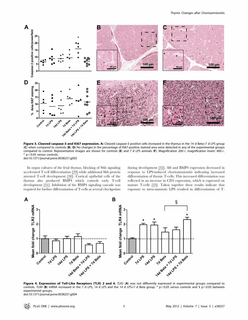
Proliferation and apoptosisCleaved caspase-3 positive cells, an indicator of apoptotic cells,
increased in the thymus of animals which received betamethasone
prior to the LPS exposure when compared to controls (Figure 3A).
No changes in Ki67 expression were detected in any of the
experimental groups compared to control (Figure 3D).
TLR expression in the fetal thymusTLR2 mRNA levels did not change in the experimental groups
compared to control (Figure 4A). TLR4 mRNA almost doubled in
animals which were exposed to LPS either 7 or 14 days before
delivery (Figure 4B). Treatment with betamethasone 7 days after
the LPS exposure did not attenuate the rise in TLR4 mRNA levels.
CD-3 positive thymic T-cellsThe percentage of CD-3 positive stained area increased
significantly 7 days (Figure 5B) and 14 days (Figure 5C) after
LPS exposure compared to controls (Figure 5A) and was primarily
located in the thymic medulla (Figure 5C). Betamethasone
treatment after the 14 day LPS exposure did not attenuate the
LPS-mediated increase in the percentage of CD3-positive stained
area of the thymus (Figure 5D).
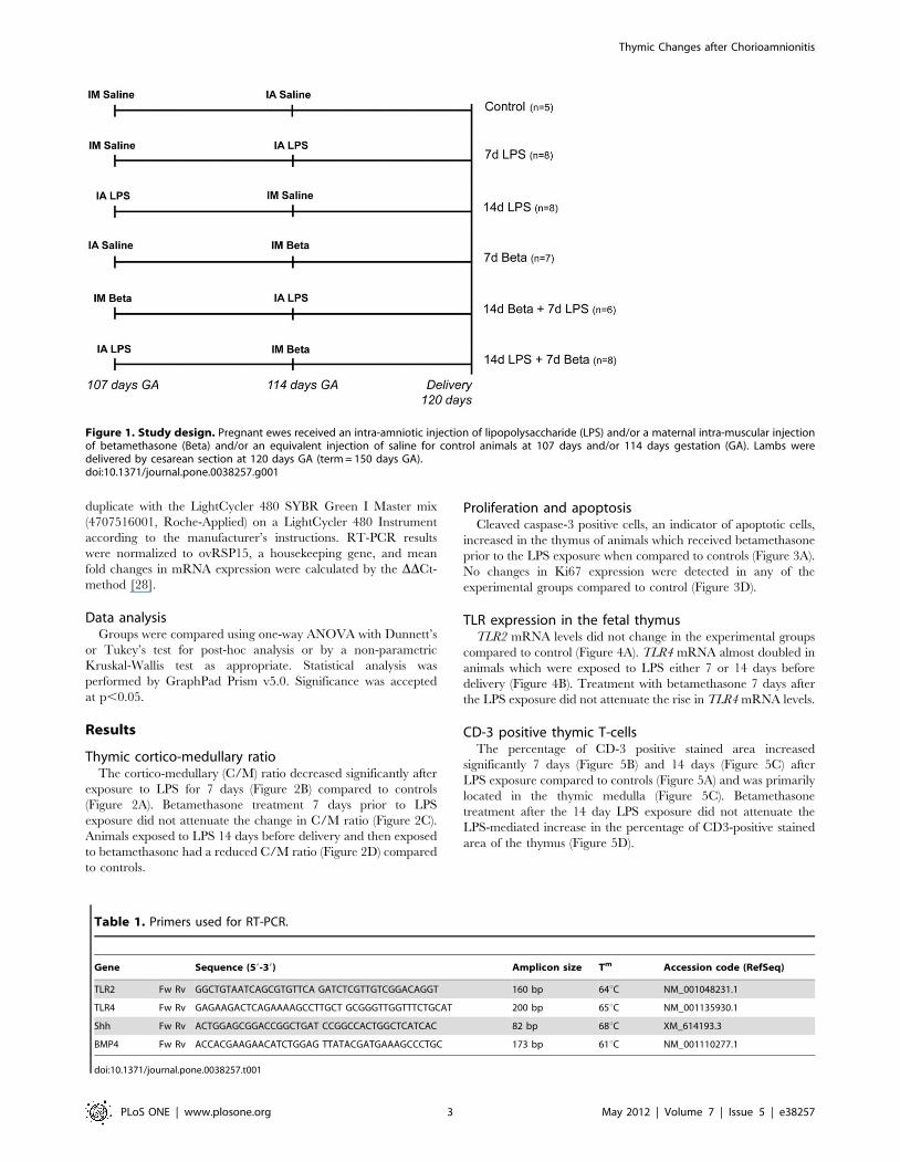
Figure 1. Study design. Pregnant ewes received an intra-amniotic injection of lipopolysaccharide (LPS) and/or a maternal intra-muscular injectionof betamethasone (Beta) and/or an equivalent injection of saline for control animals at 107 days and/or 114 days gestation (GA). Lambs weredelivered by cesarean section at 120 days GA (term = 150 days GA).doi:10.1371/journal.pone.0038257.g001
Table 1. Primers used for RT-PCR.
Gene Sequence (59-39) Amplicon size Tm Accession code (RefSeq)
TLR2 Fw Rv GGCTGTAATCAGCGTGTTCA GATCTCGTTGTCGGACAGGT 160 bp 64uC NM_001048231.1
TLR4 Fw Rv GAGAAGACTCAGAAAAGCCTTGCT GCGGGTTGGTTTCTGCAT 200 bp 65uC NM_001135930.1
Shh Fw Rv ACTGGAGCGGACCGGCTGAT CCGGCCACTGGCTCATCAC 82 bp 68uC XM_614193.3
BMP4 Fw Rv ACCACGAAGAACATCTGGAG TTATACGATGAAAGCCCTGC 173 bp 61uC NM_001110277.1
doi:10.1371/journal.pone.0038257.t001
Thymic Changes after Chorioamnionitis
PLoS ONE | www.plosone.org 3 May 2012 | Volume 7 | Issue 5 | e38257

Decreased Foxp3 expression in response to LPSThe percentage of Foxp3-positive stained area detected
primarily in the medulla, was decreased significantly 14 days after
LPS exposure (Figure 6B) compared to controls (Figure 6A)
irrespectively of betamethasone post-treatment (Figure 6C). Other
experimental groups did not show a change in thymic Foxp3
expression.
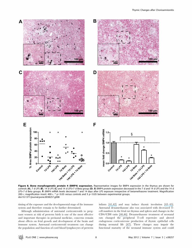
Shh and BMP4 expression in the thymusShh mRNA (Figure 7) decreased to about 20% of the control
value 7 and 14 days after LPS exposure. Similarly, BMP4
(Figure 8) mRNA and protein expression also decreased signifi-
cantly 7 and 14 days after exposure to LPS. Betamethasone
treatment before the exposure to LPS attenuated the decrease in
Shh mRNA levels and BMP4 protein. BMP4 mRNA but not
protein expression remained decreased in this group compared to
controls. The animals which received betamethasone treatment
after the LPS exposure still had decreased levels of Shh and BMP4
which were similar to the LPS effect alone.
Discussion
We investigated the responses of the fetal thymus to chorioam-
nionitis and antenatal corticosteroids, fetal exposures which are
common prior to very preterm delivery [1,3]. We found that intra-
amniotic exposure to LPS activated the fetal thymus as shown with
increased TLR4 mRNA levels and CD3 expression, decreased
Foxp3-positive cells and altered thymic structure.
Figure 2. Cortico-medullary ratio. The cortico-medullary (C/M) ratio of the thymus was measured using H&E sections. Representative images areshown for controls (A), 7 d LPS (B), 14 d betamethasone (Beta)+7 d LPS (C) and 14 d LPS+7 d Beta group (D). E: The C/M ratio decreased in the 7 dLPS group and the combined LPS and Beta groups. Red circled area: cortex, black circled area: medulla. Magnification: 406* p,0.05 versus controls.doi:10.1371/journal.pone.0038257.g002
Thymic Changes after Chorioamnionitis
PLoS ONE | www.plosone.org 4 May 2012 | Volume 7 | Issue 5 | e38257
In organ cultures of the fetal thymus, blocking of Shh signaling
accelerated T-cell differentiation [29] while additional Shh protein
arrested T-cell development [30]. Cortical epithelial cells of the
thymus also produced BMP4 which controls early T-cell
development [31]. Inhibition of the BMP4 signaling cascade was
required for further differentiation of T-cells at several checkpoints
during development [32]. Shh and BMP4 expression decreased in
response to LPS-induced chorioamnionitis indicating increased
differentiation of thymic T-cells. This increased differentiation was
reflected in an increase in CD3 expression, which is expressed on
mature T-cells [33]. Taken together these results indicate that
exposure to intra-amniotic LPS resulted in differentiation of T-
Figure 3. Cleaved caspase-3 and Ki67 expression. A: Cleaved caspase-3 positive cells increased in the thymus in the 14 d Beta+7 d LPS group(C) when compared to controls (B). D: No changes in the percentage of Ki67-positive stained area were detected in any of the experimental groupscompared to control. Representative images are shown for controls (E) and 7 d LPS animals (F). Magnification 2006; magnification insert: 4006.* p,0.05 versus controls.doi:10.1371/journal.pone.0038257.g003
Figure 4. Expression of Toll-Like Receptors (TLR) 2 and 4. TLR2 (A) was not differently expressed in experimental groups compared tocontrols. TLR4 (B) mRNA increased in the 7 d LPS, 14 d LPS and the 14 d LPS+7 d Beta group. * p,0.05 versus controls and 1 p,0.05 betweenexperimental groups.doi:10.1371/journal.pone.0038257.g004
Thymic Changes after Chorioamnionitis
PLoS ONE | www.plosone.org 5 May 2012 | Volume 7 | Issue 5 | e38257

cells with an accumulation of mature T-cells in the medulla and
depletion of early progenitor T-cells in the cortex, which was
consistent with the changed thymic structure.
Although the involution response of the fetal thymus has been
described in several human and animal studies [15,27], the
mechanistic changes behind this response remain unclear.
Kunzmann et al. [17] showed an acute thymic involution with
changes in Foxp3-positive cells in an ovine model of chorioam-
nionitis up to 5 days after exposure to LPS. Here, we further
characterized this process by demonstrating that the effects of LPS
on the thymic population and structure were detected 14 days
after the LPS exposure and were not due to changes in
proliferation or apoptosis. A persistent increase in medulla area
due to the accumulation of mature, differentiated T-cells may
explain the change in thymic structure.
Based on the anti-inflammatory properties of antenatal corti-
costeroids, a reduced inflammatory response after exposure to LPS
was expected [34]. Corticosteroids can exert anti-inflammatory
effects by upregulation of the IkB family, which are cytoplasmic
inhibitors of NF-kB, and by direct antagonism between the
glucocorticoid receptor and NF-kB, resulting in blocked tran-
scription of responsive genes. However, in our study betametha-
Figure 5. CD3-positive cells in the thymus. The percentage of CD3-positive stained area in the thymus was evaluated by immunohistochemistry.Representative images are shown for controls (A), 7 d LPS (B), 14 d LPS (C) and 14 d LPS+7 d Beta group (D). E: The percentage of CD3-positive areaincreased in the 7 d LPS, 14 d LPS and 14 d LPS+7 d Beta group. Magnification 2006; magnification insert: 4006. * p,0.05 versus controls.doi:10.1371/journal.pone.0038257.g005
Thymic Changes after Chorioamnionitis
PLoS ONE | www.plosone.org 6 May 2012 | Volume 7 | Issue 5 | e38257

sone administration after the inflammatory stimulus did not
reverse the LPS-induced increase in TLR4 and CD3 in the fetal
thymus. LPS has a half-life of 1.7 days in the amniotic fluid and
was still detectable 15 days after intra-amniotic injection [35].
Because of the slow clearance, LPS may induce a persistent
inflammatory response which is in line with measurements of
pulmonary inflammation in these animals [7].
Surprisingly, betamethasone administration 7 days before LPS
exposure attenuated activation with no signs of inflammation in
the fetal thymus. Thymic structure changed slightly due to the pro-
apoptotic properties of antenatal corticosteroids [36]. Previous
reports demonstrated only inhibitory effects of corticosteroids on
the immune system for a maximum of 48 hours [37,38]. Our
results indicate that the antenatal corticosteroids used clinically
can potentially desensitize the fetal immune system and attenuate
a response to LPS. Paradoxically, these ‘longer term’ inhibitory
effects of corticosteroids on the fetal immune system did not occur
in the animals that were exposed to LPS and then betamethasone
7 days later as the immune system remained activated. Cortico-
steroids are potent immune-modulatory hormones which can have
long term effects on the HPA-axis and subsequently on the
function of the immune system [39,40] which may be reflected in
the unresponsiveness of the fetal immune system to LPS after
corticosteroid pre-treatment. The longer-term effects of the
changes in cell composition and activation of the fetal thymus
after exposure to antenatal corticosteroids may depend on the
Figure 6. Foxp3-positive cells in the thymus. Representative images for Foxp3 expression in the thymus are shown for controls (A), 14 d LPS(B), and 14 d LPS+7 d Beta group (C). D: The percentage of Foxp3-positive stained area in the thymic medulla decreased in the animals exposed to14 days of LPS independent of Beta treatment. Magnification 2006; magnification insert: 4006. * p,0.05 versus controls.doi:10.1371/journal.pone.0038257.g006
Figure 7. Sonic Hedgehog (Shh) mRNA expression. The mRNAlevels of Shh were significantly decreased after 7 d and 14 d LPSexposures and in the 14 d LPS+7 d Beta group. * p,0.05 versuscontrols and 1 p,0.05 between experimental groups.doi:10.1371/journal.pone.0038257.g007
Thymic Changes after Chorioamnionitis
PLoS ONE | www.plosone.org 7 May 2012 | Volume 7 | Issue 5 | e38257
timing of the exposure and the developmental stage of the immune
system and therefore remain to be further determined.
Although administration of antenatal corticosteroids to preg-
nant women at risk of preterm birth is one of the most effective
and important therapies in perinatal medicine, concerns remain
about effects on fetal growth and development of the brain and
immune system. Antenatal corticosteroid treatment can change
the population and function of cord blood lymphocytes of preterm
infants [41,42] and may induce thymic involution [43–45].
Antenatal dexamethasone also was associated with decreased T-
cell numbers in the fetal rat thymus and spleen and changes in the
CD4/CD8 ratio [40,46]. Dexamethasone treatment of neonatal
rats changed the peripheral T-cell repertoire and altered
endogenous corticosterone production of thymic epithelial cells
during neonatal life [47]. These changes may impair the
functional maturity of the neonatal immune system and could
Figure 8. Bone morphogenetic protein 4 (BMP4) expression. Representative images for BMP4 expression in the thymus are shown forcontrols (A), 7 d LPS (B), 14 d LPS (C) and 14 d LPS+7 d Beta group (D). E: BMP4 protein expression decreased in the 7 d and 14 d LPS and the 14 dLPS+7 d Beta groups. F: BMP4 mRNA levels decreased 7 and 14 days after LPS exposure irrespective of betamethasone treatment. Magnification2006; magnification insert: 4006. * p,0.05 versus controls and 1 p,0.05 between experimental groups.doi:10.1371/journal.pone.0038257.g008
Thymic Changes after Chorioamnionitis
PLoS ONE | www.plosone.org 8 May 2012 | Volume 7 | Issue 5 | e38257

contribute to the increased incidence and adverse outcome of
infections [48,49].
Our findings contribute to the current concept that events
during fetal life can potentially alter the function of the immune
system [50]. The clinical associations between chorioamnionitis
and adverse outcomes in later life such as BPD [12] or asthma [51]
may be mediated in part by changes in immune responses.
In summary, our results demonstrate that fetal exposure to
intra-amniotic LPS activated the fetal thymus which was
accompanied by structural changes. Treatment with antenatal
corticosteroids before LPS partially attenuated the LPS-induced
effects but increased apoptosis in the fetal thymus. Corticosteroid
administration after the inflammatory stimulus did not inhibit the
LPS effects on the fetal thymus. However, insights into the effects
of LPS and corticosteroids on molecular pathways such as BMP4
and Shh are limited. Due to the low expression BMP4 and a lack
of specific reagents for Shh protein for ovine tissue, we were not
able to perform more detailed analysis of these pathways. Further
analysis at different time intervals of exposure are necessary to
better understand the interactive effects of chorioamnionitis and
corticosteroids on the fetal thymus. Although the design of the
study does not allow us to evaluate the dynamics of the changes
induced by LPS and corticosteroids, this report illustrates the
complicated interactions of pro- and anti-inflammatory stimuli on
the development of the fetal immune system.
Acknowledgments
We thank Richard Dalton, Joe Derwort, Masatoshi Saito, Clare Berry,
Carryn McLean, Shaofu Li, Jennifer Henderson and Anne-Sophie Warda
for excellent technical support.
Author Contributions
Conceived and designed the experiments: EK MK IN JP GP JN SK AJ
BK. Performed the experiments: EK JC MK IN JP GP JN SK AJ BK.
Analyzed the data: EK JC RJ. Contributed reagents/materials/analysis
tools: TW WG. Wrote the paper: EK JC RJ TW AJ BK.
References
1. Goldenberg RL, Culhane JF, Iams JD, Romero R (2008) Epidemiology and
causes of preterm birth. Lancet 371: 75–84.
2. Been JV, Degraeuwe PL, Kramer BW, Zimmermann LJ (2010) Antenatal
steroids and neonatal outcome after chorioamnionitis: a meta-analysis. BJOG
118: 113–122.
3. Goldenberg RL, Hauth JC, Andrews WW (2000) Intrauterine infection and
preterm delivery. N Engl J Med 342: 1500–1507.
4. Hartling L, Liang Y, Lacaze-Masmonteil T (2012) Chorioamnionitis as a risk
factor for bronchopulmonary dysplasia: a systematic review and meta-analysis.Arch Dis Child Fetal Neonatal Ed 97: F8–F17.
5. Shatrov JG, Birch SC, Lam LT, Quinlivan JA, McIntyre S, et al. (2010)Chorioamnionitis and cerebral palsy: a meta-analysis. Obstet Gynecol 116:
387–392.
6. Kallapur SG, Willet KE, Jobe AH, Ikegami M, Bachurski CJ (2001) Intra-
amniotic endotoxin: chorioamnionitis precedes lung maturation in preterm
lambs. Am J Physiol Lung Cell Mol Physiol 280: L527–536.
7. Kuypers E, Collins JJ, Kramer BW, Ofman G, Nitsos I, et al. (2012) Intra-
amniotic LPS and antenatal betamethasone: inflammation and maturation inpreterm lamb lungs. Am J Physiol Lung Cell Mol Physiol 302: L380–389.
8. Wolfs TG, Buurman WA, Zoer B, Moonen RM, Derikx JP, et al. (2009)Endotoxin induced chorioamnionitis prevents intestinal development during
gestation in fetal sheep. PLoS One 4: e5837.
9. Kemp MW, Saito M, Nitsos I, Jobe AH, Kallapur SG, et al. (2010) Exposure to
in utero lipopolysaccharide induces inflammation in the fetal ovine skin. ReprodSci 18: 88–98.
10. Gantert M, Been JV, Gavilanes AW, Garnier Y, Zimmermann LJ, et al. (2010)
Chorioamnionitis: a multiorgan disease of the fetus? J Perinatol 30 Suppl:S21–30.
11. Getahun D, Strickland D, Zeiger RS, Fassett MJ, Chen W, et al. (2010) Effect ofchorioamnionitis on early childhood asthma. Arch Pediatr Adolesc Med 164:
187–192.
12. Rosen D, Lee JH, Cuttitta F, Rafiqi F, Degan S, et al. (2006) Accelerated thymic
maturation and autoreactive T cells in bronchopulmonary dysplasia. Am J RespirCrit Care Med 174: 75–83.
13. Kramer BW, Kallapur SG, Moss TJ, Nitsos I, Newnham JP, et al. (2009) Intra-amniotic LPS modulation of TLR signaling in lung and blood monocytes of fetal
sheep. Innate Immun 15: 101–107.
14. Pearse G (2006) Normal structure, function and histology of the thymus. ToxicolPathol 34: 504–514.
15. Yinon Y, Zalel Y, Weisz B, Mazaki-Tovi S, Sivan E, et al. (2007) Fetal thymussize as a predictor of chorioamnionitis in women with preterm premature
rupture of membranes. Ultrasound Obstet Gynecol 29: 639–643.
16. De Felice C, Toti P, Santopietro R, Stumpo M, Pecciarini L, et al. (1999) Small
thymus in very low birth weight infants born to mothers with subclinicalchorioamnionitis. J Pediatr 135: 384–386.
17. Kunzmann S, Glogger K, Been JV, Kallapur SG, Nitsos I, et al. (2010) Thymicchanges after chorioamnionitis induced by intraamniotic lipopolysaccharide in
fetal sheep. Am J Obstet Gynecol 202: 476–485.
18. Kramer BW, Kallapur SG, Moss TJ, Nitsos I, Polglase GP, et al. (2009)Modulation of fetal inflammatory response on exposure to lipopolysaccharide by
chorioamnion, lung, or gut in sheep. Am J Obstet Gynecol 202: 77–86.
19. Crompton T, Outram SV, Hager-Theodorides AL (2007) Sonic hedgehog
signalling in T-cell development and activation. Nat Rev Immunol 7: 726–735.
20. Lowrey JA, Stewart GA, Lindey S, Hoyne GF, Dallman MJ, et al. (2002) Sonic
hedgehog promotes cell cycle progression in activated peripheral CD4(+) Tlymphocytes. J Immunol 169: 1869–1875.
21. Hanson ML, Brundage KM, Schafer R, Tou JC, Barnett JB (2009) Prenatal
cadmium exposure dysregulates sonic hedgehog and Wnt/beta-catenin signaling
in the thymus resulting in altered thymocyte development. Toxicol ApplPharmacol 242: 136–145.
22. Sacedon R, Varas A, Hernandez-Lopez C, Gutierrez-deFrias C, Crompton T,et al. (2003) Expression of hedgehog proteins in the human thymus. J Histochem
Cytochem 51: 1557–1566.
23. Hager-Theodorides AL, Outram SV, Shah DK, Sacedon R, Shrimpton RE,
et al. (2002) Bone morphogenetic protein 2/4 signaling regulates early
thymocyte differentiation. J Immunol 169: 5496–5504.
24. Kramer BW, Moss TJ, Willet KE, Newnham JP, Sly PD, et al. (2001) Dose and
time response after intraamniotic endotoxin in preterm lambs. Am J Respir CritCare Med 164: 982–988.
25. Crowther CA, Harding JE (2007) Repeat doses of prenatal corticosteroids for
women at risk of preterm birth for preventing neonatal respiratory disease.Cochrane Database Syst Rev. CD003935 p.
26. Ballard PL, Ballard RA (1995) Scientific basis and therapeutic regimens for useof antenatal glucocorticoids. Am J Obstet Gynecol 173: 254–262.
27. Toti P, De Felice C, Stumpo M, Schurfeld K, Di Leo L, et al. (2000) Acutethymic involution in fetuses and neonates with chorioamnionitis. Hum Pathol
31: 1121–1128.
28. Livak KJ, Schmittgen TD (2001) Analysis of relative gene expression data usingreal-time quantitative PCR and the 2(2Delta Delta C(T)) Method. Methods 25:
402–408.
29. Outram SV, Varas A, Pepicelli CV, Crompton T (2000) Hedgehog signaling
regulates differentiation from double-negative to double-positive thymocyte.
Immunity 13: 187–197.
30. Gutierrez-Frias C, Sacedon R, Hernandez-Lopez C, Cejalvo T, Crompton T,
et al. (2004) Sonic hedgehog regulates early human thymocyte differentiation bycounteracting the IL-7-induced development of CD34+ precursor cells.
J Immunol 173: 5046–5053.
31. Cejalvo T, Sacedon R, Hernandez-Lopez C, Diez B, Gutierrez-Frias C, et al.
(2007) Bone morphogenetic protein-2/4 signalling pathway components are
expressed in the human thymus and inhibit early T-cell development.Immunology 121: 94–104.
32. Graf D, Nethisinghe S, Palmer DB, Fisher AG, Merkenschlager M (2002) Thedevelopmentally regulated expression of Twisted gastrulation reveals a role for
bone morphogenetic proteins in the control of T cell development. J Exp Med
196: 163–171.
33. Dave VP (2009) Hierarchical role of CD3 chains in thymocyte development.
Immunol Rev 232: 22–33.
34. Kallapur SG, Kramer BW, Moss TJ, Newnham JP, Jobe AH, et al. (2003)
Maternal glucocorticoids increase endotoxin-induced lung inflammation in
preterm lambs. Am J Physiol Lung Cell Mol Physiol 284: L633–642.
35. Newnham JP, Kallapur SG, Kramer BW, Moss TJ, Nitsos I, et al. (2003)
Betamethasone effects on chorioamnionitis induced by intra-amniotic endotoxinin sheep. Am J Obstet Gynecol 189: 1458–1466.
36. Tonomura N, McLaughlin K, Grimm L, Goldsby RA, Osborne BA (2003)Glucocorticoid-induced apoptosis of thymocytes: requirement of proteasome-
dependent mitochondrial activity. J Immunol 170: 2469–2478.
37. Wang X, Nelin LD, Kuhlman JR, Meng X, Welty SE, et al. (2008) The role ofMAP kinase phosphatase-1 in the protective mechanism of dexamethasone
against endotoxemia. Life Sci 83: 671–680.
38. Kramer BW, Ikegami M, Moss TJ, Nitsos I, Newnham JP, et al. (2004)
Antenatal betamethasone changes cord blood monocyte responses to endotoxin
in preterm lambs. Pediatr Res 55: 764–768.
Thymic Changes after Chorioamnionitis
PLoS ONE | www.plosone.org 9 May 2012 | Volume 7 | Issue 5 | e38257

39. Sloboda DM, Newnham JP, Challis JR (2000) Effects of repeated maternal
betamethasone administration on growth and hypothalamic-pituitary-adrenal
function of the ovine fetus at term. J Endocrinol 165: 79–91.
40. Bakker JM, Schmidt ED, Kroes H, Kavelaars A, Heijnen CJ, et al. (1995) Effects
of short-term dexamethasone treatment during pregnancy on the development
of the immune system and the hypothalamo-pituitary adrenal axis in the rat.
J Neuroimmunol 63: 183–191.
41. Chabra S, Cottrill C, Rayens MK, Cross R, Lipke D, et al. (1998) Lymphocyte
subsets in cord blood of preterm infants: effect of antenatal steroids. Biol
Neonate 74: 200–207.
42. Kavelaars A, van der Pompe G, Bakker JM, van Hasselt PM, Cats B, et al.
(1999) Altered immune function in human newborns after prenatal administra-
tion of betamethasone: enhanced natural killer cell activity and decreased T cell
proliferation in cord blood. Pediatr Res 45: 306–312.
43. Wu FF, Momma K, Takao A (1993) Cardiovascular and pulmonary effects of
betamethasone during midtrimester on fetal rats. Fetal Diagn Ther 8: 89–94.
44. Quinlivan JA, Archer MA, Dunlop SA, Evans SF, Beazley LD, et al. (1998) Fetal
growth retardation, particularly within lymphoid organs, following repeated
maternal injections of betamethasone in sheep. J Obstet Gynaecol Res 24:
173–182.
45. Michie CA, Hasson N, Tulloh R (1998) The neonatal thymus and antenatal
steroids. Arch Dis Child Fetal Neonatal Ed 79: F159.46. Bakker JM, Schmidt ED, Kroes H, Kavelaars A, Heijnen CJ, et al. (1997) Effects
of neonatal dexamethasone treatment on hypothalamo-pituitary adrenal axis
and immune system of the rat. J Neuroimmunol 74: 69–76.47. Bakker JM, Kavelaars A, Kamphuis PJ, Zijlstra J, van Bel F, et al. (2001)
Neonatal dexamethasone treatment induces long-lasting changes in T-cellreceptor vbeta repertoire in rats. J Neuroimmunol 112: 47–54.
48. Bakker JM, Kavelaars A, Kamphuis PJ, Cobelens PM, van Vugt HH, et al.
(2000) Neonatal dexamethasone treatment increases susceptibility to experi-mental autoimmune disease in adult rats. J Immunol 165: 5932–5937.
49. Smolders-de Haas H, Neuvel J, Schmand B, Treffers PE, Koppe JG, et al. (1990)Physical development and medical history of children who were treated
antenatally with corticosteroids to prevent respiratory distress syndrome: a 10- to12-year follow-up. Pediatrics 86: 65–70.
50. Kramer BW, Ikegami M, Moss TJ, Nitsos I, Newnham JP, et al. (2005)
Endotoxin-induced chorioamnionitis modulates innate immunity of monocytesin preterm sheep. Am J Respir Crit Care Med 171: 73–77.
51. Kumar R, Yu Y, Story RE, Pongracic JA, Gupta R, et al. (2008) Prematurity,chorioamnionitis, and the development of recurrent wheezing: a prospective
birth cohort study. J Allergy Clin Immunol 121: 878–884 e876.
Thymic Changes after Chorioamnionitis
PLoS ONE | www.plosone.org 10 May 2012 | Volume 7 | Issue 5 | e38257