Study of the composition of the different parts of a Spanish Thymus vulgaris L. plant
Upload
independentCategory
view
1download
0
Immunologic glycosphingolipidomics and NKT cell developmentin mouse thymus
Yunsen Li1, Prakash Thapa1, David Hawke2, Yuji Kondo3, Keiko Furukawa3, KoichiFurukawa3, Fong-Fu Hsu4, Dietlind Adlercreutz5, Joel Weadge5, Monica M Palcic5, Peng G.Wang6, Steven B Levery7,*, and Dapeng Zhou1,*1 Department of Melanoma Medical Oncology, The University of Texas M.D. Anderson CancerCenter, 1515 Holcombe Boulevard, Houston, TX 77054, USA2 Mass Spectrometry Core Facility, Department of Pathology, The University of Texas M.D.Anderson Cancer Center, 1515 Holcombe Boulevard, Houston, TX 77054, USA3 Department of Biochemistry II, Nagoya University School of Medicine, Tsurumai, Showa-ku,Nagoya 466-0065, Japan4 Mass Spectrometry Resource, Division of Endocrinology, Diabetes, Metabolism, and LipidResearch, Department of Internal Medicine, Washington University School of Medicine, St. Louis,Missouri 63110, USA5 Carlsberg Laboratory, Gamle Carlsberg Vej 10, 2500 Valby, Denmark6 Department of Biochemistry, The Ohio State University, Columbus, OH 432107 Department of Cellular and Molecular Medicine, University of Copenhagen, Blegdamsvej 3, 2200Copenhagen N, Denmark
AbstractInvariant NKT cells are a hybrid cell type of Natural Killer cells and T cells, whose development isdependent on thymic positive selection mediated by double positive thymocytes through theirrecognition of natural ligands presented by CD1d, a non-polymorphic, non-MHC, MHC-like antigenpresenting molecule. Genetic evidence suggested that β-glucosylceramide derivedglycosphingolipids (GSLs) are natural ligands for NKT cells. N-butyldeoxygalactonojirimycin (NB-DGJ), a drug that specifically inhibits the glucosylceramide synthase, inhibits the endogenous ligandsfor NKT cells. Furthermore, we and others have found a β-linked glycosphingolipid,isoglobotriaosylceramide (iGb3), is a stimulatory NKT ligand. The iGb3 synthase knockout micehave a normal NKT development and function, indicating that other ligands exist and remain to beidentified. In this study, we have performed a glycosphingolipidomics study of mouse thymus, andstudied mice mutants which are deficient in β-hexosaminidase b or α-galactosidase A, twoglycosidases that are up- and down-stream agents of iGb3 turnover, respectively. Our massspectrometry methods generated a first database for glycosphingolipids expressed by mouse thymus,which are specifically regulated by rate-limiting glycosidases. Among the identified thymicglycosphingolipids, only iGb3 is a stimulatory ligand for NKT cells, suggesting that large scalefractionation, enrichment and characterization of minor species of glycosphingolipids, be necessary
*Corresponding author: E-mail: [email protected], Tel: 713-7923134, Fax: 713-5633424; E-mail: [email protected], Tel: (45)3532-7779, Fax: (45)3536-7980.The glycosphingolipid nomenclature follows the recommendations of the IUPAC-IUB Commission on Biochemical Nomenclature (CBNfor Lipids: Eur J Biochem. (1998) 257: 293).Supporting Information Available: This material is available free at http://pubs.acs.org
NIH Public AccessAuthor ManuscriptJ Proteome Res. Author manuscript; available in PMC 2010 June 5.
Published in final edited form as:J Proteome Res. 2009 June ; 8(6): 2740–2751. doi:10.1021/pr801040h.
NIH
-PA Author Manuscript
NIH
-PA Author Manuscript
NIH
-PA Author Manuscript

for identifying additional ligands for NKT cells. Our results also provide early insights into cellularlipidomics studies, with a specific focus on the important immunological functions ofglycosphingolipids.
KeywordsNatural Killer T (NKT) cells; CD1d; T cell receptor; glycomics; lipidomics; glycosphingolipids;isoglobotriaosylceramide (iGb3); iGb3 synthase; linear ion trap mass spectrometry
IntroductionGlycosphingolipids (GSLs) are a class of biomacromolecules assembled through step wiseaction of glycosyltransferases. Composed of a hydrophobic ceramide part and a hydrophiliccomplex carbohydrate part, GSLs are synthesized in the endoplasmic reticulum and Golgicomplex, where they traffic to the outside layer of plasma membrane. The cell surface GSLsare constantly recycled to the lysosome where they are degraded by glycosidases with theassistance of sphingolipid activator proteins (saposins, 1–5). In mouse and human tissues,GSLs of the globo-, isoglobo, lacto/neolacto-, ganglio-, gala-series have been identified (1–5). The globo, isoglobo, lacto/neolacto-, and ganglio-series of GSLs are assembled fromglucosylceramide, while gala-series of GSLs and sulfatides are assembled fromgalactosylceramide (6, Figure 1).
The recognition of self GLSs by T lymphocytes was first reported by De Libero’s group (7–9), who found a series of T cell clones from multiple sclerosis patients recognize sulfatides andgangliosides. These glycosphingolpid antigens are presented by CD1 molecules, a family ofnon-MHC, non-polymorphic, MHC-like antigen presenting molecules. GSLs also serve ascandidate antigens which mediate the development of a subset of innate T lymphocytes whichexpress Natural Killer cell markers, named as NKT cells. In contrast to conventional T cells,NKT cells are self reactive to mouse thymocytes (10–13), through recognition of endogenousligands present in mouse thymus. Invariant NKT cells (Type I) are the major NKT cells, whichexpress invariant, evolutionally conserved T cell receptor (Vα14 in mouse and Vα24 in human),and are defined by their recognition of α-galactosylceramide, a marine sponge derived GSL(14). However, all mammalian glycolipids stem from β-linked monohexosylceramides, β-glucosylceramide or β-galactosylceramide (1–6, Figure 1). α-linked monohexosylceramidestructures have not been found in mammals, although they have been found in pathogenicbacteria which are related to human autoimmune disease primary biliary cirrhosis(Novosphingobium aromaticivorans, 15–17). α-linked monohexosyldiacylglycerol has beenfound in Borrelia burgdorferi, which causes Lyme disease (18), while presence of suchstructure in mammals has not been reported.
The GSLs derived from β-linked monohexosylceramides were first proposed as candidates forNKT ligands by Stanic et al (19). A mutant cell line which had suppressed expression of β-glucosylceramide synthase, showed a significant defect in stimulating NKT cells. In addition,the groups of Platt and Cerundolo have reported that N-butyldeoxygalactonojirimycin (NB-DGJ), a pharmaceutical which specifically inhibits the β-glucosylceramide synthase (20),inhibits the human dendritic cells’ ability to stimulate NKT cells (21). Zhou, Teyton andBendelac first reported (22) that in vivo development of invariant NKT cells is related to GSLmetabolism; they showed that saposins, a family of lipid transfer proteins which “grab” GSLsand present them for glycosidase digestion (1), are required for NKT cell development. Invitro lipid transfer assays indicated that saposins are specific for loading of GSLs to CD1d,while the loading of the other species of membrane lipids, phospholipids, does not require theassistance of saposins (22). The results of the lipid transfer assays are consistent with the fact
Li et al. Page 2
J Proteome Res. Author manuscript; available in PMC 2010 June 5.
NIH
-PA Author Manuscript
NIH
-PA Author Manuscript
NIH
-PA Author Manuscript
that the development of type II NKT cells, which are also CD1d dependent but recognizedifferent lipids, is not impaired in saposin knockout mice. We and others also reported that thein vivo development of NKT cells may be impaired by defects in GSL trafficking to thelysosome, and general lysosomal storage of GSLs may compete with the loading of NKTligands (23–26). NPC1 mice, with a defect in glycolipid trafficking to late endosome, showedsevere defect in NKT cell development (23–25). GM1 gangliosidosis β-galactosidase KO miceshowed a defect in NKT cell development with a mechanism hypothesized to be competitionof NKT ligand loading by accumulated lysosomal gangliosides (24–25). Finally, we and othersfound that a GSL, isoglobotrihexosylceramide (Galα3Galβ4Glcβ1Cer, iGb3), is recognizedboth by mouse Vα14 and human Vα24 natural killer T (NKT) cells (27–32).
The effects of iGb3 have been found by several groups to be substantially different from thoseof α-galactosylceramide. These stimulatory properties of iGb3 are unique for the extensivelystudied TCR repertoire (Vβ8, Vβ7, and Vβ2) of endogenous ligands (29,31,33), leading to thesuggestion that iGb3 might account for the well known autoreactivity of NKT cells, and mightalso carry out important roles in NKT cell development and/or function, particularly in thenumerous non-infectious disease conditions in which they have been implicated. However, bystudying iGb3 synthase knockout mice, Porubsky et al (34) have challenged the physiologicalrelevance of iGb3, since no defect in NKT development was found in these mice.
The identities of natural ligands for invariant NKT cells remain the most critical unansweredquestions in the NKT field. A clear and comprehensive understanding of GSL and non-GSLmetabolism and distribution patterns in mouse thymus is critical for studies on NKT biology.Since we and others have already found that a GSL, iGb3, is a natural ligand for the majorityof invariant NKT cell population, we have chosen first to study the problem using sensitiveand specific mass spectrometry (MS) based glycosphingolipidomics.
It is generally accepted that MS is an indispensable method for structural glycomics studies,especially for identifying and characterizing low abundance ligands (35–38). We recentlycombined all of the potential advantages of electrospray ionization linear ion trap massspectrometry (ESI-LIT-MS) methodology, including the detection of isomeric structures usingsignature diagnostic ions, observable only in MS4 and MS5 spectra, for the highly sensitiveidentification and quantitation of GSLs present in the form of multiple isobaric mixtures (39–40). Here we have applied MALDI-TOF-MS and ESI-LIT-MSn techniques to analyze neutraland acidic GSLs purified from mouse thymus. In addition, we have studied the β-hexosamidaseb KO and Fabry mice, the two mouse models of glycolipid storage diseases that were used inour previous studies on NKT biology (22,28). We expect that glycosphingolipidomic analysisof these 2 strains of mutant mice will provide a basis for clarifying the discrepancies in resultsreported by three different laboratories (22,25,28,41).
ExperimentalMice
Hexb KO mouse strain was from Dr. Richard L Proia at NIDDK, NIH (42). Fabry mice wasfrom Dr. Ashok B. Kulkarni, NIDCR, NIH (43). Gb3 synthase KO mice was generated andmaintained as described (44). All mice were backcrossed to C57B6 background for more than10 generations. Mice aged between 6 to 8 weeks were used for experiments. Thymuses werecollected and pooled for biochemical analysis.
Fetal thymus organ cultureFetal thymuses from day 14 embryo of C57B6 mice were cultured as described (22). For drugtreatment, NB-DGJ (Toronto Research Chemicals, Canada) was added since beginning of the14 day culture period, at the concentration of 10 μg/ml and 100 μg/ml.
Li et al. Page 3
J Proteome Res. Author manuscript; available in PMC 2010 June 5.
NIH
-PA Author Manuscript
NIH
-PA Author Manuscript
NIH
-PA Author Manuscript

Bone marrow derived dendritic cellsMouse bone marrow derived dendritic cells were generated by culturing bone marrowprogenitor cells in presence of GM-CSF and IL4. The total culturing period was 14 days. Bothimmature and mature dendritic cells were studied. To mature the dendritic cells, CpG ODN(ODN 1826) was added to culture medium in the last day of culture at the concentration of 2μg/ml.
Extraction of glycosphingolipidsGSLs were extracted as described (39–40). Mouse thymus or mouse bone marrow deriveddendritic cells were stored in 16×100 mm glass tubes under −80°C. Lipids were extracted byextensive sonication for four times with mixed polarity solvents. The first and last solvent usedwas chloroform-methanol 1:1 (v/v). The second and third solvent used was isopropanol-hexane-water 55:25:20 (v/v/v, upper phase removed by aspiration before use). Sonication wasfollowed by centrifugation to pellet the insoluble material. The supernatants were pooled anddried under nitrogen stream at 40°C, and subjected to preliminary analysis by high performancethin layer chromatography (HPTLC).
Separation of neutral and acidic lipidsNeutral and acidic lipids were fractionated by anion exchange chromatography on a smallcolumn of DEAE Sephadex A-25. The acidic lipid fraction was eluted with 0.8M sodiumacetate in methanol. Both neutral and acidic fractions were dried, desalted by dialysis, dried,and analyzed by HPTLC.
Florisil fractionation of neutral GSLsThe DEAE Sephadex A-25 pass-through fraction was dried under vacuum in the presence ofP2O5 for 3 hours and peracetylated with 1 ml pyridine and 0.5 ml acetic anhydride in the darkat room temperature overnight. The peracetylated material was dried, with the addition of 2ml toluene 3x to ensure complete evaporation. A Florisil (Sigma-Aldrich, St. Louis, MO)column (30–60 mesh, 10×80 mm) was equilibrated in 1,2-dichloroethane-hexane 4:1 (v/v).The peracetylated sample was applied in this solvent, and the column was then washed with20 ml of the same solvent, followed by 20 ml 1,2-dichloroethane. Neutral peracetylated GSLswere eluted with 40 ml 1,2-dichloroethane-acetone 1:1 (v/v). The fractions were dried anddeacetylated with 1 ml 0.5 M sodium methoxide in 2 ml methanol for 3 hours at roomtemperature. The mixture was neutralized with methanolic acetic acid, dried, and then desaltedby dialysis. The GSL contents of all three fractions were compared by HPTLC.
Per-N,O-methylation of GSLsGSLs (1–20 μg) were introduced into a conical-glass vial, and dimethyl sulfoxide (150 μl) wasadded without using special drying conditions or inert gas atmosphere. Powdered sodiumhydroxide (40–60 mg) was then added to the sample solution, and was stirred at roomtemperature until completely dissolved. Iodomethane (80 μL) was added with a syringe, andthe mixture was shaked at room temperature for 1 hour. The methylation reaction was quenchedwith water (2 ml). The permethylated products were extracted 3 times by addition ofdichloromethane (2 ml). The combined dichloromethane extracts were then washed 3 timeswith water (2 mL each). Following the final wash, the samples were transferred to a new tube,and dried under nitrogen stream at 35–40°C.
MALDI-TOF mass spectrometry analysisMALDI-TOF-MS and MALDI-TOF/TOF-MS/MS experiments were performed on a MALDITOF-TOF Mass Spectrometer (Applied Biosystems 4700, Foster City, CA). The matrix was
Li et al. Page 4
J Proteome Res. Author manuscript; available in PMC 2010 June 5.
NIH
-PA Author Manuscript
NIH
-PA Author Manuscript
NIH
-PA Author Manuscript

prepared by mixing 50% volume of 10 mg/ml dihydroxybenzoic acid (DHB) in acetonitrileand 50% volume of 0.1% trifluoroacetic acid (TFA) in water. The DHB matrix was spotted onthe MALDI target (1 μL), followed by a 1 μL (containing 100 ng GSLs) spot of sampledissolved in methanol, and allowed to dry before analysis. MS experiments were acquired usingthe reflectron settings in the positive mode. MS/MS data were obtained using the 1 kV modewith argon or air as collision gas (CID cell gas pressure 3.5 × 10⁻⁶ to 9 × 10⁻⁷ torr). MS spectrawere summed from 1000 to 10000 laser shots, for MS/MS 5000 to 50000 shots were summed.
Electrospray-Ionization Ion-Trap mass spectrometry (ES-LIT-MSn) of permethylated neutralGSLs
Mass spectrometry (MS and MSn) was carried out in positive ion mode on a linear ion trapmass spectrometer (LTQ, ThermoFinnigan, San Jose, CA), using a nanoelectrospray sourcefor direct infusion of samples (dissolved in methanol) by static nanospray, at capillarytemperature 230°C, with injection time as 100.00 ms, activation time as 30 ms, activation Q-value as 0.250, isolation width as m/z 1.5, and acquisition time as 3 min to 10 min. Electrosprayvoltage was 1 kV. Static nanoelectrospray needles were from Proxeon Biosciences (Denmark).Normalized collision energies were set to leave a minimal residual abundance of precursor ion;in this case 30% was used for all product ion scans. All ions were detected as sodium adducts.To obtain MS5 spectra specifically from the characteristic internal HO-3Gal3/4Gal-1-enedisaccharides, each iGb4/Gb4 molecular species (or lipoform, observed at, e.g., m/z X), wassubjected to multistep fragmentation via the MS5 pathway as described (39). To obtain MS4spectra specifically from the characteristic nonreducing Gal3/4Gal-1-ene disaccharides, eachiGb3/Gb3 molecular species (or lipoform, observed at, e.g., m/z X), was subjected to multistepfragmentation via the MS4 pathway as described (40).
Artificial iGb3 and asiao-GM1 as quantitation standardA chemically synthesized, “short-tail” iGb3 (C8), was added as internal standard beforepermethylation experiments, as published previously (40). Asialo-GM1 (Matreya Cat. No.1064) was used as internal standard for analyzing charged GSLs.
Other GSL standards for Ion-Trap MS fragmentation assaysLactosylceramide (Matreya Cat. No. 1500), Gb3 (Matreya Cat. No. 1067), Gb4 (Matreya Cat.No. 1068), asialo-GM1 (Matreya Cat. No. 1064), GM1 (Matreya Cat. No. 1061), asialo-GM2(Matreya Cat. No. 1512), GM2 (Matreya Cat. No. 1502), GM3 (Matreya Cat. No. 1503), GM4(Matreya Cat. No. 1535), GD1a (Matreya Cat. No. 1062), GD1b (Matreya Cat. No. 1501),GD3 (Matreya Cat. No. 1504), GT1b (Matreya Cat. No. 1063), GQ1b (Matreya Cat. No. 1516),and sulfatide (Matreya Cat. No. 1049) were from Matreya, Pleasant Gap, PA. Synthetic sugarpart of Gb4, GM1, asialo-GM1, iGb3, and Gb3 were provided by Drs. Ola Blixt and JamesPaulson, Consortium of Functional Glycomics. iGb4 and digalactosylceramide (Galα1,4GalCer ), purified from cat intestine, were from Dr. Sussan Tenenberg, Gorteberg University.
Stimulation toward NKT cell hybridomasGSLs were stored in DMSO at 2 mg/ml. GSLs were pulsed to 50,000 mouse BMDCs at 100ng/ml for overnight, and mixed with 50,000 NKT cell hybridomas for overnight. IL2 releasedby NKT hybridomas were measured by CTLL assays. NKT hybridomas were DN32.D3 (22)and 23.8A (provided by Dr. Laurent Gapin, 29).
Flow cytometry analysis of NKT cellsNKT cells were stained by PBS57 (αGalCer analog) loaded mouse CD1d tetramer (APC color,provided by NIAID tetramer facility at Emory University, GA), in combination with anti-CD3and anti-CD44 monoclonal antibodies (eBiosciences, CA).
Li et al. Page 5
J Proteome Res. Author manuscript; available in PMC 2010 June 5.
NIH
-PA Author Manuscript
NIH
-PA Author Manuscript
NIH
-PA Author Manuscript

ResultsNB-DGJ treatment abolishes the development of invariant NKT cells
NB-DGJ treatment of cultured fetal thymuses completely blocked the development of NKTcells (Figure 2). In contrast, the development of conventional CD4 and CD8 T cells remainedunchanged. This was observed at both 100 μg/ml and 10 μg/ml concentration of drug treatment.
Mass spectrometry assays generate first profiles of neutral and acidic glycosphingolipidsin mouse thymus
To gain a clear and precise view of the GSL structures in mouse thymus, we used both MALDI-TOF-MS and ESI-LIT-MSn techniques. Table 1 lists the GSLs that were identified from 6–8week mouse thymi. Representative MALDI-TOF-MS profiles for neutral and acidic GSLs arepresented in Supplementary Figure 1. The identities of GSLs were verified by MALDI-TOF/TOF-MS/MS and ESI-LIT-MSn analysis, with the fragmentation patterns compared to thoseof authentic GSL standards (Supplementary Figure 2).
Glycosidases specifically regulate glycosphingolipid profiles in glycosidase mutant miceα-galactosidase and β-hexosaminidase are two enzymes involved in GSL degradation. NKTcell development has been studied in mutant mice, and contradictory findings have beenreported by different groups. To understand the effects of disruption of the enzyme activitieson the relevant components at the biochemical level, we have performedglycosphingolipidomic analysis of these mutant mice strains. To compare the relativeabundance of GSLs detected in Hexb KO mice, Fabry mice, and wild type mice, we have addedan internal standard (artificial iGb3 for neutral GSLs, and asialo-GM1 (Gg4) for acidic GSLs,respectively). Figure 3 shows the ratio of each GSL in Hexb KO mice and Fabry mice, withrespect to the same component in wild type mice, based on comparison of relative ionabundances of each detected GSL to the artificial internal standards (Supplementary Table 1).We found total Gb3+iGb3 triglycosylceramide (6.5 fold ratio to wild type), and total Gb4+iGb4tetraglycosylceramide (8 fold ratio to wild type) were accumulated in Fabry mice, along witha diglycosylceramide fraction and an additional triglycosylceramide fraction containing aterminal HexNAc residue, whose exact structure remains to be studied. In Hexb mice, we foundan accumulation of Gb4/iGb4 (2.4 fold), Lc3+ asialo-GM2 (15.5 fold), GM2 (4.2 fold), GD3(4.5 fold), and GD1 (2.2 fold). On the other hand, a severe reduction (>95%) was found foriGb3+Gb3 GSLs.
Low abundance of thymic iGb3 is only detectable by ESI-LIT-MSn technologyTo address the controversy that iGb3 is not expressed by mouse thymus (34,45), we analyzedits expression by a more sensitive mass spectrometry method using a linear ion-trap (ESI-LIT-MSn). Trihexosylceramide molecular species observed in MS1 represent both regioisomers(iGb3+Gb3), but we have found that the expression of iGb3 can be detected and measuredfrom characteristic ions produced in MS4 analysis of selected molecular ions (40, Figure 4).We observed multiple molecular ion species of iGb3+Gb3 regioisomers in MS1, due to thevariation in their fatty acyl chain lengths; each was subjected to MS4 analysis as described.The expression of iGb3 was detected as higher than 1% in molecular ions of m/z 1277 (1.5%)and m/z 1325 (3.5%) observed in MS1(Supplementary Table 2). In most othertrihexosylceramide ions observed in MS1, iGb3 was found to be less than 1%, or undetectablecompared to its regioisomer Gb3. The total iGb3 to Gb3 ratio was estimated as 0.4%.
The expression of iGb3 is undetectable in Hexb KO mice, due to more than 95% overallreduction of trihexosylceramide levels (iGb3+Gb3). The expression of iGb3 was increased inα-galactosidase A KO mice, easily detected due to the 6.5 fold overall increase of
Li et al. Page 6
J Proteome Res. Author manuscript; available in PMC 2010 June 5.
NIH
-PA Author Manuscript
NIH
-PA Author Manuscript
NIH
-PA Author Manuscript

trihexosylceramides (iGb3+Gb3). Noteworthy, the iGb3/Gb3 ratio was only slightly increasedin Fabry mice, suggesting that the iGb3 and Gb3 are digested by α-galactosidase with similarefficiency.
We further measured relative levels of iGb4, the lysosomal precursor of iGb3, by MS5 analysisof molecular ions representing regioisomers Gb4 and iGb4 (39). We found a similarly lowexpression of iGb4 in thymus of the wild type mice (Figure 4). iGb4+Gb4 was increased 2.4fold in Hexb KO mice, and 7 fold in Fabry KO mice (Figure 4).
Mass spectrometry assays reveal identities of neutral and acidic glycosphingolipids inmouse bone marrow derived dendritic cells
Bone marrow derived dendritic cells (BMDCs) have been widely used to study the biology ofNKT cells. It is generally accepted that BMDCs express natural ligands for NKT cells. To havea clear and precise view of the GSL structures expressed by BMDCs, we used both MALDI-TOF/TOF-MS/MS and ESI-LIT-MSn techniques. The GSLs identified are listed in Table 2.Representative MALDI-TOF-MS profiles for neutral GSLs and acidic GSLs are presented inSupplementary Figure 3.
CpG specifically regulate GSL profiles in dendritic cellsTo study whether the maturation process of dendritic cells correlates with alteration of GSLexpression, we compared the ratio of GSLs detected in mature DCs to those in immature DCs.Figure 5 shows increased levels of Gb3+iGb3 (2.4 fold) and Gb4+iGb4 (2.3 fold) expression,based on comparison of relative ion abundances of each detected GSL to artificial internalstandards (Supplementary Table 3). The other significant increase was in the level of Forssmanantigen (4.3 fold). No significant changes were found for any acidic GSLs in CpG maturedDCs.
The expression of iGb3 in mouse bone marrow derived dendritic cells was also investigated.The results show that iGb3 is detectable in certain trihexosylceramide molecular ions at levelsof 1 to 2.5% (Supplementary Figure 4), while in many other trihexosylceramide molecularions, iGb3 is below 1% or undetectable (Supplementary Table 4). The total iGb3 to Gb3 ratiowas estimated as 0.8% in both immature and mature DCs.
iGb3 versus Gb3 ratio increases more than 10 fold in Gb3 synthase KO miceSince the detection of iGb3 is hampered by the overwhelming abundance of its regioisomerGb3, we hypothesized that we should be able to measure higher ratios of iGb3 intrihexosylceramide molecular species if we examine Gb3 synthase knockout mice. This willalso address the possibility that Gb3 could give rise to artifactual signals resembling those fromiGb3 in ESI-LIT-MS4 spectra, i.e., through rearrangement or some other process,fragmentation of the terminal disaccharide MS3 product of Gb3 (Galα1-4Gal-1-ene) in MS4
could give rise to background levels of product ion signals putatively specific for iGb3(Galα1-3Gal-1-ene). These background signals were observed to be below 0.2% for the m/z211 product, and below 0.4% for the m/z 371 product, in multiple experiments performed inthree different mass spectrometry core facilities (40). If we have measured artifactual iGb3 ionsignals in MS4 which are solely background Gb3 fragmentation products, we should not detectan increase of the iGb3/Gb3 ratio in Gb3 synthase KO mice.
In Figure 6, we compare the iGb3/Gb3 ratio in Gb3 synthase−/−, Gb3 synthase+/−, and wildtype mice. A more than 10 fold increase of the iGb3/Gb3 ratio, from 1.7 % to 26.5%, was foundin Gb3 synthase KO mice (Supplementary Table 5), when selecting the molecular ion m/z 1325.The total iGb3 to Gb3 ratio in Gb3 synthase KO mice was estimated as 7.5%. Noteworthy, westill found a presence of Gb3 (5% versus wild type control mice) in Gb3 synthase KO mice.
Li et al. Page 7
J Proteome Res. Author manuscript; available in PMC 2010 June 5.
NIH
-PA Author Manuscript
NIH
-PA Author Manuscript
NIH
-PA Author Manuscript

A slight increase of iGb3/Gb3 ratio was found in Gb3 synthase+/− mice as well (2–3 fold),which is consistent with our previous finding that Gb3 synthase activity was decreased 50%in heterozygous mutant mice (44).
We further studied the NKT cells in the thymus of Gb3 synthase KO mice, and found normalNKT cell numbers in thymus, spleen, and bone marrow (Supplementary Figure 5). This findingis consistent with our previous report that Gb3 is not a stimulatory ligand for NKT cells (28),since Gb3 synthase KO mice have much reduced Gb3 expression not measurable by anti-Gb3antibody staining (44).
Major species of glycosphingolipids do not stimulate NKT cellsTo identify new candidate GSL antigens for NKT cells, we examined the stimulatory activitiesof all of them toward the DN32.D3 and 24.8A hybridomas. We found no detectable stimulationby any of these GSLs except iGb3 (Supplementary Figure 6).
DiscussionProfiles of neutral and acidic glycosphingolipids in mouse thymus
In this study, we report a mass spectrometry-based glycosphingolipidomic analysis, utilizingstate-of-the-art MALDI-TOF/TOF-MS/MS and ESI-LIT-MSn instrumental techniques, of themajor GSL species in mouse thymus and bone marrow derived dendritic cells. Compared toprevious studies using HPLC and fast atom bombardment mass spectrometry (FAB-MS), ourresults demonstrated a number of significant advantages:
1. we clearly observed presence of GSL structures not previously reported by others(21,34,45,46), including iGb3 and iGb4;
2. our results were generated with very small amounts of sample (107 mouse bonemarrow derived dendritic cells);
3. tedious HPLC or TLC purification of individual GSLs is avoided;
4. the method is generally applicable, and results have been reproduced in more thanone mass spectrometry core facility.
A significant part of this study required permethylation of the GSL samples, which increasesthe sensitivity and specificity of carbohydrate structure detection by mass spectrometry, butmay destroy or leave undetected sulfated GSL structures. Detection of sulfated GSLs wascarried out on native GSLs using ESI-MSn with a precursor ion scanning technique (47), whichenables specific searching for GSLs containing a sulfo group. Using this technique, weidentified sulfatide molecular species in mouse thymus (Supplementory Figure 7).
In comparison to HPLC-based quantitation methods, and to the traditional use of MS forqualitative glycan profiling and structure analysis, quantitation of glycoconjugates by MSremains a relatively less traveled field. In this study, we have compared the relative ionabundance of neutral GSLs to that of an artificial iGb3, and the acidic GSLs to asialo-GM1.This comparison allowed us to calculate the relative ratios of GSLs in Hexb KO mice and Fabrymice to wild type mice. However, it is difficult to calculate the absolute quantity of each GSL,because significantly different GSLs may have different ionization efficiencies. A GSLstandard for each specific GSL, having the same ionization efficiency as the analyte, is requiredfor absolute quantitation. For example, we have shown that the artificial iGb3 has sameionization efficiency as natural iGb3 expressed by mammalian cells (40). In the future, wepropose to synthesize additional specific “short-tail” GSL standards for quantitation purpose.
Li et al. Page 8
J Proteome Res. Author manuscript; available in PMC 2010 June 5.
NIH
-PA Author Manuscript
NIH
-PA Author Manuscript
NIH
-PA Author Manuscript

Specificity of glycosidases in regulating glycosphingolipid expressionα-galactosidase KO and Hexb KO mice display specific accumulation of GSLs terminated bysugars that are cleavable by specific glycosidases. Our results indicate that normal GSLdegradative processing is only selectively impaired in α-galactosidase KO mice or Hexb KOmice. The finding that Hexb KO mice lack iGb3 expression is consistent with our previoushypothesis that the Hexb enzyme is required for generation of iGb3 (28). We found that HexbKO cells showed a specific defect in processing and presenting HexNAc terminated glycolipidantigens (iGb4 and GalNAcβ1,3Galα1,1Cer), but not α-Gal terminated complex glycolipids(Galα1,2Galα1,1Cer) and αGalCer antigen (28). Studies from the Cerundolo lab contradictedour findings; Gadola et al (25) reported instead that Hexb KO cells could not process andpresent Gal α1,2Gal α Cer, nor could they present the surrogate antigen for NKT cells, αGalCer.These discrepancies could be explained by differences in age and genetic background betweenmouse strains. Indeed, we have already found clear biochemical differences between the HexbKO mice used by two laboratories. The Hexb KO mice used by the Cerundolo group, as studiedby Speak et al (45), showed a similar amount of Gb3 as compared to wild type mice, while weobserved more than 95% reduction of Gb3 in thymus from our Hexb KO strain.
GSL antigen processing and presentation in Fabry mice were first studied by the Kronenberggroup (41). In their study, α-galactosidase A deficient cells were completely defective inpresentation of Galα1,2Galα1,1Cer, although no defect was found in presenting αGalCer.Prigozy et al (41) found a 50% reduction of NKT cells in the periphery (spleen), but NKT cellswere normal in the thymus. When we first discovered that saposins are required for NKT celldevelopment, we compared the presentation of Galα1,2Galα1,1Cer and αGalCer by saposinKO cells to that of α-galactosidase KO cells (22). We found a defect in presenting αGalCer bysaposin KO cells, but not α-galactosidase KO cells, while neither saposin KO and α-galactosidase KO cells could present Galα1,2Galα1,1Cer. Similar to Kronenberg’s group, wecould not find a defect of NKT cells in the thymus of α-galactosidase KO mice. We have foundnormal numbers of NKT cells in thymus and spleen of 4 to 8 weeks old Fabry mice (22). Thedefect (90% reduction in thymus) reported by Gadola et al (25) may be due to a different Fabrymouse strain used in their study. They could find Gb3, but no other GSLs, in the thymus oftheir Fabry mice (45); in contrast, we observed an 8-fold increase of Gb4 expression in ourFabry mice (supplementary Figure 1). It is also interesting to note that although iGb3 expressionis increased (5 fold) in our Fabry mice, we did not find significant increase of stimulationtoward NKT cells by thymocytes from Fabry mice (22) compared to wild type mice, and theunderlying mechanisms remain to be studied.
We used Hexb KO mice in our discovery of the first natural ligand for NKT cells, through thereasoning that iGb3 is cleaved from iGb4 by β-hexosaminidases. However, we agree with mostinvestigators in the field that iGb3 may not be the only natural ligand. We have pointed thisout since our first report of the antigenic activity of iGb3, and in review articles (27,28). Sincewe found iGb3 by an indirect approach, testing GSL candidates that could be generated in thelysosome by β-hexosaminidases, we can not exclude the possibility that other natural ligandscould also be present. The exact mechanisms responsible for the defect of NKT celldevelopment in Hexb KO mice remain unclear. It may be due to defect in generating naturalligands such as iGb3, or a secondary effect of GSL accumulation (Figure 3, especiallygangliosides), or other more severe secondary effects which may even cause defect in loadingof α-galactosylceramide, as reported by Gadola et al (25) in a different Hexb KO mouse strain.A complete view of all these biochemical and cell biological parameters is still currentlymissing.
The current study is the first to describe in detail the GSL profiles of glycosidase mutantimmune cells. GSL profiles in Hexb KO and Fabry mice have been previously characterizedat the organ level, including brain, kidney, and liver. The Sandhoff group reported the
Li et al. Page 9
J Proteome Res. Author manuscript; available in PMC 2010 June 5.
NIH
-PA Author Manuscript
NIH
-PA Author Manuscript
NIH
-PA Author Manuscript

accumulation of HexNAc terminated GSLs in kidney of Hexb KO mice (48). The Shaymangroup reported that Gb3 was increased in liver and kidney of Fabry mice, while the level ofGb4, present at a similar amount to Gb3, was not significantly changed (49–50). These resultssuggest that expression of GSLs in different cell types is regulated by complex processes. Theregulation may operate at the transcriptional level of glycosyltransferases and glycosidases,through post-translational trafficking of these glyco-enzymes and their activator proteins (suchas saposins), and through lysosomal acidification (51).
Low abundance of iGb3 expression by mouse thymus and mouse dendritic cellsWe found that the level of iGb3 in mouse immune cells is overwhelmed by expression of theGb3 regioisomer. In most trihexosylceramide molecular species detectable at the MS1 level,the abundance of iGb3 MS4 product ions is less than 1% or undetectable. However, levels ofiGb3 higher than 1% were detected upon MS4 analysis of particular trihexosylceramidemolecular species corresponding to certain ceramide compositions (lipoforms). Our data areconsistent with findings by the Platt group (45), that overall the level iGb3 is below 1% of totaliGb3 & Gb3 mixture. However, because the ESI-LIT-MSn method can measure the iGb3 levelsin individual lipoforms (differing in this case by m/z increments corresponding to fatty-N-acylchain length and unsaturation), the presence of iGb3 can be differentially detected in a fewmolecular species, even when it is below the limit of detectability in the majority of them. Incontrast, the HPLC method employed by the Platt group involves cleavage of the glycan fromthe ceramides of all iGb3 and Gb3 species prior to analysis. Even if the the resulting mixtureof isomeric glycans were well resolved in the subsequent HPLC separation, the relativesensitivity of iGb3 detection in a much larger pool of Gb3 is paradoxically reduced, rather thanincreased, if its expression was confined to a limited set of lipoforms. The ability to detect iGb3expression on a single lipoform out of many is a noteworthy advantage of the MSn
methodology.
It remains to be studied whether such a low level of iGb3 expression in thymocytes anddendritic cells is sufficient for providing stimulatory signals to NKT cells at an in vivo setting,although it is generally accepted that single peptide-MHC complex expression in an antigenpresenting cell is sufficient to trigger TCR signaling. In addition to direct TCR stimulation bynatural ligands, costimulatory pathways may contribute to the full activation program of NKTcells. We and others have recently found that B7 family of co-stimulatory molecules are criticalfor the second wave of NKT cell expansion during their development (52–54).
The physiological relevance of iGb3 has been further challenged by the groups of Platt andSandrin (45,55–56), based on their finding that iGb3 is absent in mouse and human immunecells (thymus and dendritic cells). The Platt group employed an HPLC method to assayoligosaccharides cleaved from total thymic GSLs following fluorescence-labeling (45); themethod relies purely on retention behavior for identification. The Sandrin group used amonoclonal antibody (15.101) to stain human tissue sections (55–56), although the specificityof their antibody remains questionable (57). Both laboratories have concluded that iGb3 is notexpressed in humans, but in contrast to their findings, we have detected iGb3 and its lysosomalprecursor iGb4 in human thymus and dendritic cells by an ion trap mass spectrometry method(39–40, and manuscript in preparation), which is considerably more sensitive than HPLCmethod (40,45), and more specific than antibody staining (57). Christiansen et al (56) furtherconcluded that human iGb3 synthase is a pseudogene, based on negative finding by RT-PCRexperiments, and the negative finding that a hybrid rat-human iGb3 synthase enzyme failed togenerate products that react to antibody 15.101. However, the antibody staining method usedto measure glycosyltransferase activities by detection of their products has a far lowersensitivity compared to mass spectrometry methods (39–40), or conventional methods basedon measuring the transfer of radioisotope labeled sugar donors.
Li et al. Page 10
J Proteome Res. Author manuscript; available in PMC 2010 June 5.
NIH
-PA Author Manuscript
NIH
-PA Author Manuscript
NIH
-PA Author Manuscript

The enzymatic mechanism of iGb3 synthesis in mouse thymus is not clearly understood yet.We first cloned the mouse iGb3 synthase from mouse thymus (28). Subsequently, mRNAblotting analysis by Milland et al. showed that this enzyme is most abundantly expressed bymouse thymus, as compared to other organs (36). Thus it is difficult to understand why iGb3is expressed at such a low abundance in thymus. We have studied whether other α1,3-glycosyltransferases can synthesize iGb3, and found α1,3-galactosyltransferase 1 (α3GalT1)produced iGb3 when transfected to Chinese Hamster Ovary (CHO) cells (SupplementaryFigure 8). Using recombinant bovine α3GalT1 protein, we also synthesized iGb3 in a cell-freesystem (data not shown). However, the efficiency of α3GalT1 in synthesizing iGb3 is 10 foldlower than iGb3 synthase in CHO cell transfection experiments, suggesting the latter is themajor enzyme. The low level of iGb3 in mouse thymus may be due to yet unknown negativeregulators on enzyme activity. Alternatively, there may exist enzyme-cofactors required forefficient assembly of iGb3.
Stimulatory activity toward NKT cells by major species of glycosphingolipidsIn our preliminary screening for stimulatory activities of the GSLs, we have not found anyothers that are stimulatory ligands besides iGb3. Further studies are currently under way tomeasure the stimulatory activities of these ligands using additional NKT cell clones, as wellas fresh NKT cells. On the other hand, we reason that minor species of GSLs can only bepurified and characterized through large scale fractionation of GSLs from pooled mousethymuses.
Currently, there are two series of lipid species to consider as candidates for the major naturalligands of NKT cells: phospholipids and glycosphingolipids. Supporting the involvement ofphospholipids, Gumperz et al (58) reported that phosphatidylcholine and phosphatidylinositalstimulate a few NKT cell hybridomas in plate-bound assays. Evidence supporting a role forGSLs includes genetic data that glucosylceramide synthase is involved in natural ligandproduction (19); that NB-DGJ inhibits the antigen presenting cells’ ability to stimulate NKTcells (21, Figure 2); and that charged GSLs from mature dendritic cells stimulate fresh liverNKT cells in an IFN-α dependent manner (59). Although our data (Figure 2) and data from thegroups of Joyce (19) and Cerundolo (21) strongly support that GSL metabolism influences theproduction of natural ligand, it is still not conclusive that the major natural ligands must beGSLs. Specifically, glucosylceramide has been reported as critical for intracellular traffickingof certain membrane proteins. For example, in the melanoma tumor cell line B16 mutant GM95,which does not synthesize glucosylceramide due to inactivation of β-glucosylceramidesynthase, a severe defect was found in the trafficking of melanin from the endoplasmicreticulum to the melanosome compartment, leading to a “white” phenotype of the cell line(60), although the trafficking of CD1d was normal in GM95 cells (19). At this time, we cannot exclude non-GSLs as the candidates for natural ligands.
In summary, our study provides the first in depth view of GSL expression in mouse immunecells, and should provide a basis for future identification of potential thymic GSL ligands forNKT cells. This study also demonstrated the presence of iGb3, the first natural ligand, in mousethymus, and highlighted the importance of biochemical studies by sensitive, specific, andgenerally applicable methodologies.
ConclusionsThe search for natural ligands for NKT cells has been ongoing for more than a decade withoutconsiderable progress. Our study indicates these ligands contain unknown structures which arenot reported as major GSLs. Similarly, previous studies by others (61) showed that majormembrane phospholipids with known structures are not stimulatory ligands for the majoritypopulation of NKT cells. We propose a “lipidomics” approach, i.e., one that identifies new
Li et al. Page 11
J Proteome Res. Author manuscript; available in PMC 2010 June 5.
NIH
-PA Author Manuscript
NIH
-PA Author Manuscript
NIH
-PA Author Manuscript
lipid structures and tests their immunological activity, with a priority on those structures similarto known ligands of NKT cells. The generation of first lipidomics database for antigenpresenting cells in the immune system will be a fundamental step, and open up an a new avenueto identify lipid and glycolipid targets for the immune-related disorders which have heretoforebeen challenging to study.
Supplementary MaterialRefer to Web version on PubMed Central for supplementary material.
AcknowledgmentsThis work was supported by a start up fund from M.D. Anderson Cancer Center, and a Developmental Award fromNCI Joe Moakley Leukemia SPORE grant to D.Z. and an NIH grant (R21 RR20355) to S.B.L. The authors thank Drs.Ola Blixt and Jim Paulson (Consortium of Functional Glycomics, the Scripps Research Institute) for syntheticcarbohydrate standards; Dr. Vernon N. Reinhold for providing the MS facilities of the UNH Center for StructuralBiology (NIH/NCRR grant P20 RR16459) for these studies; Drs. Jochen Mattner, Albert Bendelac, and Bhanu PrakashPappu for discussion.
Abbreviationsα1
3GT, α-1,3 galactosyltransferase I
ESI-LIT-MS electrospray ionization linear ion trap mass spectrometry
Forssman antigen GalNAcα1,3 GalNAcβ1,3Galα1,4Galβ1,4Glcβ1,1Cer
Glc-Cer glucosylceramide
Gal-Cer galactosylceramide
GM3 NeuAcα2,3Galβ1,4Glcβ1,1Cer
GM2 GalNAcβ1, 4(NeuAcα2,3)Galβ1,4Glcβ1,1Cer
GSL glycosphingolipid
iGb3 isoglobotrihexosylceramide (Galα1,3Galβ1,4Glcβ1,1Cer)
Lac-Cer lactosylceramide
Lc3 GlcNAcβ1,3Galβ1,4Glcβ1,1Cer
MALDI-TOF-MS Matrix-assisted laser desorption/ionization time-of-flight mass spectrometry
NB-DGJ
Li et al. Page 12
J Proteome Res. Author manuscript; available in PMC 2010 June 5.
NIH
-PA Author Manuscript
NIH
-PA Author Manuscript
NIH
-PA Author Manuscript

N-butyldeoxygalactonojirimycin
TLC thin layer chromatography
References1. Kolter T, Sandhoff K. Principles of lysosomal membrane digestion: stimulation of sphingolipid
degradation by sphingolipid activator proteins and anionic lysosomal lipids. Annu Rev Cell Dev Biol2005;21:81–103. [PubMed: 16212488]
2. Yamakawa T, Suzuki A, Hashimoto Y. Genetic control of glycolipid expression. Chem Phys Lipids1986 Dec 15;42(1–3):75–90. [PubMed: 3103940]
3. Schwarzmann G, Sandhoff K. Metabolism and intracellular transport of glycosphingolipids.Biochemistry 1990 Dec 11;29(49):10865–71. [PubMed: 2271686]
4. Ichikawa S, Hirabayashi Y. Glucosylceramide synthase and glycosphingolipid synthesis. Trends CellBiol 1998 May;8(5):198–202. [PubMed: 9695839]
5. Lloyd KO, Furukawa K. Biosynthesis and functions of gangliosides: recent advances. Glycoconj J1998 Jul;15(7):627–36. [PubMed: 9881769]Review. No abstract available
6. Suzuki, A. Biosynthetic pathways of glycosphingolipids. In: Taniguchi, N.; Honke, K.; Fukuda, M.,editors. Handbook of glycosyltransferases and related genes. Springer-Verlag; Tokyo: 2002. p.636-646.
7. De Libero G, Mori L. Self glycosphingolipids: new antigens recognized by autoreactive T lymphocytes.News Physiol Sci 2003 Apr;18:71–6. [PubMed: 12644623]
8. Shamshiev A, Donda A, Carena I, Mori L, Kappos L, De Libero G. Self glycolipids as T-cellautoantigens. Eur J Immunol 1999 May;29(5):1667–75. [PubMed: 10359121]
9. Shamshiev A, Donda A, Prigozy TI, Mori L, Chigorno V, Benedict CA, Kappos L, Sonnino S,Kronenberg M, De Libero G. The alphabeta T cell response to self-glycolipids shows a novelmechanism of CD1b loading and a requirement for complex oligosaccharides. Immunity 2000 Aug;13(2):255–64. [PubMed: 10981968]
10. Bendelac A. Positive selection of mouse NK1+ T cells by CD1-expressing cortical thymocytes. J ExpMed 1995 Dec 1;182(6):2091–6. [PubMed: 7500054]
11. Zimmer MI, Colmone A, Felio K, Xu H, Ma A, Wang CR. A cell-type specific CD1d expressionprogram modulates invariant NKT cell development and function. J Immunol 2006 Feb 1;176(3):1421–30. [PubMed: 16424169]
12. Brossay L, Tangri S, Bix M, Cardell S, Locksley R, Kronenberg M. Mouse CD1-autoreactive T cellshave diverse patterns of reactivity to CD1+ targets. J Immunol 1998 Apr 15;160(8):3681–8.[PubMed: 9558068]
13. Park SH, Roark JH, Bendelac A. Tissue-specific recognition of mouse CD1 molecules. J Immunol1998;160(7):3128–34. [PubMed: 9531267]
14. Kawano T, Cui J, Koezuka Y, Toura I, Kaneko Y, Motoki K, Ueno H, Nakagawa R, Sato H, KondoE, Koseki H, Taniguchi M. CD1d-restricted and TCR-mediated activation of valpha14 NKT cells byglycosylceramides. Science 1997 Nov 28;278(5343):1626–9. [PubMed: 9374463]
15. Mattner J, Debord KL, Ismail N, Goff RD, Cantu C 3rd, Zhou D, Saint-Mezard P, Wang V, Gao Y,Yin N, Hoebe K, Schneewind O, Walker D, Beutler B, Teyton L, Savage PB, Bendelac A. Exogenousand endogenous glycolipid antigens activate NKT cells during microbial infections. Nature 2005Mar 24;434(7032):525–9. [PubMed: 15791258]
16. Kinjo Y, Wu D, Kim G, Xing GW, Poles MA, Ho DD, Tsuji M, Kawahara K, Wong CH, KronenbergM. Recognition of bacterial glycosphingolipids by natural killer T cells. Nature 2005 Mar 24;434(7032):520–5. [PubMed: 15791257]
17. Sriram V, Du W, Gervay-Hague J, Brutkiewicz RR. Cell wall glycosphingolipids of Sphingomonaspaucimobilis are CD1d-specific ligands for NKT cells. Eur J Immunol 2005 Jun;35(6):1692–701.[PubMed: 15915536]
18. Kinjo Y, Tupin E, Wu D, Fujio M, Garcia-Navarro R, Benhnia MR, Zajonc DM, Ben-Menachem G,Ainge GD, Painter GF, Khurana A, Hoebe K, Behar SM, Beutler B, Wilson IA, Tsuji M, Sellati TJ,
Li et al. Page 13
J Proteome Res. Author manuscript; available in PMC 2010 June 5.
NIH
-PA Author Manuscript
NIH
-PA Author Manuscript
NIH
-PA Author Manuscript

Wong CH, Kronenberg M. Natural killer T cells recognize diacylglycerol antigens from pathogenicbacteria. Nat Immunol 2006 Sep;7(9):978–86. [PubMed: 16921381]Epub 2006 Aug 20
19. Stanic AK, De Silva AD, Park JJ, Sriram V, Ichikawa S, Hirabyashi Y, Hayakawa K, Van Kaer L,Brutkiewicz RR, Joyce S. Defective presentation of the CD1d1-restricted natural Va14Ja18 NKTlymphocyte antigen caused by beta-D-glucosylceramide synthase deficiency. Proc Natl Acad Sci US A 2003;100(4):1849–54. [PubMed: 12576547]
20. Andersson U, Butters TD, Dwek RA, Platt FM. N-butyldeoxygalactonojirimycin: a more selectiveinhibitor of glycosphingolipid biosynthesis than N-butyldeoxynojirimycin, in vitro and in vivo.Biochem Pharmacol 2000;59(7):821–9. [PubMed: 10718340]
21. Salio M, Speak AO, Shepherd D, Polzella P, Illarionov PA, Veerapen N, Besra GS, Platt FM,Cerundolo V. Modulation of human natural killer T cell ligands on TLR-mediated antigen-presentingcell activation. Proc Natl Acad Sci U S A 2007 Dec 18;104(51):20490–5. [PubMed: 18077358]Epub2007 Dec 11
22. Zhou D, Cantu C 3rd, Sagiv Y, Schrantz N, Kulkarni AB, Qi X, Mahuran DJ, Morales CR, GrabowskiGA, Benlagha K, Savage P, Bendelac A, Teyton L. Editing of CD1d-bound lipid antigens byendosomal lipid transfer proteins. Science 2004 Jan 23;303(5657):523–7. [PubMed: 14684827]Epub2003 Dec 18
23. Sagiv Y, Hudspeth K, Mattner J, Schrantz N, Stern RK, Zhou D, Savage PB, Teyton L, Bendelac A.Cutting edge: impaired glycosphingolipid trafficking and NKT cell development in mice lackingNiemann-Pick type C1 protein. J Immunol 2006 Jul 1;177(1):26–30. [PubMed: 16785493]
24. Schümann J, Facciotti F, Panza L, Michieletti M, Compostella F, Collmann A, Mori L, De Libero G.Differential alteration of lipid antigen presentation to NKT cells due to imbalances in lipidmetabolism. Eur J Immunol 2007 Jun;37(6):1431–41. [PubMed: 17492806]
25. Gadola SD, Silk JD, Jeans A, Illarionov PA, Salio M, Besra GS, Dwek R, Butters TD, Platt FM,Cerundolo V. Impaired selection of invariant natural killer T cells in diverse mouse models ofglycosphingolipid lysosomal storage diseases. J Exp Med 2006 Oct 2;203(10):2293–303. [PubMed:16982810]Epub 2006 Sep 18
26. Schrantz N, Sagiv Y, Liu Y, Savage PB, Bendelac A, Teyton L. The Niemann-Pick type C2 proteinloads isoglobotrihexosylceramide onto CD1d molecules and contributes to the thymic selection ofNKT cells. J Exp Med 2007 Apr 16;204(4):841–52. [PubMed: 17389239]Epub 2007 Mar 26
27. Zhou D. The immunological function of iGb3. Curr Protein Pept Sci 2006 Aug;7(4):325–33.[PubMed: 16918447]Review
28. Zhou D, Mattner J, Cantu C 3rd, Schrantz N, Yin N, Gao Y, Sagiv Y, Hudspeth K, Wu YP, YamashitaT, Teneberg S, Wang D, Proia RL, Levery SB, Savage PB, Teyton L, Bendelac A. Lysosomalglycosphingolipid recognition by NKT cells. Science 2004;306:1786–9. [PubMed: 15539565]
29. Scott-Browne JP, Matsuda JL, Mallevaey T, White J, Borg NA, McCluskey J, Rossjohn J, KapplerJ, Marrack P, Gapin L. Germline-encoded recognition of diverse glycolipids by natural killer T cells.Nat Immunol 2007 Oct;8(10):1105–13. [PubMed: 17828267]Epub 2007 Sep 9
30. Xia C, Yao Q, Schumann J, Rossy E, Chen W, Zhu L, Zhang W, De Libero G, Wang PG. Synthesisand biological evaluation of alpha-galactosylceramide (KRN7000) and isoglobotrihexosylceramide(iGb3). Bioorg Med Chem Lett 2006 Apr 15;16(8):2195–9. [PubMed: 16458002]Epub 206 Feb 2
31. Schumann J, Mycko MP, Dellabona P, Casorati G, MacDonald HR. Cutting edge: influence of theTCR Vbeta domain on the selection of semi-invariant NKT cells by endogenous ligands. J Immunol2006 Feb 15;176(4):2064–8. [PubMed: 16455960]
32. Yu KO, Im JS, Illarionov PA, Ndonye RM, Howell AR, Besra GS, Porcelli SA. Production andcharacterization of monoclonal antibodies against complexes of the NKT cell ligand alpha-galactosylceramide bound to mouse CD1d. J Immunol Methods 2007 May 31;323(1):11–23.[PubMed: 17442335]Epub 2007 Apr
33. Wei DG, Curran SA, Savage PB, Teyton L, Bendelac A. Mechanisms imposing the Vbeta bias ofValpha14 natural killer T cells and consequences for microbial glycolipid recognition. J Exp Med2006 May 15;203(5):1197–207. [PubMed: 16651387]Epub 2006 May 1
34. Porubsky S, Speak AO, Luckow B, Cerundolo V, Platt FM, Gröne HJ. Normal development andfunction of invariant natural killer T cells in mice with isoglobotrihexosylceramide (iGb3) deficiency.Proc Natl Acad Sci U S A 2007 Apr 3;104(14):5977–82. [PubMed: 17372206]Epub 2007 Mar 19
Li et al. Page 14
J Proteome Res. Author manuscript; available in PMC 2010 June 5.
NIH
-PA Author Manuscript
NIH
-PA Author Manuscript
NIH
-PA Author Manuscript

35. Levery SB. Glycosphingolipid structural analysis and glycosphingolipidomics. Methods Enzymol2005;405:300–69. [PubMed: 16413319]
36. Haslam SM, Julien S, Burchell JM, Monk CR, Ceroni A, Garden OA, Dell A. Characterizing theglycome of the mammalian immune system. Immunol Cell Biol. 2008 Aug 26;[Epub ahead of print]
37. Ashline D, Singh S, Hanneman A, Reinhold V. Congruent strategies for carbohydrate sequencing. 1.Mining structural details by MSn. Anal Chem 2005 Oct 1;77(19):6250–62. [PubMed: 16194086]
38. Reinhold VN, Reinhold BB, Costello CE. Carbohydrate molecular weight profiling, sequence,linkage, and branching data: ES-MS and CID. Anal Chem 1995 Jun 1;67(11):1772–84. [PubMed:9306731]Review
39. Li Y, Teneberg S, Thapa P, Bendelac A, Levery SB, Zhou D. Sensitive detection of isoglobo andglobo series tetraglycosylceramides in human thymus by ion trap mass spectrometry. Glycobiology2008 Feb;18(2):158–65. [PubMed: 18056651]Epub 2007 Dec 3
40. Li Y, Zhou D, Xia C, Wang PG, Levery SB. Sensitive quantitation of isoglobotriaosylceramide inthe presence of isobaric components using electrospray ionization-ion trap mass spectrometry.Glycobiology 2008 Feb;18(2):166–76. [PubMed: 18048405]Epub 2007 Nov 28
41. Prigozy TI, Naidenko O, Qasba P, Elewaut D, Brossay L, Khurana A, Natori T, Koezuka Y, KulkarniA, Kronenberg M. Glycolipid antigen processing for presentation by CD1d molecules. Science 2001Jan 26;291(5504):664–7. [PubMed: 11158680]
42. Sango K, McDonald MP, Crawley JN, Mack ML, Tifft CJ, Skop E, Starr CM, Hoffmann A, SandhoffK, Suzuki K, Proia RL. Mice lacking both subunits of lysosomal beta-hexosaminidase displaygangliosidosis and mucopolysaccharidosis. Nat Genet 1996 Nov;14(3):348–52. [PubMed: 8896570]
43. Ohshima T, Murray GJ, Swaim WD, Longenecker G, Quirk JM, Cardarelli CO, Sugimoto Y, PastanI, Gottesman MM, Brady RO, Kulkarni AB. alpha-Galactosidase A deficient mice: a model of Fabrydisease. Proc Natl Acad Sci U S A 1997 Mar 18;94(6):2540–4. [PubMed: 9122231]
44. Okuda T, Tokuda N, Numata S, Ito M, Ohta M, Kawamura K, Wiels J, Urano T, Tajima O, FurukawaK, Furukawa K. Targeted disruption of Gb3/CD77 synthase gene resulted in the complete deletionof globo-series glycosphingolipids and loss of sensitivity to verotoxins. J Biol Chem 2006 Apr 14;281(15):10230–5. [PubMed: 16476743]Epub 2006 Feb 13
45. Speak AO, Salio M, Neville DC, Fontaine J, Priestman DA, Platt N, Heare T, Butters TD, Dwek RA,Trottein F, Exley MA, Cerundolo V, Platt FM. Implications for invariant natural killer T cell ligandsdue to the restricted presence of isoglobotrihexosylceramide in mammals. Proc Natl Acad Sci U SA 2007 Apr 3;104(14):5971–6. [PubMed: 17372214]Epub 2007 Mar 19
46. Schwarting GA, Gajewski A. Glycolipids of murine lymphocyte subpopulations. Structuralcharacterization of thymus gangliosides. J Biol Chem 1983 May 10;258(9):5893–8. [PubMed:6853556]
47. Hsu FF, Turk J. Studies on sulfatides by quadrupole ion-trap mass spectrometry with electrosprayionization: structural characterization and the fragmentation processes that include an unusualinternal galactose residue loss and the classical charge-remote fragmentation. J Am Soc MassSpectrom 2004 Apr;15(4):536–46. [PubMed: 15047058]
48. Sandhoff R, Hepbildikler ST, Jennemann R, Geyer R, Gieselmann V, Proia RL, Wiegandt H, GroneHJ. Kidney sulfatides in mouse models of inherited glycosphingolipid disorders: determination bynano-electrospray ionization tandem mass spectrometry. J Biol Chem 2002 Jun 7;277(23):20386–98. [PubMed: 11919180]Epub 2002 Mar 27
49. Abe A, Arend LJ, Lee L, Lingwood C, Brady RO, Shayman JA. Glycosphingolipid depletion in fabrydisease lymphoblasts with potent inhibitors of glucosylceramide synthase. Kidney Int 2000 Feb;57(2):446–54. [PubMed: 10652021]
50. Abe A, Gregory S, Lee L, Killen PD, Brady RO, Kulkarni A, Shayman JA. Reduction ofglobotriaosylceramide in Fabry disease mice by substrate deprivation. J Clin Invest 2000 Jun;105(11):1563–71. [PubMed: 10841515]
51. Trombetta ES, Ebersold M, Garrett W, Pypaert M, Mellman I. Activation of lysosomal function duringdendritic cell maturation. Science 2003 Feb 28;299(5611):1400–3. [PubMed: 12610307]
52. Chung Y, Nurieva R, Esashi E, Wang YH, Zhou D, Gapin L, Dong C. A critical role of costimulationduring intrathymic development of invariant NK T cells. J Immunol 2008 Feb 15;180(4):2276–83.[PubMed: 18250436]
Li et al. Page 15
J Proteome Res. Author manuscript; available in PMC 2010 June 5.
NIH
-PA Author Manuscript
NIH
-PA Author Manuscript
NIH
-PA Author Manuscript

53. Williams JA, Lumsden JM, Yu X, Feigenbaum L, Zhang J, Steinberg SM, Hodes RJ. Regulation ofthymic NKT cell development by the B7-CD28 costimulatory pathway. J Immunol 2008 Jul 15;181(2):907–17. [PubMed: 18606642]
54. Zheng X, Zhang H, Yin L, Wang CR, Liu Y, Zheng P. Modulation of NKT cell development by B7-CD28 interaction: an expanding horizon for costimulation. PLoS ONE 2008 Jul 16;3(7):e2703.[PubMed: 18628995]
55. Milland J, Christiansen D, Lazarus BD, Taylor SG, Xing PX, Sandrin MS. The molecular basis forgalalpha(1,3)gal expression in animals with a deletion of the alpha1,3galactosyltransferase gene. JImmunol 2006 Feb 15;176(4):2448–54. [PubMed: 16456004]
56. Christiansen D, Milland J, Mouhtouris E, Vaughan H, Pellicci DG, McConville MJ, Godfrey DI,Sandrin MS. Humans lack iGb3 due to the absence of functional iGb3-synthase: implications forNKT cell development and transplantation. PLoS Biol 2008 Jul 15;6(7):e172. [PubMed: 18630988]
57. Zhou D, Levery SB. Response to Milland et al.: Carbohydrate residues downstream of the terminalGalalpha(1,3)Gal epitope modulate the specificity of xenoreactive antibodies. Immunol Cell Biol.2008 Sep 9;[Epub ahead of print]
58. Gumperz JE, Roy C, Makowska A, Lum D, Sugita M, Podrebarac T, Koezuka Y, Porcelli SA, CardellS, Brenner MB, Behar SM. Murine CD1d-restricted T cell recognition of cellular lipids. Immunity2000 Feb;12(2):211–21. [PubMed: 10714687]
59. Paget C, Mallevaey T, Speak AO, Torres D, Fontaine J, Sheehan KC, Capron M, Ryffel B, FaveeuwC, Leite de Moraes M, Platt F, Trottein F. Activation of invariant NKT cells by toll-like receptor 9-stimulated dendritic cells requires type I interferon and charged glycosphingolipids. Immunity 2007Oct;27(4):597–609. [PubMed: 17950005]Epub 2007 Oct 18
60. Sprong H, Degroote S, Claessens T, van Drunen J, Oorschot V, Westerink BH, Hirabayashi Y,Klumperman J, van der Sluijs P, van Meer G. Glycosphingolipids are required for sortingmelanosomal proteins in the Golgi complex. J Cell Biol 2001 Oct 29;155(3):369–80. [PubMed:11673476]Epub 2001 Oct 22
61. Parekh VV, Singh AK, Wilson MT, Olivares-Villagómez D, Bezbradica JS, Inazawa H, Ehara H,Sakai T, Serizawa I, Wu L, Wang CR, Joyce S, Van Kaer L. Quantitative and qualitative differencesin the in vivo response of NKT cells to distinct alpha- and beta-anomeric glycolipids. J Immunol2004 Sep 15;173(6):3693–706. [PubMed: 15356115]
Li et al. Page 16
J Proteome Res. Author manuscript; available in PMC 2010 June 5.
NIH
-PA Author Manuscript
NIH
-PA Author Manuscript
NIH
-PA Author Manuscript

Figure 1. Glycosphingolipid synthesis in mouseAll mouse GSLs stem from β-glucosylceramide or β-galactosylceramide, and are assembledby key glycosyltransferaes to become lacto-, ganglio-, globo, isoglobo, sulfo- series of complexglycosphingolpids. N-butyldeoxygalactonojirimycin (NB-DGJ) specifically inhibits thesynthesis of glucosylceramide derived glycosphingolipids, through inhibiting theglucosylceramide synthase. β4GalT: β1,4 galactosyltransferase 6 (lactosylceramide synthase);CST: cerebroside sulfotransferase.
Li et al. Page 17
J Proteome Res. Author manuscript; available in PMC 2010 June 5.
NIH
-PA Author Manuscript
NIH
-PA Author Manuscript
NIH
-PA Author Manuscript

Figure 2. NB-DGJ completely abolishes the NKT cell developmentFetal thymus organ culture was performed as previously described (22). Thymuses from fetalmice (Day 14 after pregnancy) were cultured in 6 well transwell plates (Corning, NY) inEHAA/RPMI medium with 10% Fetal Calf Serum. NB-DGJ (10 μg/ml and 100 μg/ml) wasadded at the beginning of culture. The thymuses were cultured to allow the development ofNKT cells. After 14 days, the thymocytes were harvested and analyzed by flow cytometry afterstaining by αGalCer/CD1d tetramer and anti-mouse CD44 monoclonal antibody (Pharmingen,CA). To exclude the possibility of toxicity to thymocytes, the development of conventionalCD4 and CD8 T cells were used as controls. No reduction of CD4 or CD8 numbers was foundin either percentage or total cell numbers.
Li et al. Page 18
J Proteome Res. Author manuscript; available in PMC 2010 June 5.
NIH
-PA Author Manuscript
NIH
-PA Author Manuscript
NIH
-PA Author Manuscript

Figure 3. Specific alterations of glycosphingolipids in Hexb KO mice and Fabry miceThymic GSLs from Hexb KO, Fabry, and wild type mice were extracted, permethylated, andanalyzed by MALDI-MS. The relative ion abundance of MS1 ions of Hex-Hex-Cer (Gal β1,4GlcCer and Gal α1,4 GalCer), Hex-Hex-Hex-Cer (Gb3, Gal α1,4 Gal β1,4 GlcCer and iGb3,Gal α1,3 Gal β1,4 GlcCer), HexNAc-Hex-Hex-Hex-Cer (Gb4, Gal β1,4 Gal α1,4 Gal β1,4GlcCer, and iGb4, Gal β1,4 Gal α1,3 Gal β1,4 GlcCer), HexNAc-Hex-Hex-Cer (Lc3, GlcNAcβ1,3 Gal β1,4 GlcCer, and asialo-GM2, GalNAc β1,4 Gal β1,4 GlcCer), GM2, GD3, GM1,GD1, GT1, and GQ1 were compared to internal standards (“short tail” iGb3 for neutral GSLs,and asialo-GM1 for acidic GSLs). The ratio of above GSLs in Hexb mice and Fabry miceversus wild type mice were calculated according to Supplementary Table 1. Gal-terminatedGSLs were accumulated in Fabry mice. HexNAc terminated GSLs were accumulated in HexbKO mice. Gb3 and iGb3 were absent in Hexb KO mice.
Li et al. Page 19
J Proteome Res. Author manuscript; available in PMC 2010 June 5.
NIH
-PA Author Manuscript
NIH
-PA Author Manuscript
NIH
-PA Author Manuscript

Figure 4. Expression of iGb3 and iGb4 in mouse thymusNeutral GSLs from wild type, Hexb KO, and Fabry mice were extracted, permethylated, andanalyzed by ion trap mass spectrometry. MS1 ions representing regioisomers of iGb3/Gb3 wereselected for MS4 analysis according to published method (40). MS1 ions representingregioisomers of iGb4/Gb4 were selected for MS5 analysis (39). Percentage of iGb3 in iGb3/Gb3 mixtures were calculated according to standard curve as described (40).
Li et al. Page 20
J Proteome Res. Author manuscript; available in PMC 2010 June 5.
NIH
-PA Author Manuscript
NIH
-PA Author Manuscript
NIH
-PA Author Manuscript
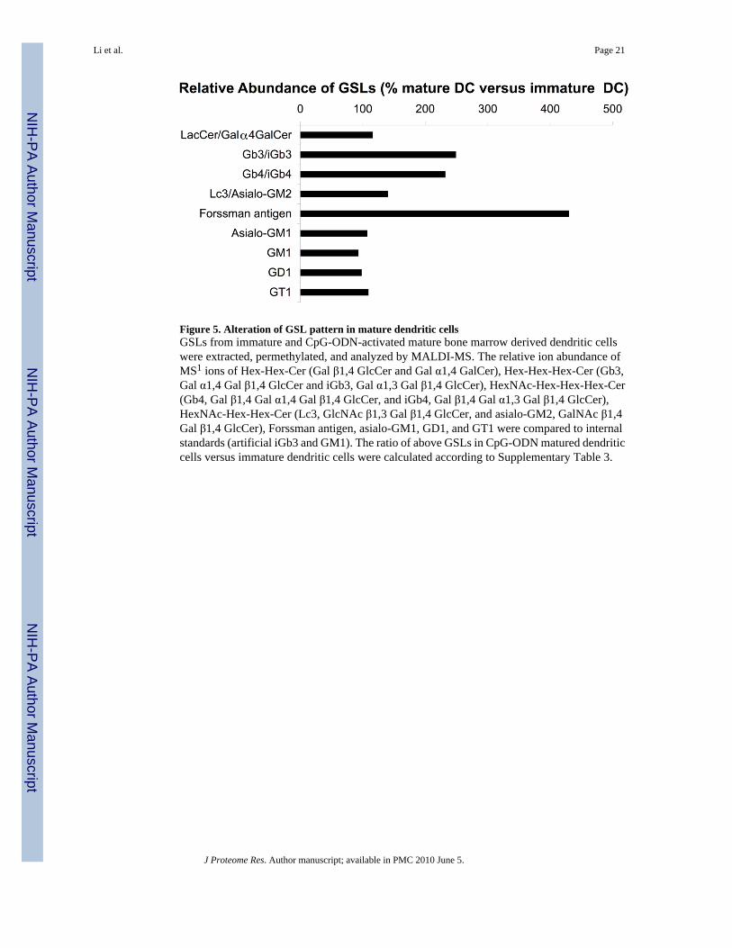
Figure 5. Alteration of GSL pattern in mature dendritic cellsGSLs from immature and CpG-ODN-activated mature bone marrow derived dendritic cellswere extracted, permethylated, and analyzed by MALDI-MS. The relative ion abundance ofMS1 ions of Hex-Hex-Cer (Gal β1,4 GlcCer and Gal α1,4 GalCer), Hex-Hex-Hex-Cer (Gb3,Gal α1,4 Gal β1,4 GlcCer and iGb3, Gal α1,3 Gal β1,4 GlcCer), HexNAc-Hex-Hex-Hex-Cer(Gb4, Gal β1,4 Gal α1,4 Gal β1,4 GlcCer, and iGb4, Gal β1,4 Gal α1,3 Gal β1,4 GlcCer),HexNAc-Hex-Hex-Cer (Lc3, GlcNAc β1,3 Gal β1,4 GlcCer, and asialo-GM2, GalNAc β1,4Gal β1,4 GlcCer), Forssman antigen, asialo-GM1, GD1, and GT1 were compared to internalstandards (artificial iGb3 and GM1). The ratio of above GSLs in CpG-ODN matured dendriticcells versus immature dendritic cells were calculated according to Supplementary Table 3.
Li et al. Page 21
J Proteome Res. Author manuscript; available in PMC 2010 June 5.
NIH
-PA Author Manuscript
NIH
-PA Author Manuscript
NIH
-PA Author Manuscript

Figure 6. Increased ratio of iGb3 in Gb3 synthase KO miceNeutral GSLs from wild type, Gb3 synthase+/− and Gb3 synthase−/− mice were extracted,permethylated, and analyzed by ion trap mass spectrometry. MS1 ions representingregioisomers of iGb3 & Gb3 were selected for MS4 analysis. Percentage of iGb3 in iGb3 &Gb3 mixtures were calculated according to standard curve as described (40).
Li et al. Page 22
J Proteome Res. Author manuscript; available in PMC 2010 June 5.
NIH
-PA Author Manuscript
NIH
-PA Author Manuscript
NIH
-PA Author Manuscript

NIH
-PA Author Manuscript
NIH
-PA Author Manuscript
NIH
-PA Author Manuscript
Li et al. Page 23
Table 1Glycosphingolipids identified in mouse thymus.
Neutral GSLs
Monosaccharide-ceramidea (Hex-Cer) Glc-Cer (Glc β1 Cer)
Disaccharide-ceramideb (Hex-Hex-Cer) Lac-Cer (Gal β4 Glc β1Cer)Digalactosylceramide (Gal α4 Gal β1Cer)
Trisaccharide-ceramide (Hex-Hex-Hex-Cer) iGb3 (Gal α3 Gal β4 Glc Cer)Gb3 (Gal α4 Gal β4 Glc Cer)
Trisaccharide-ceramidec (HexNAc-Hex-Hex-Cer) Lc3 (GlcNAc β3 Gal β4 Glc Cer)Asialo-GM2 (GalNAc β4 Gal β4 Glc Cer)
Tetrasaccharide-ceramide (HexNAc-Hex-Hex-Hex-Cer) iGb4 (GalNAc β3 Gal α3 Gal β4 Glc Cer)Gb4 (GalNAc β3 Gal α4 Gal β4 Glc Cer)
Acidic GSLs
Monosialyl-gangliosides GM3 (NeuAc α3 Gal β4 Glc Cer)GM2 (GalNAc β4 <NeuAc α3> Gal β4 Glc Cer)GM1 (Gal β3 GalNAc β4 <NeuAc α3> Gal β4 Glc Cer)GM1b (NeuAc α3 Gal β3 GalNAc β4 Gal β4 Glc Cer)
Disialyl-gangliosides GD1a (<NeuAc α3> Gal β3 GalNAc β4 <NeuAc α3> Gal β4 Glc Cer)GD1b (Gal β3 GalNAc β4 (<NeuAc α8 NeuAc α3> Gal β4 Glc Cer)
Trisialy-gangliosides GT1a (<NeuAc 8 NeuAc α3> Gal β3 GalNAc β4 <NeuAc α3> Gal β4 Glc Cer)GT1b (<NeuAc α3> Gal β3 GalNAc β4 < NeuAc 8 NeuAc α3> Gal β4 Glc Cer)
Tetrasialyl-gangliosides GQ1b (<NeuAc 8 NeuAc α3> Gal β3 GalNAc β4 < NeuAc 8 NeuAc α3> Gal β4 GlcCer)
a: The presence of Glc-Cer in the thymus of C57BL6 strain of mice was previous reported (52). We have found Glc β1 Cer is the major structure by NMR
analysis (data not shown).
b: The disaccharide-ceramide structure in mouse thymus is a mixture of Lac-Cer and Digalactosylceramide. This was verified by comparing the MS3 ion
patterns, analyzed by ion trap mass spectrometry to standard Lac-Cer and Digalactosylceramide (Supplementary online Figure 2).
c: The trisaccharide-ceramide structures of mouse thymus remain to be studied. Preliminary analysis by ion trap mass spectrometry suggested the structures
of Lc3 and asialo-GM2 (data not shown).
J Proteome Res. Author manuscript; available in PMC 2010 June 5.

NIH
-PA Author Manuscript
NIH
-PA Author Manuscript
NIH
-PA Author Manuscript
Li et al. Page 24
Table 2Glycosphingolipids identified in mouse dendritic cells.
Neutral GSLs
Disaccharide-ceramide (Hex-Hex-Cer) Lac-Cer (Gal β4 Glc β1Cer)Digalactosylceramide (Gal α4 Gal β1Cer)
Trisaccharide-ceramide (Hex-Hex-Hex-Cer) iGb3 (Gal α3 Gal β4 Glc Cer)Gb3 (Gal α4 Gal β4 Glc Cer)
Trisaccharide-ceramide (HexNAc-Hex-Hex-Cer) Lc3 (GlcNAc β3 Gal β4 Glc Cer)Asialo-GM2 (GalNAc β4 Gal β4 Glc Cer)
Tetrasaccharide-ceramide (HexNAc-Hex-Hex-Hex-Cer) iGb4 (GalNAc β3 Gal α3 Gal β4 Glc Cer)Gb4 (GalNAc β3 Gal α4 Gal β4 Glc Cer)
Tetrasaccharide-ceramide (Hex-HexNAc-Hex-Hex-Cer) Asialo-GM1 (Gal β3 GalNAc β4 Gal β4 Glc Cer)
Fossman antigen (HexNAc)3-(Hex)2-Cer GalNAc α3 GalNAc β3 Gal α4 Gal β4 Glc Cer
Acidic GSLs
Monosialyl-gangliosides GM1 (Gal β3 GalNAc β4 <NeuAc α3> Gal β4 Glc Cer)GM1b (NeuAc α3 Gal β3 GalNAc β4 Gal β4 Glc Cer)
Disialyl-gangliosides GD1a (<NeuAc α3> Gal β3 GalNAc β4 <NeuAc α3> Gal β4 Glc Cer)GD1b (Gal β3 GalNAc β4 (<NeuAc α8 NeuAc α3> Gal β4 Glc Cer)GD3 (NeuAc α8 NeuAc α3 Gal β4 Glc Cer)
Trisialyl-gangliosides GT1a (<NeuAc 8 NeuAc α3> Gal β3 GalNAc β4 <NeuAc α3> Gal β4 Glc Cer)GT1b (<NeuAc α3> Gal β3 GalNAc β4 < NeuAc 8 NeuAc α3> Gal β4 Glc Cer)
J Proteome Res. Author manuscript; available in PMC 2010 June 5.
Copyright © 2022 FDOKUMEN