Major histocompatibility antigens and antigen-processing molecules in retinoblastoma
Upload
independentCategory
view
0download
0
doi:10.1182/blood-2007-09-114769Prepublished online January 14, 2008;2008 111: 4588-4595
Douglas C. Palmer, Nicholas P. Restifo, Pierre-Alain Clavien and Burkhard LudewigBeatrice Bolinger, Philippe Krebs, Yinghua Tian, Daniel Engeler, Elke Scandella, Simone Miller, histocompatibility antigenImmunologic ignorance of vascular endothelial cells expressing minor
http://bloodjournal.hematologylibrary.org/content/111/9/4588.full.htmlUpdated information and services can be found at:
(2497 articles)Hemostasis, Thrombosis, and Vascular Biology �Articles on similar topics can be found in the following Blood collections
http://bloodjournal.hematologylibrary.org/site/misc/rights.xhtml#repub_requestsInformation about reproducing this article in parts or in its entirety may be found online at:
http://bloodjournal.hematologylibrary.org/site/misc/rights.xhtml#reprintsInformation about ordering reprints may be found online at:
http://bloodjournal.hematologylibrary.org/site/subscriptions/index.xhtmlInformation about subscriptions and ASH membership may be found online at:
Copyright 2011 by The American Society of Hematology; all rights reserved.Washington DC 20036.by the American Society of Hematology, 2021 L St, NW, Suite 900, Blood (print ISSN 0006-4971, online ISSN 1528-0020), is published weekly
For personal use only. at NATIONAL INSTITUTES OF HEALTH LIB on February 24, 2013. bloodjournal.hematologylibrary.orgFrom

HEMOSTASIS, THROMBOSIS, AND VASCULAR BIOLOGY
Immunologic ignorance of vascular endothelial cells expressing minorhistocompatibility antigen*Beatrice Bolinger,1 *Philippe Krebs,1 Yinghua Tian,2 Daniel Engeler,1 Elke Scandella,1 Simone Miller,1 Douglas C. Palmer,3
Nicholas P. Restifo,3 Pierre-Alain Clavien,2 and Burkhard Ludewig1
1Research Department, Kantonal Hospital, St Gallen, Switzerland; 2Departement of Visceral Surgery, University Hospital Zurich, Zurich, Switzerland; and3National Cancer Institute (NCI), National Institutes of Health (NIH), Bethesda, MD
Endothelial cells (ECs) presenting minorhistocompatibility antigen (mhAg) are ma-jor target cells for alloreactive effectorCD8� T cells during chronic transplantrejection and graft-versus-host disease(GVHD). The contribution of ECs to T-cellactivation, however, is still a controver-sial issue. In this study, we have as-sessed the antigen-presenting capacityof ECs in vivo using a transgenic mousemodel with beta-galactosidase (�-gal) ex-pression confined to the vascular endo-
thelium (Tie2-LacZ mice). In a GVHD-likesetting with adoptive transfer of �-gal–specific T-cell receptor–transgenic T cells,�-gal expression by ECs was not suffi-cient to either activate or tolerize CD8�
T cells. Likewise, transplantation of fullyvascularized heart or liver grafts fromTie2-LacZ mice into nontransgenic recipi-ents did not suffice to activate �-gal–specific CD8� T cells, indicating that CD8�
T-cell responses against mhAg cannot beinitiated by ECs. Moreover, we could show
that spontaneous activation of �-gal–specific CD8� T cells in Tie2-LacZ micewas exclusively dependent on CD11c�
dendritic cells (DCs), demonstrating thatmhAgs presented by ECs remain immuno-logically ignored unless presentation byDCs is granted. (Blood. 2008;111:4588-4595)
Introduction
Endothelial cells (ECs) act as the major interface between bloodand tissues. Forming the inner lining of blood vessels, they areuniquely positioned between circulating lymphocytes and theperiphery and thereby regulate the trafficking of T lymphocytesfrom the bloodstream to sites of infection and inflammation.Following transplantation of vascularized organs, ECs are thefirst graft cells encountered by activated host lymphocytes andare therefore primary targets of alloreactive cytotoxic T lympho-cytes (CTLs).1,2 Since donor ECs persist in vascularized organtransplants, they may contribute to chronic immune stimulationand thereby fuel the process of chronic rejection. Such late graftfailure is a major problem in transplantation medicine thatfrequently necessitates retransplantation.3 Furthermore, ECs areimportant target cells for activated alloreactive CTLs duringgraft-versus-host disease (GVHD),4 which is characterized bylarge numbers of circulating minor histocompatibility antigen(mhAg)–specific CTLs.5
ECs can act as antigen-presenting cells (APCs) to CD8� T cellsboth via the direct pathway (ie, recognition of allo–major histocom-patibility complex [MHC]/peptide complexes) or via the indirectpathway involving cross-presentation of exogenous antigens.6-8 Invitro studies have demonstrated that both human9 and murine10
ECs can activate resting allogeneic CD8� T cells, suggesting thatECs critically contribute to the initial stimulation of alloreactiveT lymphocytes.1 Moreover, ECs exhibit important functions ofprofessional APCs, including expression of MHC class II andcostimulatory molecules11 and cross-presentation of minor histo-
compatibility antigens.6,8 The notion that ECs may under particularcircumstances act as professional APCs has been supported by thefinding that nonhematopoietic cells within vascularized grafts—presumably ECs—are able to initiate CTL responses that mediateallograft rejection.12
There are, however, a number of reports challenging the viewthat ECs may act as immune activators. Murine lung ECs, forexample, have been shown to negatively regulate CD8� T-cellfunction.13 Furthermore, liver sinusoidal ECs can induce CD8�
T-cell tolerance to soluble8,14 or tumor-derived antigens.15 A thirdpossible form of EC-CTL interaction is that of immunologicignorance. Indeed, aly/aly mice lacking secondary lymphoid or-gans fail to reject vascularized organ transplants, even in anallogeneic setting,16 suggesting that the environment of organizedlymphoid tissues is critical for primary activation of T-cell re-sponses. To a large extent, these contradictory findings can beexplained by the use of in vitro coculture systems or the lack of anappropriate in vivo model with truly EC-restricted antigen presen-tation. An experimental in vivo system with expression of well-defined antigens exclusively in vascular ECs may therefore behelpful to solve the question whether antigen presentation byvascular ECs can lead to activation or tolerization of antigen-specific CD8� T cells.
The use of antigen-transgenic mice combined with the power ofT-cell receptor (TCR)–transgenic animals has provided importantinsight into the basic principles of autoimmunity17,18 and tumorimmunity.19,20 Recently, similar systems have been exploited to
Submitted September 24, 2007; accepted January 5, 2008. Prepublishedonline as Blood First Edition paper, January 14, 2008; DOI 10.1182/blood-2007-09-114769.
*B.B. and P.K. contributed equally to this work.
An Inside Blood analysis of this article appears at the front of this issue.
The online version of this article contains a data supplement.
The publication costs of this article were defrayed in part by page chargepayment. Therefore, and solely to indicate this fact, this article is herebymarked ‘‘advertisement’’ in accordance with 18 USC section 1734.
4588 BLOOD, 1 MAY 2008 � VOLUME 111, NUMBER 9
For personal use only. at NATIONAL INSTITUTES OF HEALTH LIB on February 24, 2013. bloodjournal.hematologylibrary.orgFrom

analyze T-cell responses in different allograft transplantation6,21-23
and GVHD models.24 However, despite significant advances in ourunderstanding of the antigen-presenting function of nonhematopoi-etic cells during allograft reactions21 or the importance of T-cellfrequencies for solid organ graft rejection,23,25 the precise role ofmhAg presentation by ECs has remained elusive. We have usedhere Tie2-LacZ mice to model mhAg presentation by ECs. In thesemice, the tie2 promotor drives the expression of the beta-galactosidase (�-gal) antigen in ECs in all tissues.26 In vivoanalysis of antigen-specific interaction between ECs and CD8�
T cells has been facilitated by using high-affinity �-gal–specificTCR-transgenic CD8� T cells (Bg1 cells). Adoptive transfer of Bg1CD8� T cells into Tie2-LacZ mice revealed that mhAg presentationby ECs did not suffice to activate or to tolerize CD8� T cells.Furthermore, �-gal expression by ECs in heterotopically trans-planted Tie2-LacZ hearts or orthotopically transplanted Tie2-LacZlivers did not result in CD8� T-cell activation in naive recipients.Finally, generation of bone marrow (BM) chimeric mice thatfacilitated selective ablation of CD11c� dendritic cells (DCs)revealed that EC-associated mhAg has to be cross-presented byDCs in order to elicit CD8� T-cell activation.
Methods
Mice
Male and female C57BL/6 mice were obtained from Charles River(Sulzfeld, Germany). Tie2-LacZ mice26 had been backcrossed with C57BL/6mice at least 14 times. B6.C-H2bm1 mice were provided by Christian Kurts(University of Bonn, Germany). Bg1 mice were produced with TCRcassette vectors generously provided by Dr Diane Mathis (Brigham andWomen’s Hospital, Boston, MA). RNA was isolated from a �-gal96-103–specific CD8� T-cell clone, generated by limiting dilution, using silicamatrix columns (Qiagen, Valencia, CA). Known TCR � and � constantregion sequences were used to perform 5� rapid amplification of cDNA ends(Invitrogen, Carlsbad, CA), and TCR sequences were then cloned intopCR4TOPO TA cloning sequencing vectors (Invitrogen). The TCR � and �transcripts were sequenced using an ABI Prism (Perkin-Elmer, Wellesley,MA), and these sequences were compared with available sequences todevelop genomic cloning polymerase chain reaction (PCR) primers. Thesecloning primers provide amplification of the variable domains consisting of10 to 20 bp upstream of the start codon through 200 to 300 bp of intronicsequence downstream of the junctional regions, thereby preserving splicedonor/acceptor sites. The � and � genomic variable domains werePCR-amplified (Perkin-Elmer) and TA-cloned into a sequencing vector(Invitrogen). The genomic variable domains were sequenced (V�1/J�TA13/C� and V�7S1/J�2S4/C�2) and subcloned into the TCR cassettevectors. The � and � cassette vectors were coinjected into fertilizedC57BL/6 embryos (SAIC, Frederick, MD), and founders were obtained.The resulting mice, named Bg1, were maintained as heterozygotes, as ahigh rate of lymphoma in homozygotes reduced their life span. Heterozy-gotes were bred to B6.SJL mice and transgene expression was monitored bystaining of blood cells with anti-V�7 by flow cytometry. Bg1 mice werefurther crossed with C57BL/6 mice expressing the congenic marker Thy1.1.Mice expressing the human high-affinity diphteria toxin receptor (DTR)under the control of the CD11c promoter27 were provided by Steffen Jung(The Weizmann Institute of Science, Rehovot, Israel). The presence of the�-gal and DTR transgenes was determined by PCR from genomic DNA;the presence of the H2-Kbm1 molecule was determined by flow cytometry ofblood lymphocytes using the 5F1 antibody.28 All animals were kept underconventional conditions in individually ventilated cages and fed withnormal chow diet. Experiments were carried out with age-matched(6-8 weeks) and sex-matched animals. Experiments were performed inaccordance with Swiss Kantonal and federal legislations and were approvedby the Veterinary Officer of the Kanton of St Gallen.
Viruses and peptides
Recombinant murine cytomegalovirus (MCMV) expressing the �-galprotein under the transcriptional control of the human CMV ie1/ie2promoter-enhancer (MCMV-LacZ RM42729) was kindly provided by ProfE. S. Mocarski (Stanford University, Stanford, CA). MCMV-LacZ waspropagated and titrated on NIH 3T3 cells (European Collection of CellCultures, Salisbury, United Kingdom) and injected intravenously at a doseof 2 � 106 pfu. �-gal96-103 (DAPIYTNV),30 �-gal497-504 (ICPMYARV),31
and MCMV M45985-993 (HGIRNASFI)32 peptides were purchased fromNeosystem (Strasbourg, France).
Generation of BM chimeric mice
Recipient mice were lethally irradiated with 9 Gy (900 rad) from a linearaccelerator (Clinic of Radio-Oncology, Kantonal Hospital, St Gallen) andintravenously injected 1 day later with 2 � 107 of the indicated donorBM cells. Chimeric mice were maintained on antibiotic water containingsulfadoxin and trimethoprim (Veterinaria, Zurich, Switzerland) for thefollowing 3 weeks. Recipient mice carrying the Kbm1 mutation receivedCD4� and CD8� T cell–depleted BM and were further depleted of NK 1.1�
cells by intraperitoneal injection of 20 �L anti-asialo GM1 antibody (WakoPure Chemical Industries, Tokyo, Japan) on the day before and in weeklyintervals for 6 weeks following irradiation. Mice were used for experiments8 to 10 weeks after BM reconstitution.
Antibodies and flow cytometry
Anti-CD8–FITC, anti-CD4–PerCP, anti-V�7–FITC, anti-CD90.1–PE, anti-CD44–PE, and anti-IFN�–PE were obtained from BD PharMingen (Basel,Switzerland). Anti-CD8–allophycocyanin was obtained from Biolegend(LuBioScience, Lucerne, Switzerland). Anti-CD62L–PE was obtained fromImmunoTools (Friesoythe, Germany). For flow cytometry, single-cellsuspensions were generated from the indicated organs and 106 cells wereincubated with the indicated mAb at 4°C for 20 minutes. For peripheralblood lymphocyte (PBL) samples, erythrocytes were lysed with fluorescence-activated cell sorter (FACS) Lysing Solution (BD PharMingen). Cells wereanalyzed with a FACScalibur flow cytometer using the CellQuest software(BD Biosciences). The cells were analyzed by flow cytometry, gating onviable leukocytes using 7-aminoactinomycin D (Sigma, Buchs, Switzerland).
Construction of tetrameric MHC class I peptide complexes andflow cytometry
MHC class I monomers complexed with �-gal (H-2Kb) or M45 peptides(H-2Db) were produced as previously described33 and tetramerized byaddition of streptavidin-PE (Molecular Probes, Eugene, OR). At theindicated time points following infection, organs were removed andsingle-cell suspensions were prepared. Aliquots of 5 � 106 cells or 300 �Lof blood were stained using 50 �L of a solution containing tetrameric class Ipeptide complexes at 37°C for 10 minutes, followed by staining withanti-CD8–FITC (BD PharMingen) at 4°C for 20 minutes. Absolute cellcounts were determined by counting leukocytes in an improved Neubauerchamber (Sigma).
Chromium release assay
EL-4 cells pulsed with peptide or without peptide (negative control) wereused as target cells in a standard 51Cr release assay. Cells were labeled with7.4 MBq (200 �Ci) 51Cr (EGT Chemie, Tagerig, Switzerland) for 1 hour at37°C. A total of 104 target cells/well were incubated for 5 hours in 96-wellround bottom plates with 3-fold serial dilutions of effector cells. Spleno-cytes from MCMV-LacZ–infected mice that were restimulated with theindicated peptides for 5 days were tested for their cytolytic activity.Spontaneous chromium release was always below 15%.
CFSE labeling of TCR-transgenic T cells and adoptive transfer
Single-cell suspensions from the spleens of Bg1 mice were subjected tohypotonic red blood cell lysis and stained with CFSE (Molecular Probes,
IGNORANCE OF VASCULAR ENDOTHELIAL CELLS 4589BLOOD, 1 MAY 2008 � VOLUME 111, NUMBER 9 For personal use only. at NATIONAL INSTITUTES OF HEALTH LIB on February 24, 2013. bloodjournal.hematologylibrary.orgFrom

Leiden, The Netherlands). A maximum concentration of 2.5 � 107 cells/mLwere incubated in 5 �M CFSE in phosphate-buffered saline (PBS) for10 minutes at 37°C. Cells were washed twice with ice-cold balanced saltsolution (BSS) and resuspended in BSS at a concentration of 1.5 � 107
splenocytes/mL. Recipient B6, Tie2-LacZ, and the different subsets ofchimeric mice were injected intravenously with 1.5 � 107 Bg1-Thy1.1�
splenocytes in 500 �L BSS.
Immunohistology
Freshly removed organs were immersed in Hanks BSS (HBSS) andsnap-frozen in liquid nitrogen. Frozen tissue sections were cut in a cryostatand fixed in acetone for 10 minutes. Sections were incubated withantibodies against �-gal (MP Biomedicals, Irvine, CA) and CD8 (cloneYTS169.4.2), followed by goat anti-rat Ig (Caltag Laboratories, Burlin-game, CA) and alkaline phosphatase–labeled donkey anti-goat Ig (JacksonImmunoResearch Laboratories, West Grove, PA). Alkaline phophatase wasvisualized by using AS-BI phosphate/new fuchsin; sections were counter-stained with hemalum, and images were acquired using a Leica DM Rmicroscope equipped with a Leica DC300 FX camera (Leica, Heerbrugg,Switzerland). Digital images were processed using Adobe Photoshop(Adobe, San Jose, CA).
Surgical procedure for liver transplantation
Donor procedure, back-table preparation, and recipient procedure were per-formed as described previously with minor modifications.34 Briefly, all vesselsand ligaments of the liver were dissected in the donor after midline laparotomy. Insitu perfusion of the liver was performed using cold (4°C) Ringer solution.Subsequently, the liver was separated from its retroperitoneal attachments andremoved. The graft was stored in cold (4°C) Ringer solution for 60 minutes untilimplantation into the recipient. Following hepatectomy of the native liver in therecipient, the donor liver was implanted in an orthotopic position. The anhepatictime in the recipient was consistently kept below 20 minutes. The portal vein wasreconstructed and the liver was reperfused after completing the anastomosisbetween the suprahepatic inferior vena cava of the recipient and donor. Arterialrecirculation was established by an end-to-side anastomosis between the recipientaorta and an aortic segment attached to the hepatic artery of the graft. A singlesubcutaneous injection of 5 mg cefazolin provided antibiotic prophylaxis.
Heterotopic heart transplantation
Heterotopic vascularized cardiac transplantation was performed accordingto the method described by Corry et al.35 Donor hearts were explanted fromeither male Tie2-LacZ or male C57BL/6 mice. The donor heart wasremoved from the chest after intracaval injection of 1 mL heparin(100 U/mL), rinsed with NaCl 0.9%, and placed on ice. After isolation ofthe recipient’s abdominal aorta and inferior vena cava, the donor ascendingaorta and pulmonary artery were joined end-to-side to the recipient’s aortaand vena cava, respectively, using 10-0 nylon running suture. The abdomenwas closed with individual running sutures to musculofascial layer andskins. The recipient mouse was then warmed for a few hours duringrecovery from anesthesia and had free access to water and food. Thefunction of the transplanted heart was assessed daily by abdominalpalpation.
Statistical data analysis
To evaluate statistically significant differences, the unpaired 2-tailed Student testwas used. P values smaller than .05 were considered statistically significant.Statistical data analysis was performed using GraphPad Prism version 4.03 forWindows (GraphPad Software, San Diego CA).
Results
CD8 T-cell tolerance in Tie2-LacZ mice
Currently, a number of transgenic mouse lines are available thatexhibit EC-restricted transgene expression: von Willebrand factor–
LacZ36 and thrombomodulin-LacZ37 mice, which both show patchytransgene distribution in some arteries; and tie2-H-2Kb mice,14
which express the H2-Kb molecule as a transgene. In this study,Tie2-LacZ mice26 backcrossed to the C57BL/6 background havebeen used because of the uniform �-gal Ag expression in ECs of allorgans. It is noteworthy that the intensity of �-gal expression inTie2-LacZ mice is most pronounced in small and large arteries, butclearly detectable in venous and capillary ECs (Figure 1A; FigureS1, available on the Blood website; see the Supplemental Materialslink at the top of the online article). Furthermore, expression levelsof �-gal mRNA in various organs were comparable (Figure S2),indicating that these mice are well suited to study EC–CD8� T-cell
Figure 1. CD8� T-cell reactivity in Tie2-LacZ mice. (A) Heart and thymus sectionsof naive Tie2-LacZ mice were stained for �-gal and CD8. Images were acquired witha 25�/0.65 objective (magnification: �162). (B,C) C57BL/6 (B6) and Tie2-LacZ (T2)mice were infected intravenously with 106 pfu MCMV-LacZ. (B) Tetramer analysis forthe indicated �-gal– and MCMV-derived M45 epitopes was performed on day 6 afterinfection. Mean percentage of tetramer-positive cells within the CD8 compartmentare indicated (� SEM; n 3-4). (C) Lysis of peptide-pulsed EL-4 cells by MCMV-LacZ-induced CTLs. On day 6 after infection, splenocytes from the indicated mouse strainswere restimulated in vitro for 5 days with �-gal497-504, �-gal96-103, or M45985-993 peptideand tested in a standard chromium release assay.
4590 BOLINGER et al BLOOD, 1 MAY 2008 � VOLUME 111, NUMBER 9 For personal use only. at NATIONAL INSTITUTES OF HEALTH LIB on February 24, 2013. bloodjournal.hematologylibrary.orgFrom

interaction in vivo. Indeed, in a previous study, Rothermel et al38
used Tie2-LacZ mice on the FVB genetic background to assessCD4� and CD8� T-cell responsiveness under conditions of persist-ing Ag expression in ECs. Whereas this previous investigationsuggested that Tie2-LacZ mice can mount CD8� T-cell responsesagainst the EC-restricted �-gal Ag,38 the results obtained in thepresent study indicate that CD8� T cells in Tie2-LacZ mice aretolerant to the �-gal antigen. This is shown by the fact thatTie2-LacZ mice failed to mount �-gal–specific CTL responsesfollowing infection with �-gal–recombinant MCMV (MCMV-LacZ), whereas CD8� T-cell responses against the viral M45epitope were not influenced by the EC-specific transgene expres-sion (Figure 1B,C).
We next addressed whether the apparent �-gal–specific CD8� T-celltolerance in Tie2-LacZ mice is mediated by thymic negative selection orby peripheral tolerizing mechanisms. To this end, Tie2-LacZ mice werecrossed with TCR-transgenic Bg1 mice, which possess CD8� T cellsthat recognize the H2-Kb–restricted �-gal96-103 epitope.30 A total of 60%to 70% of the Bg1 CD8� T cells bind H2-Kb/�-gal96-103 tetramers(Figure 2), and Bg1 CD8� T cells possess a high functional avidity forthe �-gal96-103 epitope as shown by target cell recognition and prolifera-tion assays (Figure S4). In the thymus of Tie2-LacZ � Bg1 mice, thenumbers of transgenic V�7 chain–positive and tetramer-binding CD8�
cells were reduced to 40% (Figure 2), suggesting that central toleranceled to partial deletion of �-gal–specific T cells. Interestingly, inperipheral lymphoid organs such as the spleen, the numbers of V�7�
and �-gal96-103 tetramer-binding CD8� T cells was further reduced from60% to 70% in Bg1 to less than 10% in Tie2-LacZ � Bg1 mice. Thus, ata first glance, it appears that EC-specific Ag expression in Tie2-LacZmice precipitated both central and peripheral tolerance.
EC-independent peripheral CD8� T-cell tolerance
Peripheral CD8� T-cell tolerance can be induced via different celltypes, including circulating hematopoietic cells expressingmhAg,39,40 BM-derived APC cross-presenting antigen derived from
parenchymal tissues,41,42 or particular subsets of ECs that alsopossess the ability to cross-present circulating antigens.8,14 In orderto assess truly EC-mediated peripheral tolerance induction, weestablished first a highly sensitive in vivo restimulation assay todetect very low amounts of circulating Bg1 cells. To this end,graded numbers of sorted CD8�Thy1.1� cells from naive Bg1 micewere transferred into Thy1.2� Tie2-LacZ and C57BL/6 mice. At6 days following adoptive transfer, mice were infected withMCMV-LacZ, and the expansion of Bg1 cells was assessed 6 dayslater. As shown in Figure 3A, �-gal–specific CD8� T cellsexpanded in C57BL/6 but not in Tie2-LacZ mice, confirming thatBg1 cells encounter their antigen in Tie2-LacZ mice outside of the
Figure 2. CD8� T cell tolerance in Tie2-LacZ mice. Tie2-LacZ mice were crossedwith TCR-trangenic Bg1 mice. Thymocytes from Tie2-LacZ (T2; top row), Bg1 (middlerow), and Bg1 � Tie2-LacZ mice (Bg1 � T2; bottom row) were stained for CD4 andCD8 expression. The expression of the transgenic V�7 chain and binding of theH2-Kb-�-gal96-103 tetramer was determined by gating on CD8� T cells. Values in thetop right quadrants indicate mean frequencies of CD4/CD8-positive cells in thymo-cytes or percentage of antigen-specific cells in single CD8� thymocytes, respectively(T2, n 2; Bg1, n 3; Bg1 � T2, n 7). Splenocytes were assessed for V�7 andCD8 expression. Percentage of �-gal96-103–specific cells was determined by V�7 andH2-Kb-�-gal96-103 tetramer staining, gating on CD8� T cells (T2, n 3; Bg1, n 5;Bg1 � T2, n 9).
Figure 3. Loss of adoptively transferred Bg1 CD8� T cells in Tie2-LacZ mice isnot dependent on �-gal expression by ECs. (A) Graded numbers of CD8� Bg1cells expressing the congenic marker Thy1.1 were adoptively transferred intoThy1.2� C57BL/6 or Tie2-LacZ recipient mice. At 6 days later, mice were challengedwith 2 � 106 pfu MCMV-LacZ, and the proliferation of Bg1 CD8� T cells wasdetermined on day 6 following immunization by staining for CD8, Thy1.1, and thetransgenic V�7 chain. Representative data from one of 2 independent experimentsare shown. (B,C) Adoptive transfer of Bg1 CD8� T cells in BM chimeric mice. A total of5 � 104 (B) or 105 (C) TCR transgenic Thy1.1� Bg1 cells were adoptively transferredintravenously into the indicated Thy1.2� BM chimeric mice. At 9 days (B) or 30 days(C) later, mice were challenged with 2 � 106 pfu MCMV-LacZ, and proliferation ofBg1 CD8� T cells was determined on day 6 following MCMV-LacZ challenge in theindicated organs. Values represent mean percentage (� SEM) of Thy1.1�V�7� cellswithin the CD8 T-cell compartment.
IGNORANCE OF VASCULAR ENDOTHELIAL CELLS 4591BLOOD, 1 MAY 2008 � VOLUME 111, NUMBER 9 For personal use only. at NATIONAL INSTITUTES OF HEALTH LIB on February 24, 2013. bloodjournal.hematologylibrary.orgFrom

thymus, and that this interaction leads to their deletion. However,reconstituting Tie2-LacZ mice with C57BL/6 BM (B63 T2)revealed that CD8� T-cell tolerance in Tie2-LacZ mice was solely
dependent on �-gal expression within the BM (Figure 3B,C). Inaddition, �-gal expression by ECs in B63 T2 chimeric mice didnot affect the pattern of responsiveness of adoptively transferredBg1 cells following MCMV-LacZ infection (Figure S5). Takentogether, these data indicate that expression of an mhAg by ECsalone is not sufficient to directly tolerize CD8� T cells, nor is thisantigen available to BM-derived APCs in a way that would lead toCD8� T-cell tolerance.
ECs fail to directly activate naive CD8� T cells in vivo
Deletional tolerization of CD8� T cells via activation-induced celldeath is usually associated with a transient period of T-cellactivation and proliferation.24,43 Furthermore, it is possible that ECsin Tie2-LacZ mice might directly interact with CD8� T cells in away that leads to T-cell activation and/or proliferation. In order toassess a potential spontaneous T-cell activation by ECs in aGVHD-like model situation, 3 � 106 CFSE-labeled TCR-transgenic CD8� T cells were adoptively transferred into Tie2-LacZ mice, and T-cell activation was monitored as CD44 up-regulation on proliferating Bg1 cells (Figure 4A). Furthermore,monitoring of CD44 up-regulation, expression of annexin V, andenumeration of the total numbers of Bg1 cells in spleens at latertime points revealed that Bg1 T cells did not progress towardactivation-induced cell death (Figure S6). Quantification of �-gal–dependent T-cell proliferation was achieved by adoptive transfer ofCSFE-labeled, Thy1.1� CD8� Bg1 cells into either C57BL/6 orTie2-LacZ mice (Figure 3B). This sensitive read-out system wasthen used to assess whether antigen presentation by ECs alone issufficient to mediate CD8� T-cell activation or whether BM-derived APCs, in particular DCs, contribute to the observed initialCD8� T-cell triggering.
An array of BM chimeric mice was generated using differentcombinations between C57BL/6 (B6) and Tie2-LacZ (T2) controls,Tie2-LacZ mice on the C57BL/6bm1 background (T2bm1) exhibit-ing a mutated H2-Kb molecule that precludes H2-Kb-restrictedpresentation, and CD11c-DTR mice,27 which facilitate the specificablation of CD11c� DCs in lymphoid organs. As expected, Bg1cells were not activated in B63 B6 chimeras (Figure 5A),
Figure 4. Activation of Bg1 CD8� T cells in Tie2-LacZ mice. (A) A total of 1.5 � 107
CFSE-labeled splenocytes (corresponding to 3 � 106 CD8� TCR-transgenic T cells)from Bg1 mice were adoptively transferred into C57BL/6 or Tie2-LacZ mice. Micewere killed on day 4 following transfer and cells from blood, spleen, and lymph nodeswere analyzed by flow cytometry for CFSE dilution and CD44 up-regulation on CD8�
lymphocytes. Representative FACS plots from one representative of 2 independentexperiments are shown. (B) Quantification of Bg1 T-cell proliferation. A total of1.5 � 107 CFSE-labeled Bg1 Thy1.1� splenocytes were injected into C57BL/6 ornaive Tie2-LacZ mice. Mice were killed on day 4 following transfer and cells fromblood, spleen, and lymph nodes were analyzed by flow cytometry. Values representmean percentage (� SEM, n 7; pooled data from 2 independent experiments) ofproliferating CD8�Thy1.1� cells (*P .05; **P .005; ***P .001).
Figure 5. In vivo proliferation of Bg1 CD8� T cells in BM chimeric mice. A total of 1.5 � 107 CFSE-labeled splenocytes (corresponding to 3 � 106 CD8� TCR-transgenicT cells) expressing the congenic marker Thy1.1 were adoptively transferred into the indicated Thy1.2� bone marrow chimeras. (A) C57BL/63C57BL/6 (B63 B6).(B) Tie2-LacZ3 Tie2LacZ (T23 T2). (C) C57BL/63 Tie2-LacZ (B63 T2). (D) Tie2-LacZ � B6.C-H2bm13 Tie2-LacZ (T2 � bm13 T2). (E) Tie2-LacZ � B6.C-H2bm13 Tie2-LacZ � B6.C-H2bm1 (T2 � bm1 3 T2 � bm1). (F) C57BL/6 3 Tie2-LacZ � B6.C-H2bm1 (B6 3 T2 � bm1). (G) CD11c DTR 3 Tie2-LacZ � B6.C-H2bm1
(CD11cDTR3 T2 � bm1). CD11c-DTR BM recipients had been injected intraperitoneally with 4 ng/g body weight diphteria toxin (DT), which led to a 95% to 98% depletion ofCD11c� cells for more than 48 hours. Mice were killed on day 4 following adoptive transfer and cells from blood and spleens were analyzed by flow cytometry. Values in thehistograms represent mean percentages (� SEM, n 5-7; pooled data from 3 independent experiments) of proliferating CD8�Thy1.1� cells.
4592 BOLINGER et al BLOOD, 1 MAY 2008 � VOLUME 111, NUMBER 9 For personal use only. at NATIONAL INSTITUTES OF HEALTH LIB on February 24, 2013. bloodjournal.hematologylibrary.orgFrom
whereas transgenic CD8� T cells proliferated in T23 T2 chimericmice (Figure 5B). Proliferation of Bg1 cells in B63 T2 chimerasindicated that BM-derived nontransgenic APCs had activated thetransgenic T cells (Figure 5C). This interpretation is supported bythe fact that Bg1 proliferation was aborted in T2bm13 T2chimeras where direct and cross-presentation via BM-derivedAPCs is abolished, and only ECs can present the �-gal epitope(Figure 5D). Experiments with T2bm13 T2bm1 chimeras con-firmed that Bg1 cells do not proliferate in the absence of theappropriate H2 restriction element (Figure 5E). Bg1 activationcould be restored in B63 T2bm1 chimeras, confirming thatBM-derived APCs are crucial in this setting (Figure 5F). Finally,reconstitution of T2bm1 mice with BM from CD11c-DTR micetogether with diphteria toxin–mediated ablation of DCs showedthat the proliferation of Bg1 cells depended strictly on thecross-presentation of �-gal Ag by DCs (Figure 5G). It is notewor-thy that Bg1 activation in B63 T2bm1 chimeras (Figure 5F) wassomewhat reduced compared with the proliferative activity ob-served in B63 T2 chimeras (Figure 5C), suggesting that ECsmight to some extent contribute to the observed effects. Neverthe-less, as a whole, the presented results indicate that �-gal–presenting vascular ECs remain immunologically ignored by CD8�
T cells, and that activation and proliferation of CD8� T cellsrecognizing the mhAg in Tie2-LacZ mice is almost exclusivelydependent on cross-presentation of Ag by BM-derived DCs.
Immunologic ignorance of antigen-expressing ECs invascularized organ transplants
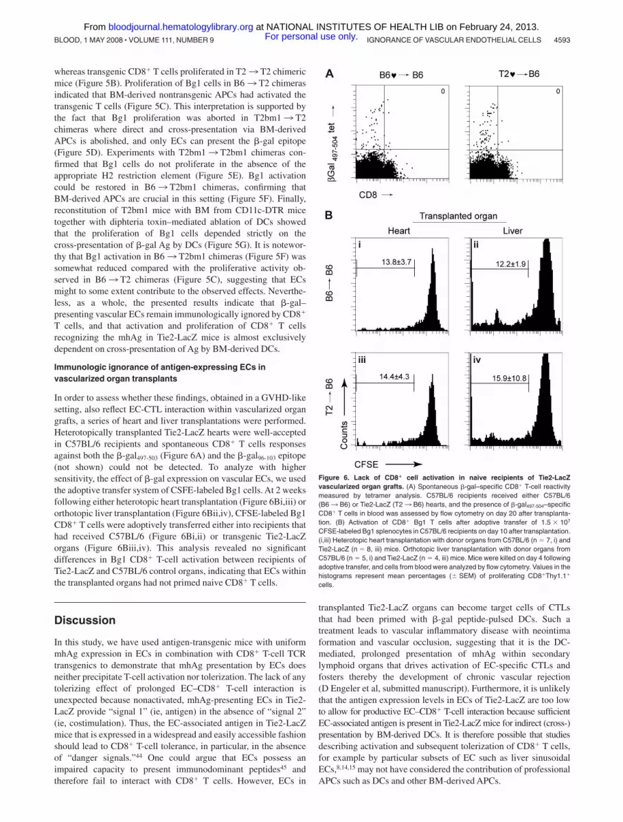
In order to assess whether these findings, obtained in a GVHD-likesetting, also reflect EC-CTL interaction within vascularized organgrafts, a series of heart and liver transplantations were performed.Heterotopically transplanted Tie2-LacZ hearts were well-acceptedin C57BL/6 recipients and spontaneous CD8� T cells responsesagainst both the �-gal497-503 (Figure 6A) and the �-gal96-103 epitope(not shown) could not be detected. To analyze with highersensitivity, the effect of �-gal expression on vascular ECs, we usedthe adoptive transfer system of CSFE-labeled Bg1 cells. At 2 weeksfollowing either heterotopic heart transplantation (Figure 6Bi,iii) ororthotopic liver transplantation (Figure 6Bii,iv), CFSE-labeled Bg1CD8� T cells were adoptively transferred either into recipients thathad received C57BL/6 (Figure 6Bi,ii) or transgenic Tie2-LacZorgans (Figure 6Biii,iv). This analysis revealed no significantdifferences in Bg1 CD8� T-cell activation between recipients ofTie2-LacZ and C57BL/6 control organs, indicating that ECs withinthe transplanted organs had not primed naive CD8� T cells.
Discussion
In this study, we have used antigen-transgenic mice with uniformmhAg expression in ECs in combination with CD8� T-cell TCRtransgenics to demonstrate that mhAg presentation by ECs doesneither precipitate T-cell activation nor tolerization. The lack of anytolerizing effect of prolonged EC–CD8� T-cell interaction isunexpected because nonactivated, mhAg-presenting ECs in Tie2-LacZ provide “signal 1” (ie, antigen) in the absence of “signal 2”(ie, costimulation). Thus, the EC-associated antigen in Tie2-LacZmice that is expressed in a widespread and easily accessible fashionshould lead to CD8� T-cell tolerance, in particular, in the absenceof “danger signals.”44 One could argue that ECs possess animpaired capacity to present immunodominant peptides45 andtherefore fail to interact with CD8� T cells. However, ECs in
transplanted Tie2-LacZ organs can become target cells of CTLsthat had been primed with �-gal peptide-pulsed DCs. Such atreatment leads to vascular inflammatory disease with neointimaformation and vascular occlusion, suggesting that it is the DC-mediated, prolonged presentation of mhAg within secondarylymphoid organs that drives activation of EC-specific CTLs andfosters thereby the development of chronic vascular rejection(D Engeler et al, submitted manuscript). Furthermore, it is unlikelythat the antigen expression levels in ECs of Tie2-LacZ are too lowto allow for productive EC–CD8� T-cell interaction because sufficientEC-associated antigen is present in Tie2-LacZ mice for indirect (cross-)presentation by BM-derived DCs. It is therefore possible that studiesdescribing activation and subsequent tolerization of CD8� T cells,for example by particular subsets of EC such as liver sinusoidalECs,8,14,15 may not have considered the contribution of professionalAPCs such as DCs and other BM-derived APCs.
Figure 6. Lack of CD8� cell activation in naive recipients of Tie2-LacZvascularized organ grafts. (A) Spontaneous �-gal–specific CD8� T-cell reactivitymeasured by tetramer analysis. C57BL/6 recipients received either C57BL/6(B63 B6) or Tie2-LacZ (T23 B6) hearts, and the presence of �-gal497-504–specificCD8� T cells in blood was assessed by flow cytometry on day 20 after transplanta-tion. (B) Activation of CD8� Bg1 T cells after adoptive transfer of 1.5 � 107
CFSE-labeled Bg1 splenocytes in C57BL/6 recipients on day 10 after transplantation.(i,iii) Heterotopic heart transplantation with donor organs from C57BL/6 (n 7, i) andTie2-LacZ (n 8, iii) mice. Orthotopic liver transplantation with donor organs fromC57BL/6 (n 5, i) and Tie2-LacZ (n 4, iii) mice. Mice were killed on day 4 followingadoptive transfer, and cells from blood were analyzed by flow cytometry. Values in thehistograms represent mean percentages (� SEM) of proliferating CD8�Thy1.1�
cells.
IGNORANCE OF VASCULAR ENDOTHELIAL CELLS 4593BLOOD, 1 MAY 2008 � VOLUME 111, NUMBER 9 For personal use only. at NATIONAL INSTITUTES OF HEALTH LIB on February 24, 2013. bloodjournal.hematologylibrary.orgFrom

Indeed, the complexity of the multicellular processes involvedin EC-mediated antigen presentation in vivo requires carefulconsideration of possible confounding factors. Rothermel et al38
have suggested that immune recognition of ECs is context depen-dent, with antigen expressed in hearts of Tie2-LacZ mice beingimmunologically ignored, whereas ECs presenting �-gal antigen inskin are immunogenic and thus elicit T-cell responses capable ofrejecting skin grafts. The results of our study clearly confirm thenotion that direct presentation of mhAg by ECs is accompanied byimmunologic ignorance. However, in the context of mhAg presen-tation in transplant vasculopathy and GVHD, DCs are probably themost important cell population that cross-presents the antigen in animmunogenic fashion.
Our study revealed further details that could confound theanalysis of T-cell activation/tolerization in Tie2-LacZ mice:�-gal–specific CD8� T cells were effectively tolerized innonirradiated Tie2-LacZ and in T23 T2 BM chimeric mice.We conclude from these findings that cells within the BM, butnot professional APCs that descend from BM precursors, exert atolerizing stimulus in Tie2-LacZ mice. Hematopoietic stem cells(HSCs) express the angiopoetin 1 receptor Tie2,46 and recentstudies have shown that the tie2 promoter that has been used inTie2-LacZ26 mice is active in HSCs during embryonic develop-ment.47 Likewise, in BM of adult Tie2-LacZ mice, LacZtranscripts were most abundant in HSCs (Figure S3). Thus, sincecirculating lymphocytes presenting mhAg efficiently tolerizenaive CD8� T cells,24,40 it is reasonable to assume that naiveCD8� T cells traveling through the BM can receive tolerizingstimuli within this compartment. Using Tie2-LacZ BM chime-ras, it will be feasible to further characterize those cells withinthe BM that are highly efficient in inducing tolerance to mhAg.
Taken together, this study identifies the initial priming ofmhAg-specific CD8� T cells via DCs as a critical step in thegeneration of alloimmune responses. Therefore, it appears to be
crucial that therapeutic intervention should aim at preventing or atleast reducing the initial T-cell activation against mhAg. Indeed,blockade of essential costimulatory pathways such as CD40-CD15423,48 or CD28-CD80/8649 interaction during initial DC-mediated CD8� T-cell stimulation bear a high potential for clinicalapplication. It may well be that a combination of costimulatoryblockade before and during priming of EC-specific CD8� cellresponses together with the induction of regulatory T cells49 willhelp to protect ECs from injury following transplantation ofvascularized organs or during GVHD.
Acknowledgments
We would like to thank Silvia Behnke and Andre Fitsche for helpwith immunohistochemistry and Ton Rolink for help with FACSsorting of bone marrow cells.
This project received support from the Swiss National ScienceFoundation, the Velux Foundation (Zurich), and the Kanton of StGallen.
Authorship
Contribution: B.L. designed the study and wrote the paper; B.B.performed research and wrote the paper; P.-A.C. designed theexperimental procedure of liver transplantation; N.P.R. and D.C.P.provided mice; and E.S., P.K., D.E., Y.T., and S.M. performedresearch.
Conflict-of-interest disclosure: The authors declare no compet-ing financial interests.
Correspondence: Burkhard Ludewig, Research Department,Kantonsspital St Gallen, Rorschacherstrasse 95, CH-9007 StGallen, Switzerland; e-mail: [email protected].
References
1. Briscoe DM, Alexander SI, Lichtman AH. Interac-tions between T lymphocytes and endothelialcells in allograft rejection. Curr Opin Immunol.1998;10:525-531.
2. Libby P, Pober JS. Chronic rejection. Immunity.2001;14:387-397.
3. Weis M, von Scheidt W. Cardiac allograft vascu-lopathy: a review. Circulation. 1997;96:2069-2077.
4. Biedermann BC, Sahner S, Gregor M, et al. En-dothelial injury mediated by cytotoxic T lympho-cytes and loss of microvessels in chronic graftversus host disease. Lancet. 2002;359:2078-2083.
5. Mutis T, Gillespie G, Schrama E, et al. TetramericHLA class I-minor histocompatibility antigen pep-tide complexes demonstrate minor histocompat-ibility antigen-specific cytotoxic T lymphocytes inpatients with graft-versus-host disease. Nat Med.1999;5:839-842.
6. Valujskikh A, Lantz O, Celli S, Matzinger P, Hee-ger PS. Cross-primed CD8(�) T cells mediategraft rejection via a distinct effector pathway. NatImmunol. 2002;3:844-851.
7. Bagai R, Valujskikh A, Canaday DH, et al. Mouseendothelial cells cross-present lymphocyte-derived antigen on class I MHC via a TAP1- andproteasome-dependent pathway. J Immunol.2005;174:7711-7715.
8. Limmer A, Ohl J, Kurts C, et al. Efficient presenta-tion of exogenous antigen by liver endothelialcells to CD8� T cells results in antigen-specificT-cell tolerance. Nat Med. 2000;6:1348-1354.
9. Epperson DE, Pober JS. Antigen-presentingfunction of human endothelial cells: direct activa-tion of resting CD8 T cells. J Immunol. 1994;153:5402-5412.
10. Kreisel D, Krupnick AS, Balsara KR, et al. Mousevascular endothelium activates CD8� T lympho-cytes in a B7-dependent fashion. J Immunol.2002;169:6154-6161.
11. Rose ML. Endothelial cells as antigen-presentingcells: role in human transplant rejection. Cell MolLife Sci. 1998;54:965-978.
12. Kreisel D, Krupnick AS, Gelman AE, et al. Non-hematopoietic allograft cells directly activateCD8� T cells and trigger acute rejection: an alter-native mechanism of allorecognition. Nat Med.2002;8:233-239.
13. Marelli-Berg FM, Scott D, Bartok I, et al. Activatedmurine endothelial cells have reduced immuno-genicity for CD8� T cells: a mechanism of immu-noregulation? J Immunol. 2000;165:4182-4189.
14. Limmer A, Ohl J, Wingender G, et al. Cross-presentation of oral antigens by liver sinusoidalendothelial cells leads to CD8 T cell tolerance.Eur J Immunol. 2005;35:2970-2981.
15. Berg M, Wingender G, Djandji D, et al. Cross-presentation of antigens from apoptotic tumorcells by liver sinusoidal endothelial cells leads totumor-specific CD8� T cell tolerance. Eur J Im-munol. 2006;36:2960-2970.
16. Lakkis FG, Arakelov A, Konieczny BT, Inoue Y.Immunologic ‘ignorance’ of vascularized organtransplants in the absence of secondary lymphoidtissue. Nat Med. 2000;6:686-688.
17. Ohashi PS, Oehen S, Buerki K, et al. Ablation of“tolerance” and induction of diabetes by virus in-fection in viral antigen transgenic mice. Cell.1991;65:305-317.
18. von Herrath MG, Oldstone MB. Virus-inducedautoimmune disease. Curr Opin Immunol. 1996;8:878-885.
19. Morgan DJ, Kreuwel HT, Fleck S, et al. Activationof low avidity CTL specific for a self epitope re-sults in tumor rejection but not autoimmunity.J Immunol. 1998;160:643-651.
20. Speiser DE, Miranda R, Zakarian A, et al. Selfantigens expressed by solid tumors do not effi-ciently stimulate naive or activated T cells: impli-cations for immunotherapy. J Exp Med. 1997;186:645-653.
21. Kreisel D, Krasinskas AM, Krupnick AS, et al.Vascular endothelium does not activate CD4�direct allorecognition in graft rejection. J Immunol.2004;173:3027-3034.
22. Valujskikh A, Zhang Q, Heeger PS. CD8 T cellsspecific for a donor-derived, self-restricted trans-plant antigen are nonpathogenic bystanders aftervascularized heart transplantation in mice. J Im-munol. 2006;176:2190-2196.
23. Ford ML, Koehn BH, Wagener ME, et al. Antigen-specific precursor frequency impacts T cell prolif-eration, differentiation, and requirement for co-stimulation. J Exp Med. 2007;204:299-309.
24. Ehl S, Hombach J, Aichele P, et al. Viral and bac-terial infections interfere with peripheral toleranceinduction and activate CD8� T cells to cause im-munopathology. J Exp Med. 1998;187:763-774.
4594 BOLINGER et al BLOOD, 1 MAY 2008 � VOLUME 111, NUMBER 9 For personal use only. at NATIONAL INSTITUTES OF HEALTH LIB on February 24, 2013. bloodjournal.hematologylibrary.orgFrom

25. He C, Schenk S, Zhang Q, et al. Effects of T cellfrequency and graft size on transplant outcome inmice. J Immunol. 2004;172:240-247.
26. Schlaeger TM, Bartunkova S, Lawitts JA, et al.Uniform vascular-endothelial-cell-specific geneexpression in both embryonic and adult trans-genic mice. Proc Natl Acad Sci U S A. 1997;94:3058-3063.
27. Jung S, Unutmaz D, Wong P, et al. In vivo de-pletion of CD11c(�) dendritic cells abrogatespriming of CD8(�) T cells by exogenous cell-associated antigens. Immunity. 2002;17:211-220.
28. Sherman LA, Randolph CP. Monoclonal anti-H-2Kb antibodies detect serological differences be-tween H-2Kb mutants. Immunogenetics. 1981;12:183-186.
29. Manning WC, Mocarski ES. Insertional mutagen-esis of the murine cytomegalovirus genome: oneprominent alpha gene (ie2) is dispensable forgrowth. Virology. 1988;167:477-484.
30. Overwijk WW, Surman DR, Tsung K, Restifo NP.Identification of a Kb-restricted CTL epitope ofbeta-galactosidase: potential use in developmentof immunization protocols for “self ” antigens.Methods. 1997;12:117-123.
31. Oukka M, Cohen-Tannoudji M, Tanaka Y, BabinetC, Kosmatopoulos K. Medullary thymic epithelialcells induce tolerance to intracellular proteins.J Immunol. 1996;156:968-975.
32. Gold MC, Munks MW, Wagner M, et al. The mu-rine cytomegalovirus immunomodulatory genem152 prevents recognition of infected cells byM45-specific CTL but does not alter the immu-nodominance of the M45-specific CD8 T cell re-sponse in vivo. J Immunol. 2002;169:359-365.
33. Altman JD, Moss PAH, Goulder PJR, et al. Phe-
notypic analysis of antigen-specific T lympho-cytes. Science. 1996;274:94-96.
34. Tian Y, Rudiger HA, Jochum W, Clavien PA. Com-parison of arterialized and nonarterialized ortho-topic liver transplantation in mice: prowess or rel-evant model? Transplantation. 2002;74:1242-1246.
35. Corry RJ, Winn HJ, Russell PS. Primarily vascu-larized allografts of hearts in mice: the role ofH-2D, H-2K and non-H-2 antigens in rejection.Transplantation. 1973;16:343-350.
36. Aird WC, Jahroudi N, Weiler Guettler H, RayburnHB, Rosenberg RD. Human von Willebrand factorgene sequences target expression to a subpopu-lation of endothelial cells in transgenic mice. ProcNatl Acad Sci U S A. 1995;92:4567-4571.
37. Weiler-Guettler H, Aird WC, Husain M, RayburnH, Rosenberg RD. Targeting of transgene expres-sion to the vascular endothelium of mice by ho-mologous recombination at the thrombomodulinlocus. Circ Res. 1996;78:180-187.
38. Rothermel AL, Wang Y, Schechner J, et al. Endo-thelial cells present antigens in vivo. BMC Immu-nol. 2004;5:5.
39. Ehl S, Aichele P, Ramseier H, et al. Antigen per-sistence and time of T-cell tolerization determinethe efficacy of tolerization protocols for preventionof skin graft rejection. Nat Med. 1998;4:1015-1019.
40. Bonilla WV, Geuking MB, Aichele P, et al. Micro-chimerism maintains deletion of the donor cell-specific CD8� T cell repertoire. J Clin Invest.2006;116:156-162.
41. Kurts C, Kosaka H, Carbone FR, Miller JF, HeathWR. Class I-restricted cross-presentation of ex-
ogenous self-antigens leads to deletion of autore-active CD8(�) T cells. J Exp Med. 1997;186:239-245.
42. Kurts C, Heath WR, Kosaka H, Miller JF, CarboneFR. The peripheral deletion of autoreactiveCD8� T cells induced by cross-presentation ofself-antigens involves signaling through CD95(Fas, Apo- 1). J Exp Med. 1998;188:415-420.
43. Kyburz D, Aichele P, Speiser DE, et al. T cell im-munity after a viral infection versus T cell toler-ance induced by soluble viral peptides. Eur J Im-munol. 1993;23:1956-1962.
44. Matzinger P. Tolerance, danger, and the extendedfamily. Annu Rev Immunol. 1994;12:991-1045.
45. Kummer M, Lev A, Reiter Y, Biedermann BC.Vascular endothelial cells have impaired capacityto present immunodominant, antigenic peptides:a mechanism of cell type-specific immune es-cape. J Immunol. 2005;174:1947-1953.
46. Arai F, Hirao A, Ohmura M, et al. Tie2/angiopoietin-1 signaling regulates hematopoieticstem cell quiescence in the bone marrow niche.Cell. 2004;118:149-161.
47. Schlaeger TM, Mikkola HK, Gekas C, HelgadottirHB, Orkin SH. Tie2Cre-mediated gene ablationdefines the stem-cell leukemia gene (SCL/tal1)-dependent window during hematopoietic stem-cell development. Blood. 2005;105:3871-3874.
48. Larsen CP, Elwood ET, Alexander DZ, et al. Long-term acceptance of skin and cardiac allograftsafter blocking CD40 and CD28 pathways. Nature.1996;381:434-438.
49. Salomon B, Bluestone JA. Complexities of CD28/B7: CTLA-4 costimulatory pathways in autoimmu-nity and transplantation. Annu Rev Immunol.2001;19:225-252.
IGNORANCE OF VASCULAR ENDOTHELIAL CELLS 4595BLOOD, 1 MAY 2008 � VOLUME 111, NUMBER 9 For personal use only. at NATIONAL INSTITUTES OF HEALTH LIB on February 24, 2013. bloodjournal.hematologylibrary.orgFrom
Copyright © 2022 FDOKUMEN