Theoretical prediction of a peptide binding to major histocompatibility complex II
6
Journal of Molecular Graphics and Modelling 29 (2010) 240–245 Contents lists available at ScienceDirect Journal of Molecular Graphics and Modelling journal homepage: www.elsevier.com/locate/JMGM Theoretical prediction of a peptide binding to major histocompatibility complex II Sarah Aldulaijan, James A. Platts ∗ School of Chemistry, Cardiff University, Park Place, Cardiff CF10 3AT, UK article info Article history: Received 24 March 2010 Received in revised form 27 May 2010 Accepted 28 May 2010 Available online 8 June 2010 Keywords: Non-covalent interactions Peptides Semi-empirical methods Dispersion abstract Prediction of the binding energy of a peptide implicated in multipole sclerosis to its major histocom- patibility complex (MHC) receptor is reported using numerous ab initio, density functional (DFT) and semi-empirical theoretical methods. Using the crystalline coordinates taken from the protein databank, two ab initio methods are shown to be in good agreement for pairwise interaction of amino acids. These data are then used to benchmark more approximate DFT and semi-empirical approaches, which are shown to have substantial errors. However, in some cases significant improvement is apparent on inclu- sion of an empirical correction to account for dispersion interactions. Most promising among these cases is RM1, a re-parameterisation of the popular AM1 method for atoms typically found in organic and bio- logical molecules. Together with the dispersion correction, this reproduces ab initio data with a mean unsigned error of 1.36 kcal/mol. This approach is used to predict binding for progressively larger model systems, up to binding of the peptide with the entire MHC receptor, and is then applied to multiple snapshots taken from molecular dynamics simulation. © 2010 Elsevier Inc. All rights reserved. 1. Introduction In order to create and develop new drugs, we have to under- stand the way that a drug interacts with its receptor in order to affect the biological system in the body’s cells. One important concept is understanding the chemical interactions between the drug and the receptor [1–3]. In most cases, the most significant interactions between drugs and their biological receptors are non- covalent interactions [4]. Although, non-covalent interactions are typically weaker than covalent interactions, collectively they exert important influence in many properties of biomacromolecules, for example they are well known to affect the structure of proteins, DNA and RNA [5–8]. Accurate and efficient theoretical description of non-covalent interactions is an intense and ongoing area of research [9–11]. Coupled-cluster methods, especially CCSD(T), is widely considered to be a benchmark for non-covalent interaction energies, how- ever; they are not suitable for large complexes calculation due to unfavourable scaling with molecular size [12–15]. MØller–Plesset second-order perturbation theory (MP2), is applicable to larger sys- tems and successfully describes many non-covalent interactions such as hydrogen bonds. However, it is now recognised as not being appropriate for study of stacking interactions, since it over- ∗ Corresponding author. Tel.: +44 2920 874950; fax: +44 2920 874030. E-mail addresses: [email protected], [email protected] (J.A. Platts). estimates binding energy in cases where dispersion plays a major role [16–18]. According to Sponer, several of the shortcomings of MP2, including those with stacking interactions, can be success- fully overcome by use of relatively small basis sets with expanded, diffuse polarisation functions, such as 6-31G(0.25d) [17]. Density fitted, local MP2 (DF-LMP2) makes use of the local nature of electron correlation to further reduce the computa- tional resources required for MP2 calculations [19]. Importantly for the study of non-covalent interactions, this also effectively eliminates basis set superposition error (BSSE), thereby remov- ing the need for potentially expensive counterpoise corrections [20–22]. Spin-component scaling (SCS) MP2 was introduced by Grimme to systematically improve conventional MP2 by sep- arate scaling of the electron correlation due to singlet and triplet electron pairs [23]. More recently, ourselves and others showed that re-parameterisation of this method specifically for non-covalent interactions drastically reduces errors in binding energies. Spin-component scaled for nucleobases (SCSN) [18] and spin-component scaled for molecular interactions (SCS(MI)) [24] methods gave errors of around 0.3 kcal/mol [18] relative to bench- mark CCSD(T) calculations for a set of 22 complexes containing a diverse set of interactions, widely denoted as the “S22 set” [6]. Density functional theory (DFT) is widely used in study of large molecules due to its computational efficiency and general accu- racy for a wide range of properties [4,8–10,12,25–27]. For instance, DFT has been shown to be highly suitable for study of hydro- gen bonding [15,27]. Unfortunately, this method does not succeed 1093-3263/$ – see front matter © 2010 Elsevier Inc. All rights reserved. doi:10.1016/j.jmgm.2010.05.010
Transcript of Theoretical prediction of a peptide binding to major histocompatibility complex II
Th
SS
a
ARRAA
KNPSD
1
stcdictieD
iCteustsb
1d
Journal of Molecular Graphics and Modelling 29 (2010) 240–245
Contents lists available at ScienceDirect
Journal of Molecular Graphics and Modelling
journa l homepage: www.e lsev ier .com/ locate /JMGM
heoretical prediction of a peptide binding to majoristocompatibility complex II
arah Aldulaijan, James A. Platts ∗
chool of Chemistry, Cardiff University, Park Place, Cardiff CF10 3AT, UK
r t i c l e i n f o
rticle history:eceived 24 March 2010eceived in revised form 27 May 2010ccepted 28 May 2010vailable online 8 June 2010
a b s t r a c t
Prediction of the binding energy of a peptide implicated in multipole sclerosis to its major histocom-patibility complex (MHC) receptor is reported using numerous ab initio, density functional (DFT) andsemi-empirical theoretical methods. Using the crystalline coordinates taken from the protein databank,two ab initio methods are shown to be in good agreement for pairwise interaction of amino acids. Thesedata are then used to benchmark more approximate DFT and semi-empirical approaches, which are
eywords:on-covalent interactionseptidesemi-empirical methodsispersion
shown to have substantial errors. However, in some cases significant improvement is apparent on inclu-sion of an empirical correction to account for dispersion interactions. Most promising among these casesis RM1, a re-parameterisation of the popular AM1 method for atoms typically found in organic and bio-logical molecules. Together with the dispersion correction, this reproduces ab initio data with a meanunsigned error of 1.36 kcal/mol. This approach is used to predict binding for progressively larger modelsystems, up to binding of the peptide with the entire MHC receptor, and is then applied to multiple
lecul
snapshots taken from mo. Introduction
In order to create and develop new drugs, we have to under-tand the way that a drug interacts with its receptor in ordero affect the biological system in the body’s cells. One importantoncept is understanding the chemical interactions between therug and the receptor [1–3]. In most cases, the most significant
nteractions between drugs and their biological receptors are non-ovalent interactions [4]. Although, non-covalent interactions areypically weaker than covalent interactions, collectively they exertmportant influence in many properties of biomacromolecules, forxample they are well known to affect the structure of proteins,NA and RNA [5–8].
Accurate and efficient theoretical description of non-covalentnteractions is an intense and ongoing area of research [9–11].oupled-cluster methods, especially CCSD(T), is widely consideredo be a benchmark for non-covalent interaction energies, how-ver; they are not suitable for large complexes calculation due tonfavourable scaling with molecular size [12–15]. MØller–Plesset
econd-order perturbation theory (MP2), is applicable to larger sys-ems and successfully describes many non-covalent interactionsuch as hydrogen bonds. However, it is now recognised as noteing appropriate for study of stacking interactions, since it over-∗ Corresponding author. Tel.: +44 2920 874950; fax: +44 2920 874030.E-mail addresses: [email protected], [email protected] (J.A. Platts).
093-3263/$ – see front matter © 2010 Elsevier Inc. All rights reserved.oi:10.1016/j.jmgm.2010.05.010
ar dynamics simulation.© 2010 Elsevier Inc. All rights reserved.
estimates binding energy in cases where dispersion plays a majorrole [16–18]. According to Sponer, several of the shortcomings ofMP2, including those with stacking interactions, can be success-fully overcome by use of relatively small basis sets with expanded,diffuse polarisation functions, such as 6-31G(0.25d) [17].
Density fitted, local MP2 (DF-LMP2) makes use of the localnature of electron correlation to further reduce the computa-tional resources required for MP2 calculations [19]. Importantlyfor the study of non-covalent interactions, this also effectivelyeliminates basis set superposition error (BSSE), thereby remov-ing the need for potentially expensive counterpoise corrections[20–22]. Spin-component scaling (SCS) MP2 was introduced byGrimme to systematically improve conventional MP2 by sep-arate scaling of the electron correlation due to singlet andtriplet electron pairs [23]. More recently, ourselves and othersshowed that re-parameterisation of this method specifically fornon-covalent interactions drastically reduces errors in bindingenergies. Spin-component scaled for nucleobases (SCSN) [18] andspin-component scaled for molecular interactions (SCS(MI)) [24]methods gave errors of around 0.3 kcal/mol [18] relative to bench-mark CCSD(T) calculations for a set of 22 complexes containing adiverse set of interactions, widely denoted as the “S22 set” [6].
Density functional theory (DFT) is widely used in study of largemolecules due to its computational efficiency and general accu-racy for a wide range of properties [4,8–10,12,25–27]. For instance,DFT has been shown to be highly suitable for study of hydro-gen bonding [15,27]. Unfortunately, this method does not succeed
S. Aldulaijan, J.A. Platts / Journal of Molecular Graphics and Modelling 29 (2010) 240–245 241
tide is
trte[daa
piHionfpmaafcTc
alAaaPwseiaM(oCt5oiu
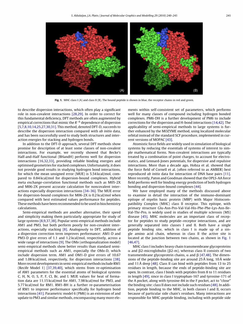
Fig. 1. MHC class I (A) and class II (B). The bound pep
o describe dispersion interactions, which often play a significantole in non-covalent interactions [28,29]. In order to correct forhis fundamental deficiency, DFT methods are often augmented bympirical corrections that mimic the R−6 dependence of dispersion5,7,8,10,14,25,27,30,31]. This method, denoted DFT-D, succeeds toescribe the dispersion interaction compared with ab initio data,nd has been successfully used to study both structures and inter-ction energies for stacking and hydrogen bonds.
In addition to the DFT-D approach, several DFT methods showromise for description of at least some classes of non-covalent
nteractions. For example, we recently showed that Becke’salf-and-Half functional (BHandH) performs well for dispersion
nteractions [16,32,33], providing reliable binding energies andptimised geometries for stacked complexes. Unfortunately, it doesot provide good results in studying hydrogen bond interactions,
or which the mean unsigned error (MUE) is 5.54 kcal/mol, com-ared to 0.84 kcal/mol for dispersion-bound complexes. Hybrideta exchange-correlation functional methods such as M05-2X
nd M06-2X present accurate calculation for noncovalent inter-ctions especially dispersion interactions [34–36]. The MUE erroror dispersion-bound complexes for M06-2X is 0.2 kcal/mol whenompared with best estimated values performance for peptides.hese methods have been recommended to be used in biochemistryalculations.
Semi-empirical methods are another alternative, their speednd simplicity making them particularly appropriate for study ofarge systems [8,9,37,38]. Popular semi-empirical methods includeM1 and PM3, but both perform poorly for non-covalent inter-ctions, especially stacking [8]. Analogously to DFT, addition ofdispersion correction term improves performance: AM1-D and
M3-D give errors of 1.1 and 1.2 kcal/mol, respectively, across aide range of interactions [9]. The OMx (orthogonalization model)
emi-empirical methods show better results than standard semi-mpirical methods such as AM1, especially the OMx-D, whichnclude dispersion term. AM1 and OM1-D give errors of 10.67nd 1.08 kcal/mol, respectively, for dispersion interactions [38].ore recent developments in semi-empirical methods include RM1
Recife Model 1) [37,39,40], which stems from re-optimisationf AM1 parameters for the essential atoms of biological systems, H, N, O, S, P, F, Cl, Br, and I. MUE values for heat of forma-ion data are 11.15 kcal/mol for AM1, 7.98 kcal/mol for PM3, and
.77 kcal/mol for RM1. RM1-BH is a further re-parameterisationf RM1 to improve performance specifically for hydrogen bondnteractions [41]. Parametric model 6 (PM6) is an extension of andpdate to PM3 and similar methods, encompassing many more ele-shown in blue, the receptor chains in red and green.
ments within self-consistent set of parameters, which performswell for many classes of compound including hydrogen bondedcomplexes. PM6-DH is a further development of PM6 to includecorrections for the dispersion and H-bond interactions [14,42]. Theapplicability of semi-empirical methods to large systems is fur-ther enhanced by the MOZYME method, using localized molecularorbital instead of the standard SCF procedure, implemented in cur-rent versions of MOPAC [43].
Atomistic force fields are widely used in simulation of biologicalsystems by reducing the essentials of systems of interest to sim-ple mathematical forms. Non-covalent interactions are typicallytreated by a combination of point charges, to account for electro-statics, and Lennard-Jones potentials, for dispersive and repulsiveinteractions. More than a decade ago, Hobza et al. showed thatthe force field of Cornell et al. (often referred to as AMBER) bestreproduced ab initio data for interaction of DNA base pairs [11].More recently, Paton and Goodman showed that the OPLS-AA forcefield performs well for binding energy prediction of both hydrogenbonding and dispersion-bound complexes [44].
We have employed many of the methods discussed aboveto examine in detail the interaction of the immuno-dominantepitope of myelin basic protein (MBP) with Major Histocom-patibility Complex (MHC) class II receptor. This epitope, withprimary structure Glu-Asn-Pro-Val-Val-His-Phe-Phe-Lys-Asn-Ile-Val-Thr-Pro, is widely used in studies of multiple sclerosis (MS)disease [45]. MHC molecules are an important class of recep-tor for peptides to study peptide–receptor interactions, and aregenerally separated into classes I and II. Both have a singlepeptide binding site, which in class I is made up of a sin-gle amino acid chain, whereas in class II the active site islocated at the junction between two chains, as shown in Fig. 1[46,47].
MHC class I includes heavy chain transmembrane glycoproteins� and �2-microglobulin (�2-m), whereas class II consists of twotransmembrane glycoprotein chains, � and � [47,48]. The dimen-sions of the peptide-binding site are around 25 Å long, 10 Å wideand 11 Å deep [48]. Class II can bind with peptides from 13 to 25residues in length, because the ends of peptide-binding site areopen. In contrast, class I binds with peptides from 8 to 11 residuesin length [49], since in class I tryptophan-167 and tyrosine-171 ofthe A pocket, along with tyrosine-84 in the F pocket, act to “close”
the binding site: class II does not include such residues [48]. In addi-tion, peptide binding to the MHC, in both classes I and II, occursbecause of particular side chain’s residues. Many interactions areresponsible for MHC-peptide binding, including with peptide side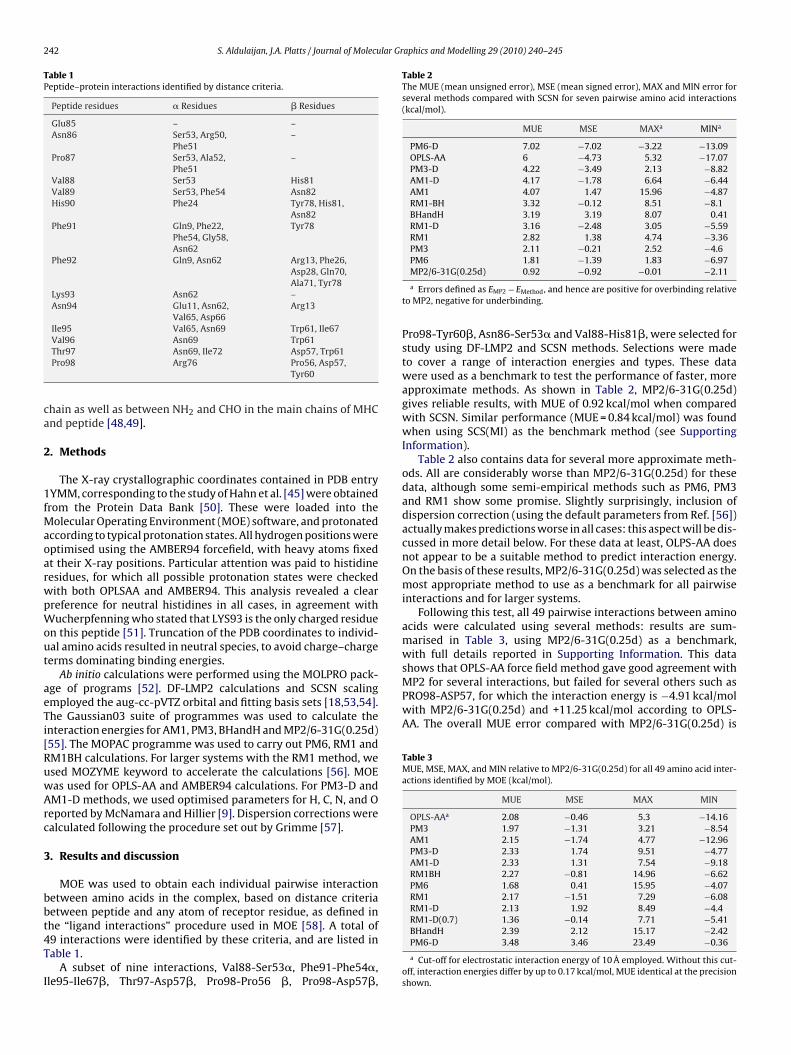
242 S. Aldulaijan, J.A. Platts / Journal of Molecular Graphics and Modelling 29 (2010) 240–245
Table 1Peptide–protein interactions identified by distance criteria.
Peptide residues � Residues � Residues
Glu85 – –Asn86 Ser53, Arg50,
Phe51–
Pro87 Ser53, Ala52,Phe51
–
Val88 Ser53 His81Val89 Ser53, Phe54 Asn82His90 Phe24 Tyr78, His81,
Asn82Phe91 Gln9, Phe22,
Phe54, Gly58,Asn62
Tyr78
Phe92 Gln9, Asn62 Arg13, Phe26,Asp28, Gln70,Ala71, Tyr78
Lys93 Asn62 –Asn94 Glu11, Asn62,
Val65, Asp66Arg13
Ile95 Val65, Asn69 Trp61, Ile67
ca
2
1fMaoarwpWout
aeTi[RuwArc
3
bbt4T
I
Table 2The MUE (mean unsigned error), MSE (mean signed error), MAX and MIN error forseveral methods compared with SCSN for seven pairwise amino acid interactions(kcal/mol).
MUE MSE MAXa MINa
PM6-D 7.02 −7.02 −3.22 −13.09OPLS-AA 6 −4.73 5.32 −17.07PM3-D 4.22 −3.49 2.13 −8.82AM1-D 4.17 −1.78 6.64 −6.44AM1 4.07 1.47 15.96 −4.87RM1-BH 3.32 −0.12 8.51 −8.1BHandH 3.19 3.19 8.07 0.41RM1-D 3.16 −2.48 3.05 −5.59RM1 2.82 1.38 4.74 −3.36PM3 2.11 −0.21 2.52 −4.6PM6 1.81 −1.39 1.83 −6.97
MP2 for several interactions, but failed for several others such asPRO98-ASP57, for which the interaction energy is −4.91 kcal/molwith MP2/6-31G(0.25d) and +11.25 kcal/mol according to OPLS-AA. The overall MUE error compared with MP2/6-31G(0.25d) is
Table 3MUE, MSE, MAX, and MIN relative to MP2/6-31G(0.25d) for all 49 amino acid inter-actions identified by MOE (kcal/mol).
MUE MSE MAX MIN
OPLS-AAa 2.08 −0.46 5.3 −14.16PM3 1.97 −1.31 3.21 −8.54AM1 2.15 −1.74 4.77 −12.96PM3-D 2.33 1.74 9.51 −4.77AM1-D 2.33 1.31 7.54 −9.18RM1BH 2.27 −0.81 14.96 −6.62PM6 1.68 0.41 15.95 −4.07RM1 2.17 −1.51 7.29 −6.08RM1-D 2.13 1.92 8.49 −4.4RM1-D(0.7) 1.36 −0.14 7.71 −5.41
Val96 Asn69 Trp61Thr97 Asn69, Ile72 Asp57, Trp61Pro98 Arg76 Pro56, Asp57,
Tyr60
hain as well as between NH2 and CHO in the main chains of MHCnd peptide [48,49].
. Methods
The X-ray crystallographic coordinates contained in PDB entryYMM, corresponding to the study of Hahn et al. [45] were obtainedrom the Protein Data Bank [50]. These were loaded into the
olecular Operating Environment (MOE) software, and protonatedccording to typical protonation states. All hydrogen positions wereptimised using the AMBER94 forcefield, with heavy atoms fixedt their X-ray positions. Particular attention was paid to histidineesidues, for which all possible protonation states were checkedith both OPLSAA and AMBER94. This analysis revealed a clearreference for neutral histidines in all cases, in agreement withucherpfenning who stated that LYS93 is the only charged residue
n this peptide [51]. Truncation of the PDB coordinates to individ-al amino acids resulted in neutral species, to avoid charge–chargeerms dominating binding energies.
Ab initio calculations were performed using the MOLPRO pack-ge of programs [52]. DF-LMP2 calculations and SCSN scalingmployed the aug-cc-pVTZ orbital and fitting basis sets [18,53,54].he Gaussian03 suite of programmes was used to calculate thenteraction energies for AM1, PM3, BHandH and MP2/6-31G(0.25d)55]. The MOPAC programme was used to carry out PM6, RM1 andM1BH calculations. For larger systems with the RM1 method, wesed MOZYME keyword to accelerate the calculations [56]. MOEas used for OPLS-AA and AMBER94 calculations. For PM3-D andM1-D methods, we used optimised parameters for H, C, N, and Oeported by McNamara and Hillier [9]. Dispersion corrections werealculated following the procedure set out by Grimme [57].
. Results and discussion
MOE was used to obtain each individual pairwise interactionetween amino acids in the complex, based on distance criteriaetween peptide and any atom of receptor residue, as defined in
he “ligand interactions” procedure used in MOE [58]. A total of9 interactions were identified by these criteria, and are listed inable 1.A subset of nine interactions, Val88-Ser53�, Phe91-Phe54�,le95-Ile67�, Thr97-Asp57�, Pro98-Pro56 �, Pro98-Asp57�,
MP2/6-31G(0.25d) 0.92 −0.92 −0.01 −2.11
a Errors defined as EMP2 − EMethod, and hence are positive for overbinding relativeto MP2, negative for underbinding.
Pro98-Tyr60�, Asn86-Ser53� and Val88-His81�, were selected forstudy using DF-LMP2 and SCSN methods. Selections were madeto cover a range of interaction energies and types. These datawere used as a benchmark to test the performance of faster, moreapproximate methods. As shown in Table 2, MP2/6-31G(0.25d)gives reliable results, with MUE of 0.92 kcal/mol when comparedwith SCSN. Similar performance (MUE = 0.84 kcal/mol) was foundwhen using SCS(MI) as the benchmark method (see SupportingInformation).
Table 2 also contains data for several more approximate meth-ods. All are considerably worse than MP2/6-31G(0.25d) for thesedata, although some semi-empirical methods such as PM6, PM3and RM1 show some promise. Slightly surprisingly, inclusion ofdispersion correction (using the default parameters from Ref. [56])actually makes predictions worse in all cases: this aspect will be dis-cussed in more detail below. For these data at least, OLPS-AA doesnot appear to be a suitable method to predict interaction energy.On the basis of these results, MP2/6-31G(0.25d) was selected as themost appropriate method to use as a benchmark for all pairwiseinteractions and for larger systems.
Following this test, all 49 pairwise interactions between aminoacids were calculated using several methods: results are sum-marised in Table 3, using MP2/6-31G(0.25d) as a benchmark,with full details reported in Supporting Information. This datashows that OPLS-AA force field method gave good agreement with
BHandH 2.39 2.12 15.17 −2.42PM6-D 3.48 3.46 23.49 −0.36
a Cut-off for electrostatic interaction energy of 10 Å employed. Without this cut-off, interaction energies differ by up to 0.17 kcal/mol, MUE identical at the precisionshown.

S. Aldulaijan, J.A. Platts / Journal of Molecular Graphics and Modelling 29 (2010) 240–245 243
2Tg
ijcam1tis
amTdaaTarmb
E
wmf
TS
Table 5Interaction energies of larger models (kcal/mol).
DipeptidesGlu85-Asn86 10.62Pro87-Val88 −6.34Val89-His90 −7.45Phe91-Phe92 −17.88Lys93-Asn94 −58.84Ile95-Val96 −7.63Thr97-Pro98 1.72Sum −85.79
Fig. 2. Response of RM1-D error as a function of global scaling parameter s6.
.08 kcal/mol, which although rather smaller than that reported inable 2 (for fewer interactions) is rather greater than the 1 kcal/molenerally accepted as “chemical accuracy”.
BHandH results in overestimated interaction energies, fornstance �E for PHE92-GLN9 is −12.3 kcal/mol using BHandH andust −5.01 kcal/mol with MP2/6-31G(0.25d). The overall MUE erroromparing with MP2/6-31G(0.25) calculations is 2.39 kcal/mol,gain rather larger than required for our purposes. Semi-empiricalethods PM3 and AM1 give similar overall errors, with MUE of
.97 kca/mol for PM3 and 2.15 kcal/mol for AM1, and more oftenhan not underestimate interaction energies. Similar performances found for re-parameterisations RM1 and RM1-BH, whereas PM6hows a slight improvement over other related methods.
Including a dispersion correction, and modifying parametersccording to McNamara and Hillier [9], does not improve perfor-ance, with MUE of 2.33 kcal/mol for both AM1-D and PM3-D.
o analyse these data in more detail, errors for hydrogen bonded,ispersion bound, and charged interactions were calculated sep-rately, and found to be 1.92, 2.41 and 2.82 kcal/mol for PM3-D,nd 2.36, 2.23, and 3.07 kcal/mol for AM1-D, respectively. Notably,able 3 shows that the mean signed error (MSE) changes sign onddition of dispersion, suggesting that the default dispersion cor-ection overcompensates for the shortcomings of the underlyingethods in these cases. The form of the dispersion correction given
y Grimme is shown in Eq. (1):
disp = −s6
n−1∑ n∑ Cij66
fdmp(Rij) (1)
i=1 j=i+1R
ij
here s6 is a global scaling factor that must be optimised for theethod to be corrected. Tables 2 and 3 employed the default scaling
actor of 1.4, as in the initial reports of AM1-D and PM3-D method.
able 4tabilisation energies due to each residue in peptide (kcal/mol).
Sum of pairwise Direct Many body
MP2 RM1-D RM1-D
Glu85 – – – –Asn86 +18.44 +11.51 +14.20 +2.69Pro87 −3.67 −3.30 −3.21 +0.09Val88 −4.77 −4.05 −2.09 +1.96Val89 −4.56 −3.41 −3.52 −0.11His90 −7.59 −4.77 −3.76 +1.01Phe91 −6.52 −8.05 −8.36 −0.31Phe92 −21.93 −13.62 −17.85 −4.23Lys93 −6.12 −5.86 −5.86 0.00Asn94 −15.71 −21.08 −16.77 +4.33Ile95 −1.73 −2.86 −1.60 +1.26Val96 −6.70 −3.93 −3.84 +0.09Thr97 +2.22 −2.87 −5.36 −2.49Pro98 +1.97 +3.28 +0.70 −2.58
HeptapeptidesGlu85-Phe91 −20.16Phe92-Pro98 −81.83Sum −101.99
Varying the s6 parameter, the optimal combination of method andglobal scaling was located to be RM1 with s6 = 0.7, for which anoverall MUE of 1.36 kcal/mol was obtained. The response of thisoverall error to the value of s6 is shown in Fig. 2. Repeating this pro-cedure with other semi-empirical methods gave similar curves butslightly higher MUE values, with the exception of PM6 for whichthe optimal value of s6 was zero, i.e. any attempt to improve onpredictions by adding a dispersion term actually gave worse per-formance.
Previous studies [46,51,59] indicate that the most importantresidues for interaction of this MBP epitope with MHC-II are thecentral residues Val88 to Asn94. In general, our calculations are inagreement with these findings, indicating that most stabilisationof the complex stems from these residues’ interactions. Table 4reports the sum of individual pairwise interaction energies fromboth MP2/6-31G(0.25d) and RM1-D with s6 = 0.7. Both methodsshow that Phe92 and Asn94 are particularly strongly stabilising,with significant further contributions from all except the terminalresidues. Table 4 also reports interaction energies of each pep-tide residue with all receptor residues identified as interacting,calculated with RM1-D. These data show very similar trends tothose from pairwise data, with Phe92 and Asn94 considerablymore stabilising than other residues, and terminal Asn86 and Pro98destabilising the complex. The difference between pairwise anddirect interaction energies is the many-body term: no clear trendis apparent in this data, but the size of this term in some cases,e.g. more than 4 kcal/mol for Phe92, suggests that conclusions frompairwise calculations should be treated with caution. Summingeither pairwise or direct interaction energies gives estimates of theoverall stabilisation of the complex of −59.0 and −57.3 kcal/mol,respectively.
The speed of semi-empirical methods such as RM1 allows us toexamine the interaction energies of larger models of the peptidethan single amino acids. The peptide was cut into seven dipeptides,as well as two heptapeptides, and the interaction energy of thepeptide with all receptor residues identified in Table 1 calculated,as shown in Table 5. These data further indicate that stabilisationcomes mainly from the central residues, with the dipeptide Lys93-Asn94 particularly strongly bound. Table 5 also shows that muchgreater binding results from the heptapeptide Phe92-Pro98 thanfrom Glu85-Phe91, perhaps unsurprisingly given that this containsboth of the most strongly bound single residues.
These data also show that the estimated overall stabilisa-tion of the complex increases as the size of the model peptidesincrease. Considering only single amino acids, RM1-D estimate is−57.3 kcal/mol, rising to −85.8 kcal/mol from dipeptides and to
−102.0 kcal/mol from heptapeptides. Calculating all 14 residuesas one molecule with all 29 receptor residues gives an interactionenergy of −110.0 kcal/mol, i.e. larger still. From these data it seemsclear that cutting the peptide into individual amino acids causessignificant error in prediction of overall binding energy. The size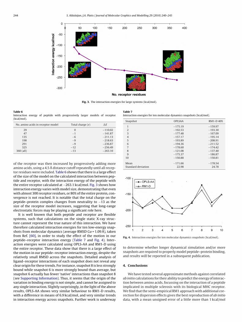
244 S. Aldulaijan, J.A. Platts / Journal of Molecular Graphics and Modelling 29 (2010) 240–245
Fig. 3. The interaction energies for large systems (kcal/mol).
Table 6Interaction energy of peptide with progressively larger models of receptor(kcal/mol).
No. amino acids in receptor model Total charge (e) �E
29 0 −110.0247 −1 −141.87
135 −6 −211.13198 −9 −218.63
oatottiwvpse
sttsfpattrlcbs(varwi
Table 7Interaction energies for ten molecular dynamics snapshots (kcal/mol).
Snapshot OPLSAA RM1-D 40%
1 −175.19 −159.972 −192.53 −193.303 −177.48 −167.094 −157.17 −195.145 −193.89 −208.916 −194.36 −211.527 −178.60 −174.428 −121.08 −137.409 −175.37 −186.87
10 −150.88 −150.81
Mean −171.66 −178.54Standard deviation 22.98 24.78
291 −9 −236.87325 −12 −256.49
360 (all) −13 −263.10
f the receptor was then increased by progressively adding moremino acids, using a 4.5 Å distance cutoff repeatedly until all recep-or residues were included. Table 6 shows that there is a large effectf the size of the model on the calculated interaction between pep-ide and receptor, with the interaction energy of the peptide withhe entire receptor calculated at −263.1 kcal/mol. Fig. 3 shows hownteraction energy varies with model size, demonstrating that even
ith almost 300 receptor residues, or 80% of the entire protein, con-ergence is not reached. It is notable that the total charge on theeptide–protein complex changes from neutrality to −13 as theize of the receptor model increases, suggesting that long-rangelectrostatic forces may be playing a significant role here.
It is well known that both peptide and receptor are flexibleystems, such that calculations on the single static X-ray struc-ure cannot represent the true nature of this interaction. We haveherefore calculated interaction energies for ten low-energy snap-hots from molecular dynamics (average RMSD C˛ = 1.09 Å), takenrom Ref. [60], in order to study the effect of the motion in oureptide–receptor interaction energy (Table 7 and Fig. 4). Inter-ction energies were calculated using OPLS-AA and RM1-D usinghe entire receptor. These data show that there is a large effect ofhe motion in our peptide–receptor interaction energy, despite theelatively small RMSD across the snapshots. Detailed analysis ofigand–receptor interactions of each snapshot does not reveal anylear origin for these trends. For instance, snapshot 8 is less stronglyound while snapshot 6 is more strongly bound than average, butnapshot 6 actually has fewer ‘native’ interactions than snapshot 8see Supporting Information). Thus, it seems that the origin of theariation in binding energy is not simple, and cannot be assigned to
ny single interaction. Slightly surprisingly, in the light of the aboveesults, OPLS-AA shows very similar behaviour to RM1-D results,ith a difference in means of 6.9 kcal/mol, and very similar trendsn interaction energy across snapshots. Further work is underway
Fig. 4. Interaction energies for ten molecular dynamics snapshots (kcal/mol).
to determine whether longer dynamical simulation and/or moresnapshots are required to properly model peptide–protein binding,and results will be reported in a subsequent publication.
4. Conclusions
We have tested several approximate methods against correlatedab initio calculations for their ability to predict the energy of interac-tion between amino acids, focussing on the interaction of a peptide
implicated in multiple sclerosis with its biological MHC receptor.We find that the semi-empirical RM1 approach with additional cor-rection for dispersion effects gives the best reproduction of ab initiodata, with a mean unsigned error of a little more than 1 kcal/mol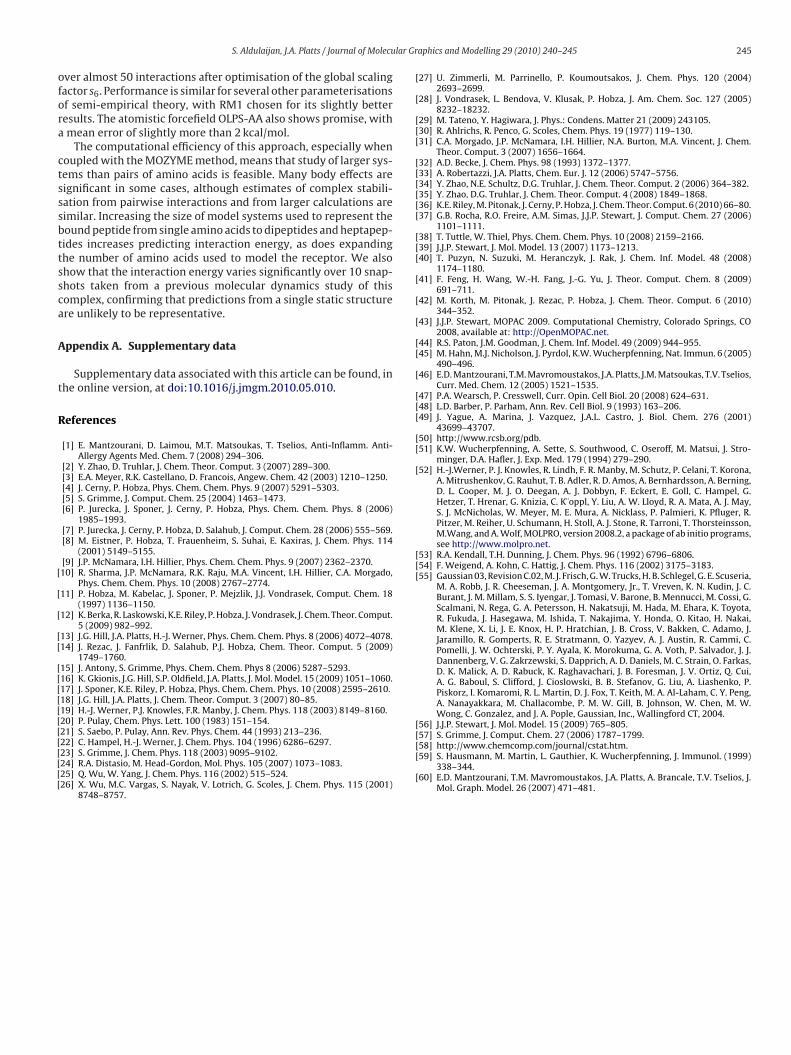
lar Gr
ofora
ctsssbttssca
A
t
R
[
[
[
[[
[[[[[[[[[[[[
[
[
[[[
[[[[[[
[[[
[
[
[
[[
[
[[[
[[
[
[[[
[
S. Aldulaijan, J.A. Platts / Journal of Molecu
ver almost 50 interactions after optimisation of the global scalingactor s6. Performance is similar for several other parameterisationsf semi-empirical theory, with RM1 chosen for its slightly betteresults. The atomistic forcefield OLPS-AA also shows promise, withmean error of slightly more than 2 kcal/mol.
The computational efficiency of this approach, especially whenoupled with the MOZYME method, means that study of larger sys-ems than pairs of amino acids is feasible. Many body effects areignificant in some cases, although estimates of complex stabili-ation from pairwise interactions and from larger calculations areimilar. Increasing the size of model systems used to represent theound peptide from single amino acids to dipeptides and heptapep-ides increases predicting interaction energy, as does expandinghe number of amino acids used to model the receptor. We alsohow that the interaction energy varies significantly over 10 snap-hots taken from a previous molecular dynamics study of thisomplex, confirming that predictions from a single static structurere unlikely to be representative.
ppendix A. Supplementary data
Supplementary data associated with this article can be found, inhe online version, at doi:10.1016/j.jmgm.2010.05.010.
eferences
[1] E. Mantzourani, D. Laimou, M.T. Matsoukas, T. Tselios, Anti-Inflamm. Anti-Allergy Agents Med. Chem. 7 (2008) 294–306.
[2] Y. Zhao, D. Truhlar, J. Chem. Theor. Comput. 3 (2007) 289–300.[3] E.A. Meyer, R.K. Castellano, D. Francois, Angew. Chem. 42 (2003) 1210–1250.[4] J. Cerny, P. Hobza, Phys. Chem. Chem. Phys. 9 (2007) 5291–5303.[5] S. Grimme, J. Comput. Chem. 25 (2004) 1463–1473.[6] P. Jurecka, J. Sponer, J. Cerny, P. Hobza, Phys. Chem. Chem. Phys. 8 (2006)
1985–1993.[7] P. Jurecka, J. Cerny, P. Hobza, D. Salahub, J. Comput. Chem. 28 (2006) 555–569.[8] M. Eistner, P. Hobza, T. Frauenheim, S. Suhai, E. Kaxiras, J. Chem. Phys. 114
(2001) 5149–5155.[9] J.P. McNamara, I.H. Hillier, Phys. Chem. Chem. Phys. 9 (2007) 2362–2370.10] R. Sharma, J.P. McNamara, R.K. Raju, M.A. Vincent, I.H. Hillier, C.A. Morgado,
Phys. Chem. Chem. Phys. 10 (2008) 2767–2774.11] P. Hobza, M. Kabelac, J. Sponer, P. Mejzlik, J.J. Vondrasek, Comput. Chem. 18
(1997) 1136–1150.12] K. Berka, R. Laskowski, K.E. Riley, P. Hobza, J. Vondrasek, J. Chem. Theor. Comput.
5 (2009) 982–992.13] J.G. Hill, J.A. Platts, H.-J. Werner, Phys. Chem. Chem. Phys. 8 (2006) 4072–4078.14] J. Rezac, J. Fanfrlik, D. Salahub, P.J. Hobza, Chem. Theor. Comput. 5 (2009)
1749–1760.15] J. Antony, S. Grimme, Phys. Chem. Chem. Phys 8 (2006) 5287–5293.16] K. Gkionis, J.G. Hill, S.P. Oldfield, J.A. Platts, J. Mol. Model. 15 (2009) 1051–1060.17] J. Sponer, K.E. Riley, P. Hobza, Phys. Chem. Chem. Phys. 10 (2008) 2595–2610.18] J.G. Hill, J.A. Platts, J. Chem. Theor. Comput. 3 (2007) 80–85.19] H.-J. Werner, P.J. Knowles, F.R. Manby, J. Chem. Phys. 118 (2003) 8149–8160.20] P. Pulay, Chem. Phys. Lett. 100 (1983) 151–154.21] S. Saebo, P. Pulay, Ann. Rev. Phys. Chem. 44 (1993) 213–236.
22] C. Hampel, H.-J. Werner, J. Chem. Phys. 104 (1996) 6286–6297.23] S. Grimme, J. Chem. Phys. 118 (2003) 9095–9102.24] R.A. Distasio, M. Head-Gordon, Mol. Phys. 105 (2007) 1073–1083.25] Q. Wu, W. Yang, J. Chem. Phys. 116 (2002) 515–524.26] X. Wu, M.C. Vargas, S. Nayak, V. Lotrich, G. Scoles, J. Chem. Phys. 115 (2001)8748–8757.
[[[
[
aphics and Modelling 29 (2010) 240–245 245
27] U. Zimmerli, M. Parrinello, P. Koumoutsakos, J. Chem. Phys. 120 (2004)2693–2699.
28] J. Vondrasek, L. Bendova, V. Klusak, P. Hobza, J. Am. Chem. Soc. 127 (2005)8232–18232.
29] M. Tateno, Y. Hagiwara, J. Phys.: Condens. Matter 21 (2009) 243105.30] R. Ahlrichs, R. Penco, G. Scoles, Chem. Phys. 19 (1977) 119–130.31] C.A. Morgado, J.P. McNamara, I.H. Hillier, N.A. Burton, M.A. Vincent, J. Chem.
Theor. Comput. 3 (2007) 1656–1664.32] A.D. Becke, J. Chem. Phys. 98 (1993) 1372–1377.33] A. Robertazzi, J.A. Platts, Chem. Eur. J. 12 (2006) 5747–5756.34] Y. Zhao, N.E. Schultz, D.G. Truhlar, J. Chem. Theor. Comput. 2 (2006) 364–382.35] Y. Zhao, D.G. Truhlar, J. Chem. Theor. Comput. 4 (2008) 1849–1868.36] K.E. Riley, M. Pitonak, J. Cerny, P. Hobza, J. Chem. Theor. Comput. 6 (2010) 66–80.37] G.B. Rocha, R.O. Freire, A.M. Simas, J.J.P. Stewart, J. Comput. Chem. 27 (2006)
1101–1111.38] T. Tuttle, W. Thiel, Phys. Chem. Chem. Phys. 10 (2008) 2159–2166.39] J.J.P. Stewart, J. Mol. Model. 13 (2007) 1173–1213.40] T. Puzyn, N. Suzuki, M. Heranczyk, J. Rak, J. Chem. Inf. Model. 48 (2008)
1174–1180.41] F. Feng, H. Wang, W.-H. Fang, J.-G. Yu, J. Theor. Comput. Chem. 8 (2009)
691–711.42] M. Korth, M. Pitonak, J. Rezac, P. Hobza, J. Chem. Theor. Comput. 6 (2010)
344–352.43] J.J.P. Stewart, MOPAC 2009. Computational Chemistry, Colorado Springs, CO
2008, available at: http://OpenMOPAC.net.44] R.S. Paton, J.M. Goodman, J. Chem. Inf. Model. 49 (2009) 944–955.45] M. Hahn, M.J. Nicholson, J. Pyrdol, K.W. Wucherpfenning, Nat. Immun. 6 (2005)
490–496.46] E.D. Mantzourani, T.M. Mavromoustakos, J.A. Platts, J.M. Matsoukas, T.V. Tselios,
Curr. Med. Chem. 12 (2005) 1521–1535.47] P.A. Wearsch, P. Cresswell, Curr. Opin. Cell Biol. 20 (2008) 624–631.48] L.D. Barber, P. Parham, Ann. Rev. Cell Biol. 9 (1993) 163–206.49] J. Yague, A. Marina, J. Vazquez, J.A.L. Castro, J. Biol. Chem. 276 (2001)
43699–43707.50] http://www.rcsb.org/pdb.51] K.W. Wucherpfenning, A. Sette, S. Southwood, C. Oseroff, M. Matsui, J. Stro-
minger, D.A. Hafler, J. Exp. Med. 179 (1994) 279–290.52] H.-J.Werner, P. J. Knowles, R. Lindh, F. R. Manby, M. Schutz, P. Celani, T. Korona,
A. Mitrushenkov, G. Rauhut, T. B. Adler, R. D. Amos, A. Bernhardsson, A. Berning,D. L. Cooper, M. J. O. Deegan, A. J. Dobbyn, F. Eckert, E. Goll, C. Hampel, G.Hetzer, T. Hrenar, G. Knizia, C. K¨oppl, Y. Liu, A. W. Lloyd, R. A. Mata, A. J. May,S. J. McNicholas, W. Meyer, M. E. Mura, A. Nicklass, P. Palmieri, K. Pfluger, R.Pitzer, M. Reiher, U. Schumann, H. Stoll, A. J. Stone, R. Tarroni, T. Thorsteinsson,M.Wang, and A. Wolf, MOLPRO, version 2008.2, a package of ab initio programs,see http://www.molpro.net.
53] R.A. Kendall, T.H. Dunning, J. Chem. Phys. 96 (1992) 6796–6806.54] F. Weigend, A. Kohn, C. Hattig, J. Chem. Phys. 116 (2002) 3175–3183.55] Gaussian 03, Revision C.02, M. J. Frisch, G. W. Trucks, H. B. Schlegel, G. E. Scuseria,
M. A. Robb, J. R. Cheeseman, J. A. Montgomery, Jr., T. Vreven, K. N. Kudin, J. C.Burant, J. M. Millam, S. S. Iyengar, J. Tomasi, V. Barone, B. Mennucci, M. Cossi, G.Scalmani, N. Rega, G. A. Petersson, H. Nakatsuji, M. Hada, M. Ehara, K. Toyota,R. Fukuda, J. Hasegawa, M. Ishida, T. Nakajima, Y. Honda, O. Kitao, H. Nakai,M. Klene, X. Li, J. E. Knox, H. P. Hratchian, J. B. Cross, V. Bakken, C. Adamo, J.Jaramillo, R. Gomperts, R. E. Stratmann, O. Yazyev, A. J. Austin, R. Cammi, C.Pomelli, J. W. Ochterski, P. Y. Ayala, K. Morokuma, G. A. Voth, P. Salvador, J. J.Dannenberg, V. G. Zakrzewski, S. Dapprich, A. D. Daniels, M. C. Strain, O. Farkas,D. K. Malick, A. D. Rabuck, K. Raghavachari, J. B. Foresman, J. V. Ortiz, Q. Cui,A. G. Baboul, S. Clifford, J. Cioslowski, B. B. Stefanov, G. Liu, A. Liashenko, P.Piskorz, I. Komaromi, R. L. Martin, D. J. Fox, T. Keith, M. A. Al-Laham, C. Y. Peng,A. Nanayakkara, M. Challacombe, P. M. W. Gill, B. Johnson, W. Chen, M. W.Wong, C. Gonzalez, and J. A. Pople, Gaussian, Inc., Wallingford CT, 2004.
56] J.J.P. Stewart, J. Mol. Model. 15 (2009) 765–805.
57] S. Grimme, J. Comput. Chem. 27 (2006) 1787–1799.58] http://www.chemcomp.com/journal/cstat.htm.59] S. Hausmann, M. Martin, L. Gauthier, K. Wucherpfenning, J. Immunol. (1999)338–344.60] E.D. Mantzourani, T.M. Mavromoustakos, J.A. Platts, A. Brancale, T.V. Tselios, J.
Mol. Graph. Model. 26 (2007) 471–481.


![Noninvasive Molecular Imaging of MYC mRNA Expression in Human Breast Cancer Xenografts with a [ 99m Tc]Peptide−Peptide Nucleic Acid−Peptide Chimera](https://static.fdokumen.com/doc/165x107/63214cddbc33ec48b20e4a4a/noninvasive-molecular-imaging-of-myc-mrna-expression-in-human-breast-cancer-xenografts.jpg)