Processing of major histocompatibility class I-restricted antigens in the endoplasmic reticulum
11
Processing of Major Histocompatibility Class I-restricted Antigens in the Endoplasmic Reticulum By Tim EUiott,* Anthony WiUis,r Vincenzo Cerundolo,* and Alain Townsend* From the "Molecular Immunology Group, Institute for Molecular Medicine,John Radcliffe Hospital, Oxford OX3 9DU; and ~Department of Biochemistry, University of Oxford, Oxford 0)(I, United Kingdom SumlTlal~ We have introduced long precursor peptides directly into the endoplasmic reticulum (ER) of a mutant cell line (T2-D b) that lacks the ability to transport peptides from the cytosol to the ER in a transporter associated with antigen processing (TAP)-dependent way. This was done by expressing various influenza A-derived peptides containing the naturally processed epitope ASNENMDAM (366-374) preceded by the influenza hemagglutinin ER translocation sequence. Peptides derived from these minigenes that became associated with D b were isolated and identified by combined reversed phase liquid chromatography and detection by cytotoxic T lym- phocytes. Our results establish that NH2-terminal extensions of at least 40 residues can be trimmed from peptides entering the ER, but that proteolysis of larger proteins may be limited. C TLs recognize peptide epitopes of 8-10 amino acids, bound to polymorphic receptors encoded by the major histocompatibility complex (1-3). The MHC peptide/class I complex is formed in the endoplasmic reticulum (ER) 1 be- fore it translocates to the call surface. Peptide binding to newly synthesized class I molecules is an integral part of the stable assembly of the MHC molecule itself and may be a prerequi- site for transit of the complex to the cell surface (4, 5). The production of most peptides destined for presenta- tion by class I MHC begins in the cytosol with the limited hydrolysis of antigenic proteins. The proteases involved in this step have not yet been identified, but attention has re- cently focused on the proteasome- a multicatalytic complex in the cytosol with one ATP-dependent proteolytic activity, specific for ubiquitinated substrates, and three ATP-indepen- dent activities (6). The involvement of the former activity in antigen processing has been implied in two experimental systems (7, 8). To date, there is no evidence that proteasomes generate peptides of a size that is optimal for binding to class I. However, recent investigations into the regulation of pro- teasome specificity by its two MHC-encoded subunits has led to the speculation that proteasomes may generate pep- tides with COOH termini that are appropriate for binding 1Abbreviationsusedin thispaper: ~2m, B2-microglobin;BFA, brefeldin A; ER, endoplasmic reticulum; HA, hemagglutinin; NP, nucleoprotein; RPLC, reverse phase liquid chromatography;TAP, transporter associated with antigen processingcomplex; TBS, Tris-bufferedsaline; TLCK, Nct- p-tosyl-t-chloromethyl ketone. to class I (9, 10) and that these may be trimmed in the ER at their NH2 termini, as suggested by Falk et al. (11). The topological paradox posed by the observations that peptide production and peptide binding to class I occur in two distinct intraceltular compartments can be resolved only if there exists a mechanism for translocating peptides across the ER membrane in a signal sequence-independent way (12, 13). Recently, a polymorphic, heterodimeric protein encoded in the MHC class II region has been identified that fulfills this function (14-17). The complex, known as TAP (trans- porter associated with antigen processing [reviewed in refer- ence 18]), belongs to a family of ATP-binding cassette pro- teins and has been shown to transport peptides from the cytosolic to the lumenal side of the ER membrane in an ATP- dependent way (19, 20). The processes by which short peptides are selected for pre- sentation at the cell surface are not fully understood. There is ample evidence for the involvement of the class I molecule itself in selecting peptides, with which it forms the most stable complexes, during the assembly of the heavy chain:3z microglobin (~zm):peptide heterotrimer (11 and reviewed in reference 21). However, the observation that few of the total number of class I-binding peptides that could be generated from any given viral protein are actually selected for use as epitopes suggests that other processes could contribute to epitope selection in vivo. This notion is supported by examples in which different peptide epitopes are presented by the same class I molecule depending on the cellular context in which that class I molecule is expressed (22). Attention has there- fore turned to the influence of proteases and the peptide trans- porters on selecting peptides for class I binding. 1481 J. Exp. Med. 9 The Rockefeller University Press 9 0022-1007/95/04/1481/11 $2.00 Volume 181 April 1995 1481-1491 on June 24, 2015 jem.rupress.org Downloaded from Published April 1, 1995
Transcript of Processing of major histocompatibility class I-restricted antigens in the endoplasmic reticulum

Processing of Major Histocompatibility Class I-restricted Antigens in the Endoplasmic Reticulum By Tim EUiott,* Anthony WiUis,r Vincenzo Cerundolo,* and Alain Townsend*
From the "Molecular Immunology Group, Institute for Molecular Medicine, John Radcliffe Hospital, Oxford OX3 9DU; and ~Department of Biochemistry, University of Oxford, Oxford 0)(I, United Kingdom
SumlTlal~
We have introduced long precursor peptides directly into the endoplasmic reticulum (ER) of a mutant cell line (T2-D b) that lacks the ability to transport peptides from the cytosol to the ER in a transporter associated with antigen processing (TAP)-dependent way. This was done by expressing various influenza A-derived peptides containing the naturally processed epitope ASNENMDAM (366-374) preceded by the influenza hemagglutinin ER translocation sequence. Peptides derived from these minigenes that became associated with D b were isolated and identified by combined reversed phase liquid chromatography and detection by cytotoxic T lym- phocytes. Our results establish that NH2-terminal extensions of at least 40 residues can be trimmed from peptides entering the ER, but that proteolysis of larger proteins may be limited.
C T L s recognize peptide epitopes of 8-10 amino acids, bound to polymorphic receptors encoded by the major
histocompatibility complex (1-3). The MHC peptide/class I complex is formed in the endoplasmic reticulum (ER) 1 be- fore it translocates to the call surface. Peptide binding to newly synthesized class I molecules is an integral part of the stable assembly of the MHC molecule itself and may be a prerequi- site for transit of the complex to the cell surface (4, 5).
The production of most peptides destined for presenta- tion by class I MHC begins in the cytosol with the limited hydrolysis of antigenic proteins. The proteases involved in this step have not yet been identified, but attention has re- cently focused on the proteasome- a multicatalytic complex in the cytosol with one ATP-dependent proteolytic activity, specific for ubiquitinated substrates, and three ATP-indepen- dent activities (6). The involvement of the former activity in antigen processing has been implied in two experimental systems (7, 8). To date, there is no evidence that proteasomes generate peptides of a size that is optimal for binding to class I. However, recent investigations into the regulation of pro- teasome specificity by its two MHC-encoded subunits has led to the speculation that proteasomes may generate pep- tides with COOH termini that are appropriate for binding
1 Abbreviations used in this paper: ~2m, B2-microglobin; BFA, brefeldin A; ER, endoplasmic reticulum; HA, hemagglutinin; NP, nucleoprotein; RPLC, reverse phase liquid chromatography; TAP, transporter associated with antigen processing complex; TBS, Tris-buffered saline; TLCK, Nct- p-tosyl-t-chloromethyl ketone.
to class I (9, 10) and that these may be trimmed in the ER at their NH2 termini, as suggested by Falk et al. (11).
The topological paradox posed by the observations that peptide production and peptide binding to class I occur in two distinct intraceltular compartments can be resolved only if there exists a mechanism for translocating peptides across the ER membrane in a signal sequence-independent way (12, 13). Recently, a polymorphic, heterodimeric protein encoded in the MHC class II region has been identified that fulfills this function (14-17). The complex, known as TAP (trans- porter associated with antigen processing [reviewed in refer- ence 18]), belongs to a family of ATP-binding cassette pro- teins and has been shown to transport peptides from the cytosolic to the lumenal side of the ER membrane in an ATP- dependent way (19, 20).
The processes by which short peptides are selected for pre- sentation at the cell surface are not fully understood. There is ample evidence for the involvement of the class I molecule itself in selecting peptides, with which it forms the most stable complexes, during the assembly of the heavy chain:3z microglobin (~zm):peptide heterotrimer (11 and reviewed in reference 21). However, the observation that few of the total number of class I-binding peptides that could be generated from any given viral protein are actually selected for use as epitopes suggests that other processes could contribute to epitope selection in vivo. This notion is supported by examples in which different peptide epitopes are presented by the same class I molecule depending on the cellular context in which that class I molecule is expressed (22). Attention has there- fore turned to the influence of proteases and the peptide trans- porters on selecting peptides for class I binding.
1481 J. Exp. Med. �9 The Rockefeller University Press �9 0022-1007/95/04/1481/11 $2.00 Volume 181 April 1995 1481-1491
on June 24, 2015jem
.rupress.orgD
ownloaded from
Published April 1, 1995
Recent evidence has shown that peptides longer than 9 amino acids can be efficiently transported into the ER. The first indication for this came from the demonstration that peptides of up to 11 amino acids, derived from cytosolic and nuclear proteins could be isolated from class I molecules (23). In addition, Udaka et al. (24) have isolated a 16-amino acid peptide from H-2L a that is derived from the cytosolicaUy synthesized enzyme 2-oxoglutarate dehydrogenase and that is recognized by allospecific CTLs. Recently, direct measure- ments of the ATP-dependent transport function of the TAP complex have been made (19, 20). These show that long peptides (up to 23 amino acids) are capable of competing against shorter peptides for transport (20). Further indirect evidence for the transport of long peptides by the TAP com- plex has come from Eisenlohr et al. (25), who have shown that an H-2Kb-restricted epitope could be generated from a 12-amino acid precursor synthesized as a minigene only if (a) an appropriate proteolytic enzyme specificity was co- expressed in the ER and (b) the TAP complex was functional. These results could be explained only if the longer precursor peptide was translocated into the ER by the TAP complex, followed by cleavage to the nonamer.
We demonstrate here that in ceils lacking TAP proteins, large fragments of influenza nucleoprotein (NP) translocated into the ER via a hydrophobic signal sequence can be trimmed to the authentic 9-residue epitope recognized by CTLs. At least 40 amino acids between the signal sequence and the epi- tope can be removed, although larger extensions inhibit trim- ming. These results establish that an NH2-terminal trim- ruing mechanism does exist in the ER lumen and show that assembly of the resulting short peptide with class I mole- cules under these conditions can occur in the absence of the TAP proteins.
Materials and Methods Cells, Antibodies and Peptides. The TAP-defective cell line T2
was transfected with H-2D b and H-2K k as previously described (26). Murine L and L-D b cells were maintained in RPMI 1640 plus 10% FCS (R10) as described previously (1). CTL clone F5 recog- nizes the Db-restricted influenza NP epitope ASNENMDAM (cor- responding to residues 366-374), as well as longer peptides con- taining this core sequence (1, 3). HA8 is a CTL clone derived by Gould et al. (27) that recognizes the hemagglutinin (HA)-derived peptide IEGGWTGMI (HA1 residues 354-362) restricted by H-2K k (27). A polyclonal CTL line recognizing the Kk-restricted influenza NP epitope SDYEGRLI (50-57) was generated from the splenocytes of CBA mice infected with influenza strain E61-13-H17 (H17) and restimulated in vitro with NP50-57 as previously de- scribed (28).
The mAb B22.249, specific for the o~1 domain of H-2D b, has been described previously (29). This antibody has been shown to stabilize the interaction between D r heavy chain and ~/2m (30, 31) and, when bound to peptide: D b complexes, slows the rate of dis- sociation of bound peptides by around 10-fold (32).
Peptides MQIASNENMDAM, QIASNENMDAM, IAS- NENMDAM, and ASNENMDAM were synthesized manually using fmoc chemistry. Cleavage and deprotection was with TFA. Peptides were purified by ether precipitation followed by reverse phase liquid chromatography (RPI.C). Peptides IEGGWTGMI and
SDYEGRLI were prepared commercially. All peptides used in this investigation were judged to be >95% pure by HPLC. Determi- nation of peptide concentration was by colorimetric assay (BCA; Pierce, Rockford, IL).
Recombinant Vaccinias. Recombinant vaccinias were made by ho- mologous recombination into the thymidine kinase gene using the shuttle vector psc11.30R.2. This was constructed by blunt-end cloning a synthetic polylinker into the SmaI site of psc11 (33) con- taining the Kozak sequence, NcoI (containing the ATG codon), SmaI, and StuI sites in two additional reading frames, followed by stop codons in all three reading frames (34). Vaccinias were propagated in tk143 cells and isolated by standard procedures. Virus titers were determined on monolayers of tk143 cells. The recom- binant genes introduced into vaccinia for use in this investigation are shown in Fig. 1.
psc11L+M366 and psc11L+M364 were made by first cloning a synthetic minigene coding for the HA-1 leader sequence MKA- NLLVLLCALAAADA into the unique NcoI site of psc11.30R.2 in such a way as to preserve a unique NcoI site immediately 3' of the inserted leader sequence. The orientation and sequence of the new plasmid (pscllL+) were confirmed by dideoxy sequencing. Oligos encoding either MASNENMDAM or MQIASNENMDAM were constructed with a 5' NcoI site (encoding Met at position 1) and a BamHI site at the 3' end. These were cloned into NcoI/BgllI- cut psc11L+, psc11L+ NP was made in a similar way, by ligating the NcoI-SalI fragment of ptac85-NP (7) containing the full-length influenza NP with NcoI/SmaI-cut psc11L+, after creating a blunt end at the 3' end of NP. psc11L+ IMP was made by cloning a syn- thetic minigene coding for MKANLLVLLCALAAADAMNAT, which contains the HA leader sequence, into the EcoRI/Pvull- cleaved fragment of ptac85-NP (7). The EcoRI-SalI fragment of this plasmid containing the leader plus residues 328-498 of NP was then blunt-end cloned into SmaI-cut psc11. Generation of 365-379 vacc, IMP vacc, and NP vacc have been described pre- viously (7, 35).
CTL Assays. 4-h CTL assays were performed using a standard protocol. When sensitization by recombinant vaccinias was tested, 106 target cells were incubated with 5 x 10⁶ PFU of virus and 20/~Ci of 5~Cr (as sodium chromate) for 1 h at 37~ The cell pellet was then washed twice with R10, resuspended in 200/zl of the same, and incubated for an additional 2.5-3 h at 37~ After three washes with R10, cells were dispensed at 10,000 cells per well for use in the assay. For influenza infection, targets were washed three times in PBS before infection with 200/zl of allantoic fluid containing influenza E61-13-H17 virus in the presence of SlCr. When peptides were tested, they were added to target and effector cells and were present throughout the CTL assay. When pharmaco- logical interference with processing was tested, target cells were labeled and then preincubated with either 0.5/zg of brefeldin A (BFA) or 80 #M chloroquine for 20 min at room temperature be- fore the addition of virus. They were then washed with R10 made either 1 #M BFA or 60/~M chloroquine before infection in the same. CTL assays were performed in the presence of 1/~M BFA or 5 /~M chloroquine.
Large-Scale Infection of Cells and Immunoprecipitation of D b from Infected Cells. For peptide elution experiments, 109 T2-D b or L-D b cells were harvested and resuspended in RPMI 1640 supplemented with glutamine at a final concentration of 108 cells per ml, in the presence of 5 x 10⁹ PFU of the appropriate purified vaccinia. After 2 h at 37~ with continual rolling, the cell pellet was washed once with R10, resuspended in 50 ml of the same, and incubated on a roller platform for 4-6 h at 37~ (Expression of vaccinia- encoded genes was confirmed by monitoring 3-galactosidase ex-
1482 Antigen Processing in the Endoplasmic Reticulum
on June 24, 2015jem
.rupress.orgD
ownloaded from
Published April 1, 1995

366 374 /4" 1 y a
1,2 328
t, I I
364 374
c ~ M -~_ QIASNENMDAM
366 374
d ~ M ASNENMDAM
498
]
498
I
Figure 1. Constructs used in this investigation. The vaccinia shuttle vector psc11.30Ik.2 containing L+ NP (a), L+IMP (b), L+ M364-374 (c), and L+M366-374 (d) were used to make recombinant vaccinias as described in Materials and Methods.
pression in aliquots of 2 x 10⁵ cells that were incubated with 6 mg/ml X-Gal for 1 h at 37~ after being lysed in 100/A of double- distilled H20. The supernatant was monitored for absorbance at 405 nm. Typically, infected cells reached a plateau of expression at OD 1.0-2.0 after around 2 h (wild-type infected cells gave an OD of 0.2). The cell pellet was then harvested, washed twice in ice-cold Tris-buffered saline (TBS), and immediately lysed by the addition of 5 ml of ice-cold TBS containing 1% NP-40, protease inhibitors (5 mM EDTA, PMSF, iodoacetamide, 50/~g/ml antipain, 2/~g/ml aprotinin, 40 #g/ml bestatin, 100/~g/ml chymostatin, 10/xg/ml E-64, 0.5/~g/ml leupeptin, 0.7/xg/ml pepstatin, and Nc~-p-tosyl-L-chloromethyl ketone [TLCK]), and 1 mg of purified B22.249. After 2 h at 4~ with continual agitation, the lysate was centrifuged at 15,000 rpm, and the cleared supernatant was har- vested. 2 ml of a 10% suspension of protein A-Sepharose 4B in TBS, 1% NP-40 was added, and incubation was continued for an additional 1 h at 4~ The immunoprecipitate was then washed twice in 10 ml of TBS, 1% NP-40 and four times in 10 mM ammo- nium acetate, pH 7.5 at 4~
Extraction and Analysis of Peptides Bound to D b. Washed precipi- tates were incubated for 5 min at room temperature with 0.1% TFA or 10% acetic acid in water. Protein A-Sepharose beads were removed by filtration through a 0.45-/~m membrane, and low mo- lecular weight material (<10,000) was isolated by membrane filtra- tion chromatography (Centricon C10; Amicon Corp., Beverly, MA). Samples of 1-2 ml recovered in this way were applied to a C8 micro- bore RPLC (Applied Biosystems, Inc., Foster City, CA) column without further processing and fractionated by gradient elution (2-50% acetonitrile in 0.07% TFA at a rate of 1% per min fol- lowed by 50-90% at a rate of 2% per min) at a flow rate of 0.2 ml/min. 1-min fractions were collected and immediately dried under vacuum. 200/A of RPMI 1640 was added to each tube, and 50 /A of the redissolved fraction was tested in duplicate for its ability to sensitize labeled T2-D b for lysis by clone F5. In addition, each fraction was tested in duplicate for its toxicity to target cells in the absence of specific CTLs.
lmmunopreciFitation of NP. 5 x 10⁶ T2-D b cells were infected for 1 h with 1.5 x 10⁷ PFU of L+NP-vacc or bNP-vacc, in 100 /A of methionine-free RPMI supplemented with 10% FCS. 50/~Ci of [35S]methionine was then added to each aliquot of cells, and metabolic labeling was allowed to proceed for 30 min at 37~ Cells were then lysed in 0.5 ml of cold "Iris buffer, pH 7.4, con- taining 0.5% NP-40 and 0.5% Mega-9 (Sigma Immunochemicals, St. Louis, MO), and the lysates were precleared overnight with fixed Staphylococcus aureus. Immunoprecipitation of NP was per- formed the following day using the anti-influenza A NP mAb 14- 7-18 as described previously (12). Endoglycosidase H digestion of immunoprecipitates was performed by resuspending the immuno-
precipitate in 30/~1 of 50 mM sodium citrate buffer, pH 5.5, con- taining 0.2% SDS, heating to 95~ for 5 min, and adding 2 U of endoglycodidase H after cooling to 37~ Digestion was per- formed at 37~ for 18 h before resolving the immunoprecipitates on a 10% gel.
Results
NH2-Terminal Trimming Occurs in the ER of TAP-deficient Cells. The immunodominant Db-restricted epitope of influ- enza A virus is the nonamer ASNENMDAM, corresponding to residues 366-374 of the NP (1, 3). This peptide has been isolated from D b molecules purified from lysates of influenza- infected cells (3). We have shown previously that 366-374 binds to D b with a half-life of >200 h at 4~ and that pep- tides extended at the NH2 terminus also bind but with an intermediate rate of dissociation, around 20 h at 4~ for the decamer Y366-374. By contrast, peptides extended at the COOH terminus dissociated 10-fold faster than those extended at the NH2 terminus (32). These results were consistent with the proposal by Rotszchke et al. (3) that peptides ex- tended at the NH2 terminus may bind to class I molecules in the ER, albeit with low affinity, before trimming.
To determine whether peptides longer than the optimal nonameric epitope can be trimmed by ER enzymes to pro- duce the optimal peptide, we constructed recombinant vac- cinia viruses containing minigenes encoding either the 10-amino acid peptide MASNENMDAM or the 12-amino acid peptide MQIASNENMDAM, tagged with the signal sequence derived from influenza A/PR/8 /34 HA (called L+ M366-vacc and L+ M364-vacc; see Fig. 1). Addition of a hydrophobic signal sequence has been shown to be an efficient way of by-passing the TAP defect in T2 (36). Fig. 2 b shows that these vaccinias were as efficient in sensitizing L-D b target cells for lysis by the CTL clone F5 as the minigenes synthesized without a leader sequence (L-M366-vacc and L-365-379-vacc). However, only those peptides expressed with the HA leader sequence were able to sensitize T2-D b cells for lysis by F5 (Fig. 2 d), indicating that efficient delivery of the peptide products to D b molecules was dependent on the presence of the ER translocation sequence in a cell line that lacks a functional TAP complex. This established that the fragments must have been translocated with their NH2-terminal extensions intact.
We next asked whether the predominant epitope recog-
1483 Elliott et al.
on June 24, 2015jem
.rupress.orgD
ownloaded from
Published April 1, 1995
5 0 - -
u~ 20
lO
I
0.1 1 10 100 1000
100 C
80
8- 4o
20
0
Peptide (nM)
J
0.1 1 10 100 1000
Pe~kle (riM)
03
o~
$
U3
lOO
8O
60
40
20
o ~ e-----------o~ e !
i ;
1 2 3 4 5
E:T Ratio
100I/ 6080 40
20
0
2 3 4 5
E:T Ratio
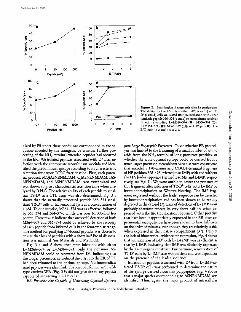
Figure 2. Sensitization of target cells with L+peptide-vacc. The ability of clone F5 to lyse either IcD b (a and b) or T2- D b (c and d) cells was tested after preincubation with either synthetic peptide 366-374 (a and c) or recombinant vaccinias (b and d) encoding L+M366-374 (0) , M366-374 (O), L+M364-374 (am), M365-379 ([]]), or HIV-pol (O). The E /T ratio in a and c was 2:1.
nized by F5 under these conditions corresponded to the se- quence encoded by the minigene, or whether further pro- cessing of the NH2-terminal extended peptides had occurred in the ER. We isolated peptides associated with D b after in- fection with the appropriate recombinant vaccinia and iden- tified the predominant epitope according to its characteristic retention time upon RPLC fractionation. First, each poten- tial product, MQIASNENMDAM, QIASNENMDAM, IAS- NENMDAM, and ASNENMDAM, was synthesized and was shown to give a characteristic retention time when ana- lyzed by RPLC. The relative ability of each peptide to sensi- tize T2-D b in a CTL assay was also determined. Fig. 3 a shows that the naturally processed peptide 366-374 sensi- tized T2-D b cells to half-maximal lysis at a concentration of I pM. To our surprise, M364-374 was as effective, followed by 365-374 and 364-374, which was over 10,000-fold less potent. These results indicate that successful detection of both M364-374 and 366-374 could be achieved by the recovery of each peptide from infected cells in the femtomolar range. The method for purifying Db-bound peptides was chosen to ensure that loss of peptides with a short half-life of dissocia- tion was minimal (see Materials and Methods).
Fig. 3 c and d show that after infection with either L+M366-374 or L+M364-374, only the nonamer AS- NENMDAM could be recovered from D b, indicating that the longer precursors, introduced directly into the ER of T2, had been trimmed to the optimal epitope. No intermediate sized peptides were identified. The control infection with wild- type vaccinia W R (Fig. 3 b) did not give rise to any peptide capable of sensitizing T2-D b cells.
ER Proteases Are Capable of Generating Optimal Epitopes
from Large Polypeptide Precursors. To see whether ER proteol- ysis was limited to the trimming of a small number of amino acids from the NH2 termini of long precursor peptides, or whether the same optimal epitope could be derived from a much larger precursor, recombinant vaccinias were constructed that encoded a 170-amino acid COOH-terminal fragment of NP (residues 328-498, referred to as IMP) with and without the HA leader sequence (termed L+ IMP and DIMP, respec- tively, see Fig. 1). We were unable to detect the presence of this fragment after infection of T2-D b cells with L+ IMP by immunoprecipitation or Western blotting. The IMP frag- ment expressed without the leader sequence can be detected by immunoprecipitation and has been shown to be rapidly degraded in the cytosol (7). Lack of detection of L+ IMP most probably therefore reflects its very short half-life when ex- pressed with the ER translocation sequence. Other proteins that have been inappropriately expressed in the ER after ex- perimental manipulation have been shown to have half-lives on the order of minutes, even though they are relatively stable when expressed in their native compartment (37). Despite the lack of biochemical evidence for expression, Fig. 4 shows that sensitization of b D b cells by L+ IMP was as e~cient as that by L-IMP, indicating that IMP was efficiently expressed by the L+ minigene construct. Furthermore, sensitization of T2-D b cells by L+ IMP-vacc was ef~cient and was dependent on the presence of the leader sequence.
Isolation of peptides associated with D b from L+ IMP-in- fected T2-D b cells was performed to determine the nature of the epitope derived from this polypeptide. Fig. 4 shows that a major species corresponding to ASNENMDAM was identified. Thus, again, the major product of intracellular
1484 Antigen Processing in the Endoplasmic Reticulum
on June 24, 2015jem
.rupress.orgD
ownloaded from
Published April 1, 1995

a t ~ ~ 1 4 ~ 3 7 4 3 4 1 4 4 7 4 M s - 3 r 4 M ~ N - 3 r 4
._ I l\ I;IZ i!l o t . . . . . 4 0 1 . . . . . 1 0 / . . . . ,
4 �9 I t 0 1 | ~411 4 (I 1 1 0 1 2 1 4 1 6 4 e | I 0 1 2 1 4 le 4 | II 1 0 1 2 1 4 l e
=0
4 o
I= !.
1o
o
b W R
to 20 30 40 5o ~1o
L 1r
L , ~ L l ( M ~ 3 7 4
�9 �9 , �9 , ,
Frmct:on number
d L §
~ l o .
V
Fract ion number
Figure 3. Characterization of peptides associated with D b isolated from recombinant vaccinia-infected T2-D b cells. (a) Titration curves for 366-374, 365-374, 364-374, and M366-374 and their characteristic reten- tion times on RPI.C. Detection of peptides by clone F5 (e) was tested after fractionation of peptides associated with Db from T2-D b cells in- fected with wild-type vaccinia (b), L+ M366-374 vacc (c), and L+ M364-374 vacc (d). In addition, the nonspecific toxicity of each fraction was tested (O).
proteolysis was the nonamer corresponding to the optimal binding peptide. These results show that proteolysis in the ER can give rise to an optimal CTL epitope contained within a 170-amino acid polypeptide, in which the epitope is ~40 residues from the NH2 terminus.
In addition, fraction 58 sensitized T2-D b cells for lysis in a CTL-dependent way, though this was not reproducible when the experiment was repeated. In the experiment shown in Fig. 4, we were unable to determine the origin of this ac- tivity, though the possibility that it represented an intermediate of proteolysis cannot be ruled out.
Trimming Does Not Occur Extracellularly. Since the pre- cursor peptides expressed with the ER translocation signal were all introduced directly into the secretory pathway, it was possible that the trimming we observed could have oc- curred in the extracellular space during infection, followed by binding to D b at the cell surface. To rule out this possi- bility, we coincubated 51Cr-labeled T2-D b cells with unla- beled T2 cells that had been infected with L+vacc. Coincu- bation was performed for 4-6 h under the same conditions as those used for infection before the isolation of D b. This mixture of cells was then exposed to clone F5 in a 4-h CTL assay. The labeled, uninfected T2-D b ceils were not sensi- tized for lysis after coincubation with infected T2 cells even when the latter were present at a 10-fold excess. In the same experiment, T2-D b cells were sensitized for lysis by infec- tion under the same conditions and by incubation with added peptides down to a concentration of 0.1 pM (data not shown).
Another explanation for the observed trimming of precursor peptides was that they were degraded in vitro after lysing the infected cells, before immunoprecipitation. We addressed this possibility by lysing 109 T2-D b cells in the presence of 2/xM crude M364-374. D b molecules were immunopurified. After washing the immunoprecipitate and eluting bound pep- tides, the peptide mixture was fractionated by RPLC, and each fraction was tested for its ability to sensitize T2-D b cells for lysis by clone F5. Fig. 5 shows that the predominant peak of CTL activity resided in fractions coeluting with RPLC- purified M364-374. A small peak of activity was also seen in fraction 20, which almost certainly represents the pres- ence of some 366-374. (This was confirmed by performing the same experiment with 2/zM 366-374 in the lysate, in which a large peak of activity was recovered from immuno- purified D b in fractions spanning 20; see Fig. 5.) Some of this material was already present in the M366-374 prepara- tion, since the same peak of activity was detected when the peptide was fractionated by RPLC and each fraction was tested (data not shown)�9 Inclusion of protease inhibitors in the lysis buffer was therefore sufficient to prevent the complete degra- dation of M364-374 during the peptide isolation procedure. Taken together, these results indicate that the recovery of 366-374 from L+ M364-374-infected cells was a consequence of intraceUular processing before lysis.
Intracellular Processing of L+ Constructs Occurs in a BFA-sensitive, Chtoroquine-insensitive Compartment. BFA causes a redistri- bution of intracellular compartments such that the medial and c/s-Golgi cisternae collapse into the ER, whereas the trans- Golgi network fuses with the recycling endosomal system (38). It therefore blocks class I-restricted presentation of in- tracellular antigens by inhibiting the egress of newly formed class I:peptide complexes from the ER to the trans-Golgi net- work (39, 40)�9 We tested the ability of BFA to interfere with the processing and presentation of vaccinia-encoded L+M364-374 and L+IMP. Fig. 6 b shows that 1/xM BFA was sufficient to block presentation of these polypeptides by T2-D b cells. Inhibition of presentation of both the L+ and L-polypeptides in L-D b cells, which has a functional TAP complex, was also observed (Fig. 6 a).
1485 Elliott et al.
on June 24, 2015jem
.rupress.orgD
ownloaded from
Published April 1, 1995

100
8o
40
20
a
0 - - 0 - - - 4
2 3 4 5
E:T Ratio
.~ 80 ,
4O
~e 20 i
1 2 3 4 5
E:T Ratio
35
i c L+IMP
~ 2 5
1 0
5
10 20 30 40 50 BO 70
Fraction number
Figure 4. Sensitization of target cells with L+ IMP and du- tion of peptides from L+IMP-infected T2-D b cells. The ability of clone F5 to lyse either L-D b (a) or T2-D b (b) cells was tested after infection with recombinant vaccinias encoding L+IMP (e) , IMP (O), or HIV-pol (O). (c) Peptides assodated with D b from T2-D b cells infected with L+IMP were ffac- tionated and tested for their ability to sensitize clone F5 (O) or to lyse T2-D b cells nonspecifically (O).
Leader-dependent, intracellular processing of antigens for presentation with MHC class II has been described previ- ously (41, 42), and has been shown to occur in low pH com- partments, by the ability of cloroquine to inhibit antigen pro- cessing. Although the requirement for newly synthesized MHC molecules was not demonstrated by these investiga- tions, we felt that it was necessary to investigate the role of low pH compartments in generating epitopes from our L+constructs. Fig. 6 shows that chloroquine had no effect on the processing and presentation of L+ M364 and L+ IMP in T2-D b cells (or in the control cell line L-Db).
Taken together, these results are consistent with the trim- ming of precursor polypeptides and the association of trimmed products with newly synthesized class I molecules occurring
in a pre-trans-Gol~ compartment and most likely the lumen of the ER.
CTL Epitopes Are Not Generated from the ER Degradation of Full-Length Proteins. It is unclear whether CTL epitopes
o io 20 30 4o 5o eo 70
Fraction number
Figure 5. Trimming is not due to extracellular processing. Little in vitro degradation of M366-374 occurs after cell lysis. T2-D b cells were lysed in the presence of 366-374 ( e ) or M364-374 (O), and peptides bound to D b were eluted and fractionated by RPLC.
Figure 6. The ability of L-D b (a) and T2-D b (b) cells to process L+M366-374 vacc, L+M364-374 vacc, and L+IMP vacc and to present synthetic 366-374 to clone F5 in the absence of drugs (open bars) or in the presence of BFA (dashed bars) or chloroquine (hatched Mrs).
1486 Antigen Processing in the Endoplasmic Reticulum
on June 24, 2015jem
.rupress.orgD
ownloaded from
Published April 1, 1995

can be generated in the ER from glycoproteins. Indeed, the observation that CTL epitopes appear to be derived mainly from nonglycoproteins and the demonstration that peptides derived from glycoproteins and secreted proteins occur rarely in eluates from purified class I molecules (see reference 21 for a summary) suggest that glycoproteins are protected from the action of ER proteases.
We have investigated the role of ER proteases in the pro- cessing of glycoproteins in two ways. First, the processing of influenza A HA was investigated in T2-K k. Fig. 7 shows that the HA epitope IEGGWTGMI (corresponding to residues 354-362 of HA1), recognized by the KLrestricted clone HA8 (27), was not generated in T2-K k cells when the an- tigen, was delivered after influenza (PRS) infection or infec- tion with recombinant vaccinias encoding full-length HA (1-565) or a truncated HA (1-413) (27), even though L-D b cells were sensitized for lysis by all three. Thus, HA and the truncated fragment did not appear to be susceptible to ER degradation in the same way as L+ IMP.
In a second approach, we constructed a recombinant vac- cinia encoding full-length influenza NP, tagged with the HA leader sequence (L+ NP-vacc). Infection of T2-D b cells with this vaccinia and immunoprecipitation of NP from a pulse- labeled lysate indicated that the majority of NP migrated more slowly than L-NP when analyzed by SDS-PAGE, and was sensitive to endoglycosidase H (Fig. 8). These results are con- sistent with the cotranslational transport of newly synthe- sized NP into the ER and the glycosylation of cryptic se- quons (NAT and NDT) at positions 21 and 144.
$
i u)
100
80
80
40
20
0
a
/
1 2 3 4 5 Pet~e (m.nM)
100
80
00
u~ 40 ~e
20
b _ ~ . ]
T F �9 �9 T 2 4 6 8 10
E:T Ratio
'001 c
m 40
20
I
0 1 2 3
Peplide (riM)
J
T 4 5
100
,_~ 80 -4
60q
m 40
20
o 2 4 6 8 10
E:T Ratio
Figure 7. Sensitization of target cells with endogenous influenza HA for lysis by clone HA8. L (a and b) and T2-K k (c and d) cells were sensi- tive to lysis by added peptide 354-362 (a and c) and were tested for their ability to process and present this peptide after infection with influenza A PR8 (V), HA vacc (e), KG26 vacc encoding the HA truncation 1-413 (11), and HIV-pol vacc (A).
Figure 8. Detection of NP in recombinant vaccinia-infected T2- D b cells. T2-D b cells were in- fected with L+ NP-vacc (lanes a and b) or L-NP-vacc (lanes c and d), and NP was detected 1.5 h after infection by immunoprecip- itation. Immunoprecipitates were either treated with endoglycosi- dase H (lanes b and d) or mock digested (lanes a and c).
The ability of ER proteases to generate two well-charac- terized CTL epitopes from L+NP was then tested. Fig. 9 shows that neither the K k (SDYEGRLI, 50-57 [43]) nor the D b (366-374) epitope was produced after infection. The CTLs used in this assay (F5 and a polyclonal anti-SDYEGRLI line) could nevertheless recognize T2 transfectants when pulsed with as little as 0.5 nM of the relevant peptide (Fig. 9 e).
-.//,.tblt + 1 ! + 4O
20 m
o
0 1 2 3 4 5 6 0 1 2 3 4 5 6
100
8O
|.o 2O
0 T
1 1 1 1 1 0 1 2 3 4 5
E:T ~ t ~
40-~
2o t I I I 1 I
0 1 2 3 4 5
E:TRat[o
100
!+ 40
20
0
I I I I I [ i 3 4 5 6 7 8 9 1 0 1 1
Peptide concentration (M x 10 "x)
Figure 9. Sensitization of target cells with L+NP. T2-D b (a), T2-K k (b), and TAP-competent I~D b cells, which express both D b and K k (c and d), were tested for their ability to present peptide antigen to clone F5 (a and c) and a KLrestricted polyclonal CTL line raised against NP50-57 (SDYEGRLI, b and d) after infection with recombinant vaccinias encoding L+NP (e) , I~NP (m), L+IMP (A), or HIV-pol (V). (e) In addition, the sensitivity of each of these CTLs (assayed at an E:T ratio of 2:1 for F5- and 10:1 for the KLrestricted CTL line) was tested on the appropriate T2 transfectant pulsed with synthetic peptides ASNENMDAM (e) and SDYEGRLI (m).
1487 Elliott et al.
on June 24, 2015jem
.rupress.orgD
ownloaded from
Published April 1, 1995

Discussion
Our results show that proteases in the ER (or other pre- Golgi compartments) can trim a precursor to a short peptide epitope that is optimal for presentation at the cell surface, as postulated by Falk et al. and Rotzschke et al. (3, 11).
These results raise the possibility that the enzymatic pro- cessing of intracellular antigens could continue in the same intracellular compartment as that in which peptide binding to MHC class I molecules occurs. The natural substrates for ER proteases could therefore be longer peptides, generated in the cytosol, which are then delivered to the ER lumen by the TAP complex.
Recent work has demonstrated a physical association be- tween class I molecules and the TAP complex in the ER (44, 45). The results of this study show that appropriate trim- ming and loading of peptides into class I molecules and their presentation at the cell surface can occur in the absence of the TAP complex, implying that contact of class I with TAP molecules is not essential for presentation of antigen.
Peptides encoded by the minigenes L+M364-374 and L+M366-374 (being only 23-26 amino acids) are unlikely to be synthesized on rough ER and are more likely to be posttranslationally translocated in a leader-dependent way (46). By studying the trimming of NH2-terminal extended pep- tides only, we were able to rule out any effect of cytosolic proteases, to which the gene products were undoubtedly ex- posed, since trimming of the NH2 terminus would destroy the signal sequence and COOH-terminal trimming would lead to peptides that eluted with unique retention times when analyzed by RPLC. Having also ruled out artifacts such as extracellular proteolysis in vitro and demonstrated that the leader-dependent generation of CTL epitopes is BFA sensi- tive and cloroquine insensitive, it is most likely that the trim- ming we have observed occurred in a pre-Golgi compartment of the infected cells.
It is therefore possible to envisage a situation in which cyto- solic proteases generate fairly large peptide fragments from denatured proteins in the cytosol, which are then transported into the lumen of the ER by the TAP heterodimer. Enzymes in the ER could then trim longer precursor peptides in order to optimize the NH2 termini for class I binding. The possi- bility that COOH-terminal trimming can occur in the ER cannot be ruled out, although experiments by Eisenlohr et al. (25) have implied that COOH-terminal trimming of a particular Kd-restricted epitope was not detectable.
We have also found that the 170-amino acid IMP frag- ment of NP could be processed in the ER via a BFA-sensitive, chloroquine-insensitive pathway and also gave rise to the op- timal nonameric epitope NP366-374. This result established that up to 40 amino acids can be removed from between the signal sequence and the epitope. In addition to the removal of 40 amino acids from the NH2 terminus, 120 amino acids must also be removed from the COOH terminus for this epitope to be produced. The L+IMP construct, being 170 amino acids, is more likely to be cotranslationally transported into the ER than its shorter counterparts (46). It is possible, therefore, that proteases that are normally present in the ER
are capable of degrading IMP in such a way as to reveal the correct COOH-terminal amino acid (Met-374) of the Db-re - stricted epitope, as well as being able to trim 40 amino acids from the NH2 terminus. However, it is also possible that some newly synthesized IMP is exposed to cytosolic proteases, which trim the COOH terminus before transport into the ER.
The ability of ER proteases to degrade full-length NP ex- pressed with a leader sequence revealed that this construct was a poor substrate, despite the synthesis of easily detect- able amounts of the L+ NP construct. The loss of presenta- tion could be due to an increase in the stability of NP when expressed as a full-length protein. NP might be stabilized in the ER as a result of conformational factors, or as a result of its glycosylation. Our results with influenza HA suggest that the latter may be the more likely, since neither the full- length HA, nor a truncated polypeptide (which would not be expected to adopt the same tertiary structure) gave rise to a Kk-restricted epitope in T2-K k cells. Both these poly- peptides contain glycosylation sites.
These results address the general issue of glycoprotein degra- dation in the ER and the ability of these degradation prod- ucts to assemble with class I molecules. Glycoprotein-derived CTL epitopes have been described, but the role of ER pro- teases in their generation is unclear. Townsend et al. (13) have shown that a Kk-restricted HA epitope can be efficiently generated in a leader-independent way, suggesting that a frac- tion of this glycoprotein is released from ribosomes into the cytoplasm and exposed to cytosolic proteases. Indeed, in this study, we have shown that the same epitope cannot be gener- ated in the ER, indicating that HA may be processed exclu- sively in the cytosol. The same may be true of other glycoprotein-derived CTL epitopes. Similar to our observa- tions with influenza HA, measles F protein cannot be presented to B27-restricted CTLs by infected T2-B27, which lacks a functional TAP complex, even though it is readily presented by TAP-competent cells (47). Nevertheless, Hammond et al. (48) have recently demonstrated the ability of T2 cells to pro- cess HIV-1 gp120 in a leader-dependent way, giving rise to an HLA-A3-restricted epitope, suggesting that this peptide was generated by enzymes in the ER. This may be a rela- tively infrequent occurrence and would explain why the majority of CTL epitopes described to date appear to be de- rived from proteins that do not enter the secretory pathway; it is supported by our observation that neither of the NP epitopes we studied could be generated in the ER. The im- plication is therefore that glycoproteins may be sheltered from the class I-associated processing pathway by their limited ex- posure to cytosolic proteases and by their relative resistance to proteolysis in the ER.
Very little is known about ER resident proteases. The best studied to date is leader peptidase, which cleaves the ER trans- location signal sequence from type 1 glycoproteins and has a well-defined specificity, cleaving after A, G, S, T, or C, in the general motif (A/G/S/T/C/L/WI)X(A/G/S/T/C) (46). Peptides derived from leader sequences, generated by the ac- tion of leader peptidase have been identified bound to HLA-
1488 Antigen Processing in the Endoplasmic Reticulum
on June 24, 2015jem
.rupress.orgD
ownloaded from
Published April 1, 1995
A2 in normal and TAP-defective cell lines (49). However, the specificity of this enzyme makes it unlikely that it is respon- sible for the NH2-terminal trimming observed in our experi- ments, since the sequences immediately preceding 366-374 in either L+ M366-374 (MQI) or L+ M364-374 (DAM) do not contain a motif susceptible to recognition by leader pep- tidase. The involvement of leader peptidase in trimming the NH2 terminus of L+IMP and L+NP is even less likely.
Selective, rapid degradation of incompletely assembled mul- tichain glycoproteins has been described for TCR o~ and and CD38 (50, 51). These polypeptides contain structural elements in their transmembrane domains that predispose them to proteolysis in the ER if they are not masked by multisubunit assembly. Although none of our L+ constructs contain this motif, the enzymes responsible for their degradation may be the same as those recruited for the degradation of TCR chains.
Recently, Wileman et al. (52) have shown that targetted degra- dation of TCR chains in the ER can be inhibited by inhibi- tors of cysteine proteases and not by specific inhibitors of tryp- sinlike serine proteases. The degradation of many other incompletely assembled or misfolded proteins in the ER has been investigated, and the involvement of calcium-dependent proteases (53) and TLCK-inhibitable serine proteases (54) has been implicated in the proteolysis of two of these. It there- fore appears that all classes of protease have been identified in association with the ER. It remains to be seen which of these activities, if any, are responsible for generating peptides suitable for assembly with class I MHC. In addition, the pos- sibility that the ER is subdivided into functionally special- ized compartments (55), with distinct regions for special- ized proteolysis, is intriguing.
The authors wish to thank Diana Hayward for her technical assistance, Dr. Geoff Smith for his advice and help in constructing recombinant vaccinias, and Dr. Keith Gould (Sir William Dunn School of Pathology, University of Oxford) for providing clone HA8 and vaccinias HA and KG26. We would also like to thank Dr. Peter Cresswell (Yale University, New Haven, CT) for providing T2 cells transfected with D b and K k and Dr. Tom Wileman for valuable discussion and for sharing his unpublished results with us.
This work was supported by the Wellcome Trust, The Medical Research Council UK, the Cancer Re- search Institute (New York), and The Howard Hughes Medical Institute.
Received for publication 18 October 1994 and in revised form 28 November 1994.
References 1. Townsend, A.R., J. tkothbard, F.M. Gotch, G. Bahadur, D.
Wraith, and A.J. McMichael. 1986. The epitopes of influenza nudeoprotein recognized by cytotoxic T lymphocytes can be defined with short synthetic peptides. Cell. 44:959-968.
2. van Bleek, G.M., and S.G. Nathenson. 1990. Isolation of an endogenously processed immunodominant viral peptide from the class I H-2Kb molecule. Nature (Lond.). 348:213-216.
3. Rotzschke, O., K. Falk, K. Deres, H. Schild, M. Norda, J. Metzger, G. Jung, and H.G. Rammensee. 1990. Isolation and analysis of naturally processed viral peptides as recognized by eytotoxic T cells. Nature (Lond.). 348:252-254.
4. Townsend, A., C. Ohlen, J. Bastin, H.G. Ljunggren, L. Foster, and K. Karre. 1989. Association of class I major histocompati- bility heavy and light chains induced by viral peptides. Nature (Lond.). 340:443-448.
5. Lapham, C.K., I. Bacik, J.W. Yewdell, K.P. Kane, and J.R. Bennink. 1993. Class I molecules retained in the endoplasmic reticulum bind antigenic peptides.J. Extx Med. 177:1633-1641.
6. Goldberg, A.L., and K.L. Rock. 1992. Proteolysis, proteasomes and antigen presentation. Nature (Lond.). 357:375-379.
7. Townsend, A., J. Bastin, K. Gould, G. Brownlee, M. Andrew, B. Coupar, D. Boyle, S. Chan, and G. Smith. 1988. Defective presentation to class I-restricted cytotoxic T lymphocytes in vaccinia-infected cells is overcome by enhanced degradation of antigen. J. Exp. Med. 168:1211-1224.
8. Michalek, M.T., E.P. Grant, C. Gramm, A.L. Goldberg, and
K.L. Rock. 1993. A role for the ubiquitin-dependent proteo- lytic pathway in MHC class I-restricted antigen presentation. Nature (Lond.). 363:552-554.
9. Driscoll, J., M.G. Brown, D. Finley, and J.J. Monaco. 1993. MHC-linked LMP gene products specifically alter peptidase activities of the proteasome. Nature (Lond.). 365:262-264.
10. Gaczynska, M., K.L. Rock, and A.L. Goldberg. 1993. Gamma- interferon and expression of MHC genes regulate peptide hy- drolysis by proteasomes, Nature (Lond.). 365:264-267.
11. Falk, K., O. Rotzschke, and H.G. Rammensee. 1990. Cellular peptide composition governed by major histocompatibility com- plex class I molecules. Nature (Lond.). 348:248-251.
12. Townsend, A.R., F.M. Gotch, andJ. Davey. 1985. Cytotoxic T cells recognize fragments of the influenza nucleoprotein. Cell. 42:457-467.
13. Townsend, A.R., J. Bastin, K. Gould, and G.G. Brownlee. 1986. Cytotoxic T lymphocytes recognize influenza haemag- glutinin that lacks a signal sequence. Nature (Lond.). 324: 575-577.
14. Monaco, J.J., S. Cho, and M. Attaya. 1990. Transport protein genes in the murine MHC: possible implications for antigen processing. Science (Wash. DC). 250:1723-1726.
15. Spies, T., M. Bresnahan, S. Bahram, D. Arnold, G. Blanck, E. Mellins, D. Pious, and R. DeMars. 1990. A gene in the human major histocompatibility complex class II region con- trolling the class I antigen presentation pathway. Nature (Lond.).
1489 Elliott et al.
on June 24, 2015jem
.rupress.orgD
ownloaded from
Published April 1, 1995

348:744-747. 16. Trowsdale, J., I. Hanson, I. Mockridge, S. Beck, A. Town-
send, and A. Kelly. 1990. Sequences encoded in the class II region of the MHC related to the 'ABC' superfamily of trans- porters. Nature (Lond.). 348:741-744.
17. Deverson, E.V., I.R. Gow, W.J. Coadwell, J.J. Monaco, G.W. Butcher, andJ.C. Howard. 1990. MHC class II region encoding proteins related to the muhidrug resistance family of trans- membrane transporters. Nature (Lond.). 348:738-741.
18. Townsend, A., and J. Trowsdale. 1993. The transporters as- sociated with antigen presentation. Semin. Cell Biol. 4:53-61.
19. Neefjes, J.J., F. Momburg, and G.J. Hammerling. 1993. Selec- tive and ATP-dependent peptide translocation of peptides by the MHC-encoded transporter. Science (Wash. DC). 261:769- 771.
20. Shepherd, J.C., T.N. Schumacher, P.G. Ashton Rickardt, S. Imaeda, H.L. Ploegh, C.A.J. Janeway, and S. Tonegawa. 1993. TAPl-dependent peptide translocation in vitro is ATP depen- dent and peptide selective. Cell. 74:577-584.
21. Elliott, T., M. Smith, P. Driscoll, and A. McMichael. 1994. Peptide selection by class I molecules of the major histocom- patibility complex. Cu~ Biol. 3:854-866.
22. Powis, S.J., E.V. Deverson, W.J. Coadwell, A. Ciruela, N.S. Huskisson, H. Smith, G.W. Butcher, and J.C. Howard. 1992. Effect of polymorphism of an MHC-linked transporter on the peptides assembled in a class I molecule. Nature (Lond.). 357:211-215.
23. Guo, H.C., T.S. Jardetzky, T.P. Garrett, W.S. Lane, J.L. Strominger, and D.C. Wiley. 1992. Different length peptides bind to HLA-Aw68 similarly at their ends but bulge out in the middle. Nature (Lond.). 360:364-366.
24. Udaka, K., T.J. Tsomides, P. Walden, N. Fukusen, and H.N. Eisen. 1993. A ubiquitous protein is the source of naturally occurring peptides that are recognized by a CDS+ T-cell clone. Proc. Natl. Acad, Sci. USA. 90:11272-11276.
25. Eisenlohr, L.C., I. Bacik, J.R. Bennink, K. Bernstein, and J.W. Yewdell. 1992. Expression of a membrane protease enhances presentation of endogenous antigens to MHC class I-restricted T lymphocytes. Cell. 71:963-972.
26. Alexander, J., J.A. Payne, R. Murray, J.A. Frelinger, and P. Cresswell. 1989. Differential transport requirements of HLA and H-2 class I glycoproteins. Immunogenetics. 29:380-388.
27. Gould, K.G., H. Scotney, and G.G. Brownlee. 1991. Charac- terization of two distinct major histocompatibility complex class I Kk-restricted T-cell epitopes within the influenza A/PR/8/34 virus hemagglutinin. J. Virol. 65:5401-5409.
28. Bastin, J., J. Rothbard, J. Davey, I. Jones, and A. Townsend. 1987. Use of synthetic peptides of influenza nucleoprotein to define epitopes recognized by class I-restricted cytotoxic T lym- phocytes. J. Exp. Med. 165:1508-1512.
29. Hammerling, G., U. Hammerling, and H. Lemke. 1979. Iso- lation of twelve monoclonal antibodies against Ia and H-2 an- tigens. Serological characterisation and reactivity with B and T lymphocytes. Immunogenetics. 8:433-439.
30. Ortiz Navarrete, V., and G.J. Hammerling. 1991. Surface ap- pearance and instability of empty H-2 class I molecules under physiological conditions. Proc. Natl. Acad, Sci. USA. 88:3594- 3597.
31. Elliott, T., V. Cerundolo, J. Elvin, and A. Townsend. 1991. Peptide-induced conformational change of the class I heavy chain. Nature (Lond.). 351:402-406.
32. Cerundolo, V., T. EUiott, J. Elvin, J. Bastin, H.G. Rammensee,
and A. Townsend. 1991. The binding affinity and dissociation rates of peptides for class I major histocompatibility complex molecules. Eur. j . Immunol. 21:2069-2075.
33. Chakrabarti, S., K. Brechling, and B. Moss. 1985. Vaccinia virus expression vector: coexpression of beta-galactosidase pro- vides visual screening of recombinant virus plaques. Mol. Cell. Biol. 5:3403-3409.
34. Elvin, J. 1992. Presentation of Peptide Antigens to Cytotoxic T Lymphocytes. Oxford University Press, Oxford, UK. p. 1.
35. Gould, K., J. Cossins, J. Bastin, G.G. Brownlee, and A. Town- send. 1989. A 15 amino acid fragment of influenza nucleo- protein synthesized in the cytoplasm is presented to class I-re- stricted cytotoxic T lymphocytes.J. Exp. Med. 170:1051-1056.
36. Anderson, K., P. Cresswell, M. Gammon, J. Hermes, A. Wil- liamson, and H. Zweerink. 1991. Endogenously synthesized peptide with an endoplasmic reticulum signal sequence sensi- tizes antigen processing mutant cells to class I-restricted cell- mediated lysis. J. Exp. Ailed. 174:489-492.
37. Stoller, T.J., and D. Shields. 1989. The propeptide of preprosomatostatin mediates intracellular transport and secre- tion of alpha-globin from mammalian cells, j . Cell Biol. 108: 1647-1655.
38. Lippincott Schwartz, J., L. Yuan, C. Tipper, M. Amherdt, L. Orci, and R.D. Klausner. 1991. Brefeldin A's effects on en- dosomes, lysosomes, and the TGN suggest a general mecha- nism for regulating organelle structure and membrane traffic. Cell. 67:601-616.
39. Yewdell, J.W., andJ.R. Bennink. 1989. Brefeldin A specifically inhibits presentation of protein antigens to cytotoxic T lym- phocytes. Science (Wash. DC). 244:1072-1075.
40. Nuchtern, J.G., J.S. Bonifacino, W.E. Biddison, and R.D. Klausner. 1989. Brefeldin A implicates egress from endoplasmic reticulum in class I restricted antigen presentation. Nature (Lond.). 339:223-226.
41. Weiss, S., and B. Bogen. 1991. MHC class lI-restricted presen- tation of intracellular antigen. Cell. 64:767-776.
42. Michalek, M.T., B. Benacerraf, and K.L. Rock. 1992. The class II MHC-restricted presentation of endogenously synthesized ovalbumin displays clonal variation, requires endosomal/lyso- somal processing, and is up-regnlated by heat shock. J. Im- munol. 148:1016-1024.
43. Gould, K.G., H. Scotney, A.R.M. Townsend, J. Bastin, and G.G. Brownlee. 1987. Mouse H-2k-restricted cytotoxic T cells recognize antigenic determinants in both the HA1 and HA2 subunits of the influenza A/PR/8/34 hemagglutinin.J. Exp. Med. 166:693-701.
44. Ortmann, B., M.J. Androlewicz, and P. Cresswell. 1994. MHC class I/beta-2 microglobulin complexes associate with TAP transporters before peptide binding. Nature (Lond.). 368:864-867.
45. Suh, W.K., M.F. Cohen-Doyle, K. Fruh, K. Wang, P.A. Peterson, and D.B. Williams. 1994. Interaction of MHC class I molecules with the transporter associated with antigen pro- cessing. Science (Wash. DC). 264:1322-1326. (Abstr.)
46. Blobel, G., and B. Dobberstein. 1975. Transfer of proteins across membranes. J. Cell Biol. 67:835-851.
47. van Binnendijk, R.S., C.A. van Baalen, M.C. Poelen, P. de Vries, J. Boes, V. Cerundolo, A.D.M.E. Osterhaus, and F.G.C.M. UytdeHaag. 1992. Measles virus transmembrane fu- sion protein synthesized de novo or presented in immuno- stimulating complexes is endogenously processed for HLA class I- and class II-restricted cytotoxic T cell recognition.
1490 Antigen Processing in the Endoplasmic Reticulum
on June 24, 2015jem
.rupress.orgD
ownloaded from
Published April 1, 1995

j . Extx Med. 176:119-128. 48. Hammond, S.A., R.C. Bollinger, T.W. Tobery, and R.F. Silli-
ciano. 1993. Transporter-independent processing of HIV-1 enve- lope protein for recognition by CD8 + T cells. Nature (Lond.). 364:158-161.
49. Wei, M.L., and P. Cresswell. 1992. HLA-A2 molecules in an antigen-processing mutant cell contain signal sequence-derived peptides. Nature (Lond.). 356:443-446.
50. Bonifacino, J.S., C.K. Suzuki, J. Lippincott Schwartz, A.M. Weissman, and R.D. Klausner. 1989. Pre-Golgi degradation of newly synthesized T-cell antigen receptor chains: intrinsic sensitivity and the role of subunit assembly. J. Cell Biol. 109:73-83.
51. Wileman, T., G.R. Carson, M. Concino, A. Ahmed, and C. Terhorst. 1990. The gamma and epsilon subunits of the CD3 complex inhibit pre-Golgi degradation of newly synthesized
T cell antigen receptors..J. Cell Biol. 110:973-986. 52. Wileman, T., L. Kane, and C. Tehorst. 1993. Degradation of
T cell receptor chains in the endoplasmic reticulum is inhibited by inhibitors of cysteine proteases. Cell Reg. 2:753-765.
53. Tsao, Y., N. Ivessa, M. Adesnik, D. Sabatini, and G. Keibich. 1992. COOH terminally truncated forms of Ribophorin I are degraded in pre-Golgi compartments by a calcium-dependent process, f Cell Biol. 116:57-67.
54. Wikstrom, L., and H.F. Lodish. 1991. Nonlysosomal, pre-Golgi degradation of unassembled asialoglycoprotein receptor subunits: a TICK- and TPCK-sensitive cleavage within the ER. f Cell Biol. 113:997-1007.
55. Wassler, M., and E. Fries. 1993. Proteolytic cleavage of hap- toglobtdin occurs in a subcompartment of the endoplasmic retic- ulum: evidence from membrane fusion in vitro. J. Cell Biol. 123:285-291.
1491 Elliott et al.
on June 24, 2015jem
.rupress.orgD
ownloaded from
Published April 1, 1995