Molecular and Cellular Mechanisms of Ecstasy-Induced Neurotoxicity: An Overview
Upload
independentCategory
view
0download
0
www.elsevier.com/locate/ynbdi
Neurobiology of Disease 23 (2006) 669–678An endoplasmic-reticulum-specific apoptotic pathway is involved inprion and amyloid-beta peptides neurotoxicity
Elisabete Ferreiro, Rosa Resende, Rui Costa, Catarina R. Oliveira, and Cláudia M.F. Pereira⁎
Center for Neuroscience and Cell Biology of Coimbra, Institute of Biochemistry, Faculty of Medicine, University of Coimbra,3004-504 Coimbra, Portugal
Received 12 January 2006; revised 5 April 2006; accepted 22 May 2006Available online 17 July 2006
Prion (PrP) and amyloid-β (Aβ) peptides are involved in the neuronalloss that occurs in Prion disorders (PrD) and Alzheimer's disease(AD), respectively, partially due to Ca2+ dysregulation. Besides, theendoplasmic reticulum (ER) stress has an active role in the neurotoxicmechanisms that lead to these pathologies. Here, we analyzed whetherthe ER-mediated apoptotic pathway is involved in the toxic effect ofsynthetic PrP and Aβ peptides. In PrP106–126- and Aβ1–40-treatedcortical neurons, the release of Ca2+ through ER ryanodine (RyR) andinositol 1,4,5-trisphosphate (IP3R) receptors induces ER stress andleads to increased cytosolic Ca2+ and reactive oxygen species (ROS)levels and subsequently to apoptotic death involving mitochondrialcytochrome c release and caspases activation. These results demon-strate that the early PrP- and Aβ-induced perturbation of ER Ca2+
homeostasis is a death message that leads to neuronal loss, suggestingthat the regulation of ER Ca2+ levels may be a potential therapeuticaltarget for PrD and AD.© 2006 Elsevier Inc. All rights reserved.
Keywords: Prion disorders; Alzheimer's disease; Prion peptide; Amyloid-βpeptide; Apoptosis; Ca2+ homeostasis; Endoplasmic reticulum; Oxidativestress
Introduction
Prion disorders (PrD) and Alzheimer's disease (AD) areprogressive neurodegenerative diseases characterized by neuronalloss, cognitive dysfunction and dementia. These diseases are linkedby the extracellular deposition of the scrapie isoform of prionprotein (PrPSc) and the amyloid-beta protein (Aβ), respectively
Abbreviations: Aβ, amyloid-beta peptide; PrP, prion peptide; AD,Alzheimer's disease; ER, endoplasmic reticulum; GRP78, glucose-regulatedprotein 78; Indo-1/AM, Indo-1 acetoxymethyl ester; IP3R, inositol 1,4,5-triphosphate receptor; PrD, Prion disorders; ROS, reactive oxygen species;RyR, ryanodine receptor.⁎ Corresponding author. Fax: +351 239822776.E-mail address: [email protected] (C.M.F. Pereira).Available online on ScienceDirect (www.sciencedirect.com).
0969-9961/$ - see front matter © 2006 Elsevier Inc. All rights reserved.doi:10.1016/j.nbd.2006.05.011
(Prusiner, 1996; Wisniewski et al., 1997). In PrD, the PrPSc resultsfrom the conversion of the normal protein PrPC, is insoluble innondenaturating detergents, aggregates easily and is partiallyresistant to protease digestion. The PrP106–126, a syntheticpeptide homologous to PrPSc, has been used as a model systemto study prion-induced neurodegeneration (Della-Bianca et al.,2001; Pérez et al., 2003). PrP106–126 is rich in β-sheet structure,forms aggregates that are detergent-insoluble and PK-resistant andcatalyzes the aggregation of endogenous PrPC to an amyloidogenicform that shares several characteristics with PrPSc (Ettaiche et al.,2000; Brown, 2000; Singh et al., 2000). In AD, the amyloidogenicmaterial is composed of amyloid-beta peptide (Aβ), derived fromβ- and γ-secretases cleavage of amyloid precursor protein (APP) inthe endoplasmic reticulum (ER), the Golgi apparatus or theendosomal–lysosomal pathway, and most is normally secreted as a1–40 (Aβ1–40) or 1–42 (Aβ1–42) amino acid peptide (Perez et al.,1999). Synthetic Aβ1–40 and Aβ1–42 are amyloidogenic andneurotoxic peptides that have been widely used to mimic in vitrothe degenerative process that occurs in the brain of AD patients(Mattson, 1997; Pereira et al., 1999).
The ER is an essential intracellular organelle involved inintracellular calcium homeostasis, in folding and processing ofproteins and in cell death activation (Baumann and Walz, 2001).Ca2+ release from the ER is mediated through ryanodine receptors(RyR) and inositol 1,4,5-triphosphate receptors (IP3R) (Berridge etal., 2000). Changes in ER Ca2+ homeostasis induce the accumula-tion of unfolded proteins and activate the ER-stress-inducedapoptosis pathway (Kaufman, 1999; Pashen, 2001). In response toER stress, expression of glucose-regulated protein (GRP) 78, anER chaperone, is induced (Kaufman, 1999; Rao et al., 2002).Furthermore, caspase-12, that is mainly located on the cytoplasmicside of the ER, is activated (Nakagawa et al., 2000; Nakagawa andYuan, 2000) and can further activate caspase-9 and -3 (Morishimaet al., 2002).
It was recently shown that caspase-12 knock-out mice areresistant to ER stress and to death caused by Aβ protein (Nakagawaet al., 2000) and also that caspase-12 activation and increasedexpression of GRP58, a chaperone with (PDI)-like activity, occurin cells treated with PrPSc (Hetz et al., 2003, 2005). These results
670 E. Ferreiro et al. / Neurobiology of Disease 23 (2006) 669–678
strongly support that ER stress is involved in the neuronal deaththat occurs in PrD and AD. In vitro studies have suggested that ERCa2+ dyshomeostasis and oxidative stress are synergisticallyrelated. While ER function is sensitive to the presence of oxidants(Dreher et al., 1995; Racay et al., 1995; Viner et al., 1996; Hayashiet al., 2005), reactive oxygen species (ROS) can also be producedintracellularly from the stress-regulated release of calcium from theER. This calcium is readily taken up by mitochondria, resulting inthe elevation of ROS (Tardif et al., 2005).
The purpose of the present study was to elucidate the role ofER-mediated apoptotic pathway in the neurotoxic effects inducedby PrP106–126 and Aβ1–40. Our results suggest that, in ER, theearly Ca2+ release through the RyR and IP3R, induced by thesepeptides, is involved in the perturbation of Ca2+ homeostasis andROS production, leading to cytochrome c release and caspase-3activation and finally to apoptotic cell death. These findings give acontribution to the elucidation of the mechanism of neuronal deaththat occurs in PrD and AD.
Materials and methods
Materials
Neurobasal medium and B27 supplement were purchased fromGibco BRL, Life Technologies (Scotland, UK). Trypsin, deoxyr-ibonuclease I (DNase I), trypsin inhibitor type II-S-soybean,protease inhibitors, phenylmethylsulfonyl fluoride (PMSF), bovineserum albumin (BSA), dantrolene and thapsigargin were obtainedfrom Sigma Chemical Co. (St. Louis, MO, USA). SYTO-13,propidium iodide (PI), Indo-1 acetoxymethyl ester (Indo-1/AM),2′,7′-dichlorodihydrofluorescein diacetate (DCFH2-DA), Mito-Tracker Green, Hoechst 33342 and Alexa Fluor 594 goat anti-mouse IgG conjugate were purchased from Molecular Probes(Leiden, Netherlands). The synthetic Aβ1–40, Aβ40–1, PrP106–126 and PrPscrambled peptides were from Bachem (Bubendorf,Switzerland). Ionomycin and xestospongin C were purchased fromCalbiochem (Darmstadt, Germany). The colorimetric substrates forcaspase-2 [N-acetyl-Val-Asp-Val-Ala-Asp-P-nitroanilide (Ac-VDVAD-pNA)], caspase-6 [N-acetyl-Val-Glu-Ile-Asp-P-nitroani-lide (Ac-VEID-pNA)] and caspase-9 [N-acetyl-Leu-Glu-His-Asp-P-nitroanilide (Ac-LEHD-pNA)] were obtained from BioSourceInternational (Nivelles, Belgium), whereas the substrate forcaspase-3 [N-acetyl-Asp-Glu-Val-Asp-P-nitroanilide (DEVD-pNA)] was from Calbiochem (Darmstadt, Germany) and forcaspase-8 [N-acetyl-Ile-Glu-Thr-Asp-P-nitroanilide (Ac-IETD-pNA)] was from Sigma Chemical Co. (St. Louis, MO, USA).Bio-Rad protein dye assay, reagents and apparatus used inimmunoblotting assays were purchased from Bio-Rad (Hercules,CA, USA). The following primary antibodies were from BDPharmingen (San Diego, CA, USA): monoclonal mouse anti-BiP/Grp78, monoclonal mouse reactive against the native form ofcytochrome c and polyclonal rabbit anti-caspase-12. The mono-clonal mouse anti-microtubule-associated protein 2 (MAP2) andthe rabbit anti-glial fibrillary acidic protein (GFAP) antibody werepurchased from Sigma Chemical Co. (St. Louis, MO, USA). Thegoat anti-rabbit IgG and goat anti-mouse IgG antibodiesconjugated to alkaline phosphatase, the enhanced chemiFluores-cence reagent (ECF) and the polyvinylidene difluoride (PVDF)membrane were obtained from Amersham Pharmacia Biotech(Buckinghamshire, UK). The DakoCytomation Fluorescent mount-ing medium was purchased from DakoCytomation (Carpinteria,
California, USA). All the other chemicals were obtained fromSigma (St. Louis, MO, USA) or from Merck kgaA (Darmstadt,Germany).
Primary cortical neuronal cultures and experimental treatments
Primary cultures of cortical neurons were prepared from 15 to16 days embryos of Wistar rats according to the method describedby Hertz et al. (1989), with some modifications (Agostinho andOliveira, 2003). Briefly, removed cortices were dissected andplaced in Ca2+- and Mg2+-free Krebs buffer (in mM): NaCl 120,KCl 4.8, KH2PO4 1.2, glucose 13 and HEPES 10 (pH 7.4)supplemented with BSA (0.3 mg/ml). Minced cortical tissues werewashed and incubated in Krebs solution supplemented with BSA,and containing trypsin (0.5 mg/ml) and DNase I (0.04 mg/ml), for10 min at 37°C. The digestion was stopped with Krebs buffercontaining trypsin inhibitor (type II-S) (0.75 mg/ml) and DNase I(0.04 mg/ml) followed by a centrifugation at 140 × g for 5 min.After washing the pellet once with Krebs buffer, the cells wereresuspended in fresh Neurobasal medium supplemented with 2 mML-glutamine, 2% B27 supplement, penicillin (100 U/ml) andstreptomycin (100 μg/ml) and were dissociated mechanically.Cortical cells were plated on poly-L-lysine (0.1 mg/ml)-coatedglass coverslips at a density of 0.10 × 10⁶ cells/cm2 forimmunocytochemistry experiments and nuclear morphology stu-dies or 0.40 × 10⁶ cells/cm2 for measurement of intracellular Ca2+
or ROS levels. For caspase-like activity assays and immunoblot-ting, neurons were mounted on poly-L-lysine (0.1 mg/ml)-coateddishes at a density of 0.45 × 10⁶ cells/cm2. The cultures weremaintained at 37°C in a humidified atmosphere of 5% CO2/95% airand were used for experiments after 5 to 7 days in vitro.Differentiated cortical neurons were treated with Aβ1–40 or Aβ40–1 (0.5 μM) or with PrP106–126 or PrPscrambled (25 μM) inserum-free Neurobasal medium supplemented with B27. PrPpeptides were added from a stock solution prepared in steriledistilled water, at a concentration of 1 mM. Aβ peptides weredissolved in sterile distilled water at a concentration of 6 mg/mland diluted to 1 mg/ml (231.5 μM) with phosphate saline buffer(PBS) and then incubated for 5–7 days to induce fibril formation.The concentrations of Aβ1−40 and PrP106–126 used were chosenbased on previous results demonstrating that, on cortical neurons,0.5 μM Aβ1–40 and 25 μM PrP106–126 induced a maximal toxiceffect (50% decrease in cell viability). Before the addition of Aβ1–40 or PrP106–126, cells were preincubated for 1 h with dantrolene(10 μM) or xestospongin C (1 μM), concentrations that wereshown to exert a maximal protective effect, without being toxic byitself (data not shown).
Assessment of neuronal injury
After treatment of cortical cells with Aβ1–40 or PrP106–126,neuronal injury was assessed as described in Ferreiro et al. (2004).Reverse Aβ1–40 (Aβ40–1) or scrambled PrP was also used inorder to analyze the specificity of the amino acids sequence of bothpeptides. Neurons were stained with PI (2.5 μg/ml) and SYTO-13(3.8 μM), and the nuclear morphology was analyzed byfluorescence microscopy. Cortical cells cultured on glass coverslipswere treated with Aβ1–40 or PrP106–126, in the absence or in thepresence of dantrolene or xestospongin C. Cells were examinedand scored with a Nikon Diaphot TMP microscope, using a tripleXF-63 Omega filter. All experiments were performed in duplicate,
671E. Ferreiro et al. / Neurobiology of Disease 23 (2006) 669–678
and a minimum of 300 cells were scored for each coverslip. Thenumber of viable, necrotic and apoptotic cells was expressed as thepercentage (%) of the total number of cells in the microscope field.
Measurement of intracellular Ca2+ concentration
Cortical cells cultured on glass coverslips, in the presence or inthe absence of peptides, were incubated in the dark with 3 μMIndo-1/AM in a salt solution without phosphate containing (inmM): NaCl 132, KCl 4, CaCl2 1, MgCl2 1.4, glucose 6, HEPES–Na 10, pH 7.4 for 45 min at 37°C. The cells were furtherincubated, for 15 min, in the absence of Indo-1/AM, to ensure acomplete hydrolysis of the acetoxymethyl ester of Indo-1. Afterwashing, cells were mounted in a special holder, and the Indo-1fluorescence was measured with excitation wavelength of 335 nmand 410 nm emission. The free intracellular Ca2+ concentration([Ca2+]i) was calculated as previously described (Bandeira-Duarteet al., 1990).
Caspases activity assay
Untreated or treated cortical cells were lysed with a buffercontaining (in mM): HEPES–Na 25, MgCl2 2, EDTA 1, EGTA 1supplemented with 100 μM phenylmethylsulphonyl fluoride(PMSF), 2 mM DTT and a protease inhibitor cocktail (containing1 μg/ml leupeptin, pepstatin A, chymostatin and antipain). Thecellular suspension was rapidly frozen/defrosted three times andthen centrifuged for 10 min at 20,200 × g. The supernatant wascollected and assayed for protein content using the Bio-Rad proteindye assay reagent. To measure caspases activity, aliquots of cellextracts containing 25 μg or 40 μg of protein were incubated for2 h at 37°C, in a reaction buffer containing 25 mM HEPES–Na,10 mM DTT, 10% sucrose and 0.1% CHAPS (pH 7.4) with100 μM Ac-VDVAD-pNA, Ac-DEVD-pNA, Ac-VEID-pNA, Ac-IETD-pNA or Ac-LEHD-pNA, chromogenic substrates forcaspase-2, -3, -6, -8 and -9, respectively (Cregan et al., 1999).Caspases activity was determined by measuring substrate cleavageat 405 nm using a microplater reader, and results were expressed asthe increase above control absorbance at 405 nm.
Western blotting analysis
For the preparation of total cell extracts, cells which were eithertreated or untreated with the peptides were scraped in Neurobasalmedium and centrifuged at 140×g. The pellet was resuspended in200 μl of ice-cold lysis buffer containing (in mM): HEPES–Na 25,MgCl2 2, EDTA 1, EGTA 1, supplemented with 100 μM PMSF,2 mM DTT and protease inhibitor cocktail (containing 1 μg/mlleupeptin, pepstatin A, chymostatin and antipain). Cell lysateswere frozen three times in liquid N2 and were centrifuged at 140 × gto remove nuclei and large debris. Protein concentration in thesupernatant was measured using the Bio-Rad protein dye assayreagent. Samples were denaturated at 95°C for 3 min in a 6×concentrated sample buffer (mM): Tris 500, DTT 600, 10.3%SDS, 30% glycerol and 0.012% bromophenol blue. Equal amountof each sample of protein was separated by electrophoresis on a10% SDS-polyacrylamide gels (SDS-PAGE) and electroblottedonto PVDF membranes. The identification of proteins of interestwas facilitated by the usage of a prestained precision proteinstandard (Bio-Rad) which was run simultaneously. After theproteins were electrophoretically transferred, the membranes were
incubated for 1 h at room temperature (RT) in Tris buffer (mM;NaCl 150, Tris–HCl 25 (pH 7.6) with 0.1% Tween 20 (TBS-T)containing 5% nonfat dry milk) to eliminate nonspecific binding,and were next incubated overnight at 4°C in TBS-T containing1% nonfat dry milk with a rabbit polyclonal primary antibodyagainst caspase-12 (1:1000 dilution) or a mouse monoclonalprimary antibody against GRP78 (1:250 dilution). The mem-branes were washed several times and then incubated in TBS-Twith 1% nonfat dry milk for 2 h at RT, with the appropriatealkaline-phosphatase-conjugated anti-rabbit or anti-mouse second-ary antibody at a dilution of 1:25,000 or 1:20,000, respectively.Immunoreactive bands were detected after incubation of mem-branes with ECF reagent for 5–10 min, on a Bio-Rad Versa Doc3000 Imaging System.
Immunocytochemistry
Control, Aβ1–40- or PrP106–126-treated cells were washedtwo times in PBS buffer (pH 7.4) and were fixed with 4%paraformaldehyde for 30 min at RT. For cytochrome c releaseexperiments, cells were incubated with 750 nM MitoTracker Greenfor 45 min at 37°C, in the dark. Then, the cells were permeabilizedfor 2 min at RTwith 0.2% Triton-X100 in PBS buffer (pH 7.4), andthe nonspecific binding sites were blocked for 30 min at RT in PBScontaining 3% BSA. Cells were incubated for 1 h with a mousemonoclonal anti-cyt c antibody that recognizes the native form ofthe protein (1:100 dilution), or with a mouse monoclonal anti-MAP2 (1:500 dilution) and a rabbit monoclonal anti-GFAPantibody (1:200 dilution), prepared in PBS containing 3% ofBSA and incubated with Alexa Fluor 594 goat anti-mouse IgGantibody conjugated (1:100 dilution in 3% BSA/PBS) or withAlexa Fluor 488 goat anti-rabbit IgG antibody conjugated (1:100dilution in 3% BSA/PBS) for 1 h at RT. Cells labeled with anti-MAP2 and anti-GFAP were also incubated with Hoechst 33342(15 μg/ml dilution in PBS). Finally, the cells were washed andtreated with DakoCytomation Fluorescent mounting solution on amicroscope slide, and neurons were visualized in a confocalmicroscope (Bio-Rad MRC 600, Cambridge, MA, UK) or in anAxiovert 200 fluorescence microscope (Zeiss, Germany).
Measurement of intracellular reactive oxygen species
The levels of ROS, in particular of intracellular hydroperoxides,were assessed using the oxidant-sensitive dye 2′,7′-dichlorodihy-drofluorescin diacetate (DCFH2-DA), a cell-permeant nonfluor-escent compound that is converted to the acid 2′,7′-dichlorodihydrofluorescin (DCFH2) when the acetate groups areremoved by intracellular esterases. This ionized acid is trapped intothe cells and can be oxidized to fluorescent 2′,7′-dichlorofluorescin(DCF) (Cathcart et al., 1993). Control cells treated with Aβ1–40 orPrP106–126 were incubated with 5 μM DCFH2-DA in sodiummedium for 20 min at 37°C. After DCFH2-DA incubation, cellswere washed and further incubated in sodium medium for 10 minto allow its desterification. Then, the glass coverslips containingthe loaded cells were mounted in a special holder (Perkin-ElmerL225008) and placed inside a temperature-controlled (37°C)cuvette containing sodium medium, with an alignment of 60° tothe excitation beam (to minimize the effect of light reflection). Thefluorescence signals, corresponding to intracellular ROS, weremonitored for 30 min at 502 nm excitation and 550 nm emission,using a temperature-controlled SPEX Fluorolog spectrometer.
672 E. Ferreiro et al. / Neurobiology of Disease 23 (2006) 669–678
Statistical analysis
Results are expressed as means ± SEM of the number ofexperiments indicated in the figure captions. Statistical significancewas performed using an analysis of variance (ANOVA) followedby Dunnett's post hoc tests for multiple comparisons or by theunpaired two-tailed Student's t test. A P < 0.05 value wasconsidered statistically significant.
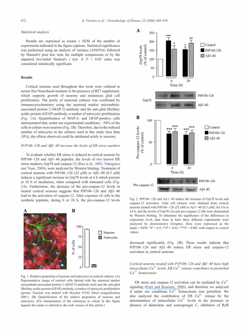
Fig. 2. PrP106–126 and Aβ1–40 induce the increase of Grp78 levels and
Results
Cortical neurons used throughout this work were cultured inserum-free Neurobasal medium in the presence of B27 supplement,which supports growth of neurons and minimizes glial cellproliferation. The purity of neuronal cultures was confirmed byimmunocytochemistry using the neuronal marker microtubule-associated protein 2 (MAP-2) antibody and the anti-glial fibrillaryacidic protein (GFAP) antibody, a marker of astrocytic proliferation(Fig. 1A). Quantification of MAP-2- and GFAP-positive cellsdemonstrated that, under our experimental conditions, ~94% of thecells in culture were neurons (Fig. 1B). Therefore, due to the reducednumber of astrocytes in the cultures used in this study (less than10%), the effects observed could be attributed solely to neurons.
PrP106–126 and Aβ1–40 increase the levels of ER stress markers
To evaluate whether ER stress is induced in cortical neurons byPrP106–126 and Aβ1–40 peptides, the levels of two known ERstress markers, Grp78 and caspase-12 (Rao et al., 2002; Nakagawaand Yuan, 2000), were analyzed by Western blotting. Treatment ofcortical neurons with PrP106–126 (25 μM) or Aβ1–40 (0.5 μM)induces a significant increase in Grp78 levels at 6 h which persistsat 24 h of incubation, when compared with untreated cells (Fig.2A). Furthermore, the decrease of the pro-caspase-12 levels intreated cortical neurons suggests that PrP106–126 and Aβ1–40lead to the activation of caspase-12. After exposure of cells to thesynthetic peptides, during 6 or 24 h, the pro-caspase-12 levels
Fig. 1. Relative proportion of neurons and astrocytes in cortical cultures. (A)Representative image of cortical cells labeled with the neuronal markermicrotubule-associated protein 2 (MAP-2) antibody (red) and the anti-glialfibrillary acidic protein (GFAP) antibody, a marker of astrocytic proliferation(green). Nucleus was stained with Hoechst 33342 (blue) (magnification400×). (B) Quantification of the relative proportion of neurons andastrocytes. (For interpretation of the references to colour in this figurelegend, the reader is referred to the web version of this article.)
caspase-12 activation. Total cell extracts were obtained from corticalneurons treated with PrP106–126 (25 μM) or Aβ1–40 (0.5 μM), for 6 h or24 h, and the levels of Grp78 (A) and pro-caspase-12 (B) were determinedby Western blotting. To determine the significance of the differences inexpression level, data from at least three different experiments wereanalyzed by densitometry (Graphs). Data were expressed as themean ± SEM. *P < 0.5; **P < 0.01; ***P < 0.001 with respect to controlvalues.
decreased significantly (Fig. 2B). These results indicate thatPrP106–126 and Aβ1–40 induce ER stress and caspase-12activation in cortical neurons.
Cortical neurons treated with PrP106–126 and Aβ1–40 have highintracellular Ca2+ levels. ER Ca2+ release contributes to perturbedCa2+ homeostasis
ER stress and caspase-12 activation can be mediated by Ca2+
signaling (Ferri and Kroemer, 2000), and therefore we analyzedif under our conditions Ca2+ homeostasis was perturbed. Wealso analyzed the contribution of ER Ca2+ release by thedetermination of intracellular Ca2+ levels in the presence orabsence of dantrolene and xestospongin C, inhibitors of RyR
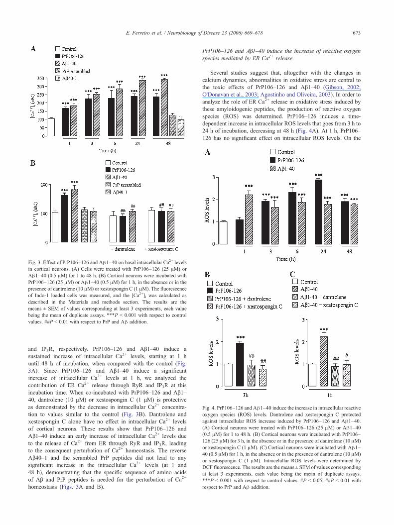
Fig. 3. Effect of PrP106–126 and Aβ1–40 on basal intracellular Ca2+ levelsin cortical neurons. (A) Cells were treated with PrP106–126 (25 μM) orAβ1–40 (0.5 μM) for 1 to 48 h. (B) Cortical neurons were incubated withPrP106–126 (25 μM) or Aβ1–40 (0.5 μM) for 1 h, in the absence or in thepresence of dantrolene (10 μM) or xestospongin C (1 μM). The fluorescenceof Indo-1 loaded cells was measured, and the [Ca2+]i was calculated asdescribed in the Materials and methods section. The results are themeans ± SEM of values corresponding at least 3 experiments, each valuebeing the mean of duplicate assays. ***P < 0.001 with respect to controlvalues. ##P < 0.01 with respect to PrP and Aβ addition.
Fig. 4. PrP106–126 and Aβ1–40 induce the increase in intracellular reactiveoxygen species (ROS) levels. Dantrolene and xestospongin C protectedagainst intracellular ROS increase induced by PrP106–126 and Aβ1–40.(A) Cortical neurons were treated with PrP106–126 (25 μM) or Aβ1–40(0.5 μM) for 1 to 48 h. (B) Cortical neurons were incubated with PrP106–126 (25 μM) for 3 h, in the absence or in the presence of dantrolene (10 μM)or xestospongin C (1 μM). (C) Cortical neurons were incubated with Aβ1–40 (0.5 μM) for 1 h, in the absence or in the presence of dantrolene (10 μM)or xestospongin C (1 μM). Intracelullar ROS levels were determined byDCF fluorescence. The results are the means ± SEM of values correspondingat least 3 experiments, each value being the mean of duplicate assays.***P < 0.001 with respect to control values. #P < 0.05; ##P < 0.01 withrespect to PrP and Aβ addition.
673E. Ferreiro et al. / Neurobiology of Disease 23 (2006) 669–678
and IP3R, respectively. PrP106–126 and Aβ1–40 induce asustained increase of intracellular Ca2+ levels, starting at 1 huntil 48 h of incubation, when compared with the control (Fig.3A). Since PrP106–126 and Aβ1–40 induce a significantincrease of intracellular Ca2+ levels at 1 h, we analyzed thecontribution of ER Ca2+ release through RyR and IP3R at thisincubation time. When co-incubated with PrP106–126 and Aβ1–40, dantrolene (10 μM) or xestospongin C (1 μM) is protectiveas demonstrated by the decrease in intracellular Ca2+ concentra-tion to values similar to the control (Fig. 3B). Dantrolene andxestospongin C alone have no effect in intracellular Ca2+ levelsof cortical neurons. These results show that PrP106–126 andAβ1–40 induce an early increase of intracellular Ca2+ levels dueto the release of Ca2+ from ER through RyR and IP3R, leadingto the consequent perturbation of Ca2+ homeostasis. The reverseAβ40–1 and the scrambled PrP peptides did not lead to anysignificant increase in the intracellular Ca2+ levels (at 1 and48 h), demonstrating that the specific sequence of amino acidsof Aβ and PrP peptides is needed for the perturbation of Ca2+
homeostasis (Figs. 3A and B).
PrP106–126 and Aβ1–40 induce the increase of reactive oxygenspecies mediated by ER Ca2+ release
Several studies suggest that, altogether with the changes incalcium dynamics, abnormalities in oxidative stress are central tothe toxic effects of PrP106–126 and Aβ1–40 (Gibson, 2002;O'Donavan et al., 2003; Agostinho and Oliveira, 2003). In order toanalyze the role of ER Ca2+ release in oxidative stress induced bythese amyloidogenic peptides, the production of reactive oxygenspecies (ROS) was determined. PrP106–126 induces a time-dependent increase in intracellular ROS levels that goes from 3 h to24 h of incubation, decreasing at 48 h (Fig. 4A). At 1 h, PrP106–126 has no significant effect on intracellular ROS levels. On the
674 E. Ferreiro et al. / Neurobiology of Disease 23 (2006) 669–678
other hand, Aβ1–40 induces a significant increase of ROS levels at1 h, maintaining these levels until 48 h of incubation (Fig. 4A).When cortical cells were treated for 3 h with PrP106–126 or for 1 hwith Aβ1–40, in the presence of dantrolene or xestospongin C, asignificant decrease in ROS levels is observed, demonstrating thatRyR- and IP3R-mediated ER Ca2+ release is involved in oxidativestress triggered by PrP and Aβ peptides in cortical neurons (Figs.4B and C). Dantrolene or xestospongin C per se did not affectintracellular ROS levels (data not shown).
PrP106–126 and Aβ1–40 induce cytochrome c release andcaspase-2, -3, -6, -8 and -9 activation upon ER Ca2+ release
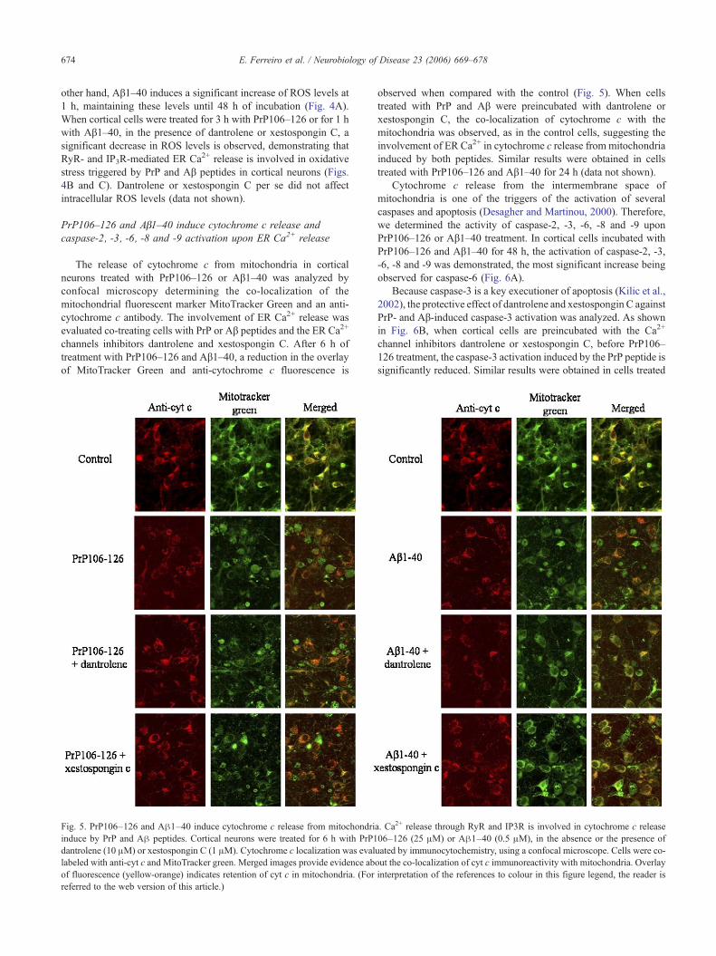
The release of cytochrome c from mitochondria in corticalneurons treated with PrP106–126 or Aβ1–40 was analyzed byconfocal microscopy determining the co-localization of themitochondrial fluorescent marker MitoTracker Green and an anti-cytochrome c antibody. The involvement of ER Ca2+ release wasevaluated co-treating cells with PrP or Aβ peptides and the ER Ca2+
channels inhibitors dantrolene and xestospongin C. After 6 h oftreatment with PrP106–126 and Aβ1–40, a reduction in the overlayof MitoTracker Green and anti-cytochrome c fluorescence is
Fig. 5. PrP106–126 and Aβ1–40 induce cytochrome c release from mitochondriinduce by PrP and Aβ peptides. Cortical neurons were treated for 6 h with PrP1dantrolene (10 μM) or xestospongin C (1 μM). Cytochrome c localization was evallabeled with anti-cyt c and MitoTracker green. Merged images provide evidence abof fluorescence (yellow-orange) indicates retention of cyt c in mitochondria. (Forreferred to the web version of this article.)
observed when compared with the control (Fig. 5). When cellstreated with PrP and Aβ were preincubated with dantrolene orxestospongin C, the co-localization of cytochrome c with themitochondria was observed, as in the control cells, suggesting theinvolvement of ER Ca2+ in cytochrome c release from mitochondriainduced by both peptides. Similar results were obtained in cellstreated with PrP106–126 and Aβ1–40 for 24 h (data not shown).
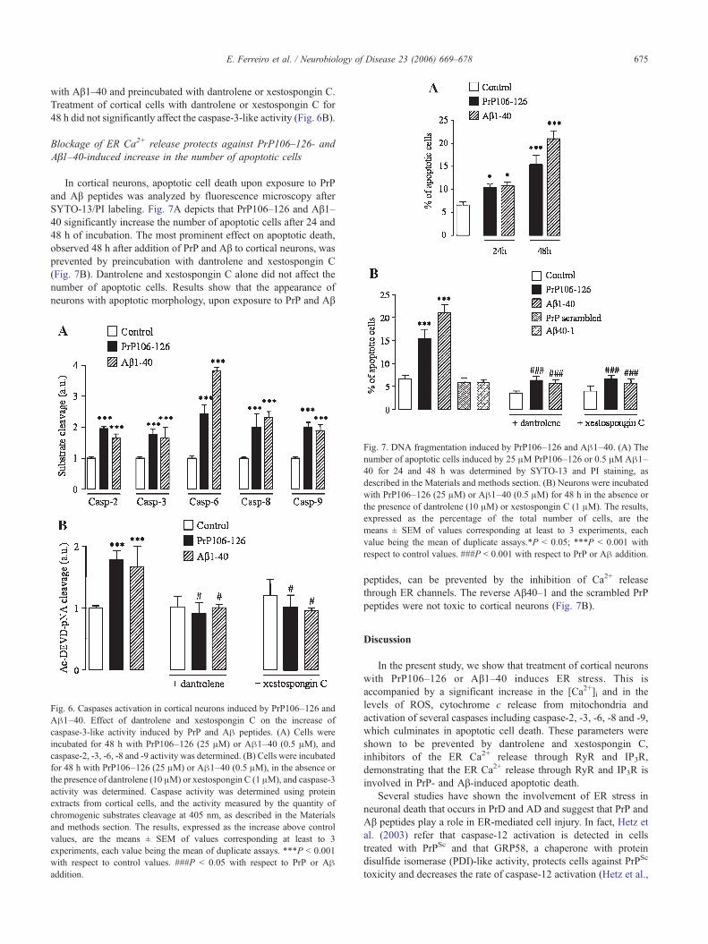
Cytochrome c release from the intermembrane space ofmitochondria is one of the triggers of the activation of severalcaspases and apoptosis (Desagher and Martinou, 2000). Therefore,we determined the activity of caspase-2, -3, -6, -8 and -9 uponPrP106–126 or Aβ1–40 treatment. In cortical cells incubated withPrP106–126 and Aβ1–40 for 48 h, the activation of caspase-2, -3,-6, -8 and -9 was demonstrated, the most significant increase beingobserved for caspase-6 (Fig. 6A).
Because caspase-3 is a key executioner of apoptosis (Kilic et al.,2002), the protective effect of dantrolene and xestospongin C againstPrP- and Aβ-induced caspase-3 activation was analyzed. As shownin Fig. 6B, when cortical cells are preincubated with the Ca2+
channel inhibitors dantrolene or xestospongin C, before PrP106–126 treatment, the caspase-3 activation induced by the PrP peptide issignificantly reduced. Similar results were obtained in cells treated
a. Ca2+ release through RyR and IP3R is involved in cytochrome c release06–126 (25 μM) or Aβ1–40 (0.5 μM), in the absence or the presence ofuated by immunocytochemistry, using a confocal microscope. Cells were co-out the co-localization of cyt c immunoreactivity with mitochondria. Overlayinterpretation of the references to colour in this figure legend, the reader is
675E. Ferreiro et al. / Neurobiology of Disease 23 (2006) 669–678
with Aβ1–40 and preincubated with dantrolene or xestospongin C.Treatment of cortical cells with dantrolene or xestospongin C for48 h did not significantly affect the caspase-3-like activity (Fig. 6B).
Blockage of ER Ca2+ release protects against PrP106–126- andAβ1–40-induced increase in the number of apoptotic cells
In cortical neurons, apoptotic cell death upon exposure to PrPand Aβ peptides was analyzed by fluorescence microscopy afterSYTO-13/PI labeling. Fig. 7A depicts that PrP106–126 and Aβ1–40 significantly increase the number of apoptotic cells after 24 and48 h of incubation. The most prominent effect on apoptotic death,observed 48 h after addition of PrP and Aβ to cortical neurons, wasprevented by preincubation with dantrolene and xestospongin C(Fig. 7B). Dantrolene and xestospongin C alone did not affect thenumber of apoptotic cells. Results show that the appearance ofneurons with apoptotic morphology, upon exposure to PrP and Aβ
Fig. 6. Caspases activation in cortical neurons induced by PrP106–126 andAβ1–40. Effect of dantrolene and xestospongin C on the increase ofcaspase-3-like activity induced by PrP and Aβ peptides. (A) Cells wereincubated for 48 h with PrP106–126 (25 μM) or Aβ1–40 (0.5 μM), andcaspase-2, -3, -6, -8 and -9 activity was determined. (B) Cells were incubatedfor 48 h with PrP106–126 (25 μM) or Aβ1–40 (0.5 μM), in the absence orthe presence of dantrolene (10 μM) or xestospongin C (1 μM), and caspase-3activity was determined. Caspase activity was determined using proteinextracts from cortical cells, and the activity measured by the quantity ofchromogenic substrates cleavage at 405 nm, as described in the Materialsand methods section. The results, expressed as the increase above controlvalues, are the means ± SEM of values corresponding at least to 3experiments, each value being the mean of duplicate assays. ***P < 0.001with respect to control values. ###P < 0.05 with respect to PrP or Aβaddition.
Fig. 7. DNA fragmentation induced by PrP106–126 and Aβ1–40. (A) Thenumber of apoptotic cells induced by 25 μM PrP106–126 or 0.5 μM Aβ1–40 for 24 and 48 h was determined by SYTO-13 and PI staining, asdescribed in the Materials and methods section. (B) Neurons were incubatedwith PrP106–126 (25 μM) or Aβ1–40 (0.5 μM) for 48 h in the absence orthe presence of dantrolene (10 μM) or xestospongin C (1 μM). The results,expressed as the percentage of the total number of cells, are themeans ± SEM of values corresponding at least to 3 experiments, eachvalue being the mean of duplicate assays.*P < 0.05; ***P < 0.001 withrespect to control values. ###P < 0.001 with respect to PrP or Aβ addition.
peptides, can be prevented by the inhibition of Ca2+ releasethrough ER channels. The reverse Aβ40–1 and the scrambled PrPpeptides were not toxic to cortical neurons (Fig. 7B).
Discussion
In the present study, we show that treatment of cortical neuronswith PrP106–126 or Aβ1–40 induces ER stress. This isaccompanied by a significant increase in the [Ca2+]i and in thelevels of ROS, cytochrome c release from mitochondria andactivation of several caspases including caspase-2, -3, -6, -8 and -9,which culminates in apoptotic cell death. These parameters wereshown to be prevented by dantrolene and xestospongin C,inhibitors of the ER Ca2+ release through RyR and IP3R,demonstrating that the ER Ca2+ release through RyR and IP3R isinvolved in PrP- and Aβ-induced apoptotic death.
Several studies have shown the involvement of ER stress inneuronal death that occurs in PrD and AD and suggest that PrP andAβ peptides play a role in ER-mediated cell injury. In fact, Hetz etal. (2003) refer that caspase-12 activation is detected in cellstreated with PrPSc and that GRP58, a chaperone with proteindisulfide isomerase (PDI)-like activity, protects cells against PrPSc
toxicity and decreases the rate of caspase-12 activation (Hetz et al.,

676 E. Ferreiro et al. / Neurobiology of Disease 23 (2006) 669–678
2005). In addition, Nakagawa et al. (2000) point out that corticalneurons deficient in caspase-12 are more resistant to Aβ proteinneurotoxicity. Furthermore, cells that express presenilin 1 mutants,linked to familial AD, have increased susceptibility to ER stress(Katayama et al., 2004; Imaizumi et al., 2001).
Our results show that PrP106–126 and Aβ1–40 induce theincrease of GRP78 levels and caspase-12 activation in corticalneurons, indicating that these peptides induce ER stress, which canbe activated by the unfolded protein response (UPR) and bydisturbance of Ca2+ homeostasis (Kaufman, 1999). The balancebetween intracellular Ca2+ levels and the ER Ca2+ content isguaranteed by two different processes: the pumping in the ER bySERCA ATPases and the release by the opening of IP3R or RyRCa2+ release channels (Berridge et al., 2000). These Ca2+ releasechannels of the ER participate in the signal transduction pathwayof apoptosis (Guo et al., 1997; Jayaraman and Marks, 1997; Pan etal., 2000). In agreement with results obtained by other authorsusing PC12 cells (Guo et al., 1996, 1997), we have recentlydemonstrated that Ca2+ release from RyR but also from IP3R isinvolved in Aβ25–35 toxicity in cortical neurons (Ferreiro et al.,2004). IP3-mediated Ca2+ release from the ER was previouslydemonstrated to trigger apoptosis in response to diverse signals(Distelhorst and Roderick, 2003). It has been shown in ourlaboratory that Aβ25–35/1–40 and PrP106–126 can induce theincrease of cytosolic Ca2+ levels in cortical neurons following 24 hof incubation (Ferreiro et al., 2004; Agostinho and Oliveira, 2003).In the present study, we show that in cortical neurons treated withPrP106–126 and Aβ1–40 intracellular Ca2+ levels increase before24 h of incubation. In fact, at 1 h of incubation, a significant rise incytosolic Ca2+ concentration was measured. The Ca2+ releasethrough the RyR and IP3R contributes to this early [Ca2+]i increase.
Several studies indicate that, in addition to perturbed Ca2+
homeostasis, oxidative stress also occurs in PrD and AD (reviewedin Brown et al., 1996; Agostinho and Oliveira, 2003; Fernaeus andLand, 2005; Sheehan et al., 1997; Zhu et al., 2004). In fact, the ERis especially vulnerable to oxidative stress since it is one of the cellorganelles that produce ROS (Hayashi et al., 2005). Furthermore,oxidative stress may lead to perturbations of the ER Ca2+
homeostasis because many of the regulatory proteins of ER Ca2+,such as IP3R and RyR, the Ca2+ ATPases and the ER-residentproteins, are sensitive to oxidants (Huang et al., 2004). In addition,Ca2+ that results from the depletion of ER Ca2+ stores can be takenup by juxtaposed mitochondria, inducing ROS formation (Tardif etal., 2005). Our study demonstrates that PrP106–126 and Aβ1–40induce ROS formation in cortical neurons, and this effect wasprevented by dantrolene and xestospongin C, indicating that Ca2+
release through RyR and IP3R is involved in PrP- and Aβ-inducedROS formation. In the case of PrP106–126, the intracellular Ca2+
increase precedes the ROS formation, indicating that intracellularCa2+ rise is an early event in PrP peptide toxicity.
ER Ca2+ release has been shown to have immediate effects onmitochondrial function, leading to rapid Ca2+ accumulation inmitochondria which promotes cytochrome c release and activationof downstream caspase pathways in cells exposed to proapoptoticagents (Nutt et al., 2002). In the cytosol, cytochrome c binds Apaf-1 and dATP to form a complex that activates caspase-9, which canactivate the executioner caspase-3 (Desagher and Martinou, 2000).Cytochrome c has been shown to be involved in the neurotoxicityof Aβ25–35 and PrP106–126 (Cardoso et al., 2002; Agostinho andOliveira, 2003). Our results show that, beyond the fact that Aβ andPrP peptides induce cytochrome c release from mitochondria, ER
Ca2+ release through RyR and IP3R is involved in the process,demonstrating that mitochondria and the ER cooperate in theneuronal death induced by Aβ and PrP peptides. These results arein agreement with data obtained in cells treated with ER stressorsin which ER/mitochondria crosstalk was demonstrated (Häcki etal., 2000; Boya et al., 2002). Although our results demonstrate thatER stress induced by PrP and Aβ peptides occurs upstream ofmitochondrial dysfunction, since cytochrome c release is preventedby dantrolene and xestospongin C, we cannot rule out thepossibility that mitochondrial dysfunction could also trigger ERstress, propagating the initial injury. Recently, it was demonstratedthat both full-length APP and Aβ accumulate in the mitochondriacompartment causing mitochondrial dysfunction (Anandatheertha-varada et al., 2003; Lustbader et al., 2004), which may in turnderegulate ER Ca2+ homeostasis. In fact, inhibition of mitochon-drial respiratory chain with rotenone was shown to induce ERstress through the phosphorylation of the key ER stress kinasesIRE1α and PKR-like ER kinase (PERK) (Ryu et al., 2002).
Several caspases have been shown to be involved in ER stress.Hitomi et al. (2003) have shown that activation of caspase-12indirectly activates cytoplasmic caspase-3 and activation of thiscaspase is completely suppressed in cells overexpressing Bcl-2targeted specifically to the ER (Häcki et al., 2000). Dahmer (2005)has shown that thapsigargin, an activator of ER stress, inducesactivation of the caspase-2, -3 and -7. He et al. (2002) had alreadyshown that thapsigargin-mediated perturbation in Ca2+ home-ostasis upregulates DR5 (death receptor 5) and TRAIL (tumornecrosis factor-related apoptosis inducing ligand), and this iscoupled with caspase-8 activation and Bid cleavage. In the presentstudy, it was demonstrated that caspase-2, -3, -6, -8 and -9activities are increased upon exposure to PrP106–126 and Aβ1–40.Also under our experimental conditions, the caspase-3 activityincreased upon treatment with PrP 106–126 and Aβ1–40 wasprevented by the ER Ca2+ release inhibitors dantrolene andxestospongin C, suggesting that perturbation of ER Ca2+ home-ostasis mediated by Ca2+ release from RyR and IP3R is involved inthe activation of caspase-3 induced by the peptides.
One possible site of initiation of apoptosis is the synapse.Perturbed synaptic ER Ca2+ homeostasis promotes activation ofapoptotic cascades (Mattson, 2000). Therefore, since PrP andAβ peptides are known to be produced and to accumulate in highamounts at the synapses (Gong et al., 2003; Sales et al., 1998;Herms et al., 1999), our results support the view that loss of ERCa2+ homeostasis induced by the peptides is involved in synapticdysfunction and loss of dendritic spines, which are early eventsthat occur in PrD and AD (Belichenko et al., 2000; Selkoe, 2002).Furthermore, perturbation of Ca2+ homeostasis induced by thesepeptides can lead to the activation of several kinases, such as GSK-3 and Cdk5, and to the consequent phosphorylation of themicrotubule-associated protein tau, leading to the formation ofneurofibrillary tangles (Pérez et al., 2003; Lee et al., 2000;reviewed in Fuentalba et al., 2004). Therefore, the ER stressinduced by PrP and Aβ in the early stages of the diseases, in whichthey are implicated, may be associated with synaptic loss and withthe presence of neurofibrillary tangles, central hallmarks of ADthat may be also related to PrD. We then suggest that drugs thatstabilize neuronal Ca2+ homeostasis, including dantrolene andxestospongin C to suppress Ca2+ release from ER, are potentialtherapeutical targets for PrD and AD.
Altogether, our in vitro results show that PrP106–126 and Aβ1–40 induce Ca2+ homeostasis deregulation, oxidative stress and
677E. Ferreiro et al. / Neurobiology of Disease 23 (2006) 669–678
apoptotic cell death by a mechanism that involves the early releaseof Ca2+ from ER through RyR and IP3R. Furthermore, the resultspresented here demonstrate that PrP- and Aβ-induced Ca2+ releasefrom ER leads to mitochondrion-mediated cell death, suggestingthat both organelles cooperate in PrP- and Aβ-induced apoptosis.
Acknowledgments
This work was supported by FCT (Portuguese ResearchCouncil) project no. POCTI/36101/NSE/2000, Elisabete Ferreiroand Rosa Resende have PhD fellowships from FCT (grant no.SFRH/BD/14108/2003 and SFRH/BD/11005/2002, respectively).
References
Agostinho, P., Oliveira, C.R., 2003. Involvement of calcineurin in theneurotoxic effects induced by amyloid-beta and prion peptides. Eur. J.Neurosci. 17, 1–8.
Anandatheerthavarada, H.K., Biswas, G., Robin, M., Avadhani, N.G., 2003.Mitochondrial targeting and a novel transmembrane arrest of Alzhei-mer's amyloid precursor protein impairs mitochondrial function inneuronal cells. J. Cell Biol. 161, 41–54.
Bandeira-Duarte, C., Carvalho, C.A.M., Cragoe, E.J., Carvalho, A.P., 1990.Influence of isolation medium on synaptosomal properties: intracellularpH, pCa and Ca2+ uptake. Neurochem. Res. 15, 313–320.
Baumann, O., Walz, B., 2001. Endoplasmic reticulum of animal cells and itsorganization into structural and functional domains. Int. Rev. Cytol. 205,149–214.
Belichenko, P.V., Brown, D., Jeffrey, M., Fraser, J.R., 2000. Dendritic andsynaptic alterations of hippocampal pyramidal neurones in scrapie-infected mice. Neuropathol. Appl. Neurobiol. 26, 143–149.
Berridge, M.J., Lipp, P., Bootman, M.D., 2000. The versality anduniversality of calcium signaling. Nat. Rev., Mol. Cell Biol. 1, 11–21.
Boya, P., Cohen, I., Zamzami, N., Vieira, H.L., Kroemer, G., 2002.Endoplasmic reticulum stress-induced cell death requires mitochondrialmembrane permeabilization. Cell Death Differ. 9, 465–467.
Brown, D.R., 2000. PrPSc-like prion protein peptide inhibits the function ofcellular prion protein. Biochem. J. 352, 511–518.
Brown, D.R., Schmidt, B., Kretzschmar, H.A., 1996. Role of microglia andhost prion protein in neurotoxicity of a prion protein fragment. Nature380, 345–347.
Cardoso, S.M., Swerdlow, R.H., Oliveira, C.R., 2002. Induction ofcytochrome c-mediated apoptosis by amyloid β 25–35 requiresfunctional mitochondria. Brain Res. 931, 117–125.
Cathcart, R., Scwiers, E., Ames, B.N., 1993. Detection of picomole levels ofhydroperoxides using a fluorescent dichlorofluorescein assay. Annal.Biochem. 134, 111–116.
Cregan, S.P., MacLaurin, J.G., Craig, C.G., Roberstson, G.S., Nicholson,D.W., Park, D.S., Slack, R.S., 1999. Bax-dependent caspase-3activation is a key determinant in p53-induced apoptosis in neurons.J. Neurosci. 19, 7860–7869.
Dahmer, M.K., 2005. Caspases-2, -3, and -7 are involved in thapsigargin-induced apoptosis of SH-SY5Y neuroblastoma cells. J. Neurosci. Res.80, 576–583.
Della-Bianca, V., Rossi, F., Armato, U., Dal-Pra, I., Costantini, C., Perini,G., Politi, V., Della Valle, G., 2001. Neurotrophin p75 receptor isinvolved in neuronal damage by prion peptide-(106–126). J. Biol.Chem. 276, 38929–38933.
Desagher, S., Martinou, J.C., 2000. Mitochondria as the central control pointof apoptosis. Trends Cell Biol. 10, 369–377.
Distelhorst, C.W., Roderick, H.L., 2003. Ins(1,4,5)P3-mediated calciumsignals and apoptosis: is there a role for Bcl-2? Biochem. Soc. Trans. 31,958–959.
Dreher, D., Jornot, L., Junod, A.F., 1995. Effects of hypoxantine–xantine
oxidase on Ca2+ stores and protein synthesis in human endothelial cells.Circ. Res. 76, 388–395.
Ettaiche, M., Pichot, R., Vincent, J.P., Chabry, J., 2000. In vivo cytotoxicityof the prion protein fragment 106–126. J. Biol. Chem. 275,36487–36490.
Fernaeus, S., Land, T., 2005. Increased iron-induced oxidative stress andtoxicity in scrapie-infected neuroblastoma cells. Neurosci. Lett. 382,217–220.
Ferreiro, E., Oliveira, C.R., Pereira, C., 2004. Involvement of endoplasmicreticulum Ca2+ release through ryanodine and inositol 1,4,5-triphosphatereceptors in the neurotoxic effects induced by the Amyloid-β peptide.J. Neurosci. Res. 76, 872–880.
Ferri, K., Kroemer, G., 2000. Organelle-specific initiation of cell deathpathway. Nat. Cell Biol. 3, E255–E263.
Fuentalba, R.A., Farias, G., Scheu, J., Bronfman, M., Marzolo, M.P.,Inestrosa, N.C., 2004. Signal transduction during amyloid-beta-peptide neurotoxicity: role in Alzheimer disease. Brain Res. 47,257–289.
Gibson, G.E., 2002. Interactions of oxidative stress with cellular calciumdynamics and glucose metabolism in Alzheimer's disease. Free RadicalBiol. Med. 32, 1061–1070.
Gong, Y., Chang, L., Viola, K.L., Lacor, P.N., Lambert, M.P., Finch, C.E.,Krafft, G.A., Klein, W.L., 2003. Alzheimer's disease-affected brain:presence of oligomeric A beta ligands (ADDLs) suggests a molecularbasis for reversible memory loss. Proc. Natl. Acad. Sci. U. S. A. 100,10417–10422.
Guo, Q., Furukawa, K., Sopher, B.L., Pham, D.G., Xie, J., Robinson, N.,Martin, G.M., Mattson, M.P., 1996. Alzheimer's PS-1 mutation perturbscalcium homeostasis and sensitizes PC12 cells to death induced byamyloid β-peptide. NeuroReport 8, 379–383.
Guo, Q., Sopher, B.L., Furukawa, K., Pham, D.G., Robinson, N., Martin,G.M., Mattson, M.P., 1997. Alzheimer's presenilin mutation sensitizesneural cells to apoptosis induced by trophic factor withdrawal andamyloid beta-peptide: involvement of calcium and oxyradicals.J. Neurosci. 17, 4212–4222.
Häcki, J., Egger, L., Monney, L., Conus, S., Rosse, T., Fellay, I., Borner, C.,2000. Apoptotic crosstalk between the endoplasmatic reticulum andmitochondria controlled by Bcl-2. Oncogene 19, 2286–2295.
Hayashi, T., Saito, A., Okuno, S., Ferrand-Drake, M., Dodd, R.L., Chan,P.H., 2005. Damage to the endoplasmic reticulum and activation ofapoptotic machinery by oxidative stress in ischemic neurons. J. Cereb.Blood Flow Metab. 25, 41–53.
He, Q., Lee, D., Rong, R., Yu, M., Luo, X., Klein, M., El-Deiry, W., Huang,Y., Hussain, A., Sheikh, M., 2002. Endoplasmic reticulum calcium pooldepletion-induced apoptosis is coupled with activation of the deathreceptor 5 pathway. Oncogene 21, 2623–2633.
Herms, J., Tings, T., Gall, S., Madlung, A., Giese, A., Siebert, H.,Schurmann, P., Windl, O., Brose, N., Kretzschmar, H., 1999. Evidenceof presynaptic location and function of the prion protein. J. Neurosci. 19,8866–8875.
Hertz, E., Yu, A.C.H., Hertz, L., Juurlink, B.H.J., Schousboe, A., 1989.Preparation of primary cultures of mouse cortical neurons. In: Shahar,A., De Vellis, J., Vernadakis, A., Haber, V. (Eds.), A Dissection andTissue Culture Manual of the Nervous System. Alan R. LissNewYork.
Hetz, C., Russelakis-Carneiro, M., Maundrell, K., Castilla, J., Soto, C.,2003. Caspase-12 and endoplasmic reticulum stress mediate neurotoxi-city of pathological prion protein. EMBO J. 22, 5435–5445.
Hetz, C., Russelakis-Carneiro, M., Walchli, S., Carboni, S., Vial-Knecht, E.,Maundrell, K., Castilla, J., Soto, C., 2005. The disulfide isomeraseGrp58 is a protective factor against prion neurotoxicity. J. Neurosci. 25,2793–2802.
Hitomi, J., Katayama, T., Taniguchi, M., Honda, A., Imaizumi, K.,Toyama, M., 2003. Apoptosis induced by endoplasmic reticulumstress depends on activation of caspase-3 via caspase-12. Neurosci.Lett. 357, 127–130.
Huang, H., Zhang, H., Ou, H., Chen, H., Gibson, G.E., 2004. α-Keto-β-
678 E. Ferreiro et al. / Neurobiology of Disease 23 (2006) 669–678
methyl-n-valeric acid diminishes reactive oxygen species and altersendoplasmic reticulum Ca2+ stores. Free. Radical Biol. Med. 37,1779–1789.
Imaizumi, K., Miyoshi, K., Katayama, T., Yoneda, T., Taniguchi, M., Kudo,T., Tohyama, M., 2001. The unfolded protein response and Alzheimer'sdisease. Biochim. Biophys. Acta 1536, 85–96.
Jayaraman, T., Marks, A.R., 1997. T cells deficient in inositol 1,4,5-triphosphate receptor are resistant to apoptosis. Mol. Cell. Biol. 17,3005–3012.
Katayama, T., Imaizumi, K., Manabe, T., Hitomi, J., Kudo, T., Tohyama, M.,2004. Induction of neuronal death by ER stress in Alzheimer's disease.J. Chem. Neuroanat. 28, 67–78.
Kaufman, R.J., 1999. Stress signaling from the lumen of the endoplasmicreticulum: coordination of gene transcriptional and translationalcontrols. Genes Dev. 13, 1211–1233.
Kilic, M., Schäfer, R., Hoppe, J., Kagerhuber, U., 2002. Formation ofnoncanonical high molecular weight caspase-3 and -6 complexes andactivation of caspase-12 during serum starvation induced apoptosis inAKR-2B mouse fibroblasts. Cell Death and Differ. 9, 125–137.
Lee, M.S., Kwon, Y.T., Li, M., Peng, J., Friedlander, R.M., Tsai, L.H., 2000.Neurotoxicity induces cleavage of p35 to p25 by calpain. Nature 405,360–364.
Lustbader, J.W., Cirilli, M., Lin, C., Xu, H.W., Takuma, K., Wang, N.,Caspersen, C., Chen, X., Pollak, S., Chaney, M., Trinchese, F., Liu, S.,Gunn-Moore, F., Lue, L., Walker, D.G., Kuppusamy, P., Zewier, A.L.,Arancio, O., Stern, D., Yan, S.S., Wu, H., 2004. ABAD directly link Abto mitochondrial toxicity in Alzheimer's disease. Science 304, 448–452.
Mattson, M.P., 1997. Cellular actions of β-amyloid precursor protein and itssoluble and fibrillogenic derivatives. Physiol. Rev. 77, 108–1132.
Mattson, M.P., 2000. Apoptosis in neurodegenerative disorders. Nat. Rev.,Mol. Cell Biol. 1, 120–129.
Morishima, N., Nakanishi, K., Takenouchi, H., Takehiko, S., Yasuhiko, Y.,2002. An endoplasmic reticulum stress-specific caspase cascade inapoptosis. J. Biol. Chem. 277, 34287–34294.
Nakagawa, T., Yuan, J., 2000. Cross-talk between two cysteine proteasefamilies: activation of caspase-12 by calpain in apoptosis. J. Cell Biol.150, 887–894.
Nakagawa, T., Zhu, H., Morishima, N., Li, E., Xu, J., Yankner, B.A.,Yuan, J., 2000. Caspase-12 mediates endoplasmic-reticulum-specificapoptosis and cytotoxicity by amyloid-β. Nature 403, 98–103.
Nutt, L.K., Chandra, J., Pataer, A., Fang, B., Roth, J.A., Swisher, G., O'Neil,R.G., McConkey, D.J., 2002. Bax-mediated Ca2+ mobilization promotescytochrome c release during apoptosis. J. Biol. Chem. 277,20301–20308.
O'Donavan, C.N., Tobin, D., Cotter, T.G., 2003. Prion protein fragment PrP-(106–126) induces apoptosis via mitochondrial disruption in humanneuronal SH-SY5Y cells. J. Biol. Chem. 276, 43516–43523.
Pan, Z., Damron, D., Nieminen, A.-L., Bhat, M.B., Ma, J., 2000. Depletionof intracellular Ca2+ by caffeine and ryanodine induces apoptosis ofChinese hamster ovary cells transfected with ryanodine receptor. J. Biol.Chem. 275, 19978–19984.
Pashen, W., 2001. Dependence of vital cell function on endoplasmic
reticulum calcium levels: implications for the mechanisms underlyingneuronal cell injury in different pathological states. Cell Calcium 29,1–11.
Pereira, C., Santos, M.S., Oliveira, C.R., 1999. Involvement of oxidativestress on the impairment of energy metabolism induced by Aβpeptides on PC12 cells: protection by antioxidants. Neurobiol. Dis. 6,209–219.
Perez, R.G., Soriano, S., Hayes, J.D., Ostaszewski, B., Xia, W., Selkoe, D.J.,Chen, X., Stokin, G.B., Koo, E.H., 1999. Mutagenesis identifies newsignals for β-amyloid precursor protein endocytosis, turnover, and thegeneration of secreted fragments, including Aβ42. J. Biol. Chem. 274,18851–18856.
Pérez, M., Rojo, A.I., Wandosell, F., Díaz-Nido, J., Avila, J., 2003. Prionpeptide induces neuronal cell death through a pathway involvingglycogen synthase kinase 3. Biochem. J. 372, 126–129.
Prusiner, S.B., 1996. Molecular biology and pathogenesis of prion diseases.Trends Biochem. Sci. 252, 482–487.
Racay, P., Kaplan, P., Lehotsky, J., Mezesova, V., 1995. Rabbit brainendoplasmic reticulum membranes as target for free radicals. Changes inCa2+-transport and protection by stobadine. Biochem. Mol. Biol. Int. 36,569–577.
Rao, R.V., Peel, A., Logvinova, A., Del Rio, G., Hermel, E., Yokota, T.,Goldsmith, P.C., Ellerby, L.M., Ellerby, H.M., Bredesen, D.E., 2002.Coupling endoplasmic reticulum stress to the cell death program: role ofthe ER chaperone GRP78. FEBS Lett. 514, 122–128.
Ryu, E.J., Harding, H.P., Angelastro, J.M., Vitolo, O.V., Ron, D., Greene,L.A., 2002. Endoplasmic reticulum stress and the unfolded proteinresponse in cellular models of Parkinson's disease. J. Neurosci. 22,10690–10698.
Sales, N., Rodolfo, K., Nassig, R., Faucheux, B., Di Giamberardino, L.,Moya, K.L., 1998. Cellular prion protein localization in rodent andprimate brain. Eur. J. Neurosci. 2464–2471.
Selkoe, D.J., 2002. Alzheimer's disease is a synaptic failure. Science 298,789–791.
Sheehan, J.P., Swerdlow, R.H., Miller, S.W., Davis, R.E., Parks, J.K., Parker,W.D., Tuttle, J.B., 1997. Calcium homeostasis and reactive oxygenspecies production in cells transformed by mitochondria fromindividuals with sporadic Alzheimer's disease. J. Neurosci. 17,4612–4622.
Singh, N., Gu, Y., Bose, S., Kalepu, S., Mishra, R.S., Verghese, S., 2000.Prion peptide 106–126 as a model for prion replication andneurotoxicity. Front. Biosci. 7, 60–71.
Tardif, K.D., Waris, G., Siddiqui, A., 2005. Hepatitis C virus, ER stress, andoxidative stress. Trends Microbiol. 13, 159–163.
Viner, R.I., Huhmer, A.F.R., Bigelow, D.J., Schoneich, C., 1996. Theoxidative inactivation of sarcoplasmic reticulum Ca2+-ATPase byperoxynitrite. Free Radical Res. 24, 243–259.
Wisniewski, T., Ghiso, J., Frangione, B., 1997. Biology of Abeta amyloid inAlzheimer's disease. Neurobiol. Aging 4, 313–328.
Zhu, X., Arun, K.R., Lee, H., Casadesus, G., Smith, M.A., Perry, G., 2004.Oxidative stress signaling in Alzheimer's disease. Brain Res. 1000,32–39.
Copyright © 2022 FDOKUMEN