Production of cattle lacking prion protein
15
Production of cattle lacking prion protein Jürgen A Richt 1,6 , Poothappillai Kasinathan 2 , Amir N Hamir 1 , Joaquin Castilla 3 , Thillai Sathiyaseelan 2 , Francisco Vargas 1 , Janaki Sathiyaseelan 2 , Hua Wu 2 , Hiroaki Matsushita 2 , Julie Koster 2 , Shinichiro Kato 4,5 , Isao Ishida 4 , Claudio Soto 3 , James M Robl 2 , and Yoshimi Kuroiwa 4,5,6 1 National Animal Disease Center, Agriculture Research Services, United States Department of Agriculture, 2300 Dayton Avenue, Ames, Iowa 50010, USA. 2 Hematech, Inc., 4401 S. Technology Drive, Sioux Falls, South Dakota 57106, USA. 3 Department of Neurology, University of Texas Medical Branch, 301 University Boulevard, Galveston, Texas, 77555-0646, USA. 4 Pharmaceutical Division, Kirin Brewery Co., Ltd., 26-1, Jingumae 6-chome, Shibuya-ku, Tokyo, Japan. 5 Gemini Science Inc., 9420 Athena Circle, La Jolla, California 92109, USA. Abstract Prion diseases are caused by propagation of misfolded forms of the normal cellular prion protein PrP C , such as PrP BSE in bovine spongiform encephalopathy (BSE) in cattle and PrP CJD in Creutzfeldt-Jakob disease (CJD) in humans 1 . Disruption of PrP C expression in mice, a species that does not naturally contract prion diseases, results in no apparent developmental abnormalities 2–5 . However, the impact of ablating PrP C function in natural host species of prion diseases is unknown. Here we report the generation and characterization of PrP C -deficient cattle produced by a sequential gene-targeting system 6 . At over 20 months of age, the cattle are clinically, physiologically, histopathologically, immunologically and reproductively normal. Brain tissue homogenates are resistant to prion propagation in vitro as assessed by protein misfolding cyclic amplification 7 . PrP C -deficient cattle may be a useful model for prion research and could provide industrial bovine products free of prion proteins. To generate PrP C -deficient (PRNP −/− ) cattle, we transfected a male Holstein primary fetal fibroblast line 6594 with first and second knockout (KO) vectors (pBPrP(H)KOneo and pBPrP (H)KOpuro vectors) 6 to sequentially disrupt the two alleles of PRNP. PRNP −/− fetal cell lines were established at 40–60 d of gestation and three of the PRNP −/− fetal cell lines (5211, 5232 and 4296) were recloned to produce calves (Table 1 and Fig. 1a). To verify that the calves possess the PRNP −/− genotype, we collected ear biopsies and established fibroblast cell lines for genotyping. Genotyping was done by genomic PCR specific to each gene targeting event 6 (primer pairs: neoF7 × neoR7 and puroF14 × puroR14, Fig. 1b), followed by sequence © 2007 Nature Publishing Group Correspondence should be addressed to Y.K. ([email protected]), J.M.R. ([email protected]) or J.A.R. ([email protected]). 6 These authors contributed equally to this work. Note: Supplementary information is available on the Nature Biotechnology website. COMPETING INTERESTS STATEMENT The authors declare competing financial interests (see the Nature Biotechnology website for details). Reprints and permissions information is available online at http://npg.nature.com/reprintsandpermissions/ NIH Public Access Author Manuscript Nat Biotechnol. Author manuscript; available in PMC 2010 January 28. Published in final edited form as: Nat Biotechnol. 2007 January ; 25(1): 132. doi:10.1038/nbt1271. NIH-PA Author Manuscript NIH-PA Author Manuscript NIH-PA Author Manuscript
Transcript of Production of cattle lacking prion protein

Production of cattle lacking prion protein
Jürgen A Richt1,6, Poothappillai Kasinathan2, Amir N Hamir1, Joaquin Castilla3, ThillaiSathiyaseelan2, Francisco Vargas1, Janaki Sathiyaseelan2, Hua Wu2, Hiroaki Matsushita2,Julie Koster2, Shinichiro Kato4,5, Isao Ishida4, Claudio Soto3, James M Robl2, and YoshimiKuroiwa4,5,61National Animal Disease Center, Agriculture Research Services, United States Department ofAgriculture, 2300 Dayton Avenue, Ames, Iowa 50010, USA.2Hematech, Inc., 4401 S. Technology Drive, Sioux Falls, South Dakota 57106, USA.3Department of Neurology, University of Texas Medical Branch, 301 University Boulevard,Galveston, Texas, 77555-0646, USA.4Pharmaceutical Division, Kirin Brewery Co., Ltd., 26-1, Jingumae 6-chome, Shibuya-ku, Tokyo,Japan.5Gemini Science Inc., 9420 Athena Circle, La Jolla, California 92109, USA.
AbstractPrion diseases are caused by propagation of misfolded forms of the normal cellular prion proteinPrPC, such as PrPBSE in bovine spongiform encephalopathy (BSE) in cattle and PrPCJD inCreutzfeldt-Jakob disease (CJD) in humans1. Disruption of PrPC expression in mice, a species thatdoes not naturally contract prion diseases, results in no apparent developmental abnormalities2–5.However, the impact of ablating PrPC function in natural host species of prion diseases is unknown.Here we report the generation and characterization of PrPC-deficient cattle produced by a sequentialgene-targeting system6. At over 20 months of age, the cattle are clinically, physiologically,histopathologically, immunologically and reproductively normal. Brain tissue homogenates areresistant to prion propagation in vitro as assessed by protein misfolding cyclic amplification7.PrPC-deficient cattle may be a useful model for prion research and could provide industrial bovineproducts free of prion proteins.
To generate PrPC-deficient (PRNP−/−) cattle, we transfected a male Holstein primary fetalfibroblast line 6594 with first and second knockout (KO) vectors (pBPrP(H)KOneo and pBPrP(H)KOpuro vectors)6 to sequentially disrupt the two alleles of PRNP. PRNP−/− fetal cell lineswere established at 40–60 d of gestation and three of the PRNP−/− fetal cell lines (5211, 5232and 4296) were recloned to produce calves (Table 1 and Fig. 1a). To verify that the calvespossess the PRNP−/− genotype, we collected ear biopsies and established fibroblast cell linesfor genotyping. Genotyping was done by genomic PCR specific to each gene targetingevent6 (primer pairs: neoF7 × neoR7 and puroF14 × puroR14, Fig. 1b), followed by sequence
© 2007 Nature Publishing GroupCorrespondence should be addressed to Y.K. ([email protected]), J.M.R. ([email protected]) or J.A.R.([email protected]).6These authors contributed equally to this work.Note: Supplementary information is available on the Nature Biotechnology website.COMPETING INTERESTS STATEMENTThe authors declare competing financial interests (see the Nature Biotechnology website for details).Reprints and permissions information is available online at http://npg.nature.com/reprintsandpermissions/
NIH Public AccessAuthor ManuscriptNat Biotechnol. Author manuscript; available in PMC 2010 January 28.
Published in final edited form as:Nat Biotechnol. 2007 January ; 25(1): 132. doi:10.1038/nbt1271.
NIH
-PA Author Manuscript
NIH
-PA Author Manuscript
NIH
-PA Author Manuscript

analysis. Negative PCR analysis6 was also carried out to confirm the absence of wild-typePRNP alleles (primer pairs: BPrPex3F × BPrPex3R, Fig. 1b). All calves born were PRNP−/−.
To demonstrate functional inactivation of the PRNP gene in these calves, we extracted mRNAand protein from the PRNP−/− fibroblasts. Wild-type calves served as controls. For mRNAexpression analysis, we performed RT-PCR6 (primer pairs: PrPmF3 × PrPmR3, Fig. 1c) andconfirmed the disruption of PRNP-specific mRNA expression in PRNP−/− calves. For proteinexpression analysis, we performed PrP-specific western blot analyses on fibroblasts (Fig. 1d),peripheral blood lymphocytes (Fig. 1e) and brain stem (Fig. 1f) from wild-type and PRNP−/−
calves using the mouse anti-bovine PrP monoclonal antibody F89. We detected PrP-specificbands in the wild-type calves, whereas no reaction was observed in PRNP−/− calves andnegative control mouse fibroblasts. These data clearly demonstrate that the PRNP gene isfunctionally inactivated in the PRNP−/− calves.
PRNP−/− cattle were monitored for growth and general health status from birth to 20 monthsof age. Mean birth weight was 46 kg and average daily gain was 0.91 kg/d to 10 months. Bothvalues were in the normal range for Holstein bulls. Serum chemistry was evaluated at 6 monthsof age and compared with published reference ranges. All the values for PRNP−/− calves (n =12) were well within the reference range (Supplementary Table 1) and obvious abnormalitieswere not observed. General physical examinations included the following parameters: bodytemperature, heart rate, heart sound, jugular vein distension, respiratory rate, respiratory sound,presence of cough, nasal discharge, eye abnormalities, appetite, general behavior (alert andactive, sluggish, hyperactive), gait, posture, joints, hooves, feces (diarrhea, constipation) andgenitalia and umbilical cord (dry, enlarged, inflamed, infected). All parameters were normalfor all PRNP−/− cattle (n = 12).
At 10 months of age, eight pairs of PRNP−/− and age-, sex- and breed-matched wild-type controlcattle were given an extensive clinical examination (consisting of 122 parameters). Theseexaminations were done according to the diagnostic evaluation of ruminants suspected oftransmissible spongiform encephalopathy (TSE) as described in the European TSE guideline“Surveillance and diagnostic of TSEs in ruminants”8,9. The clinical evaluation included ageneral examination of all organic systems and a detailed examination of the nervous system.Examination of the nervous system was focused on the following aspects: (i) evaluation ofmental status, studied by observation of animal behavior and reactions to stimulation(approaching, menace, sounds and light); (ii) evaluation of sensory function in limbs and trunk,including study of superficial sensitivity, medular reflexes and conscious proprioception; (iii)evaluation of motor function in limbs and trunk by studying muscular tone, motor irritability(presence of muscle fasciculation and tremor) and gait abnormalities and, finally, (iv)evaluation of cranial nerves by observation of disorders in the corresponding innervatedregions.
All animals (PRNP−/− and control cattle) appeared a healthy in the general clinical examination.The nervous system evaluation revealed little change other than a mild increased reaction toexternal stimulation (menace and sounds) in 3/8 PRNP−/− cattle compared to 1/8 control cattle.
Blood samples were taken for hematological analysis from five pairs of PRNP−/− and controlcattle matched for age-, sex- and breed, at 10 and 12 months of age. The means for varioushematological parameters from the two samples were compared between PRNP−/−, controlcattle and published reference ranges (Supplementary Table 2). PRNP−/− cattle had slightlylower values for mean corpuscular volume and mean corpuscular hemoglobin compared towild-type cattle; both groups were low compared to reference values. However, other measuresof erythrocyte characteristics were normal for both groups. PRNP−/− cattle had higher valuesfor white blood cell and neutrophil counts compared to controls, but values for both groups
Richt et al. Page 2
Nat Biotechnol. Author manuscript; available in PMC 2010 January 28.
NIH
-PA Author Manuscript
NIH
-PA Author Manuscript
NIH
-PA Author Manuscript
were well within the reference range. Overall, hematological analysis did not reveal obviousunusual characteristics in PRNP−/− cattle at 10 or 12 months of age, but further study will benecessary to determine whether the slight differences observed in the knockout cattle might beattributed to disruption of PRNP gene function or the presence of the knockout cassettes.
The normal prion protein, PrPC, is most abundantly expressed in the central nervous system(CNS) and lymphoid cells, and the propagation and accumulation of PrPBSE in the CNS leadsto neurodegeneration and prion disease10,11. To evaluate the impact of PrPC deletion on calfdevelopment, we carried out extensive gross and histopathological analyses on two PRNP−/−
and two age-, sex- and breed-matched wild-type cattle at 14 months of age. Representativesamples of tissues were evaluated by gross and microscopic examinations. The two groups ofcattle were of approximately similar body weights, their carcasses were in good nutritionalcondition, and no significant lesions were observed on gross examination of organs. At least14 sections of various areas of the brain (including obex, pons, colliculi, cerebellum,hippocampus, thalamus and cerebral cortex) of each animal were examined by lightmicroscopy. Two sections of spinal cord at cervical, thoracic and lumbar regions were alsoevaluated by light microscopy. No obvious abnormalities or significant lesions were observedin any tissues in either of the two groups. In particular, no plaques of spongiform tissue orneurodegeneration were detected in the obex and cerebellum of PRNP−/− cattle (Fig. 2). Thecerebellar Purkinje cells of the PRNP−/− animals at 14 months of age showed no evidence ofcell loss (Fig. 2d).
Cells of the immune system play an important role in the pathogenesis of prion diseases, andPrPC expression is readily detected in immune cells12,13. Therefore, we examined the effectsof PrPC deficiency on the immune system of PRNP−/− cattle at 12–13 months of age. Weevaluated B-cell and T-cell populations in peripheral blood lymphocytes (PBLs) of PRNP−/−
cattle by flow cytometry. No significant differences were observed in any of these cell subsetsbetween PRNP−/− and age-, sex- and breed-matched wild-type cattle (Fig. 3a–d; statisticalcomparison is provided in Supplementary Table 3).
In Prnp−/− mice, T-cell proliferation and cytokine production induced by T-cell mitogens aresignificantly affected, suggesting a role of PrPC in T-cell function14,15. Therefore, PBLs wereisolated from PRNP−/− cattle and stimulated with anti-CD3 antibody, concanavalin A andphytohemagglutinin (PHA). In contrast to Prnp−/− mice, no significant difference in T-cellproliferation after T-cell mitogen stimulation was observed for PRNP−/− cattle as compared tosimilarly treated cells from wild-type cattle (statistical analysis using Student’s t-test: anti-CD3, P = 0.9; Con A, P = 0.4; PHA, P = 0.7) (Fig. 3f). In addition, no obvious differencebetween the two groups of cattle for intracellular and secreted interferon-γ (IFNγ) productionwas observed after T-cell mitogen stimulation (Fig. 3g,h). Finally, to address immunecompetence of PRNP−/− cattle in vivo, we immunized them with ovalbumin, a T cell–dependentantigen. The ovalbumin-specific humoral immune response in PRNP−/− cattle was similar tothat of the controls (Fig. 3i). Collectively, these data indicate that ablation of PrPC expressiondoes not appear to have deleterious effects on the immune systems of cattle.
The PRNP−/− bulls reached sexual maturity at a normal age and semen was collected from twoknockout animals at 16 months of age. Sperm appeared morphologically normal(Supplementary Fig. 1a) and were capable of generating normal-appearing blastocysts(Supplementary Fig. 1b) by in vitro fertilization (IVF) with oocytes derived from wild-typecows at an efficiency similar to that of control IVF (Supplementary Table 4). Twelveblastocysts were implanted and eight cows were pregnant at 40 d of gestation. This resultindicates that PRNP−/− sperm appears to be reproductively normal. Future studies willdetermine whether the blastocysts can produce normal offspring.
Richt et al. Page 3
Nat Biotechnol. Author manuscript; available in PMC 2010 January 28.
NIH
-PA Author Manuscript
NIH
-PA Author Manuscript
NIH
-PA Author Manuscript

To determine whether the absence of endogenous bovine PrPC indeed prevents PrPBSE
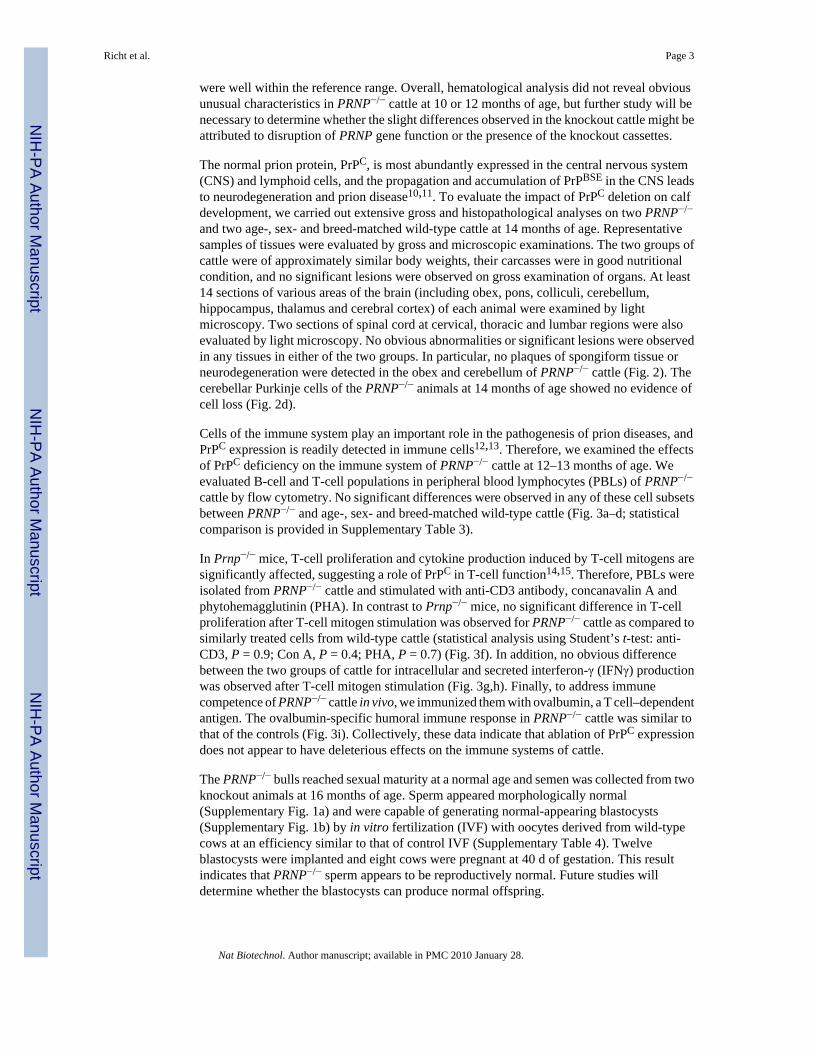
propagation in vitro, we collected two brain regions (cortex and hypothalamus) from 10-month-old PRNP−/− cattle for a protein misfolding cyclic amplification (PMCA) assay7,16–18. Ascontrol substrate for the PMCA assay, CNS tissues from the identical anatomic sites wereobtained from an age-, sex- and breed-matched wild-type calf. PMCA was carried out withbrain homogenates derived from either PRNP−/− or wild-type cattle as ‘PMCA substrates’; abrain homogenate from a BSE-infected cow was used as the PrPBSE-containing inoculum. Nopropagation of proteinase K (PK)-resistant PrPBSE was detected by western blot analysis whenbrain homogenates from the PRNP−/− cattle were used as substrates (Fig. 4a,b). In contrast,PrPBSE was readily amplified and detected in western blot analysis when brain homogenatesfrom the wild-type cattle were used as substrates (Fig. 4a,b). These results indicate that thepresence of endogenous bovine PrPC is essential for PrPBSE propagation in vitro and that anyother host-derived cellular cofactors included in the brain homogenates, such as RNA19 andsulfated glycosaminoglycan20, cannot support the in vitro PrPBSE propagation in the absenceof endogenous bovine PrPC. We also performed a similar PMCA assay using a brainhomogenate from cattle infected with transmissible mink encephalopathy (TME), anotherprion strain infectious to cattle, as inoculum and detected no propagation of the PrPTME (Fig.4c). This indicates that PRNP−/− cattle could be resistant to propagation of various prion strains.
In summary, we have demonstrated the usefulness of the sequential gene targeting system toefficiently produce PRNP−/− cattle. Excluding three animals sacrificed for thehistopathological analysis and PMCA assay, all the nine PRNP−/− cattle have remained healthyfor at least 20 months after birth without showing obvious clinical abnormalities. This indicatesthat ‘loss of function’ of bovine PrPC itself does not cause BSE and that ablation of the normalcellular prion protein PrPC function does not adversely affect normal bovine development. Ithas been reported that evolution has exerted very intense purifying selection on exon 3 ofbovine PRNP; the PRNP gene should have some indispensable function in bovine developmentbecause such strong purifying selection is usually seen only for proteins essential to eukaryoticlife21. Therefore, our findings appear to be of particular interest in supporting a generalhypothesis that PrPC function is dispensable for normal animal development.
Moreover, brain homogenates from PRNP−/− cattle were resistant to the in vitro propagationof at least two different prion strains, PrPBSE and PrPTME, by the PMCA method. PMCA hasbeen shown to closely mimic in vitro the prion propagation process that occurs in vivo, leadingto the formation of high quantities of misfolded prion protein that are infectious to wild-typeanimals16. PMCA is described to be at least as sensitive to prion propagation as in vivoinfection22 and reproduces the species barrier and prion strain phenomenon typical of the prioninfectious agent (J.C. and C.S., unpublished data).
In Prnp−/− mice, phenotypes vary depending on how the Prnp gene locus is disrupted. Forexample, ataxia and loss of Purkinje cells in aged mice have been observed in some Prnp−/−
mouse strains where, in addition to the Prnp open reading frame (ORF), 5′ flanking genomicsequences were also deleted, which results in exon skipping between Prnp and Prnd (located16 kb downstream of the murine gene Prnp and encoding Dpl protein)23–27. The cerebellarsymptoms in these Prnp−/− mouse strains were suggested to be caused by ectopic expressionof Dpl protein in brain28. In the PRNP−/− cattle described here, only the ORF of the bovinePRNP gene was disrupted by insertion of the neo and puro cassettes without any deletion ofPRNP genomic sequences, so that any splicing donor/acceptor sites remain intact6. It remainsto be determined whether or not the bovine PRND locus might be affected by the disruptionof PRNP, but if it occurs, its effect would appear to be minimal because obvious abnormalities,such as ataxia and Purkinje cell loss, were not found in PRNP−/− cattle up to 20 months of age.
Richt et al. Page 4
Nat Biotechnol. Author manuscript; available in PMC 2010 January 28.
NIH
-PA Author Manuscript
NIH
-PA Author Manuscript
NIH
-PA Author Manuscript

Consistent with our observations in the PRNP−/− cattle, Prnp−/− mice with exclusive disruptionon the Prnp ORF remained healthy2,3 and showed only slightly abnormal phenotypes, such asaltered synaptic function29,30 and sleep-wake circadian rhythms31,32. However, the phenotypein the synaptic function appears to be normal in other murine genetic backgrounds33.We havemonitored sleep-wake activity in the knockout cattle, along with age-, sex- and breed-matchedwild-type controls, at frequent intervals throughout the day and night for one week, but did notobserve any obvious alterations. Some other Prnp−/− mouse models show subtle abnormalities,such as learning differences34, and deletion of parts of the murine Prnp ORF have more severeeffects35, suggesting that complete ablation of PrPC function as done in this study may haveless detrimental phenotypes than the partial manipulation or rearrangement of the Prnp ORF.
PRNP−/− cattle are likely a more relevant model for elucidating PrPC function and the basicmechanisms of prion pathogenesis than mice, as cattle are a natural host of prion diseases. Inparticular, PRNP−/− cattle allow in vivo tests of resistance to prion propagation. We haveundertaken such tests, which will require at least 3 years to complete.
PRNP−/− cattle could be a preferred source of a wide variety of bovine-derived products thathave been extensively used in biotechnology, such as milk, gelatin, collagen, serum and plasma.In addition to the PRNP−/− cattle described here, we have also generated healthyPRNP−/−IGHM−/− (ref. 6) double-knockout cattle (Supplementary Fig. 2). This indicates thatother genetic modifications can be added to the PRNP−/− background by means of consecutiverounds of gene modification and recloning, alleviating time-consuming breeding of livestock.Additional genetic modifications to PRNP−/− background could be useful for production ofprion protein–free therapeutic recombinant human proteins, tissue and organs in transgeniclivestock for biomedical applications.
Although a ban on feeding cattle ruminant-derived meat-bone meal has greatly reduced BSEinfections in cattle, the possibility cannot be completely excluded that some PrPBSE strainsmight have originated from ‘spontaneous’ misfolding of the endogenous PrPC protein. Thisview is supported by recent reports suggesting the presence of atypical PrPBSE strains36–38.Two cattle recently identified in the United States (Texas and Alabama) appear to show anatypical PrPBSE pattern, and one animal with atypical PrPBSE characteristics was born afterthe feed ban37. Thus, the ban may not completely alleviate concerns about BSE. ThePRNP−/− cattle produced in this study would prevent BSE due to spontaneous misfoldingbecause of a complete lack of endogenous PrPC.
METHODSEmbryonic cloning
Cloned fetuses and calves were produced using the chromatin transfer procedure asdescribed6. In vitro embryo development rate is provided in Supplementary Table 5. All animalwork described in this section was done following a protocol approved by the TransovaGenetics Institutional Animal Care and Use Committee.
Generation of PRNP−/− fetal cell linesSequential gene targeting was carried out as described previously6. Two types of knockoutvectors were used (pBPrP(H)KOneo and pBPrP(H)KOpuro vectors) to sequentially disruptboth alleles of PRNP. To provide more specific information on the bovine PRNP genomicDNA, we used ~8.3 kb of BamHI-BamHI region (base position 65605–73896 of GenBankaccession no. AJ298878) for the 3′ homologous arm and ~1.2 kb of BglII-BamHI region (baseposition 64494–65604 of AJ298878) for the 5′ homologous arm. Male Holstein primary fetalfibroblast line 6594 was electroporated at 550 V and 50 µF with the first knockout vector
Richt et al. Page 5
Nat Biotechnol. Author manuscript; available in PMC 2010 January 28.
NIH
-PA Author Manuscript
NIH
-PA Author Manuscript
NIH
-PA Author Manuscript

(pBPrP(H)KOneo). We screened 94 colonies resistant to G418 (500 µg/ml) by PCR to identifyhomologous recombinants (primer pair; neoF7 × neoR7) and then homologous recombinantswere identified (40/94: 43%). Based on their morphology, we selected seven colonies andcloned embryos to generate fetuses. At 40–60 d of gestation, five fetuses were collected andthree of them (2180, 3560-1 and 3560-2) were confirmed to be PRNP−/+ (primer pair; neoF7× neoR7). The heterozygous PRNP−/+ cell line, 3560-2, was electroporated with the secondknockout vector (pBPrP(H)KOpuro), and 182 colonies resistant to puromycin (1 µg/ml) werescreened by PCR (primer pair; puroF14 × puroR14) to identify homozygously targetedcolonies. Six colonies were identified to be PRNP−/−, four of which were used for embryoniccloning to generate recloned fetuses. At 40–75 d of gestation, ten fetuses were collected andthen fibroblast cell lines were established. All of them were confirmed to be homozygousPRNP−/− by the targeting event-specific (puroF14 × puroR14 and neoF7 × neoR7) and negative(BPrPex3-F × BPrPex3-R) PCR analyses. We also performed Southern hybridization analysison SphI and BamHI-double digested genomic DNA extracted from PRNP−/− fibroblast celllines using the coding region of the neo or puro gene as a probe, which showed the expectedband size and a single-site integration of the knockout cassettes.
RT-PCRRNA was extracted from PRNP+/+ and PRNP−/− calf fibroblasts by using RNeasy mini kit(Qiagen), and first strand cDNA synthesis was done by using superscript first-strand synthesissystem for RT-PCR (Invitrogen). PCR was done as previously described6.
Western blottingProtein was extracted from PRNP+/+ and PRNP−/− calf fibroblasts, peripheral bloodlymphocytes and brain stem. The protein content was quantified with Bio-Rad protein assayreagent. Western blot analysis was carried out by running ~75 µg of protein sample on a 12%SDS-PAGE gel under nonreducing conditions. The proteins were transferred to a nitrocellulosemembrane and the membrane was stained by the mouse anti-bovine prion protein monoclonalantibody (F 89/160.1.5 from Alexis Biochemicals) as a primary antibody and, second, stainedwith peroxidase-labeled affinity-purified antibody to mouse IgG (H+L). The stained membranewas developed by ECL plus western blot analysis detection system (Amersham Bioscience)and exposed to Biomax light film by film developer. Detection limit for PrPC protein in thiswestern blot analysis was estimated to be ~1.2 µg of brain homogenate from the wild type,which was ~60-fold less than the protein amount (75 µg total brain protein) used in Figure 1.
Ovalbumin immunizationFour PRNP−/− and four control wild-type calves 12–13 months old were immunized withovalbumin antigen (Sigma) at 1 mg/dose formulated with Montanide ISA 25 adjuvant (Seppic)as water-in-oil emulsion. The calves were immunized twice at 3-week intervals (primaryimmunization followed by first booster after 3 weeks). Vaccine was administered byintramuscular injection (2-ml dose containing 1 mg/ml ovalbumin plus 1 ml of ISA-25adjuvant) in the neck region. Serum samples were collected before each immunization (V1 andV2) and 7 d and 14 d after each immunization for antibody titer analysis. Blood was drawninto serum separator tubes (tiger-top), allowed to clot and serum was separated bycentrifugation. Serum was then divided into 0.5- to 1-ml aliquots and stored frozen until assayswere performed. Anti-ovalbumin antibody titers were determined by ovalbumin-specific IgGenzyme-linked immunosorbent assay (ELISA).
Flow cytometryPeripheral blood was collected from four PRNP−/− and four control wild-type calves 12–13months old by jugular venipuncture into heparinized tubes. Whole white blood cells
Richt et al. Page 6
Nat Biotechnol. Author manuscript; available in PMC 2010 January 28.
NIH
-PA Author Manuscript
NIH
-PA Author Manuscript
NIH
-PA Author Manuscript

(leukocytes) were isolated from heparinized blood using red blood cell lysis (RBC-lysis bufferfrom Sigma) followed by two washes with PBS. Sheep anti-bovine IgM-FITC (BethylLaboratories) was used to label bovine surface IgM (sIgM) on the B cells. Mouse anti-bovineCD21 (Clone MCA1424 from Serotec) antibody or anti-bovine lambda light chain (Clone:BIG501E, VMRD) followed by anti-mouse IgG1-PE secondary antibody (CaltagLaboratories) was used to label surface CD21 marker on bovine B cells. For T-cell analysis,anti-CD3 (Clone MM1A, VMRD), anti-CD4 (Clone IL-A11, VMRD), anti-CD8 (Clone BAQ111A, VMRD) and anti-γδ TCR (Clone GB21A, VMRD) monoclonal antibodies were used,followed by fluorochrome labeled isotype-specific secondary antibodies (IgG1-PE; IgG2a-FITC; IgM-PE; IgG2b-TC, purchased from Caltag). Staining was done by a standard protocoland ~10,000 gated lymphocytes were analyzed by FACScan flow cytometer (BD Biosciences).
In vitro T-cell responses to mitogen stimulationHeparinized blood was collected from four PRNP−/− and four age-, breed- and sex-matchedwild-type control calves 12–13 months old, and PBLs were isolated using Ficoll gradientcentrifugation. After three washes with sterile HBSS, cells were resuspended in completeRPMI medium (Sigma) with 10% FBS (Hyclone) and cultured with medium only (control), 5µg/ml Con A (Sigma), 2.5 µg/ml PHA (Sigma) or 5 µg/ml purified anti-CD3 mAb (GB21-Afrom VMRD) immobilized on culture wells. Separate cultures were set up in triplicatemicrotiter wells or 48-well plate for proliferation and cytokine assays. For proliferation assays,cultures were pulsed at 48 h and 72 h with 0.5 µCi of 3H thymidine (Amersham BioSciences)for 4 h and incorporation of 3H thymidine (proliferation) was measured by liquid scintillationcounter as c.p.m. units. For IFN-γ ELISA, cultures were left for 72 h and culture supernatantwas analyzed for secreted IFN-γ protein using calibrated Bovine IFN-γ ELISA Kit (MabTech).Intracellular IFN-γ production in T cells was measured by dual-color intracellularimmunofluorescent staining. Brefeldin and Monensin (Sigma) were added to 48-well culturesat 66 h to stop secretion of cytokines and cells were cultured for a further 6–8 h. Cells wereharvested and stained for surface CD3 marker using anti-CD3 monoclonal antibody (GB21-Afrom VMRD). Cells were then fixed, permeabilized and stained by anti- IFN-γ-Biotin antibodyand Streptavidin-Tricolor for intracellular IFN-γ protein (MabTech). Approximately 10,000gated lymphocytes were analyzed by FACSaria cell sorter (Becton Dickinson) and WinMDIsoftware (Scripps Research Institute).
Gross and microscopic examinationsFour calves (2 wild-type and 2 PRNP−/− calves) were killed with pentobarbital and weresubjected to complete necropsy examinations. Representative samples of skin, nasal turbinate,lung, liver, kidney, spleen, salivary gland, thyroid gland, tonsils (pharyngeal, palatine), thymus,reticulum, rumen, omasum, abomasum, intestines (ileum, colon), adrenal gland, pancreas,urinary bladder, lymph nodes (retropharyngeal, prescapular, mesenteric, popliteal), aorta,striated muscles (heart, tongue, masseter, diaphragm, triceps, psoas major, biceps femoris),testicle (from two animals), nictitating membrane, sciatic nerve, both trigeminal nerves andganglia, pituitary gland, spinal cord (cervical, thoracic, lumbar), one eye with its optic nerveand the whole brain were evaluated by gross and microscopic examinations. The calves wereof similar body weights and the mean ratio of brain weight (g)/body weight (kg) was 1.10 and1.25 for PRNP−/− and the control cattle, respectively. The gyri of the knockout calves wereslightly narrower when compared to the controls. The samples were immersion-fixed in 10%neutral buffered formalin. One eye with its optic nerve was immersion-fixed in 2% Bouin’sfluid. The fixed brain was cut into 2- to 4-mm wide coronal sections for examination. The fixedtissues were processed for routine histopathology, embedded in paraffin wax, sectioned at 5µm and stained with hematoxylin and eosin for examination by light microscopy. Theexamination was done by a board-certified, experienced bovine pathologist (A.N.H.) in side-by side comparisons of the tissues using multiple levels of magnification.
Richt et al. Page 7
Nat Biotechnol. Author manuscript; available in PMC 2010 January 28.
NIH
-PA Author Manuscript
NIH
-PA Author Manuscript
NIH
-PA Author Manuscript

In vitro fertilizationBovine cumulus oocyte complexes (COCs) collected from slaughter house ovaries and maturedfor approximately 24 h were fertilized in fertilization medium. Fresh semen collected fromPRNP−/− bulls were prepared using Percoll gradient separation method and COCs werecultured with sperm (2 × 10⁶ motile sperm/ml) for 18 h at 39 °C in an atmosphere of 5%CO2 in air, after which they were stripped by vortexing in TL-HEPES for 2 min to remove thecumulus cells. Fertilized oocytes were cultured similarly to cloned embryos.
PMCA procedureTen percent brain homogenates (wt/vol) were prepared from the cortex or hypothalamus ofeither BSE or TME-affected animals, wild-type or PRNP−/− cattle. The homogenates wereprepared in conversion buffer (PBS containing 150 mM NaCl, 1.0% Triton X-100, 4 mMEDTA and the complete protease inhibitor cocktail from Boehringer Mannheim). The sampleswere clarified by a brief, low-speed centrifugation (1,500 rpm for 30 s) using an Eppendorfcentrifuge, model 5414. Dilutions of this brain homogenate were done in conversion bufferand they are expressed in relation to the brain, for example a 100-fold dilution is equivalent toa 1% brain homogenate. Aliquots of wild-type, PRNP−/− and BSE or TME brain homogenateprepared in conversion buffer were mixed and either immediately frozen or subjected to 48cycles of PMCA. For PMCA, tubes were positioned on an adaptor placed on the plate holderof a microsonicator (Misonix Model 3000) and programmed to perform cycles of 30 minincubation at 37 °C followed by a 20 s pulse of sonication set at 60% potency. The detailedprotocol, including troubleshooting, has been recently published elsewhere7,16–18,22.Samples were incubated with 50 µg/ml of proteinase K (PK) for 60 min at 45 °C. The digestionwas stopped by adding electrophoresis sample buffer. Proteins were fractionated by SDS-PAGE under reducing conditions, electroblotted into nitrocellulose membrane, and probedwith 6H4 antibody (Prionics) diluted 1:5,000 in PBS. The immunoreactive bands werevisualized by enhanced chemiluminescence assay (Amersham).
Statistical analysisStatistical analysis was performed by Student’s t-test using both confidence interval estimateanalysis and t score probability hypothesis testing method for two independent sample groups.Both methods of analysis showed that there were no significant differences betweenPRNP−/− and WT control cattle groups at α level of 0.05 and P > 0.2 or 0.1. However, there isa considerable probability of statistical error due to the limited numbers of subjects in thisstudy.
Supplementary MaterialRefer to Web version on PubMed Central for supplementary material.
AcknowledgmentsWe thank the team at Transova Genetics for their efforts in embryo transfer and Todd Stahl, Rebecca Cuperus, MariaDiaz, Cliff Mazour for their assistance in calf delivery and care. We thank Melanie Nichols, Jason Griffin, MelissaBien, Molly Ahlers, Rachael Paulson, Sarah Viet, Cassandra Voss for their assistance in gene targeting, cell cultureand embryonic cloning. We thank Kevin Hassall for an additional western blotting. We also thank Ralph Kubo andTomoyuki Tahara for their useful comments on the immunological study. Soto is part of the European Communityproject TSELAB and was supported in part by National Institutes of Health grants NS050349 and NS049173.
References1. Prusiner SB. Detecting mad cow disease. Sci. Am 2004;291:86–93. [PubMed: 15255592]
Richt et al. Page 8
Nat Biotechnol. Author manuscript; available in PMC 2010 January 28.
NIH
-PA Author Manuscript
NIH
-PA Author Manuscript
NIH
-PA Author Manuscript
2. Bueler H, et al. Normal development and behavior of mice lacking the neural cell-surface PrP protein.Nature 1992;356:577–582. [PubMed: 1373228]
3. Manson JC, et al. 129/Ola mice carrying a null mutation in PrP that abolishes mRNA production aredevelopmentally normal. Mol. Neurobiol 1994;8:121–127. [PubMed: 7999308]
4. Prusiner SB, et al. Ablation of the prion protein (PrP) gene in mice prevents scrapie and facilitatesproduction of anti-PrP antibodies. Proc. Natl. Acad. Sci. USA 1993;90:10608–10612. [PubMed:7902565]
5. Bueler H, et al. Mice devoid of PrP are resistant to scapie. Cell 1993;73:1339–1347. [PubMed:8100741]
6. Kuroiwa Y, et al. Sequential targeting of the genes encoding immunoglobulin-μ and prion protein incattle. Nat. Genet 2004;36:775–780. [PubMed: 15184897]
7. Saborio GP, Permmane B, Soto C. Sensitive detection of pathological prion protein by cyclicamplification of protein misfolding. Nature 2001;411:810–813. [PubMed: 11459061]
8. Berthelin, BC. Scrapie in Sheep: Clinical Features and Differential Diagnostic. VIth InternationalWorkshop on the Diagnosis of Spongiform Encephalopathies; November 25–29, 2002; Weybridge,UK. Weybridge, UK: Veterinary Laboratories Agency; 2002.
9. Vargas F, et al. Detection and clinical evolution of scrapie in sheep using third eyelid biopsy. J. Vet.Intern. Med 2006;20:187–193. [PubMed: 16496940]
10. Chesebro B. Prion protein and the transmissible spongiform encephalopathy diseases. Neuron1999;24:503–506. [PubMed: 10595502]
11. Prusiner SB. Prions. Proc. Natl. Acad. Sci. USA 1998;95:13363–13383. [PubMed: 9811807]12. Mabbott N, Turner M. Prions and the blood and immune systems. Haematologica 2005;90:542–548.
[PubMed: 15820951]13. Aguzzi A. Prion disease, blood and the immune system: concerns and realty. Haematologica
2000;85:3–10. [PubMed: 10627667]14. Bainbridge J, Walker KB. The normal cellular form of prion protein modulates T cell responses.
Immunol. Lett 2005;96:147–150. [PubMed: 15585317]15. Mabbott NA, Brown KL, Manson J, Bruce ME. T-lymphocyte activation and the cellular form of the
prion protein. Immunology 1997;92:161–165. [PubMed: 9415021]16. Castilla J, Saa P, Hetz C, Soto C. In vitro generation of infectious scrapie prions. Cell 2005;121:195–
206. [PubMed: 15851027]17. Saa P, Castilla J, Soto C. Cyclic amplification of protein misfolding and aggregation. Methods Mol.
Biol 2005;299:53–65. [PubMed: 15980595]18. Castilla, J.; Saa, P.; Soto, C. Methods and Tools in Bioscience and Medicine. Lehmann, S.; Grassi,
J., editors. Basel: Birkhauser Verlag; 2004. p. 198-213.19. Deleault NR, Lucassen RW, Supattapone S. RNA molecules stimulate prion protein conversion.
Nature 2003;425:717–720. [PubMed: 14562104]20. Wong C, et al. Sulfated glycans and elevated temperature stimulate PrPSc-dependent cell-free
formation of protease-resistant prion protein. EMBO J 2001;20:377–386. [PubMed: 11157745]21. Seabury CM, et al. Prion protein gene (PRNP) variants and evidence for strong purifying selection
in functionally important regions of bovine exon 3. Proc. Natl. Acad. Sci. USA 2004;101:15142–15147. [PubMed: 15477588]
22. Castilla J, Saa P, Soto C. Detection of prions in blood. Nat. Med 2005;11:982–985. [PubMed:16127436]
23. Sakaguchi S, et al. Loss of cerebellar Purkinje cells in aged mice homozygous for a disrupted PrPgene. Nature 1996;380:528–531. [PubMed: 8606772]
24. Moore RC, et al. Ataxia in prion protein (PrP)-deficient mice is associated with upregulation of thenovel PrP-like protein doppel. J. Mol. Biol 1999;292:797–817. [PubMed: 10525406]
25. Rossi D, et al. Onset of ataxia and Purkinje cell loss in PrP null mice inversely correlated with Dpllevel in brain. EMBO J 2001;20:694–702. [PubMed: 11179214]
26. Silverman GL, et al. Doppel is an N-glycosylated, glycosylphosphatidylinositol-anchored protein.Expression in testis and ectopic production in the brains of Prnp0/0 mice predisposed to Purkinje cellloss. J. Biol. Chem 2000;275:26834–26841. [PubMed: 10842180]
Richt et al. Page 9
Nat Biotechnol. Author manuscript; available in PMC 2010 January 28.
NIH
-PA Author Manuscript
NIH
-PA Author Manuscript
NIH
-PA Author Manuscript

27. Weissmann C, Enari M, Klohn PC, Rossi D, Flechsig E. Transmission of prions. Proc. Natl. Acad.Sci. USA 2002;99:16378–16383. [PubMed: 12181490]
28. Li A, et al. Identification of a novel gene encoding a PrP-like protein expressed as chimeric transcriptsfused to PrP exon 1/2 in ataxic mouse line with a disrupted PrP gene. Cell. Mol. Neurobiol2000;20:553–567. [PubMed: 10930132]
29. Collinge J, et al. Prion protein is necessary for normal synaptic function. Nature 1994;370:295–297.[PubMed: 8035877]
30. Mallucci GR, et al. Post-natal knockout of prion protein alters hippocampal CA1 properties, but doesnot result in neurodegeneration. EMBO J 2002;21:202–210. [PubMed: 11823413]
31. Tobler I, Deboer T, Fischer M. Sleep and sleep regulation in normal and prion protein-deficient mice.J. Neurosci 1997;17:1869–1879. [PubMed: 9030645]
32. Tobler I, et al. Altered circadian activity rhythms and sleep in mice devoid of prion protein. Nature1996;380:639–642. [PubMed: 8602267]
33. Lledo PM, et al. Mice deficient for prion protein exhibit normal neuronal excitability and synaptictransmission in the hippocampus. Proc. Natl. Acad. Sci. USA 1996;93:2403–2407. [PubMed:8637886]
34. Criado JR, et al. Mice devoid of prion protein have cognitive deficits that are rescued by reconstitutionof PrP in neurons. Neurobiol. Dis 2005;19:255–265. [PubMed: 15837581]
35. Shmerling D, et al. Expression of amino-terminally truncated PrP in the mouse leading to ataxia andspecific cerebellar lesions. Cell 1998;93:203–214. [PubMed: 9568713]
36. Biacabe AG, Laplanche JL, Ryder S, Baron T. Distinct molecular phenotypes in bovine prion diseases.EMBO Rep 2003;5:110–115. [PubMed: 14710195]
37. Yamakawa Y, et al. Atypical Proteinase K-resistant prion protein (PrPres) observed in an apparentlyhealthy23-month-oldHolstein steer. Jpn. J. Infect. Dis 2003;56:221–222. [PubMed: 14695437]
38. Casalone C, et al. Identification of a second bovine amyloidotic spongiform encephalopathy:molecular similarities with sporadic Creutzfeldt-Jakob disease. Proc. Natl. Acad. Sci. USA2004;101:3065–3070. [PubMed: 14970340]
Richt et al. Page 10
Nat Biotechnol. Author manuscript; available in PMC 2010 January 28.
NIH
-PA Author Manuscript
NIH
-PA Author Manuscript
NIH
-PA Author Manuscript

Figure 1.Generation of PRNP−/− cattle. (a) PRNP−/− cattle at 13 months of age. (b) Verification ofPRNP−/− genotype in the ear biopsy fibroblasts by genomic PCR. P, positive control6; N,negative control6. The PRNP−/− calf is positive with puroF14 × puroR14 (middle) and neoF7× neoR7 (top) primers, which are specific to the targeting events at both alleles of PRNP6. Thecalf is negative with wild-type alleles of PRNP amplified with BPrPex3F × BPrPex3R (bottom)primers6. (c) RT-PCR analysis on the PRNP−/− calves shows disruption of mRNA expressionin fibroblasts of PRNP−/− calves. Primers:PrPmF3 × PrPmR3 (ref. 6). (d) Western blot analysison the PRNP−/− calf shows absence of PrPC protein in fibroblasts. As a positive control, a wild-type (WT) calf was analyzed. As a negative control, protein extracts from mouse fibroblastswere used because the monoclonal antibody used is claimed to be specific to bovine PrPC
protein. Protein extracts from wild-type calf show the presence of 33–35 kDa of bovinePrPC protein in size, but no positive band from the PRNP−/− calf. Its replica blot was probedwith anti-β actin antibody and served as an internal positive control. (e) Absence of PrPC proteinin peripheral blood lymphocytes (PBLs) of PRNP−/− calf by western blot analysis. (f) Absenceof PrPC protein in brain stem of PRNP−/− calves by western blot analysis.
Richt et al. Page 11
Nat Biotechnol. Author manuscript; available in PMC 2010 January 28.
NIH
-PA Author Manuscript
NIH
-PA Author Manuscript
NIH
-PA Author Manuscript

Figure 2.Histopathological analysis of obex and cerebellum of 14-month-old cattle. (a,b) PRNP+/+
dorsal motor nucleus of vagus (a) and molecular layer, granular layer and white matter (b).(c,d) PRNP−/− dorsal motor nucleus of vagus (c) and molecular layer, granular layer and whitematter (d). There are neither plaques of spongiform tissues nor apparent neurodegeneration inthe tissues. H & E stain. Scale bars, 100 µm.
Richt et al. Page 12
Nat Biotechnol. Author manuscript; available in PMC 2010 January 28.
NIH
-PA Author Manuscript
NIH
-PA Author Manuscript
NIH
-PA Author Manuscript

Figure 3.Comparative analysis of immune system of PRNP−/− and wild-type (WT) control cattle at 12–13 months old. (a) Flow cytometry in peripheral blood lymphocyte (PBL), stained with anti-IgM and anti-CD21 antibodies. (b) PBLs stained with anti-IgM and anti-lambda light-chainantibodies. (c) PBLs stained with anti-CD4 and anti-CD8 antibodies. (d) PBLs stained withanti-CD3 and anti-γδ T cell–receptor antibodies. (e) Secondary antibody isotype controlstaining. (f) In vitro mitogenic response of T cells in PRNP−/− and WT cattle. PBLs from fourPRNP−/− and four WT cattle were cultured with medium only (Med) or stimulated withimmobilized anti-CD3 monoclonal antibody (CD3), Con A (concanavalin A) or PHA(phytohemagglutinin) mitogens for 48 h and proliferation was measured by 3H thymidineincorporation. Mean of T-cell response of PRNP−/− group and WT group cattle and their s.e.m.are shown. No significant differences were found. (g) Intracellular cytokine analysis of IFNγexpression in PRNP−/− and WT control cattle by dual-color flow cytometry. PBLs werestimulated by immobilized anti-CD3 monoclonal antibody for 72 h and intracellular IFNγproduction was analyzed by surface CD3 and intracellular IFNγ (positive, green; negative, red)dual-color immunofluorescent staining. Percentage of IFNγ+ T cells are shown in the upperright quadrant. (h) In vitro IFNγ production by PBLs in PRNP−/− and WT cattle. PBLs isolatedfrom four PRNP−/− and four WT cattle were stimulated by (i) immobilized anti-CD3monoclonal antibody or (ii) Con A for 72 h and secreted IFNγ in the culture supernatant wasanalyzed by calibrated bovine IFNγ ELISA. Mean of the IFNγ production (ng/ml) inPRNP−/− group and WT control cattle and their s.e.m. are shown. Statistical analysis usingStudent’s t-test showed no significant difference between PRNP−/− and WT cattle (P = 0.5).(i) Humoral immune response to ovalbumin protein antigen in PRNP−/− and WT cattle. FourPRNP−/− and four WT cattle were immunized with ovalbumin twice at day 0 (V1) and day 21(V2) and ovalbumin-specific IgG antibody titers at 7 d after V2 were determined. Meanantibody titers of PRNP−/− group and WT group cattle and their s.e.m. are shown. Statisticalanalysis using Student’s t-test showed no significant difference between PRNP−/− and WTcattle (P = 0.9).
Richt et al. Page 13
Nat Biotechnol. Author manuscript; available in PMC 2010 January 28.
NIH
-PA Author Manuscript
NIH
-PA Author Manuscript
NIH
-PA Author Manuscript

Figure 4.In vitro propagation of PrPBSE and PrPTME in PRNP−/− and PRNP+/+ wild-type (WT) cattlebrain homogenates. (a,b) In vitro propagation of PrPBSE in 10% homogenates from cortex(a) or hypothalamus (b). (c) in vitro propagation of PrPTME in 10% homogenates from cortex(c). The pathological form of the prion protein, PrPBSE or PrPTME, in the inoculum, was derivedfrom BSE- or TME-affected cattle, respectively. We used 1:50, 1:100 and 1:200 dilutions ofthe infectious material. Samples were either frozen immediately after mixture (F) or subjectedto 48 PMCA amplification cycles (S). The appearance of PrPBSE or PrPTME was assessed bywestern blot analysis after proteinase K (PK) digestion. Samples from PRNP+/+ wild-type(WT-Bo), PRNP−/− (KO-Bo) cattle (substrates) and the BSE-positive control brainhomogenate (inoculum) are shown for comparison with and without PK treatment.
Richt et al. Page 14
Nat Biotechnol. Author manuscript; available in PMC 2010 January 28.
NIH
-PA Author Manuscript
NIH
-PA Author Manuscript
NIH
-PA Author Manuscript

NIH
-PA Author Manuscript
NIH
-PA Author Manuscript
NIH
-PA Author Manuscript
Richt et al. Page 15
Tabl
e 1
Prod
uctio
n of
clo
ned
calv
es fr
om P
RNP−
/− fi
brob
last
cel
l lin
es
Preg
nant
at (
%)a
Liv
e an
imal
s at
6 m
onth
s (%
)aC
ell l
ine
IDE
mbr
yos i
mpl
ante
dR
ecip
ient
s40
d90
d15
0 d
270
d
5211
4530
17 (3
8)7
(16)
7 (1
6)6
(13)
5 (1
1)
5232
2121
7 (3
3)4
(19)
4 (1
9)3
(14)
2 (1
0)
4296
1919
9 (4
7)7
(37)
6 (3
2)5
(26)
5 (2
6)
Tota
l85
7033
(47)
18 (2
6)17
(24)
14 (2
0)12
(14)
a Perc
enta
ges w
ere
calc
ulat
ed b
y di
vidi
ng th
e nu
mbe
r of f
etus
es o
r cal
ves b
y th
at o
f em
bryo
s im
plan
ted.
Nat Biotechnol. Author manuscript; available in PMC 2010 January 28.