Clinical and ultrasonographic differences between cattle and ...
Upload
khangminh22Category
view
0download
0
arasites of Cattle
CONTENTS
1 Stages in the gut and faeces . ............ 24
• 2 Stages in the blood and circulatory system . .................... 55
• 3 Stages in the urogenital system ........ . . 83 . 4 Stages in internaiorgans . ............... 85
4.1 Locomotory system .................. 85 4.7 . 7 Muscles ...................... 85 4.7.2 Tendons . . .................... 90
4.2 Liver ............................. 90
4.3 Respiratory system ................... 97 4.4 Abdominal cavity .................. . 101 4.5 Pancreas ......................... 102
4.6 Central nervous system .............. 103
• 5 Stages on the body surface . ............ 105 5.1 Skin and co at ..................... 105 5.2 Eyes ............................. 143
J. Kaufmann, Parasitic Infections of Domestic Animals© Springer Basel AG 1996
1 Stages In the gut and taeces
, Stages in the gut and faeces
PROTOZOA • Protozoa oocysts found in the faeces . .. 24
HELMINTHS • Trematoda eggs found in the
faeces and adult trematodes living in the gastrointestinal tract . ..... .. 29
• Cestoda eggs found in the faeces and adult cestodes living in the gastrointestinal tract ...... .. ... . . 32
• Nematoda eggs found in the faeces, adult nematodes living in the gastrointestinal tract and first-stage larvae of Dictyocaulus viviparus . ..... 35
PROTOZOA
• Protozoa oocysts found in the faeces
Eimeria spp. Bovine coccidiosis
Location: Terminal ileum, caecum, colon Hosts: Cattle Species description: Of the 21 species of
Eimeria that infect cattle, E. hovis and E. zuerni are most ofren associared wirh c1inical disease. Coccidiosis is commonly a disease of young cattle and occurs especially in management systems (night holding place, limited water source, paddocks) which concentrate the hosts and the parasites within a restricted area. Bloody diarrhoea, progressive weight loss and death, especially in animals after weaning can result from heavy infections. Ruminants do normally excrete a few Eimeria spp. oocysts. The condition called coccidiosis incl udes both severe dysentery and excretion of a high number of oocysts.
Geographie distribution: World-wide Symptoms: Diarrhoea lasting for a few hours,
followed by a self eure or development of severe dysentery accompanied by a haemorrhagic and viscous diarrhoea, dehydration
24
and para lysis. Death can occur rapidly, mainly in calves. Another form of coccidiosis is characterized by persisting, non-ha emorrhagic diarrhoea with continuous weight loss until cachexia. This condition may last for several weeks. Animals that survive severe illness can have significant weight loss that is not quickly regained, or can remain permanently stunted.
Significance: E. hovis and E. zuerni are most commonly involved in c1inical coccidiosis of cattle.
Diagnosis: Clinical signs and extremely high numbers of oocysts per gram of faeces (50,000-500,000).
Therapy: The drugs that are commonly used to treat c1inical coccidiosis in ruminants are listed in Table 1. These include sulfonamids, nitrofurazone, amprolium (10 mg/kg po. > 10 days) , monensin (1 mg/kg po. for 10-30 days) and lasalocid (0.5-1 mg/kg po. for up to 6 weeks). Sulfadimidine (33% solution for intravenous injection), sulfamethazine (50-100 mg/kg, po. daily for 4 days), sulfaquinoxaline (15 mg/kg, po. for 3 days) may be used to treat siek animals. Sulfaguanidine is less effective than the soluble sulfonamides which can be administered orally or parenterally . Sulfonamides, combined wirh trimethoprime can also be used to treat c1inical coccidiosis. Toltrazuril (1 x 20 mg/kg) is highly effective to treat c1inical coccidiosis in ruminants.
Prophylaxis: Young animals should be kept in clean and dry quarters, and watering devices should be prorecred from faecal contamination. Decoquinate, amprolium and ionophorous antibiotics can be used in catt1e fo r prophylaxis (Table 3). Neonates should receive colostrum within 6 ho urs after birth and stress (e.g. weaning, sudden change in feed, etc.) should be minimized.
(Figures 14, 15, 16, 17, 18, 19, 20, 21, 23, Table 3)

Fig. 14 Oocyst of Eimeria bovis (26-32 x 18-:-21 J.Im)
Fig. 15 Oocysts of Eimeria zuerni (16-20 x 15-18 J.Im)
Fig. 16 Oocyst of Eimeria brasiliensis (33-42 x 23-30 J.Im)
Fig. 17 Oocyst of Eimeria alabamensis (16-24 x 12-16)
Fig. 18 Oocyst of Eimeria auburnensis (36-42 x 19-26 J.Im)
Fig. 19 Oocyst of Eimeria bukidnonensis (44 x 32 J.Im)
25

1 Stage5 in the gut and faece5
E_ ENt< .. E .... bu,~ E."h~
Fig. 20 Coccidia found in cattle [11
Fig. 21 Bull with an acute coccidiosis
26
e ..... dnu E._ ....
Fig. 22 Rumen flukes found in the rumen o(
N'Dama canle
Fig. 23 Haemorrhage in the small intestine of a
calf with an acute coccidiosis (Eimeria bovis)
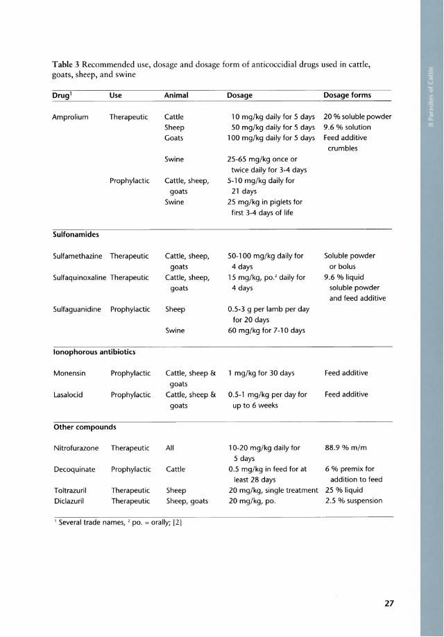
Table 3 Recommended use, dosage and dosage form of anticoccidial drugs used in cattle, goats, sheep, and swine
Drug 1 Use Animal Dosage Dosage forms
Amprolium Therapeutic Cattle 10 mg/kg daily for 5 days 20 % soluble powder Sheep 50 mg/kg daily for 5 days 9.6 % solution Goats 100 mg/kg daily for 5 days Feed additive
crumbles Swine 25-65 mg/kg onee or
twiee daily for 3-4 days Prophylaetie Cattle, sheep, 5-10 mg/kg daily for
goats 21 days Swine 25 mg/kg in piglets for
first 3-4 days of life
Sulfonamides
Sulfamethazine Therapeutie Cattle, sheep, 50-100 mg/kg daily for Soluble powder goats 4 days or bolus
Sulfaquinoxaline Therapeutic Cattle, sheep, 15 mg/kg, po.' daily for 9.6 % liquid goats 4 days soluble powder
and feed additive Sulfaguanidine Prophylaetie Sheep 0.5-3 9 per lamb per day
for 20 days Swine 60 mg/kg for 7-10 days
lonophorous antibiotics
Monensin Prophylactie Cattle, sheep & 1 mg/kg for 30 days Feed additive
goats Lasaloeid Prophylaetic Cattle, sheep & 0.5-1 mg/kg per day for Feed additive
goats up to 6 weeks
Other eompounds
Nitrofurazone Therapeutie All 10-20 mg/kg daily for 88.9 % mim 5 days
Deeoquinate Prophylaetie Cattle 0.5 mg/kg in feed for at 6 % premix for
least 28 days addition to feed Toltrazuril Therapeutie Sheep 20 mg/kg, single treatment 25 % liquid Diclazuril Therapeutie Sheep, goats 20 mg/kg, po. 2.5 % suspension
1 Several trade names, ' po. = orally; [2]
27
1 Stages in lhe gut and faece.s
Cryptosporidium parvum (syn. C. bovis)
Loeation: Small and large intestine Hosts: All domestie animals Speeies deseription: Spherieal oocysts 4.5-5 flm
in diameter are passed in the faeces . Especially calves younger than 8 weeks may be affected, showing different degrees of diarrhoea, associated with oedema and hyperplasia of the mesenteric Iymph nodes. Cryptosporidial diarrhoea in both immunodeficient and non -immunodeficient human beings have been reported.
Geographie distribution: World-wide Symptoms: Persistent diarrhoea in calves 5-35
days old that does not respond to therapy. Signifieanee: Cryptosporidium parvum is
pathogenic for calves, sheep, pig, rat and man. Bovine eryptosporidiosis is a zoonosis. Diarrhoea due to Cryptosporidium parvum alone is often mild and self-Iimiting, although the severity may be related to the general strength of the calf and the density of the pathogen in the environment and the intensity of the ex pos ure to the organism. Combination of the infection with rota- and/or coronavirus are common and result in persistent diarrhoea, emaciation and death.
Diagnosis: Oocysts may be demonstrated in Ziehl-Neelsen's carbol-fuchsin stained smears of diarrhoeie faeces (1& METHODS,
1.10). Therapy: No effective specific treatment
known. Supportive therapy, such as rehydra tion and antibiotics may help in mild cases . Chemoprophylaxis of bovine cryptosporidiosis can be made experimentally with halofuginone lactate (60-120 flg/kg/ day, po. for 7 days) or by paromomycin (100 mg/kg/day for 11 days).
Prophylaxis: Animals with a massive oocyst excretion should be separated from other animals and man. Contamination with faeces should be avoided. Oocysts are resistant to many disinfectants (exception 5% formalin). Emphasis should therefore be on regular removal oE contaminated faecal material.
28
Note: Trophozoites of Giardia bovis (small intestine) and Entamoeba bovis (rumen, intesrines) occur world-wide in the faeces of cattle, but are cOl1sidered to be 11011-pathogenic.
(Figures 24, 25)
Fig. 24 Oocysts of Cryptosporidium parvum (4.5-5 11m) stained wirh carbol-fuchsine (KW
METHODS 1.10)
Fig. 25 Oocysts of Cryptosporidium parvum (4.5-5 11m ) stained wirh the modified Ziehl Neelsen's carbol-fllchsine (1& METHOIlS, 1.10)
Buxtonella spp.
Remarks: This is a protozoan parasite foul1d occasionally in the large intestine of cartle. Oocysts are found in the faeces. The parasite is 110n-pathogenic.
(Figure 26)

Fig. 26 eyst of Buxtonella sp. (50-131 pm)
HF.L:\H~THS
• Trematoda eggs found in the faeces and adult trema todes living in the gastrointestinal tract
PARAMPHISTOMA TIDAE
Loeation: Adult flukes in the rumen; immature f1ukes in the small intestine
Hosts: Domestie and wild ruminants Speeies deseription: A great number of speeies
of this family has been deseribed in Afriea. Indireet life eycle with development in freshwater snails (Bulinus spp.; Planorbis spp.). The eereariae eneyst on herbage whieh is in contaet with water and develop into the infeetive stages (metaeereariae) . Infeetion is aequired by ingestion of herbage eontaminated with metaeereariae, espeeially around water holes. The adult flukes are generally non-pathogenie (>80% of adult eatde are infeeted in endemie areas). Disease oeeurs when masses of immature worms attaeh to the intestinal mueosa after exeystment, causing destruetion and inflammation. Peaks usually at the end of the dry season. Game animals, using the same water holes, may be a souree of reinfeetion. Standing water, troughs and other water bodies are preferred habitats of the intermediate hosts.
Geographie distribution: World-wide Symptoms: Enteritis with extensive diarrhoea,
hypoproteinaemia, weakness during the intestinal phase, when immature worms irritate the small intestinal mueosa. Severe symptoms mainly in young stoek. Irregular rumination and progressive degeneration of the animal's eondition may be found. Infeetions with adult rumen f1ukes are generally inapparent, despite the high numbers of adult parasites in the rumen. Rumenitis may oeeur due to adult flukes, espeeially if Carmyerius spp. are involved. Adult flukes are generally non-pathogenic.
Signifieanee: Severe diarrhoea and weakness, generally eaused by the immature intestinal f1ukes, may lead to death.
Diagnosis: This is based on the demonstration of immature flukes in the diarrhoeie faeees and the presenee of previous eases in the area.
Cave: In aeute paramphistomidosis the large, clear, opereulated eggs may not be found in the faeees. In many areas > 80% of the adult ruminants pass paramphistome eggs in the faeees. The presenee of eggs without clinieal signs does not neeessarily indieate paramphistomidosis.
Therapy: Niclosamide (90 mg/kg po.), resorantel (65 mg/kg po.) and closantel are aetive against immature forms. Triclabendazole (12 mg/kg) and albendazole (10 mg/kg) mayaiso be used against immature flukes. Resorantel is aetive only against mature f1ukes (~Table 4).
Prophylaxis: Avoiding infeetion by supplying the herds with clean water, e.g. bore holes or raised, snail-free troughs.
T able 4 Recommended drugs against Paramphistomum spp. infeetions in eatde
Drug Dosage Immat. Mature
(mg/kg) flukes flukes
Niclosamide 160.0' + Oxyclozanide 18.7
and Levamisole 19.4' + Oxyclozanide 15 + Resorantel 65 + 'at 3·day intervals
29

1 Stage5 in the gut and faece5
Remarks: Rumen f1ukes in Afriea belong to the families of Paramphistomatidae (Bothriophoron bothriophoron; Stephanopharynx compactus; Cotylophoron cotylophorum and other Cotylophoron spp.; Paramphistomum spp. [8 speeies] and Cotylophoron spp.; [CO daubneyi and C. microbothrium are the most frequent speeies]) and Gastrothylaeidae (Carmyerius spp. l c. spatiosus; C. papillatus; C. dollfusi [Zebu only in Madagasear]).
(Figures 27, 28, 29)
Fig. 27 Rumen flukes found in the rumen of N'Dama cattle
Fig. 28 Paramphistomum sp. attached to the rumen mucosa
30
Fig. 29 Egg of a rumen fluk e (Paramphiswmatidae)
livcr f1ukc
F. hepatica Large liver fluke of temperate areas and high altitude regions in East Afriea
L can n: dulr fluke In bdiary du r ; egg 10
ehe fae e ( 1111" • 4.2) (Figurc 0 I )
Fig. 30 Egg of Fasciola hepatica (130-150 x 63-90 ~m)
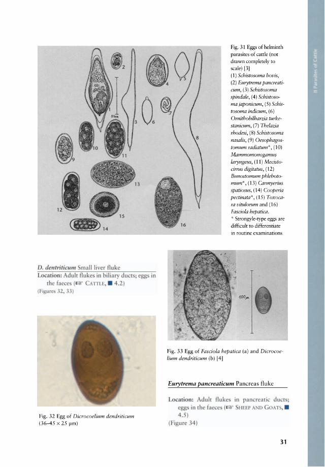
Fig. 32 Egg of Dicrocoelium dendriticum (36-45 x 25 pm)
Fig. 31 Eggs of helminth parasites of cattle (not drawn completely to scale) [3] (1) Schistosoma bovis, (2) Eurytrema pancreaticum, (3) Schistosoma spindale, (4) Schistosoma japonicum, (5) Schistosoma indicum, (6)
Ornithobilharzia turkestanicum, (7) Thelazia rhodesi, (8) Schistosoma nasalis, (9) Oesophagostomum radiatum'", (10) Mammomonogamus laryngeus, (11) Mecistocirrus digitatus, (12)
Bunostomum phlebotomum':·, (13) Carmyerius spatiosus, (14) Cooperia pectinata", (15) Toxocara vitulorum and (16)
Fasciola hepatica. ,:. Strongyle-type eggs are
difficult to differentiate in routine examinations
Fig. 33 Egg of Fasciola hepatica (a) and Dicrocoelium dendriticum (b) [4]
Eurytrema pancreaticum Pancreas fluke
arion:
e in rlle fac c 4.5 )
(Fi ure 4)
31

1 Stages In lhe gUl and laeces
Fig. 34 Egg of Eurytrema pancreaticum (40-50 x 23-34 flm )
Schistosoma spp. (S. bovis, S. mattheei, S. curassoni) Blood flukes
Lo ation: Adult fluke in me enteri ein' egg in the inte tinal wall and fae e
TIL • 2) (Figure 3 - 6)
Fig. 35 Egg of Schistosoma bovis (180 x 60 flm)
Characteristics
Length
Width
Moniezia expansa
1-6 m
16 mm
Moniezia benedini
26mm
26mm
Fig. 36 Eggs of Schistosoma spindale (300 x 80 flm) [4]
• Cestoda eggs found in the faeces and adult cestodes living in the gastrointestinal tract
(Figure 37)
Moniezia expansa and Moniezia benedeni Common tapeworms
Loeation: Small intestine Hosts: Cattle, sheep, goat and many other
ruminants Speeies deseription: Worms are up to 6 m long
and 1.6 em wide (M. expansa). M. benedeni whieh oeeurs more often in eattle is broader (up to 2.6 em). The life eyde of Moniezia is indireet, induding many speeies of oribatid mites as intermediate hosts. Ruminants are infeeted by ingestion of the
Avetellina spp.
3m
3mm
Thysaniezia giardi
Stilesia hepatica
2 m 20-50 cm
12 mm 2 mm
Egg Semi-triangle Diamond-shaped Oval Oval Round
Genitalia Double Double Single Single Single
Interproglottid glands Follicular Compact
Paruterine organs One: lemon- Numerous: Two:
in gravid segment shaped onion-shaped round
Fig. 37 Morphological identification of common cestodes in ruminants [5]
32
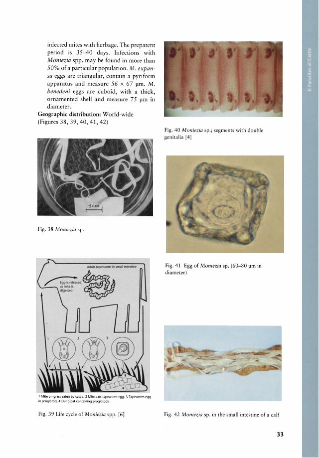
infeeted mites with herbage. The prepatent period is 35-40 days. Infeetions with Moniezia spp. may be found in more than 50% of a partieular population. M. expansa eggs are triangular, eontain a pyriform apparatus and measure 56 x 67 11m. M. benedeni eggs are euboid, with a thiek, ornamented shell and measure 75 11m 10
diameter. Geographie distribution: World-wide (Figures 38, 39, 40, 41, 42)
Fig. 38 Moniezia sp.
1 Mite on grass eaten by cattle, 2 Mite eats tapeworm egg, 3 Tapeworm egg in proglottid, 4 Dung pat containing proglottids
Fig. 39 Life eyde of Moniezia spp. [6]
Fig. 40 Moniezia sp.; segments with double genitalia [4]
Fig.41 Egg of Moniezia sp. (60-80 11m in diameter)
Fig. 42 Moniezia sp. in the small intestine of a ealf
33
1 Stages In lhe gut and faeces
P::tJ~i:~:~ ., UPA
AVITELLINA
HO
• srltESIA
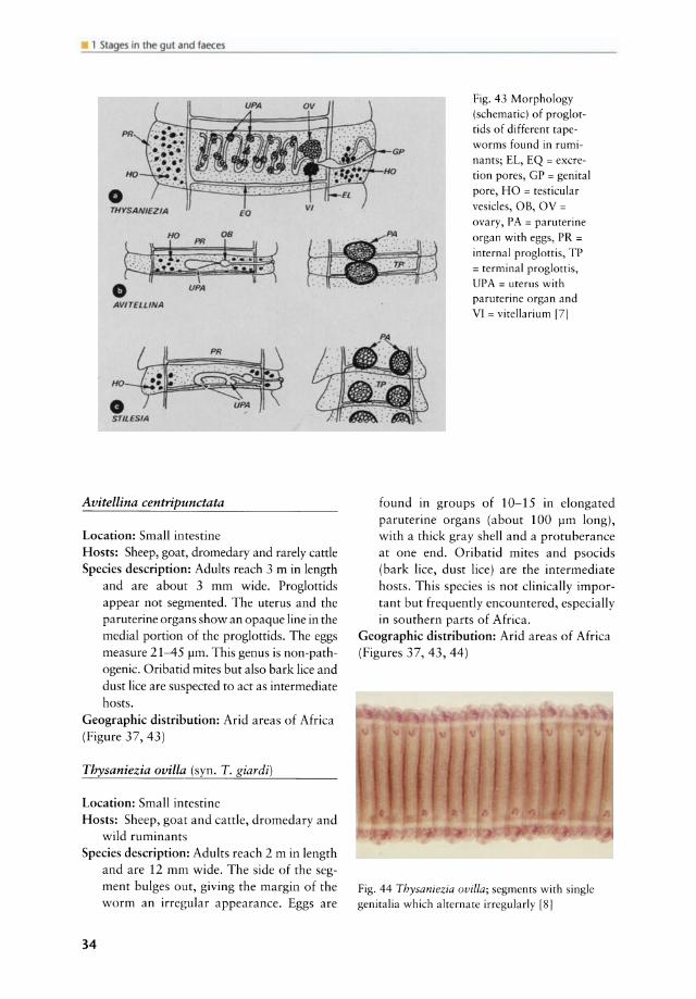
Avitellina centripunctata
Loeation: Sm all intestine Hosts: Sheep, goat, dromedary and rarely eattle Speeies deseription: Adults reaeh 3 m in length
and are about 3 mm wide. Proglottids appear not segmented. The uterus and the paruterine organs show an opaque line in the medial portion of the proglottids. The eggs measure 21-45 pm. This genus is non-pathogenie. Oribatid mites but also bark liee and dust lice are suspected to act as intermediate hosts.
Geographie distribution: Arid areas of Africa (Figure 37, 43)
Thysaniezia ovilla (syn. T. giardi)
Loeation: Small intestine Hosts: Sheep, goat and cattle, dromedary and
wild ruminants Speeies deseription: Adults reach 2 m in length
and are 12 mm wide. The side of the segment bulges out, giving the margin of the worm an irregular appearance. Eggs are
34
Fig. 43 Morphology (schematic) of proglottids o f different tapeworms found in rumi
nants; EL, EQ = excretion po res, GP = genital pore, HO = testicular
vesicles, OB, OV = ovary, PA = paruterine organ with eggs, PR =
internal proglottis, TP = terminal proglottis, UP A = uterus with paruterine organ and VI = vitellarium [71
found in groups of 10-15 in elongated paruterine organs (about 100 pm long), with a thick gray shell and a protuberance at one end. Oribatid mites and psoeids (bark liee, dust liee) are the intermediate hosts. This species is not clinically important but frequently eneountered, especially in southern parts of Africa.
Geographie distribution: Arid areas of Africa (Figures 37, 43, 44)
Fig. 44 Thysaniezia ovilla; segments with single genitalia wh ich alternate irregularly [81

Stilesia globipunctata
ur in rh mall
Fig. 45 Stilesia sp.; proglottids (a) and scolex (b); PA = paruterine organ [4)
Fig. 46 Stilesia globipunctata; immature segments [8)
• General features of intestinal tapeworms (Moniezia spp., Thysaniezia spp., Avitellina spp. and Stilesia spp.) of ruminants
Symptoms: Generally inapparent infections; heavy infections are found only in young animals and can cause reduced weight
gain. Masses of Moniezia can cause obstruction of the intestine.
Cave: Because of the large size of the tapeworms their presence is obvious and the underlying cause of parasitism, the small trichostrongylids, easily overlooked.
Significance: Tapeworms are widespread in ruminants, but their pathogenicity has not been proved conclusive!y and it seems that they are re!ative!y low-pathogenic.
Diagnosis: Proglottids wh ich look Iike cooked rice grains, containing typical thick-shelled or imperfectly rounded eggs, appear in the faeces. Eggs mayaiso appear isolated in the faeces. The presence of tapeworms in the small intestine at slaughter is conclusive.
Therapy: Niclosamide (75-90 mg/kg) and praziquantel (5 mg/kg, sheep only) are specific cestodicidal drugs. In addition the following benzimidazoles are effective against tapeworms: albendazole (7.5 mg/kg), fenbendazole (5-10 mg/kg), oxfendazole (5 mg/ kg), mebendazole (15-20 mg/kg, po.), netobimin (7.5-20 mg/kg, po.), febante! (5-7.5 mg/kg, po.). Special attention should be given to the control of tapeworms in lambs to avoid los ses in heavily infected populations.
Prophylaxis: Reduction of the mite population is not feasible and emphasis should be given to anthelmintic treatments when losses due to tapeworms occur.
• Nematoda eggs found in the faeces, adult nematodes living in the gastrointestinal tract and first-stage larvae of Dictyocoulus viviparus
Gongylonema pulchrum Gullet worm, zigzag worm
Location: Mucosa of oesophagus and forestomachs
Hosts: Sheep, goat and less frequently cattle Species description: Eggs laid by adult worms
are passed in faeces and hatch into larvae when swallowed by manure-eating beetles or cockroaches. Larvae in cattle may be liberated in the stornach and migrate towards
35

1 Stages in Lhe gut and faeces
the oesophagus. Adult worms are 6 to 14 cm in length and a number of oval cuticular thickenings appear in longitudinal rows at the anterior end. The worms are embedded in the oesophageal lining in a zigzag pattern.
Geographie distribution: World-wide Symptoms: Rarely observed Significance: This is a harmless parasite and
mainly found at autopsy. Irritation of the oesophageal and gastric mucosae may occur in infected animals.
Diagnosis: Mainly at necropsy; eggs occur in the faeces.
Therapy: Generally not indicated Prophylaxis: Keeping the animals on dry, well
drained grounds or concrete floors has been described to be effective in controlling infection.
(Figures 47, 48, 49)
Fig. 47 Gongylonema pulchrum; typical zigzag pattern [41
Fig. 48 Gongylonema pulchrum; anterior end with cuticula r plaques [8]
36
lrlHM-...3<uticular plaques
Fig. 49 Gongylonema pulchrum; anterior end with cuticular plaques [5]
Gongylonema verrucosum Rumen gullet worm
Location: Mucosa of rumen, reticulum, omasum
Hosts: Cattle, sheep and goat Species description: Not weil known; dung
beetles are intermediate hosts. The worms have a reddish colour when fresh. Males are 32-41 mm long and females 70-95 mm. A festooned cervical ala as weil as cuticular bosses on one side only are typical.
Geographie distribution: South Africa, India, USA
Symptoms: Inapparent Significance: Non-pathogenic Diagnosis: Casually at necropsy Therapy: Unknown Prophylaxis: Il@f above G. pulchrum
Parabronema skrjabini
Location: Abomasum Hosts: Cattle, sheep, goat and dromedary Species description: This parasite belongs to
the Spiruridae. Stomoxys spp. and L yperosia spp. are intermediate hosts. Infective thirdstage larvae develop in the flies and are deposited on the final host where the larvae are ingested. Males are 15-18 mm long, with one spicule measuring 600-710 fJm and the other one 290-310 fJm. Female

A
B
Fig. 50 Parabronema rhodesiense; anterior end (a ), lateral view of the tail end of a male (b), ventral view of the tail of a male (c) [9]
worms are> 36 mm long and the vulva is situated at 5 mm distance to the hind end. The anterior end is characterised by pseudo-lips, which are wound around the buccal opening. Four papillae are found on these lips. The tail end of the male is reeled to a spiral and four pairs of preanal papillae are found. A group of terminal papillae is present as it is in all the spirurids.
Geographic distribution: Asia, Central and East Africa
Symptoms: Unspecific; abomasitis may be found .
Significance: Unknown; P. skrjabini may play a role together with other gastrointestinal nematodes
Diagnosis: At necropsy, adult worms may be found in abornasal scrapings.
Therapy and Prophylaxis: A specific therapy and prophylaxis is not described and probably also not necessary. For further information w below THERAPY OF NEMATODE INFECTIONS, p. 53
(Figure 50)
Haemonchus contorlus Large stornach worm, twisted wire worm, barber's pole worm
Loeation: Abomasum Hosts: Cattle, sheep, goat and other ruminants Speeies deseription: H. contortus is known as
the " red stomach-worm" or "wire worm " of ruminants and one of their most prevalent and most pathogenic parasites. Adults are 10 to 30 mm long. Males are shorter than females and have a reddish colour and a bursa with an asymmetrical dorsal lobe and barbed spicules. Females are identified as "barber's pole worms" because their white uteri are wound around their red blood-filled intestine. The vulva f1ap is prominent in female worms. The Iife cycle is direct and typical for the strongyle nematodes. The prepatent period is 19-21 days but it can be shortened by immunosuppressive pathogens such as concomitant trypanosome infections and stress factors. In arid areas, the parasite survives the dry season as inhibited fourth-stage larvae within the abornasal mucosa of the host. The inhibited larvae resurne their development a few weeks before the onset of the new rainy season. This phenomenon is accompanied by a drastic increase of the egg output of infected animals before the wet season starts ("rains rise") .
Geographie distribution: World-wide Symptoms: Anaemia, oedema ("botde jaw"),
rough coat, weight loss or retarded growth Signifieanee: H. contortus is a very common
parasite and one of the most pathogenic nematodes of ruminants. Heavy infections cause death in young animals, whereas chronic infections cause anaemia, hypoproteinaemia, progressive emaClation. These effects are generally compensated during the rainy season and only appear progressively during the following dry season when previously heavily infected animals may die.
Diagnosis: Eggs of strongyle-type appear in the faeces . In acute infections anaemia and death may occur be fore the worms reach maturity. No eggs are found in this case in the faeces and only the examination of the abomasum at necropsy allows an exact diagnosis.
Therapy and Prophylaxis: w below THERAPY OF NEMATODE INFECTIONS, p. 53
(Figures 51, 52, 53, 54, 55, 56, 57, 58, 59, 60)
37

1 Stage5 in the gut and faece5
Fig. 51 Haemonchus contortus; adult parasites
Fig. 52 Haemonchus contortus; twisted unteri and intesine [1 OJ
Fig. 53 Haemonchus contortus; prominent cervical papillae (50x)
38
Fig. 54 Haemonchus contortus; bursa copula trix (schematic) with barbed spicules (490-540 !-Im long) and asymmetrical dorsal lobe [3]
Fig. 55 Haemonchus conlortus; vulva fla p
Fig. 56 N'Dama calf showing emaciation a nd "botde jaw" due to a chronic haemonchosis
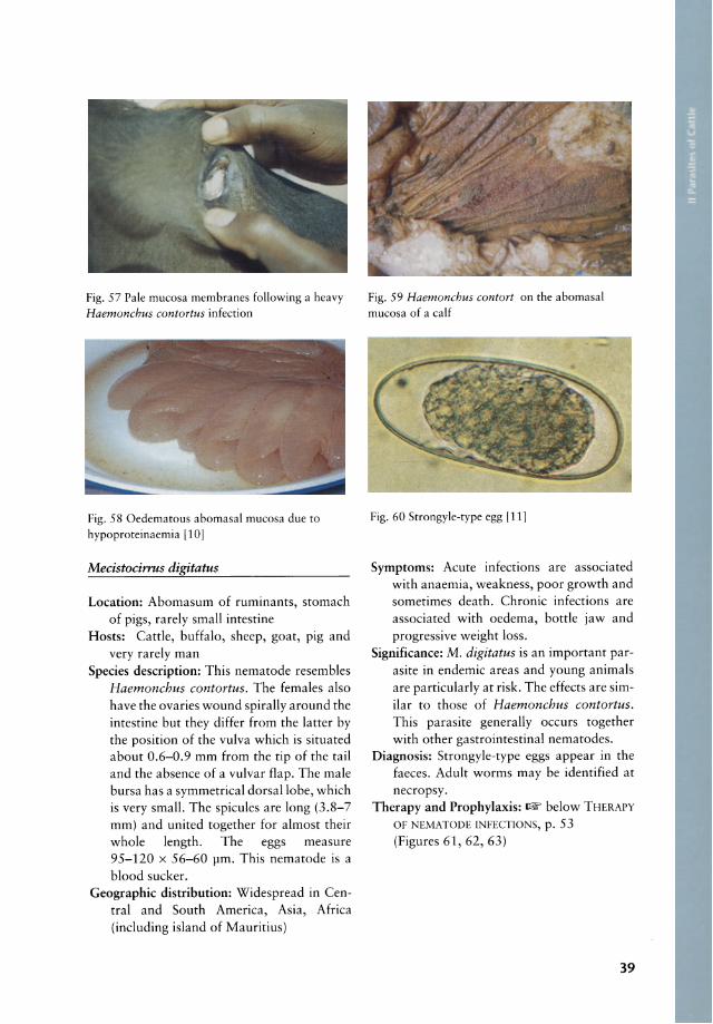
Fig. 57 Pale mucosa membranes following a heavy Haemonchus contortus infection
Fig. 58 Oedematous abornasal mucosa due to hypoproteinaemia 1101
Mecistocinus digitatus
Location: Abomasum of ruminants, stomaeh of pigs, rarely small intestine
Hosts: Cattle, buffalo, sheep, goat, pig and very rarely man
Speeies description: This nematode resembles Haemonchus contortus. The females also have the ova ries wound spirally around the intestine but they differ from the latter by the position of the vulva whieh is situated about 0.6-0.9 mm from the tip of the tail and the absence of a vulvar flap. The male bursa has a symmetrical dorsal lobe, which is very small. The spieules are long (3.8-7 mm) and united together for almost their whole length. The eggs measure 95-120 x 56-60 flm. This nematode is a blood sueker.
Geographie distribution: Widespread in Central and South Ameriea, Asia, Afriea (including island of Mauritius)
'\ ., .. ,-~ ,/
... . 0/.' .. ,,~
'./. :f
Ci!"" '~ .. , ,I • # : ., . . '. _. -
........ . ,jf~. \ *:~,
• ,~ .. J ... ~~. Fig. 59 Haemonchus contort on the abornasal mucosa of a calf
Fig. 60 Strongyle-type egg [11)
Symptoms: Aeute infeetions are assoeiated with anaemia, weakness, poor growth and sometimes death. Chronie infeetions are assoeiated with oedema, bottle jaw and progressive weight loss.
Signifieance: M. digitatus is an important parasite in endemie areas and young animals are particularly at risk. The effeets are similar to those of Haemonchus contortus. This parasite generally oeeurs together with other gastrointestinal nematodes.
Diagnosis: Strongyle-type eggs appear in the faeees. Adult worms may be identified at neeropsy.
Therapy and Prophylaxis: ~ below THERAPY OF NEMATODE INFECTIONS, p. 53 (Figures 61, 62, 63)
39

1 Stages in the gut and faeces
Fig. 61 Mecistocirrus digitatus; anterior end 181
Fig. 62 Mecistocirrus digitatus; bursa copulatrix [3]
Fig. 63 Mecistocirrus digitatus; fern ale worm (19-43 mm long; 470 flm in diameter) 191
40
Trichostrongylus axei Stomach hair worm
Location: Abomasum and occasionally small intestine
Hosts: T. axei oecurs in the abomasum of sheep, goat, eattle, wild ruminants and in the stomach of pig, horse donkey and man.
Species description: Direct nematode life eyde; males are 2.5-6 mm long and females are 3.5-8 mm long. The spieules are dissimilar: one measures 74-98 pm and the other 96-128 pm. The gubernaeulum length is 50-6] 11m. The eggs measure 79-92 x 31-41 pm.
Geographie distribution: World-wide Symptoms: Weight loss and poor growth oeeur
when animals are heavily infected espeeialIy in mixed infections with other triehostrongyles.
Significance: Serious weight loss and poor growth oeeur when animals are heavily infected especially in mixed infeetions with Haemonchus, Ostertagia and heavy Cooperia burdens.
Diagnosis: Detection of strongyle-type eggs in the sediment or flotation of faeees. An accurate diagnosis of the species can only be obtained by mieroseopie examimination of adult speeimens at necropsy.
Therapy and Prophylaxis: - below THERAPY OF NEMATODE INFECTIONS, p. 53
(Figures 64, 65, 66)
Fig. 64 Trichostrongylus axei; spicula (74-98flm and 96-128 flm) and gubernaculum (50-61 flrn)
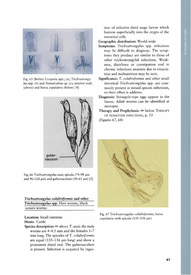
Fig. 65 (Berlin) Cooperia spp.; (a), Trichostrongylus spp. (b) and Nematodirus sp. (c); anterior ends (above) and bursa copulatrix (below) [4J
gubernaculum
Fig. 66 Trichostrongylus axei; spicula (74-98 pm and 96-128 pm) and gubernaculum (50-61 pm) [5)
Trichostrongylus coluhriformis and other Trichostrongylus spp. Hair worms, black scours worms
Location: Small intestine Hosts: Cattle Species description: IEF above T. axei; the male
worms are 4-4.5 mm and the females 5-7 mm long. The spicules of T. colubriformis are equal (135-156 pm long) and show a prominent distal end. The gubernaculum is present. Infection is acquired by inges-
ti on of infective third stage larvae which burrow superficially into the crypts of the intestinal cells.
Geographie distribution: World-wide Symptoms: Trichostrongylus spp. infections
may be difficult to diagnose. The symptoms they produce are similar to those of other trichostrongylid infections. Weakness, diarrhoea or constipation and in chronic infections anaemia due to emaciati on and malnutrition may be seen.
Significance: T. coluhriformis and other small intestinal Trichostrongylus spp. are commonly present in mixed-species infections, so their effect is additive.
Diagnosis: Strongyle-type eggs appear in the faeces. Adult worms can be identified at necropsy.
Therapy and Prophylaxis: IEF below THERAPY OF NEMATODE INFECTIONS, p. 53
(Figures 67, 68)
Fig. 67 Trichostrongylus colubriformis; bursa copulatrix with spicula (135-156 pm)
41

1 Stages In the gut and taeces
gubernaculum
Fig. 68 Trichostrongylus colubriformis; spicula (135-156 pm) and gubernaculum (65-75 pm) [51
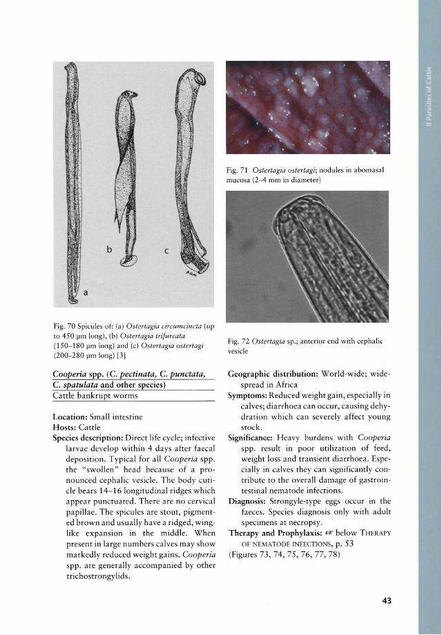
Osterlagia spp_ (0. osterlagi, O. [syn. Skrjabinagia] lyra ta and other species) Brown stomaeh worms
Location: Abomasum, upper small intestine Hosts: Cattle, sheep and goat Species description: O. ostertagi is the most
important triehostrongylid of eattle worldwide. In Africa it plays a role in imported eattle and autoehthonous infeetions are reported from East Afriea. O. circumcincta was reported from sheep and goats in East Afriea (Kenya, Ethiopia, Uganda, Zambia) and South Afriea, and o. pinnata was only reported from sheep in Kenya. O. lyrata (syn. Skrjabinagia lyrata or Grosspiculagia lyrata) oeeurs in eattle in Afriea. Ir is very similar to O. ostertagi whieh aeeounts for enormous losses world-wide in eattle rasing areas. Adult worms of the genus Ostertagia are brownish, thread-like, and grow to 9 mm in length. They all have a restrieted and small eephalie veside, small eervieal papillae projeeted from the body surfaee and pronouneed longitudinal, eutieular ridges. The life eyde is direet and typieal for roundworms. Environmental conditions of cold or exeessive dryness may trigger a eondition known as hypobiosis, in whieh larval development in the abomasal mueosa is arrested and maturation is resumed several months later. Ingested
42
larvae enter the glands of the abomasum, eausing erosion of the eells, maldigestion, protein losses with the eonsequenee of weight loss, diarrhoea and hypoproteinaemla.
Geographie distribution: World-wide Symptoms: Severe diarrhoea, oedema (bottle
jaw or aseites), weight loss leading to emaeiation.
Significance: Ostertagia spp. are widespread parasites of eattle. Affeeted eattle not only lose weight but often die of dinieal ostertagiosis. Ostertagia spp. infeetions are generally aeeompanied by other gastrointestinal nematodes.
Diagnosis: Strongyle-type eggs appear in the faeees. Eggs per gramm> 1000 in ealves indieate a harmful eondition. lt should be eontrolled with an anthelmintie treament.
Therapy and Prophylaxis: Avoid overstoeking, use pa sture management to avoid aeeumulation of infeetive larvae on herbage and soil; regular or strategie use of anthelminties llW below THERAPY 01' NEM
ATODE INFECTIONS, p. 53 (Figures 69, 70, 71, 72)
Fig. 69 Ostertagia ostertagi; bursa copularrix wirh spiculum (200-280 pm) [12]
, ~ .
b c
a
Fig. 70 Spicules of: (a) Ostertagia circumcincta (up to 450 pm long), (b) Ostertagia trifurcata (150-180 pm long) and (c) Ostertagia ostertagi (200-280 pm long) [3)
Cooperia spp. (c. pectinata, C. punctata, C. spatulata and other speeies) Cattle bankrupt worms
Loeation: Small intestine Hosts: Cattle Speeies deseription: Direet life eyele; infeetive
larvae develop within 4 days after faeeal deposition. Typieal for all Cooperia spp. the "swollen" head beeause of a pronouneed eephalie vesiele. The body eutiele bears 14-16 longitudinal ridges whieh appear punetuated. There are no eervieal papillae. The spieules are stout, pigmented brown and usually have a ridged, wingIike expansion in the middle. When present in large numbers ealves may show markedly redueed weight gains. Cooperia spp. are generally aeeompanied by other triehostrongylids.
Fig. 71 Ostertagia ostertagi; nodules in abornasal mucosa (2-4 mm in diameter)
Fig. 72 Ostertagia sp.; anterior end with cephalic vesicle
Geographie distribution: World-wide; widespread in Afriea
Symptoms: Redueed weight gain, especially in calves; diarrhoea can occur, causing dehydration which can severely affect young stock.
Signifieance: Heavy burdens with Cooperia spp. result in poor utilization of feed, weight loss and transient diarrhoea. Especially in calves they can significantly contribute to the overall damage of gastrointestinal nematode infections.
Diagnosis: Strongyle-type eggs occur in the faeces. Species diagnosis only with adult specimens at necropsy.
Therapy and Prophylaxis: - below THERAPY OF NEMATODE INFECTIONS, p. 53
(Figures 73, 74, 75, 76, 77, 78)
43

1 Stage5 in the gut and faece5
Fig. 73 Cooperia spp.; adult parasites [10]
Fig. 74 Cooperia punctata; bursa copulatrix with spicules (123-145 pm)
Fig. 75 Cooperia pectinata; anterior end with enlarged cephalic vesicle
44
Fig. 76 Cooperia punctata; anterior end with enlarged cephalic vesicle
Fig. 77 Cooperia pectinata; spicules (240-390 pm)
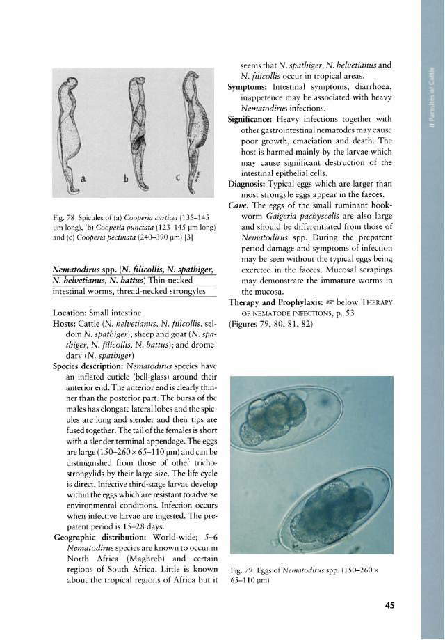
Fig.78 Spicules of (a) Cooperia curticei (135-145 11m long), (b) Cooperia punctata (123-145 11m long) and (c) Cooperia pectinata (240-390 11m) [3]
Nematodirus spp. (N. filicollis, N. spathiger, N. helvetianus, N. battus) Thin-necked intestinal worms, thread-necked strongyles
Location: Small intestine Hosts: Cattle (N. helvetianus, N. filicollis, sel
dom N. spathiger); sheep and goat (N. spathiger, N. filicollis, N. battus); and dromedary (N. spathiger)
Species description: Nematodirus species have an inflated cutide (bell-glass) around their anterior end. The anterior end is dearly thinner than the posterior part. The bursa of the males has elongate lateral lobes and the spicules are long and slender and their tips are fused together. The tail of the females is short with asiender terminal appendage. The eggs are large (150-260 x 65-110 11m) and can be distinguished from those of other trichostrongylids by their large size. The life cyde is direct. Infective third-stage larvae develop within the eggs which are resistant to adverse environmental conditions. Infection occurs when infective larvae are ingested. The prepatent period is 15-28 days.
Geographie distribution: World-wide; 5-6 Nematodirus species are known to oecur in North Afriea (Maghreb) and eertain regions of South Afriea. Little is known about the tropical regions of Africa but it
seems that N. spathiger, N. helvetianus and N. filicollis occur in tropical areas.
Symptoms: Intestinal symptoms, diarrhoea, inappetence may be associated with heavy Nematodirus infections.
Significance: Heavy infections together with other gastrointestinal nematodes may cause poor growth, emaciation and death. The host is harmed mainly by the larvae which may cause significant destruction of the intestinal epithelial cells.
Diagnosis: Typical eggs which are larger than most strongyle eggs appear in the faeces.
Cave: The eggs of the small ruminant hookworm Gaigeria pachyscelis are also large and should be differentiated from those of Nematodirus spp. During the prepatent period damage and symptoms of infection may be seen without the typical eggs being excreted in the faeces. Mucosal scrapings may demonstrate the immature worms in the mucosa.
Therapy and Prophylaxis: - below THERAPY OF NEMATODE INFECTIONS, p. 53
(Figures 79, 80, 81, 82)
Fig.79 Eggs of Nematodirus spp. (150-260 x 65-110 11m)
45

1 Stages In lhe gUl and laeces
G ©
(a) (b) I (c) (d)
Fig. 80 Posterior end of spicules of: (a) Nematodirus helvetianus, (b) N. (ilicollis, (c) N. spathiger and (d) N. battus [5[
Fig. 81 Nematodirus spathiger; bursa copulatrix
with spicules (950 ).Im long; tips fused together) [5]
46
Impalaia tuberculata
Fig. 82 Nematodirus helvetianus; anterior end with
enlarged cephalic vesicle (schematic)
Remark : Thi i mainly a para ire of (he dr m dary, rarely fzebu, artl and heep
f fri a. Ir ur in (he upper mall ince tin
1).
Dictyocaulus viviparus Cattle lungworm
Remar : The fir [- ca e larvae f Dicty call1llS
viviparus ( anle, . 4. ) i f und in rh fa e f inf ted animal. he lar ae are re vered fr f th Ba rmann [
igur
Fig. 83 First-stage larvae of Dictyocaulus viviparus (390-490 x 25 ).Im) [41

Oesophagostomum radiatum Nodular worm
Loeation: Adult worms are found in the large intestine (eaeeum and colon aseendens) . Larvae oeeur in nodules between small intestine and rectum.
Hosts: Cattle Speeies deseription: Direet life eyele; ingested
larvae penetrate the intestinal wall, forming nodules anywhere between the small intestine and the rectum. The head end of the adult worms is characterised by a large cephalic vesiele which is constricted behind its middle. Eggs appear in the faeces about 40 days after ingestion of the third-stage larvae.
Geographie distribution: World-wide Signifieanee: One of the most damaging worms
to cattle when present in high numbers (> 200 adults in calves; > 1000 adults in adult cattle). Young stock may die from nodular worm infections.
Symptoms: Heavy infections are accompanied by anaemia, oedema (hypoalbuminaemia) and diarrhoea (reduced fluid absorption). Oesophagostomum infections occur generally with other gastrointestinal nematodes.
Diagnosis: Thin-shelled strongyle-type eggs appear in the faeces and pea-shaped nodules in the intestinal wall at necropsy indica te infection with the nodular worm. The diagnosis can be confirmed by demonstrating the typicallarge worms at necropsy.
Therapy and Prophylaxis: I!F below THERAPY OF NEMATODE INFECTlONS, p. 53
(Figures 84, 85, 86, 87)
Fig. 84 Oesophagostomum spp.; adult parasites
Fig. 85 Oesophagostomum radiatum; anterior end with large, constricted cephalic vesicle
Fig. 86 Multiple nodules caused by Oesophagostomum radiatum in the caecal mucosa of a calf
Fig. 87 Multiple nodules in the wall of the large intesrine caused by Oesophagostomum radiatum; view from the serosa side
47

1 Stages In lhe gUl and laeces
Oesophagostomum multifoliatum
Remarks: This i mainly a large intestinal para ire of heep, goar bur al 0 zebu. lts occurrence ha been reponed from igeria, Kenya, Tanzanja and Zimbabwe
SHEEP AND GOATS, 1).
Oesophagostomum columbianum
Remarks: Thi para ire occurs in the colon of sheep, goar ,camel, a number of anrelope and occa ionally of zebu. Ir occur worldwide and is parricularly reporred from tropical Africa ( SHEEP AND GOATS, . 1).
Bunostomum phlebotomum Cattle hookworm
Loeation: Small intestine Hosts: Cattle Species deseription: Adults are 10-28 mm long.
There is a prominent buecal eapsule with two pairs of subventrallancers. The spieules of the male measure 3.5-4 mm. The life eyele is direet. Infeetive larvae usually enter cattle by ingestion or by skin penetration.
Signifieance: B. phlebotomum is one of the most pathogenie helminths of cattle in warm and humid areas. Especially for suekling and freshly weaned ealves, during
Fig.88 Bunostomum phlebotomum; anterior end (a) and bursa copulatrix (b) [5]
48
(a)
the rainy season it can be a major pathogen causing severe anaemia. Only 50 adult specimens in the small intestine can cause severe anaemia in calves. This parasite is generally accompanied by other gastrointestinal nematodes. Bunostomum was found to occur focall y only in certain herds of a region.
Geographie distribution: World-wide, especially in warm, humid zones
Symptoms: Penetration of the skin by larvae may cause irritation of the host: itehing of the legs and feet results in cattle stamping their feet. Iron-deficiency anaemia and hypoproteinaemia, accompanied by oedma ("bottle jaw") are the p~edominant symptoms of cattle hookworm infeetions.
Signifieanee: B. phlebotomum may be a serious threat to cattle, especially to calves. Because of its skin-penetrating infection mode, it can also affect suckling calves, killing them within a few days. Heavy infections cause acute anaemia and death. Chronie infections result in poor growth.
Diagnosis: Strongyle-type eggs in the faeces and severe anaemia especially in young calves during the rainy season may suggest hookworm infection.
Therapy and Prophylaxis: I!F below THERAPY OF NEMATODE INFECTIONS, p. 53
(Figures 88, 89)
lOOth
exlemodor- -rr-rrl SOlI ray
(b) spocu!e
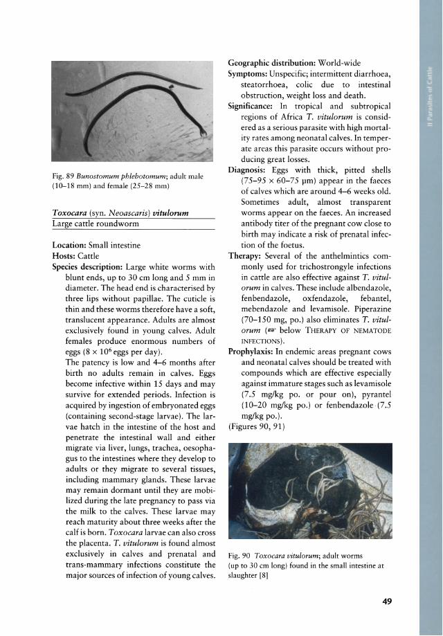
Fig. 89 Bunostomum phlebotomum; adult male (10-18 mm) and female (25-28 mm)
Toxocara (syn. Neoascaris) vitulorum Large cattle roundworm
Location: Small intestine Hosts: Cattle Species description: Large white worms with
blunt ends, up to 30 cm long and 5 mm in diameter. The head end is characterised by three Iips without papillae. The cuticle is thin and these worms therefore have a soft, translucent appearance. Adults are almost exclusively found in young calves. Adult females produce enormous numbers of eggs (8 x 10⁶ eggs per day). The patency is low and 4-6 months after birth no adults remain in calves. Eggs become infective within 15 days and may survive for extended periods. Infection is acquired by ingestion of embryonated eggs (containing second-stage larvae). The larvae hatch in the intestine of the host and penetrate the intestinal wall and either migrate via Iiver, lungs, trachea, oesophagus to the intestines where they develop to
adults or they migrate to several tissues, induding mammary glands. These larvae may remain dormant until they are mobiIized during the late pregnancy to pass via the milk to the calves. These larvae may reach maturity about three weeks after the calf is born. Toxocara larvae can also cross the placenta. T. vitulorum is found almost exclusively in calves and prenatal and trans-mammary infections constitute the major sources of infection of young calves.
Geographic distribution: World-wide Symptoms: Unspecific; intermittent diarrhoea,
steatorrhoea, colic due to intestinal obstruction, weight loss and death.
Significance: In tropical and subtropical regions of Africa T. vitulorum is considered as a serious parasite with high mortality rates among neonatal calves. In temperate areas this parasite occurs without producing great losses.
Diagnosis: Eggs with thick, pitted shells (75-95 x 60-75 pm) appear in the faeces of calves which are around 4-6 weeks old. Sometimes adult, alm ost transparent worms appear on the faeces. An increased antibody titer of the pregnant cow dose to birth may indicate a risk of prenatal infection of the foetus.
Therapy: Several of the anthelmintics commonly used for trichostrongyle infections in cattle are also effective against T. vitulorum in calves. These indude albendazole, fenbendazole, oxfendazole, febantel, mebendazole and levamisole. Piperazine (70-150 mg, po.) also e1iminates T. vitulorum (- below THERAPY OF NEMATODE INFECTIONS).
Prophylaxis: In endemic areas pregnant cows and neonatal calves should be treated with compounds which are effective especially against immature stages such as levamisole (7.5 mg/kg po. or pour on), pyrantel (10-20 mg/kg po.) or fenbendazole (7.5 mg/kg po.).
(Figures 90, 91)
Fig. 90 Toxocara vitulorum; adult worms (up to 30 cm long) found in the small intestine at slaughter [8]
49

1 Stage5 in the gut and faece5
fig. 91 Egg of Toxocara vitu/orum (69-95 x 60-77 11m ) f11]
Trichuris ovis, Trichuris globulosa and Trichuris discolor Whipworms
Location: Caecum and colon Hosts: Cattle, sheep, goat and many other
ruminant species Species description: Whip worms are 3-8 cm
long and easily identified by their long filamentous anterior portion and a thick shorter posterior portion . The male posterior end is usually tightly coiled and there is a single spicule. Direct roundworm Iife cycle. Infective larvae (second-stage larvae) develop within the eggs after at least 3 weeks on pasture. Eggs may remain infective for several years. Animals become infected by ingesting embryonated eggs, and the larvae penetrate the anterior small intestine for 2-10 days before they move to the caecum where they develop to adults. Prepatent period is 50-84 days and va ries markedly among the species. High numbers of preadult and adult worms cause irritation and inflammation of the caecum and colon.
Geographie distribution: World-wide Symptoms: Mild infections (up to 50 adult Tri
churis spp. in cattle or small ruminants) do not cause symptoms. Heavy infections (> 500 adult Trichuris spp . .) may cause colitis, diarrhoea, progressive weight loss. Oedema may occur in the neck and thoraclC regIon.
50
Significance: Whipworms are widespread but the naturally acquired infections in cattle, sheep and goats usually do not cause c1inical disease. Sheep older than 8 months show an age resistance, resulting in a resistance to reinfection 2-3 weeks after a primary infection. However, animals kept in poor condition and carrying multiple infections (e.g. trypanosomes and trichostrongylids) are oEren heavily parasitised by Trichuris spp.
Diagnosis: Demonstration of the characteristic, brown, barrel-shaped eggs with a transparent plug at either pole.
Therapy: So me of the modern benzimidazoles (at increased dose rates) may be used to
treat Trichuris spp. and Capillaria spp. infections in ruminants. These include albendazole, fenbendazole, netobimin and oxfendazole. Ivermectin (200 flg/kg, Sc.) is effective.
Prophylaxis: Difficult because of the tenacity of the infective eggs. Regular removal of faeces in the surroundings of the animals may drastically reduce the infection risk.
(Figures 92, 93, 94)
Fig. 92 Trichuris spp.; adult parasites
Fig. 93 Trichuris spp.; masses of wonns in rhe large inresrine of a calf
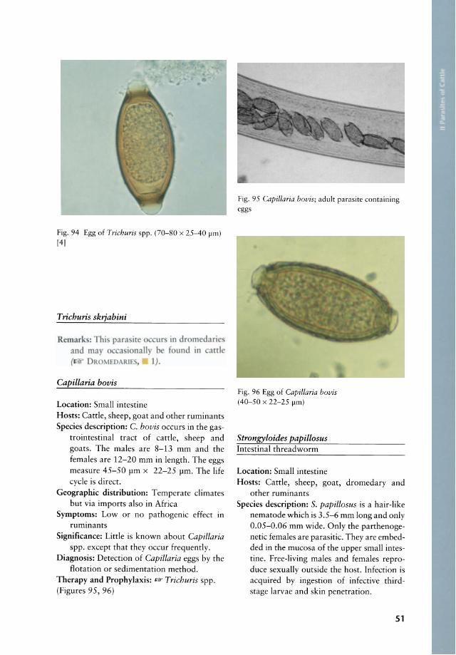
Fig. 94 Egg of Trichuris spp. (70-80 x 25-40 !-Im) [41
Trichuris skrjabini
Remark : Thi para ire ur in dromedaric naH ' be f und In anl
~ I) .
Capillaria bovis
Loeation: Small intestine Hosts: Catde, sheep, go at and other ruminants Speeies deseription: C. bovis occurs in the gas-
trointestinal tract of catde, sheep and goats. The males are 8-13 mm and the females are 12-20 mm in length. The eggs measure 45-50 flm x 22-25 flm. The life cyele is direct.
Geographie distribution: Temperate elimates but via imports also in Africa
Symptoms: Low or no pathogenic effect in ruminants
Significance: Little is known about Capillaria spp. except that they occur frequendy.
Diagnosis: Detection of Capillaria eggs by the flotation or sedimentation method.
Therapy and Prophylaxis: .." Trichuris spp. (Figures 95, 96)
Fig. 95 Capillaria bovis; adult parasite containing eggs
Fig. 96 Egg of Capillaria bovis (40-50 x 22-25 !-Im)
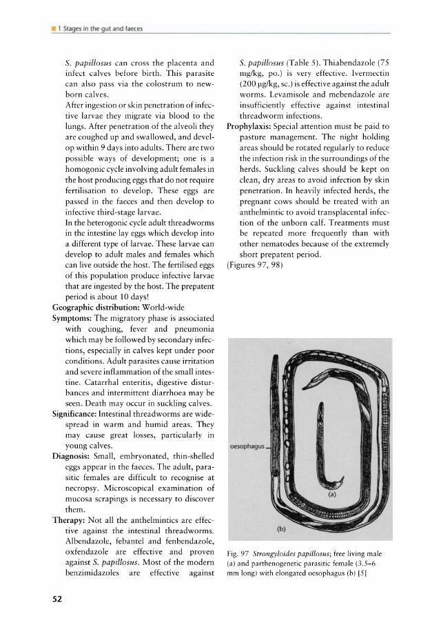
Strongyloides papillosus Intestinal threadworm
Location: Small intestine Hosts: Catde, sheep, goat, dromedary and
other ruminants Speeies deseription: S. papillosus is a hair-like
nematode wh ich is 3.5-6 mm long and only 0.05-0.06 mm wide. Only the parthenogenetic females are parasitic. They are embedded in the mucosa of the upper small intestine. Free-living males and females reproduce sexually outside the host. Infection is acquired by ingestion of infective thirdstage larvae and skin penetration.
51
1 Stages In the gut and taeces
S. papillosus can cross the placenta and infect calves before birth . This parasite can also pass via the colostrum to newborn calves. After ingestion or skin penetration of infective larvae they migrate via blood to the lungs. After penetration of the alveoli they are coughed up and swallowed, and develop within 9 days into adults. There are two possible ways of development; one is a homogonic cycle involving adult females in the host producing eggs that do not require fertilisation to develop. These eggs are passed in the faeces and then develop to infective third-stage larvae. In the heterogonic cycle adult threadworms in the intestine lay eggs which develop into a different type of larvae. These larvae can develop to adult males and females wh ich can live outside the host. The fertilised eggs of this population produce infective larvae that are ingested by the host. The prepatent period is about 10 days!
Geographie distribution: World-wide Symptoms: The migratory phase is associated
with coughing, fever and pneumonia wh ich may be followed by secondary infections, especially in calves kept under poor conditions. Adult parasites cause irritation and severe inflammation of the small intestine. Catarrhal enteritis, digestive disturbances and intermittent diarrhoea may be seen. Death may occur in suckling calves.
Significance: Intestinal threadworms are widespread in warm and humid areas. They may cause great losses, particularly in young calves.
Diagnosis: SmalI, embryonated, thin-shelled eggs appear in the faeces. The adult, parasitic females are difficult to recognise at necropsy. Microscopical examination of mucosa scrapings is necessary to discover them.
Therapy: Not all the anthelmintics are effective against the intestinal threadworms. Albendazole, febantel and fenbendazole, oxfendazole are effective and proven against S. papillosus. Most of the modern benzimidazoles are effective against
52
S. papillosus (Table 5). Thiabendazole (75 mg/kg, po.) is very effective. Ivermectin (200 pg/kg, sc.) is effective against the adult worms. Levamisole and mebendazole are insufficiently effective against intestinal threadworm infections.
Prophylaxis: Special attention must be paid to pasture management. The night holding areas should be rota ted regularly to reduce the infection risk in the surroundings of the herds. Suckling calves should be kept on clean, dry areas to avoid infection by skin penetration. In heavily infected herds, the pregnant cows should be treated with an anthelmintic to avoid transplacental infection of the unborn calf. Treatments must be repeated more frequently than with other nematodes because of the extremely short prepatent period.
(Figures 97, 98)
oesophagus
Fig. 97 Strongyloides papillosus; free living male (a) and parthenogenetic parasitic female (3.5-6 mm long) with e10ngated oesophagus (b) [51
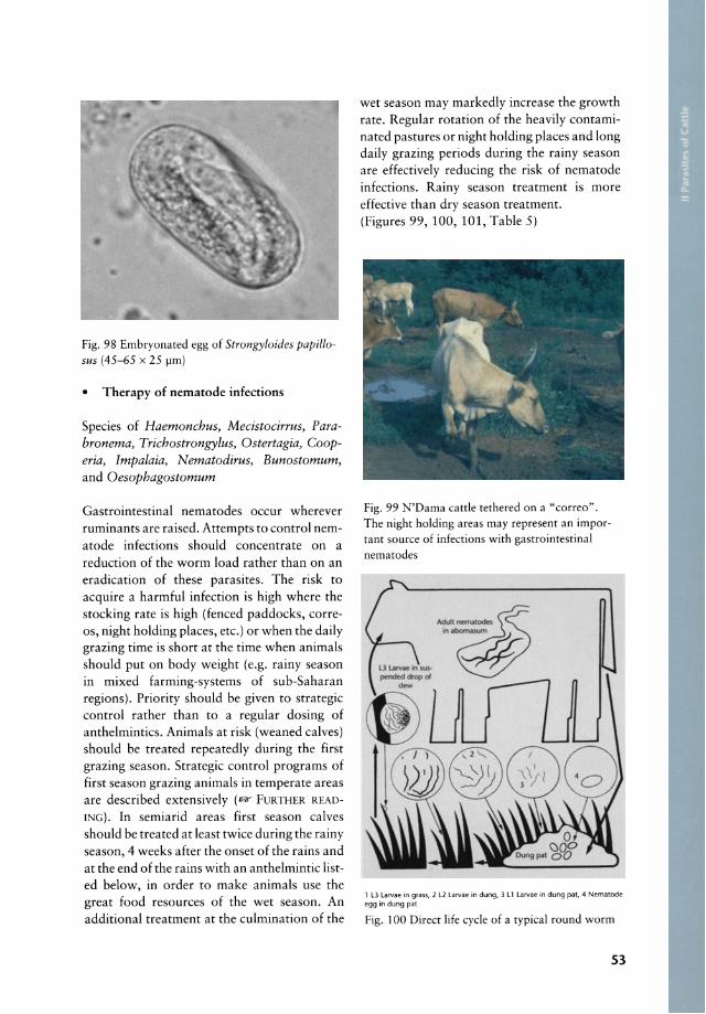
Fig. 98 Embryonated egg of Strongyloides papillosus (45-65 x 25 pm)
• Therapy of nematode infections
Speeies of Haemonchus, Mecistocirrus, Parabronema, Trichostrongylus, Ostertagia, Cooperia, Impalaia, Nematodirus, Bunostomum, and Oesophagostomum
Gastrointestinal nematodes oeeur wherever ruminants are raised. Attempts to control nematode infections should eoneentrate on a reduetion of the worm load rather than on an eradieation of these parasites. The risk to aequire a harmful infection is high where the stocking rate is high (feneed paddocks, correos, night holding plaees, ete.) or when the daily grazing time is short at the time when animals should put on body weight (e.g. rainy season in mixed farming-systems of sub-Saharan regions). Priority should be given to strategie eontrol rather than to a regular dosing of anthelminties. Animals at risk (weaned ealves) should be treated repeatedly during the first grazing season. Strategie eontrol programs of first season grazing animals in temperate areas are deseribed extensively (I&" FURTHER READING). In semiarid areas first season calves should be treated at least twiee during the rainy season, 4 weeks after the onset of the rains and at the end of the rains with an anthelmintie listed below, in order to make animals use the great food resourees of the wet season. An additional treatment at the eulmination of the
wet season may markedly inerease the growth rate. Regular rotation of the heavily eontaminated pastures or night holding plaees and long daily grazing periods during the rainy season are effectively redueing the risk of nematode infections. Rainy season treatment IS more effeetive than dry season treatment. (Figures 99, 100, 101, Table 5)
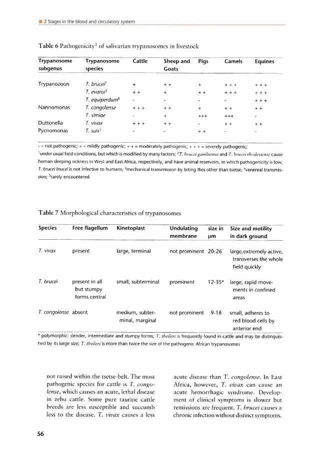
Fig. 99 N'Dama eattle tethered on a "eorreo". The night holding areas may represent an importa nt souree of infeetions with gastrointestinal nematodes
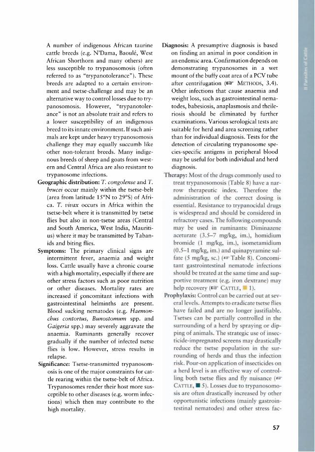
, l3 Larvae in grass, 2 l2 larvae in dung, 3 L 1 larvae in dung pat, 4 Nematode egg in dung pat
Fig. 100 Direet life eyde of a typieal round worm
53

.'·S!p:"!t!;gut.fiIKer
Table 5 Anthelmintics recommended for cattle and sheep
Anthelmintics Spectrum Dosage (mg/kg) Cattle Sheep AM
Albendazole Nematodes, cestodes, trematodes 7.5 5.0 po. Coumaphos Trichostrongylid spp. 2 (6x) po. Febantel Nematodes 7.5 5 po. Fenbendazole Nematodes, cestodes 7.5 5.0 po. Ivermectin Nematodes, arthropods 0.2 0.2 po. or sc. Ivermectin and Nematodes, trematodes, arthropods 0.2/7 po.
Clorsulon Ivermectin Nematodes, trematodes, arthropods 0.5 pouron Levamisole Nematodes 50 5.0 sc. or im. Levamisole Nematodes 7.5 7.5 po. Levamisole Nematodes 10 10 pouron Mebendazole Nematodes 20 po. Netobimin Nematodes, cestodes 7.5 7.5 po. Morantel tartrate Nematodes 10 10 po. Morantel tartrate Nematodes SRB* ir. Oxfendazole Nematodes, cestodes 4.5 5 po. Phenothiazine Nematodes 2201 po. Pyrantel tartrate Nematodes 12.5 25 po. Thiabendazole Nematodes 44-75 po. Moxidectin Nematodes, arthropods 0.2 0.2 po.
*Slow Release Bolus (rumen retention device releases about 200 mg/day for 60 days)
1 safety index very low (= 1); AM = application method, po. = orally, sc. = subcutaneously, im. = intramuscularly and
ir. = intraruminal
54

2 Stages in the blood and drculatory system
PROTOZOA .................... 55
HELMINTHS • Trematoda found in the blood and
circulatory system .............. .78 • Nematoda larvae (microfilariae) found
in the blood and adult nematodes living in the circulatory system ...... 81
PROTOZOA
TRYPANOSOMA TIDAE
Trypanosoma congolense, Trypanosoma vivax, Trypanosoma brucei Tsetse-transmitted trypanosomosis, Nagana, sleeping sickness
Hosts: Cattle, sheep, goat and many other domestic and wild animal species
Vector: Several species of tsetse flies: Glossina morsitans (savanna areas), Glossina palpalis (areas around rivers and lakes) and Glossina fusca (high forest areas) . All three species transmit trypanosomosis and feed on a wide spectrum of mammals.
Species description: The most important trypanosomes affecting cattle, sheep and goats are as follows (in order of importance): T. congolense, T. vivax and T. brucei. These three species belong to the Salivaria (Table 6). The differentiation and the morphological characteristics of these pathogenic trypanosomes are listed in Table 7. All three species are transmitted by tsetse flies. Most tsetse-transmission is cyelic and begins when blood from a trypanosome infected animal is ingested by the fly. The trypanosomes lose their surface coat, multiply in the fly, re-acquire a new surface coat and become infective. The life cyele within the tsetse fly varies among the different trypanosome species. The infective form for animals in the tsetse fly is referred to as the metacyelic form. The development in the tsetse flies may be
as short as 1 week with T. vivax or extend to a few weeks for T. brucei. Mechanical transmission requires blood containing trypanosomes being transferred from one anima I to another. Tsetse flies inoculate metacyelic trypanosomes into the skin of animals, where the trypanosomes multiply and cause swellings (chancres). They enter the blood stream either directly or through the Iymph nodes, then the bloodstream where they divide rapidly by binary fission. T. congolense attach to endothelial cells and localize in capillaries. T. brucei and T. vivax invade tissues and cause tissue damage in several organs. The necropsy findings vary and are not specific. Extensive petechiae of the serosal membranes, especially in the peritoneal cavity may occur in acute, fatal cases. The lymph nodes and the spleen are usually swollen. Chronic cases are associated with atrophy of body and organ fat, severe anaemia and swollen lymph nodes. The immune response of infected animals is vigorous, and immune complexes cause inflammation, which contributes to the elinical signs and lesions of trypanosomosis. Trypanosomes have multiple genes that code for different surface-coat glycoproteins. The number of different antigenic types of glycoprotein that can be made is unknown but exceeds several hundred. The antigenic variation results in persistence of the organisms in the host and is a way of the parasite to evade the host's immune system. Antigenic variation has prevented the development of a vaccine and permits reinfections when animals are exposed to a new antigenic type of the same trypanosome species. Animals infected with trypanosomes show some degree of immunodepression and are more susceptible to a.o. gastrointestinal helminth infections. Haemonchus contortus shows a reduced prepatent period and causes a markedly increased mortality in animals that are chronically infected with trypanosomes. Some bos indicus (Zebu) are very sensitive to trypanosomosis and they are generally
55
Iystem
Table 6 Pathogenicityl of salivarian trypanosomes in livestock
Trypanosome Trypanosome Cattle Sheep and Pigs Camels Equines subgenus species Goats
Trypanozoon T. brucel'2 + ++ + +++ +++
T. evansi1 ++ + ++ +++ +++
T. equiperdum4 +++
Nannomonas T. congolense +++ ++ + ++ ++
T. simiae + +++ +++
Duttonella T. vivax +++ ++ ++ ++
Pycnomonas T. suis5 ++
- = not pathogenic; + = mildly pathogenic; + + = moderately pathogenic; + + + = severely pathogenic;
1 under usual field conditions, but which is modified by many factors; 2T. brueei gambiense and T. brueei rhodesiense cause
human sleeping sickness in West and East Africa, respectively, and have animal reservoirs, in which pathogenicity is low;
T. brucei brucei is not infective to humans; 3mechanical transmission by biting flies other than tsetse; 4venereal transmis
sion; Srarely encountered
T able 7 Morphological characteristics of trypanosomes
Species Free flagellum Kinetoplast Undulating size in Size and motility membrane !-Im in dark ground
T. vivax present large, terminal not prominent 20-26 large,extremely active, transverses the whole field quickly
T. brucei present in all smalI, subterminal prominent 12-35* large, rapid move-but stumpy ments in confined forms central areas
T. congolense absent medium, subter- not prominent 9-18 smalI, adheres to minal, marginal red blood cells by
anterior end • polymorphie: slender, intermediate and stumpy forms; T. theileri is frequently found in cattle and may be distinguis
hed by its large size; T. theileri is more than twice the size of the pathogenic African trypanosomes
56
not raised within the tsetse-belt. The most pathogenic species for cattle is T. congolense, which causes an acute, lethaI disease in zebu cattle. Some pure taurine cattle breeds are less susceptible and succumb less to the disease. T. vivax causes a less
acute disease than T. congolense. In East Africa, however, T. vivax can cause an acute hemorrhagic syndrome. Development of clinical symptoms is slower but remissions are frequent. T. brucei causes a chronic infection without distinct symptoms.
A number of indigenous African taurine cattle breeds (e.g. N 'Dama, Baoule, West African 5horthorn and many others) are less susceptible to trypanosomosis (often referred to as "trypanotolerance"). These breeds are adapted to a certain environment and tsetse-challenge and may be an alternative way to controllosses due to trypanosomosis . However, "trypanotolerance" is not an absolute trait and refers to
a lower susceptibility of an indigenous breed to its innate environment. If such animals are kept under heavy trypanosomosis challenge they may equally succumb like other non-tolerant breeds. Many indigenous breeds of sheep and goats from western and Central Africa are also resistant to trypanosome infections.
Geographie distribution: T. congolense and T. brucei occur mainly within the tsetse-belt (area from latitude 15°N to 29°5) of Africa. T. vivax occurs in Africa within the tsetse-belt where it is transmitted by tsetse flies but also in non-tsetse areas (Central and 50uth America, West India, Mauritius) where it may be transmitted by Tabanids and biting flies.
Symptoms: The primary clinical signs are intermittent fever, anaemia and weight loss. Cattle usually have a chronic course with a high mortality, especially if there are other stress factors such as poor nutrition or other diseases. Mortality rates are increased if foncomitant infections with gastrointestinal helminths are present. Blood sucking nematodes (e.g. Haemonchus contortus, Bunostomum spp. and Gaigeria spp.) may severely aggravate the anaemia. Ruminants generally recover gradually if the number of infected tsetse flies is low. However, stress results in relapse.
Significance: Tsetse-transmitted trypanosomosis is one of the major constraints for cattle rearing within the tsetse-belt of Africa. Trypanosomes render their host more susceptible to other diseases (e.g. worm infections) wh ich then may contribute to the high mortality.
Diagnosis: A presumptive diagnosis is based on finding an anima I in poor condition in an endemie area. Confirmation depends on demonstrating trypanosomes in a wet mount of the buffy coat area of a PCV tube after centrifugation (~ METHODS, 3.4). Other infections that cause anaemia and weight loss, such as gastrointestinal nematodes, babesiosis, anaplasmosis and theileriosis should be eliminated by further examinations. Various serological tests are suitable for herd and area screening rather than for individual diagnosis. Tests for the detection of circulating trypanosome species-specific antigens in peripheral blood may be useful for both individual and herd diagnosis.
Therapy: Mo t of the drug commonly used to treat trypano mo i (Table ) have a narr w therapeutic inde . Therefore the admin' tra tion of the correct do ing i e ential. Re i tance co trypanocidal drug i wide pread and hould be con idered in refra tor ca e . The foll 'ti ing ompound may b u ed in ruminant: Diminazene aceturate (3.5-7 mglkg, im.), homidium bromide (1 mglkg im.), i omctamidium (0. --I mglkg im.) and quinapyramine ulfate (- mglkg, c.) ( Table ). on mitant ga troiare rinal nemat de infe fion hould be treated ar the ame time and up
p rtive treatment (e.g. iron de trane) may help re overy ( ATILE, L) .
Prophyla 'i : ntrol can be carried our at e -eral level. ttempt to eradicare r er e flie have failed and are n longer ju tifiablc. T er e an be partially conrrolled in rhe urrounding of a herd by pra ing or dip
ping of animal . The [rare i u e f in ccricide-impregnared reen may dra tically reduce the r er e population in the urrounding of berd and thu rhe infecti n risk. Po ur-on appli ari n of in e ti ide n a herd leve l is an effe tive way of oorr 1-ling both tserse flies and fly nui an e (
ATILE . 5). Losses due to rrypano omoi are onen drasrically increa ed by other
opportunistic infections (mainly ga trointe tinal nematodes) and other srre fac-
57
• 2 Stages in the blood and circulatory system
Fig. 101 Trypanosoma vivax (20-26 ~m); Giemsa-stained bloodsmear [4]
58
tors. Control measures should also include these faetors. The prophylaetie use of trypanoeidal drugs requires a regular applieation of the drug and includes the risk of ereating resistanee. It should therefore be limited to emergeneies, sueh as eattle trekking through infested areas and herds seasonally exposed to high trypanosomosis risk. Isometamidium (1-2 mg/kg, im.) proteets eattle for 2 to 6 months. Prophylaetie use of trypanoeidal drugs should be avoided in eattle prior to slaughtering, sinee drug
Fig. 102 Trypanosoma brucei (12-35 ~m); Giemsa-stained bloodsmear; this speeies is polymorphie: slender form (left), stumpy form (right)
[131
F
Fig. 103 Important Trypanosoma species of livestoek: (a) Trypanosoma theileri (60-70 ~m), (b) T. cruzi, (e) T. congolense (9-18 ~m), (d) T. vivax (20-26 ~m), (e) T. equiperdum (25 ~m) and (f) T. brucei (12-35 ~m, polymorphie) [14J
residues may be detrimental to human health. Proper nutrition is essential in all eases and the strategie applieation of erop residues at the late dry season may signifieantly improve the nutritional status and thus the resistanee of animals in endemie areas. The exploitation and propagation of indigenous breeds ean be a promising way of animal produetion in endemie areas.
(Figures 101, 102, 103, 104, 105, Table 6, Table 7, Table 8)

VI
'D
Ta
ble
8 G
en
eri
c a
nd
tra
de
na
me
s o
f tr
ypa
no
cid
es
for
the
th
e t
rea
tme
nt
an
d p
reve
nti
on
of
an
ima
l tr
ypa
no
som
osi
s
Co
mp
ou
nd
T
rad
e n
am
e
Ma
nu
fact
ure
r A
ctio
n
Ran
ge o
f d
osa
ge
R
ou
te o
f R
emar
ks
ge
ne
ric
na
me
R
ates
(m
g/k
g)
ad
min
istr
ati
on
Dim
inaz
ene
acet
urat
e B
eren
il H
oech
st A
G,
Ger
man
y T
3.
5-7.
0 IM
A
lso
babe
siac
idal
; to
xic
to h
orse
s,
SC
donk
eys,
dog
s, a
nd c
amel
s G
anas
ag
Squ
ibb,
US
A
T
3.5-
7.0
IM
or
SC
Ho
mid
ium
bro
mid
e
Eth
idiu
m
CA
MC
O A
nim
al H
ealth
, U
K
T(P
) 1.
0 IM
Ho
mid
ium
ch
lori
de
N
ovi
diu
m
Rh6
ne-M
erie
ux,
Fra
nce
T(P
) 1.
0 IM
Iso
me
tam
idiu
m
Sam
orin
R
h6ne
-Mer
ieux
, F
ranc
e P/
T 0.
25-1
.0
IM,I
V3
Tox
ie a
bove
2 m
g/kg
; h
igh
ly
irri
tan
t; a
void
SC
ad
min
istr
atio
n
Try
pa
mid
ium
R
h6ne
-Pou
lenc
San
te F
ranc
e P/
T 0.
25-1
.0
M,I
V3
Ou
ina
pyr
am
ine
A
ntry
eide
C
oope
rs A
nim
al H
ealth
T
3.
0-5.
0 SC
R
est
anim
als
befo
re a
nd a
fter
su
lfate
' *
Ltd,
UK
tr
ea
tme
nt
Ou
ina
pyr
am
ine
A
ntry
cide
2 C
oope
rs A
nim
al H
ealth
PT
3.
0-5
.0
SC
Dos
age
eale
ulat
ed a
s su
lfate
pros
alt'
R.F
. Lt
d, U
K
Sur
amin
* N
agan
ol
Bay
er A
G,
Ger
man
y T
10
.0
IV
Sev
ere
loea
l re
actio
ns b
y o
the
r A
ntr
ypo
l ro
utes
Mel
amin
ophe
nyla
rsin
e C
ymel
arsa
n R
h6ne
-Mer
ieux
, F
ranc
e T
0.
25-0
.5
IM,
SC
The
IM
ro
ute
is p
refe
rred
in
dih
ydro
eh
lori
de
eq
uine
s. T
. ev
ansi
and
T.
brue
ei
Man
y o
f th
ese
prep
arat
ions
are
sol
d u
nd
er
a va
riety
of t
rade
nam
es.
Con
sult
publ
icat
ions
suc
h as
Vet
erin
ary
Pha
rmac
eutic
als
an
d B
iola
gica
ls,
6th
ed
. Le
nexa
, KS
, V
eter
inar
y
Med
icin
e P
ublis
hing
Co,
19
89
/19
90
;
1 Rei
ntro
duce
d in
198
5 to
tre
at m
ainl
y T.
evan
si in
fect
ions
;
2Pro
salt.
Thi
s is
a m
ixtu
re o
f su
lfate
and
chl
orid
e sa
lts o
f qui
napy
ram
ine;
3Giv
en b
y ve
ry s
low
inj
ectio
n o
f 1 %
W/V
sol
utio
n at
0.5
mg
/kg;
T =
the
rape
utic
act
ion;
P =
pro
phyl
actic
act
ion;
(P
) =
sho
rt p
rop
hyl
act
ic a
ctiv
ity;
IM =
int
ram
uscu
lar;
IV
= i
ntra
veno
us;
SC =
sub
cuta
neou
s;
* no
lon
ger
com
mer
cial
ly a
vaila
ble

• 2 Stages in the blood and circulatory system
Fig. 104 Trypanosoma congolense (9-18 11m); Giemsa-stained bloodsmear [15J
Trypanosoma evansi (syn. T. brucei evansi) Surra
Rcmark : T. Cl/allS; ur in buffa!
60
Fig. 105 Comparison of a trypano-tolerant N'Dama catrle (Jeft) and susceptible zebu (right ) after 15 weeks' exposure to a middle-grade tsetse challenge. Both animals were chronically infected with T rypanosoma congolense [361
rhe ub enu rypan Z n ( DR IfR-
DARIE, 2).
Trypanosoma theileri
Hosts: Cattle Vector: Tabanid flies (Tabanus spp. and Hae
matopota spp.) Species description: This is a large species
(60-70 flm, sometimes up to 120 flm in length=twice the size of the pathogenic trypanosomes) and belongs to the Stercoraria . The undulating membrane is weil developed and the free flagellum is weil defined. Although the parasite is considered as nonpathogenic it may assume increased significance when stress conditions arise or when concurrent infections with other pathogens are present.
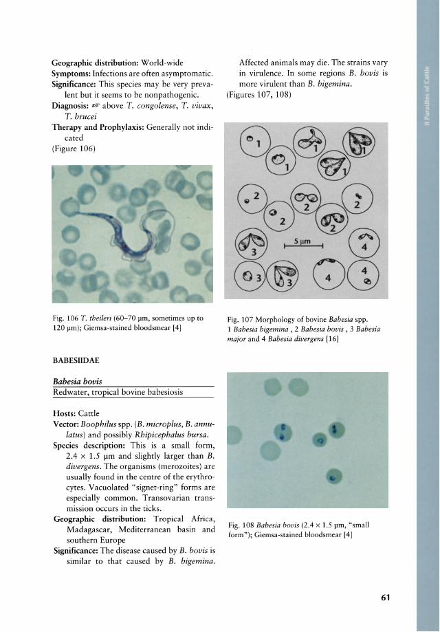
Geographie distribution: World-wide Symptoms: Infeetions are often asymptomatie. Signifieanee: This speeies may be very preva-
lent but it seems to be nonpathogenie. Diagnosis: trF above T. congolense, T. vivax,
T. brucei Therapy and Prophylaxis: Generally not indi
eated (Figure 106)
Fig. 106 T. theileri (60-70 pm, sometimes up to 120 pm); Giemsa-stained bloodsmear [4)
BABESIIDAE
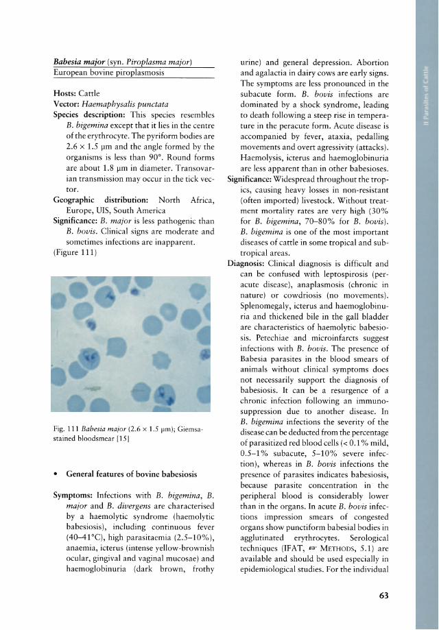
Babesia bovis Redwater, tropieal bovine babesiosis
Hosts: Cattle Veetor: Boophilus spp. (B. microplus, B. annu
latus) and possibly Rhipicephalus bursa. Speeies deseription: This is a small form,
2.4 x 1.5 pm and slightly larger than B. divergens . The organisms (merozoites) are usually found in the centre of the erythrocytes. Vacuolated "signet-ring" forms are especially common. Transovarian transmission occurs in the ticks.
Geographie distribution: Tropical Africa, Madagascar, Mediterranean basin and southern Europe
Signifieanee: The disease caused by B. bovis is similar to that caused by B. bigemina.
Affected animals may die. The strains vary in virulence. In some regions B. bovis is more virulent than B. bigemina.
(Figures 107, 108)
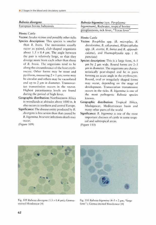
Fig. 107 Morphology of bovine Babesia spp. 1 Babesia bigemina , 2 Babesia bovis , 3 Babesia major and 4 Babesia divergens [16)
Fig. 108 Babesia bovis (2.4 x 1.5 pm, "small form"); Giemsa-stained bloodsmear [4)
61
• 2 Stages in the blood and circulatory system
Babesia divergens European bovine babesiosis
Hosts: Cattle Veetor: Ixodes ricinus and possibly other ticks Speeies deseription: This speeies is smaller
than B. bovis. The merozoites usually oeeur as paired, c1ub-shaped organisms about 1.5 x 0.4 pm. The angle between the pair is relatively large, so that they diverge more from eaeh other than those of B. bovis. The organisms tend to Iie along the eireumferenee of the host erythroeyte. Other forms may be stout and pyriform, measuring 2 x 1 pm; some may be eireular and others may be vaeuolated and up to 2 pm in diameter. Transovarian transmission oeeurs in the veetor. Highest parasitaemia levels are found during the period of high fever.
Geographie distribution: Northwestern Afriea in woodlands at altitudes above 1000 m. It also oeeurs in northern and eentral Europe.
Signifieanee: The disease entity produeed by B. divergens is less severe than that eaused by B. bigemina. In severe infeetions death may oeeur.
(Figure 109)
Fig. 109 Bahesia divergens (1.5 x 0.4 )lm); Giemsa
sta in ed bloodsmear 14]
62
Babesia bigemina (syn. Piroplasma bigeminum), Redwater, tropieal bovine piroplasmosis, tiek fever, "Texas fever"
Hosts: Cattle Veetor: Boophilus spp. (B. microplus, B.
decoloratus, B. calcaratus) , Rhipicephalus spp. (R. evertsi, R. bursa and R. appendiculatus), and Haemaphysalis spp. ( H. punctata)
Speeies deseription: This is a large form, 4-5 pm by 2 pm wide. Round forms are 2-3 pm in diameter. The organisms are eharaeteristieally pear-shaped and lie in pairs forming a n aeute angle in the erythroeyte. Round, oval or irregularly shaped forms may oeeur, depending on the stage of development. Transovarian transmission oeeurs in the tieks. B. bigemina is one of the most pathogenie Babesia speCles known.
Geographie distribution: Tropieal Afriea, Madagasear, Mediterranean basin and many other parts of the world
Signifieanee: B. bigemina is one of the most important diseases of eattle in some tropieal and subtropieal areas.
(Figure 110)
Fig. 110 Bahesia higemina (4-5 x 2 )lm, "Iarge form"); Giemsa-sta ined bloodsmear [4]
Babesia major (syn. Piroplasma major) European bovine piroplasmosis
Hosts: Catde Veetor: Haemaphysalis punctata Speeies deseription: This speeies resembles
B. bigemina except that it lies in the centre of the erythroeyte. The pyriform bodies are 2.6 x 1.5 pm and the angle formed by the organisms is less than 90°. Round forms are about 1.8 pm in diameter. Transovarian transmission may oecur in the tick vector.
Geographie distribution: North Africa, Europe, UIS, South America
Signifieanee: B. major is less pathogenic than B. bovis. Clinieal signs are moderate and sometimes infections are inapparent.
(Figure 111)
Fig. 111 Babesia maior (2.6 x 1.5 firn); Giernsastained bloodsrnear [15 J
• General features of bovine babesiosis
Symptoms: Infections with B. bigemina, B. major and B. divergens are charaeterised by a haemolytic syndrome (haemolytic babesiosis), including continuous fever (40-41°C), high parasitaemia (2.5-10%), anaemia, icterus (intense yellow-brownish oeular, gingival and vaginal mucosae) and haemoglobinuria (dark brown, frothy
urine) and general depression. Abortion and agalactia in dairy eows are early signs. The symptoms are less pronounced in the subaeute form. B. bovis infeetions are domina ted by a shock syndrome, leading to death following a steep rise in temperature in the peracute form. Acute disease is accompanied by fever, ataxia, pedalling movements and overt agressivity (attaeks). Haemolysis, icterus and haemoglobinuria are less apparent than in other babesioses.
Significance: Widespread throughout the tropics, causing heavy losses in non-resistant (often imported) livestock. Without treatment mortality rates are very high (30% for B. bigemina, 70-80% for B. bovis). B. bigemina is one of the most important diseases of catde in some tropical and subtropical areas .
Diagnosis: Clinical diagnosis is difficult and can be confused with leptospirosis (peracute disease), anaplasmosis (ehronic in nature) or cowdriosis (no movements). Splenomegaly, icterus and haemoglobinuria and thickened bile in the gaU bladder are characteristics of haemolytic babesiosis. Petechiae and microinfarcts suggest infections with B. bovis. The presence of Babesia parasites in the blood smears of animals without clinical symptoms does not necessarily support the diagnosis of babesiosis. Ir can be a resurgence of a chronic infection following an immunosuppression due to another disease. In B. bigemina infections the severity of the disease can be deducted from the percentage of parasitized red blood cells « 0.1 % mild, 0.5-1 % subacute, 5-10% severe infection), whereas in B. bovis infections the presence of parasites indicates babesiosis, because parasite concentration in the peripheral blood is considerably lower than in the organs. In acute B. bovis infections impression smears of congested organs show punctiform babesial bodies in agglutinated erythrocytes. Serological techniques (IFAT, UM' METHODS, 5.1) are available and should be used especially in epidemiological studies. For the individual
63

Table 9 Drugs used in the treatment of babesiosis in cattle
Drug
Trypan blue*
Acridine derivatives
Euflavine 1 *
Diamidine derivatives
Amicarbalide
Diampron 1
Diminazene
Berenil2
Imidocarb
Phenamidine
Lomadine 1
Quinoline derivatives
Quinuronium
Babesan4
Dosage
2-3 mg/kg
4-8 ml/1 00 kg as
5% solution
5-10 mg/kg
3-5 mg/kg
1-3 mg/kg
Imizol 3
8-13.5 mg/kg
1 mg/kg
AM
IV
IV
IM
IM
SC or IM
SC or IM
SC
Parasite
B. bigemina
B. bigemina B. bovis B. divergens
B. bigemina B. divergens B. bavis
B. bigemina B. bavis B. divergens
B. bigemina B. bavis B. divergens
B. bigemina B. bovis
B. bigemina B. bovis B. divergens
Remark
Not effective against small
babesias; discouloration of
animal's flesh; can cause
severe tissue sloughing if
not given IV
Highly irritant if not given IV
Prophylactic activity up to 8 weeks depending on dose
and Babesia spp. involved;
nephrotoxic; cholinesterase
inhibitor; slowly metabolized
and eliminated-tissue
Cholinesterase inhibition
Low therapeutic index
Siow effect against B. bovis Side effects associated with
the stimulation of the para
sympathetic nervous system
Many 01 these preparations are sold under a variety 01 trade names; * no longer commercially available;
IM = intramuscular; IV = intravenous; SC = subcutaneous; AM = application method;
1 May & Baker Ltd., Dagenham, England. 2 Hoechst AG, Frankfurt, Germany. 3 Burroughs Wellcome & Co., London, England.
4 ICI-, Maccleslield, Cheshire, England
64
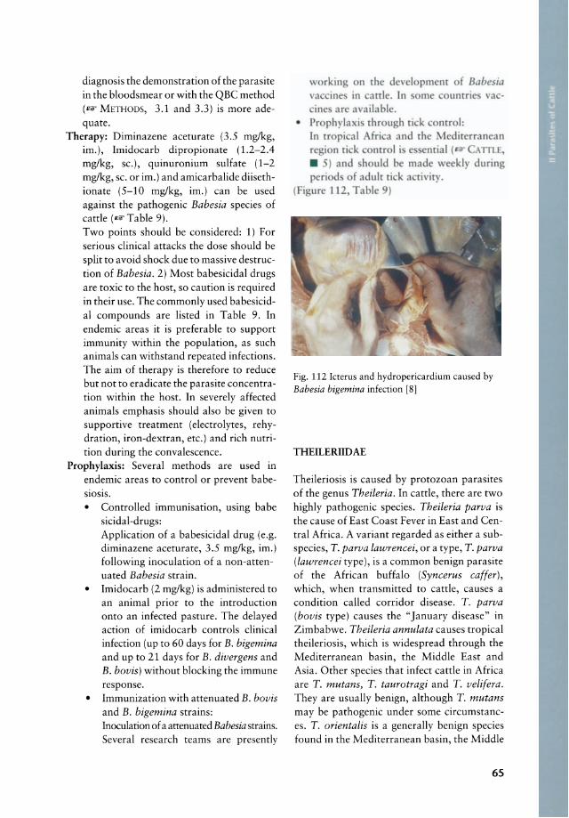
diagnosis the demonstration of the parasite in the bloodsmear or with the QBC method (1lF METHODS, 3.1 and 3.3) is more adequate.
Therapy: Diminazene aceturate (3.5 mg/kg, im.), Imidocarb dipropionate (1.2-2.4 mg/kg, sc.), quinuronium sulfate (1-2 mg/kg, sc. or im.) and amicarbalide diisethionate (5-10 mg/kg, im.) can be used against the pathogenic Babesia species of cattle (IlFTable 9). Two points should be considered: 1) For serious clinical attacks the dose should be split to avoid shock due to massive destruction of Babesia. 2) Most babesicidal drugs are toxic to the host, so caution is required in their use. The commonly used babesicidal compounds are listed in Table 9. In endemic areas it is preferable to support immunity within the population, as such animals can withstand repeated infections. The aim of therapy is therefore to reduce but not to eradicate the parasite concentration within the host. In severely affected animals emphasis should also be given to supportive treatment (electrolytes, rehydration, iron-dextran, etc.) and rich nutrition during the convalescence.
Prophylaxis: Several methods are used in endemic areas to control or prevent babesiosis. • Controlled immunisation, using babe
sicidal-drugs: Application of a babesicidal drug (e.g. diminazene aceturate, 3.5 mg/kg, im.) following inoculation of a non-attenuated Babesia strain.
• Imidocarb (2 mg/kg) is administered to an animal prior to the introduction onto an infected pasture. The delayed action of imidocarb controls clinical infection (up to 60 days for B. bigemina and up to 21 days for B. divergens and B. bovis) without blocking the immune response.
• Immunization with attenuated B. bovis and B. bigemina strains: Inoculation of a attenuated Babesia strains. Several research teams are presently
n the d vel pment of Babesia me untrie va -
riviry .
ATfLE,
during
Fig. 112 kterus and hydropericardium caused by Babesia bigemina infection [8]
THEILERIIDAE
Theileriosis is caused by protozoan parasites of the genus Theileria. In cattle, there are two highly pathogenic species. Theileria parva is the cause of East Coast Fever in East and Central Africa. A variant regarded as either a subspecies, T. parva lawrencei, or a type, T. parva (lawrencei type), is a common benign parasite of the African buffalo (Syncerus caffer), wh ich, when transmitted to cattle, causes a condition called corridor disease. T. parva (bovis type) causes the "January disease" in Zimbabwe. Theileria annulata causes tropical theileriosis, wh ich is widespread through the Mediterranean basin, the Middle East and Asia. Other species that infect cattle in Africa are T. mutans, T. taurotragi and T. velifera. They are usually benign, although T. mutans may be pathogenic under some circumstances. T. orientalis is a generally benign species found in the Mediterranean basin, the Middle
65

• 2 Stages in the blood and circulatory system
East, Asia and Australia which may cause disease in imported cattle. (Figures 113, 114, 115)
l ymph nodes stages (1-3)
Erythrocyte stages (4-6)
Fig. 113 Theileria orientalis; Giemsa-stained blood
smear; this species occurs outside of Africa and is lowly pathogenic [41
Fig. 114 Morphology of Theileria spp. stages in
mammals; stages found in the Iymph nodes:
5(0 6(2) 4~
(1) Macrosehizont with medium-sized nudei (0.4-2 ~m; Koeh 's blue bodies) , (2) Microschizont
with small nudei (0.3-0.8 ~m ) and (3) Merozoites
(0. 7-1 ~m); stages found in the erythrocytes: intra erythrocytic stages may be (4) eircular, oval or
piriform (0.8 x1.5 ~m; 80%) or (5) comma-shaped (0.5 x 2 ~m; 20%); (6) some parasites appear to
have a velum [16]
Fig. 115 Life eyde of Theileria parva [17] (I) Merogony, (2) M erozoites (3) Piroplasm in erythroeyte, (4) Macromete, (5) Syngamy, (6 ) Microgamete, (7) Zygote in gut lumen, (8) Gur epithelial cells, (9) Kinete, (10) Moult, (11) Sporogony, (12) Sporozoites, (13) Sporozoite in saliva, (14) Sporozoite enters Iymphocyte, (15) Sehizont in lymphoblast, (16) Clonal expansion, (17) Schizont
66
16 /'" .;.
I~ 15 ~J
f ®14 t
13 0
17
1\ ® @3
1--1 12~ _ ~'~I~:
11 ~ Adult Nymph /4 '~ I ~Q7 Salivary
Gland Acinus
t' I 0 IO,(
\ .§:'~ ~O 8
~1b • 9

Theileria parva (syn. T. bovis, T. lawrencei) East Coast Fever, bovine theileriosis, Corridor Disease, Rhodesian tick fever
Hosts: Cattle, water buffalo, African buffalo Vector: Rhipicephalus appendiculatus (equa
torial East Africa and southern Africa above 1200 m) and Rhipicephalus duttoni (Angola plateau). Other vectors are Rhipicephalus zambeziensis, Hyalomma excavatum, H. dromedarii, H. truncatum and some other Rhipicephalus spp.
Species description: T. parva causes a disease ca lied East Coast Fever (ECF), which is responsible for high mortality among susceptible and imported stock. The zebu in endemie areas has a high natural res istance, but animals imported into endemie areas are highly susceptible. Transmission of the parasite in all is on a stage to stage basis (trans-stadial transmission). There is no transovarial transmission in the tick vector as in Babesia spp. The organisms occur both in erythrocytes and in lymphocytes. The forms in the red blood cells are mainly rod-shaped, 1.5-2 x 0.5-1 J.Im, but round, oval, comma- and ring-shaped forms mayaiso occur (Fig. 114, 4-6). Several parasites may occur in individual erythrocytes but there is no multiplication in the red cells. The multiplying forms of the parasite occur in the cytoplasm of lymphocytes and occasionally in the endothelial cells of the lymphatic glands and the spleen. These are schizonts, being circular or irregularly shaped structures about 8 J.Im in diameter. Two forms of schizonts are recognized. Those wh ich contain large chromatin granules, 0.4-2 J.Im in diameter, are termed macroschizonts and produce macromerozoites, 2-2.5 J.Im in diameter (Fig. 114, 1). The other forms contain smaller chromatin granules, 0.3-0.8 J.Im in diameter, and are referred to as microschizonts and produce micromerozoites (Fig. 114, 2) . The latter invade the red blood cells and may represent sexual stages of the parasite. After the intraerythrocytic stages are ingested by ticks, merozoites are
liberated and differentiate into sexual stages. There is a sexual development of T. parva in the tick vector which results in infective sporozoites. Cattle are infected when vector ticks engorge on an animal and these infective particles (sporozoites) are transmitted. Most recovered animals remain carriers of infection.
Geographie distribution: East, Central and South Africa according to the distribution of the main vector Rhipicephalus appendiculatus.
Symptoms: High fever, swelling of the lymph nodes, dyspnoea and death. Fever occurs 7-10 days after the tick bite and continues throughout the course of the disease and may reach > 42°C. Lymph node swelling becomes pronounced and generalized. Anorexia, loss of condition, lacrimation and nasal discharge may occur. Terminally, dyspnoea is common. Just before death, a sharp fall in temperature occurs and pulmonary exsudate pours from the nostrils. Anaemia is not a major diagnostic sign as it is in babesiosis.
Significance: ECF is a serious disease with high morrality in susceptible stock.
Diagnosis: Lymphoblasts in Giemsa-stained lymph node biopsy smears (1& METHODS, 3.1) during the acute febrile phase contain multinuclear schizonts. Lymph node enlargement and massive pulmonary oedema and hyperaemia are the most striking post-mortem lesions. Haemorrhages are common on the serosal and mucosal surfaces of many organs.
Therapy: Cattle with clinical ECF may be treated with parvaquone (2 x 10-20 mgl kg, im. at 48 h intervals),buparvaquone (2 x 2.5 mglkg, im. at 48 h intervals) or with the coccidiostat halofuginone (2 x 1.2 mglkg, po. at 48 h intervals). Chlorretracycline and oxytetracyline are relatively ineffective. None of these drugs will sterilize infections, and recovered animals may be carriers.
Prophylaxis: Immunization of cattle using an infection-and-treatment procedure is practical and relatively effective. The compo-
67
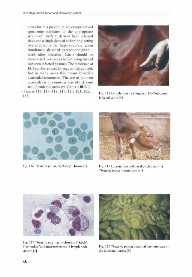
• 2 Slages in lhe blood and circulatory syslem
nent for rhi pro edure are ryopre erved porozoite tabilate of the appropriate train of Theileria derived from infected
tick and a ingle do e of eirher long-acting ytetracycline or buparvaquone given
imulraneou I or of parvaquone given I week after infe rion. aale hould be immunized -4 week bef re being turned out Onto infected pa rure. The inciden e of E F an be reduced by regular ri k control bur in many area thi mean biweekly a ari idal treatment. The u e of pur-on a ari ide i a promi ing way f ci k con-trol in ende mi area ( ATfLE . 5 .1)
(Figure 116,1 17 118,119 120 121 122 123)
Fig. 116 Theileria parva; erythrocyre forms [4J
Fig. 117 Theileria sp.; macroschizonrs (" Koch's
blue bodies"and microschizonts in Iymph node
smears [4]
68
Fig. 118 Lymph node swelling in a Theileria parva infected cattle [4]
Fig. 119 Lacrimation and nasal discharges in a
Theileria parva infected carrle [8]
Fig. 120 Theileria parva; petechial haemorrhage on the intestinal se rosa [8]
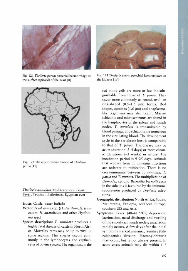
Fig. 121 Theileria parva; petechial haemorrhage on the surface (epicard) of the heart [8]
Fig. 122 The reported distribution of Theileria parva [17]
Theileria annulata Mediterranean Coast Fever, Tropical theileriosis, Egyptian ever
Hosts: Cattle, water buffalo Vector: Hyalomma spp. (H. detritum, H. trun
catum, H. anatolicum and other Hyalomma spp.)
Species description: T. annulata produces a highly fatal disease of cattle in North Africa. Mortality rates may be up to 90% in so me regions. This species oceurs commonly in the lymphocytes and erythrocytes of bovine species. The organisms in the
Fig. 123 Theileria parva; petechial haemorrhage on the kidneys [15]
red blood cells are more or less indistinguishable from those of T. parva. They occur more commonly as round, oval- or ring-shaped (0.5-1.5 flm) forms. Rod shapes, commas (1.6 flm) and anaplasmalike organisms mayaiso occur. Macroschizonts and microschizonts are found in the lymphocytes of the spleen and lymph nodes. T. annulata is transmissible by blood passage, and schizonts are numerous in the circulating blood. The development cycle in the vertebrate host is comparable to that of T. parva. The disease may be aeute (duration: 3-4 days) or more chronie (duration: 2-3 weeks) in nature. The incubation period is 9-25 days. Animals that recover from T. annulata infections are resistant to reinfection. There is no cross-immunity between T. annulata, T. parva and T. mutans. The multiplication of Demodex sp. and Besnoitia besnoiti cysts in the subcutis is favoured by the immunosuppression produeed by Theileria infections.
Geographie distribution: North Africa, Sudan, Mauretania, Ethiopia, southern Europe, southern DIS and Asia.
Symptoms: Fever (40-41.5°q, depression, laerimation, nasal discharge and swelling of the superficiallymph nodes; emaciation rapidly occurs. A few days after the initial symptoms marked anaemia, jaundice (bilirubinaemia) develop. Haemoglobinuria may oecur, but is not always present. In acute cases animals may die within 1-2
69
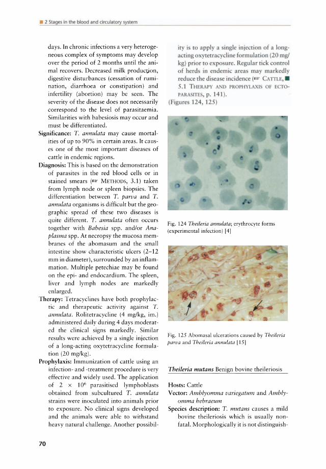
• 2 Slages in lhe blood and circulatory syslem
days. In chronic infections a very heterogeneous complex of symptoms may develop over the period of 2 months until the animal recovers. Decreased milk production, digestive disturbances (cessation of rumination, diarrhoea or constipation) and infertility (abortion) may be seen. The severity of the disease does not necessarily correspond to the level of parasitaemia. Similarities with babesiosis may occur and must be differentiated.
Significance: T. annulata may cause mortalities of up to 90% in certain areas. It causes one of the most important diseases of cattle in endemie regions.
Diagnosis: This is based on the demonstration of parasites in the red blood cells or in stained smears (1I2lf METHODS, 3.1) taken from Iymph node or spleen biopsies. The differentiation between T. parva and T. annulata organisms is difficult but the geographie spread of these two diseases is quite different. T. annulata often occurs together with Babesia spp. and/or Anaplasma spp. At necropsy the mucosa membranes of the abomasum and the small intestine show characteristic ukers (2-12 mm in diameter), surrounded by an inflammation. Multiple petechiae may be found on the epi- and endocardium. The spleen, Iiver and Iymph nodes are markedly enlarged.
Therapy: Tetracyclines have both prophylactic and therapeutic activity against T. annulata. Rolitetracycline (4 mg/kg, im.) administered daily during 4 days moderated the c1inical signs markedly. Similar results were achieved by a single injection of a long-acting oxytetracycline formulation (20 mg/kg).
Prophylaxis: Immunization of cattle using an infection- and -treatment procedure is very effective and widely used. The application of 2 x 10⁶ parasitised Iymphoblasts obtained from subcultured T. annulata strains were inoculated into animals prior to exposure. No c1inical signs developed and the animals were able to withstand heavy natural challenge. Another possibil-
70
iry i t apply a ingl injc ri n fa longa ring yr [ra y lin f rmulation (20 mg/ kg) prior t e pure. R ular ri k nrrol
f herd in endemi area may markedl redu e rhe di ca ein id n e ( TIlJ..
• 1 THfR P A D PR PHYLA I r PAR p. 141).
(Figurc 124 12 )
Fig. 124 Theileria annulata; erythrocyte forms (experimental infection) [4]
Fig. 125 Abomasal ulcerations caused by Theileria parva and Theileria annulata [15]
Theileria mutans Benign bovine theileriosis
Hosts: Cattle Vector: Amblyomma variegatum and Ambly
omma hebraeum Species description: T. mutans causes a mild
bovine theileriosis which is usually nonfatal. Morphologically it is not distinguish-

able from the pathogenic T. parva and T. annulata. The forms in the erythrocytes are round, oval, piriform, comma-shaped or Anaplasma-like. The round forms are 1-2 pm in diameter and the oval forms measure 1.5 x 0.6 pm. Binary fission occurs in the erythrocytes. The incubation period is 10-20 days.
Geographie distribution: Throughout Africa, Madagascar, Reunion, Mauritius, southern Europe, Asia, Australia, UIS, Caribbean
Symptoms: Similar to a mild form of T. annulata infection. Anaemia is the major clinical sign. kterus and lymph node swellings may occur.
Signifieanee: T. mutans is usually only slightlyor non-pathogenic for indigenous cattle in endemic areas. An aeute form may develop in cattle imported into an endemic area. The mortality is generally less than 1 %.
Diagnosis: Same as for other Theileria species Therapy: Unknown; the same drugs as for the
pathogenic Theileria species may be used. Prophylaxis: Tick control and immunisation
as for the pathogenic Theileria species. (Figures 126, 127, 128)
Fig. 126 Theileria mutans; erythrocyte forms [4]
Fig. 127 Theileria mutans; macroschizont (rarely encountered) [15]
Fig. 128 Theileria mutans; microschizonts (rarely encountered) [151
Theileria velifera
Remarks: This is another mild Theileria speeies of cattle in Africa and in the Caribbean. Its distribution coineides with that of the vector Amblyomma variegatum (Africa and Madagascar) and Amblyomma lepidum and Amblyomma hebraeum elsewhere. The parasites occur as pleomorphic or rodshaped bodies in the erythrocytes and presumably lymphocytes. Most are 1-2 pm long. The great majority have a rectangular "veil" 1.0-3.5 pm long, extending out from the side. This speeies is nonpathogenic.
(Figure 129)
71
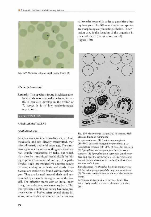
• 2 Slages in lhe blood and circulatory syslem
Fig. 129 Theileria velifera; erythrocyte forms 14]
Theileria taurotragi
Remarks: This species is found in African antelopes and can occasionally be found in cattle. lt can also develop in the vector of T. parva. It is of low epidemiological importance.
RICKElTSJAI.ES
ANAPLASMAT ACEAE
Anaplasma spp.
Anaplasmoses are infectious diseases, virulent, inoculable and not directly transmitted, that affect domestic and wild ungulates. The causative agent is a Rickettsia of the genus Anaplasma, usually transmitted by ticks, but which mayaIso be transmitted mechanically by biting Diptera (Tabanidae, Stomoxys). The pathological signs are progressive anaemia (acute or slow) ending in cachexia and death. Anaplasma are exclusively found within erythrocytes. They are located intracellularly and surrounded by a vacuolar invagination of the host cel\. The infection starts with an initial body that grows to become an elementary body. This multiplies by doubling or binary fission to produce new initial bodies. After several binary fissions, initial bodies accumulate in the vacuole
72
to leave the host cell in order to parasitize other erythrocytes. The different Anaplasma species are morphologically indistinguishable. The criterion used is the location of the organism in the erythrocyte (marginal or central). (Figure 130)
5
Fig. 130 Morphology (schematic) of various Rickettsiales found in ruminants; Anaplasmataceae: (1) Anaplasma marginale (80-90% parasites marginal or peripheral); (2) Anaplasma centrale (80-90% of parasites central); (3) Eperythrozoon wenyoni, (on the erythrocyte surface); (4) Eperythrozoon teganodes (on the su rface and near the erythrocyte); (5) Eperythrozoon tuomii (on the thrombocyte surface) and (6) Haemobartonella bovis; Ehrlichiaceae: (7) Ehrlichia bovis (in monocytes); (8) Ehrlichia phagocytophila (in granulocyte) and (9) Cowdria ruminantium (in the vascular endothelium); development stages: A = elementary body, B = initial body amd C = mass of elementary bodies [16]
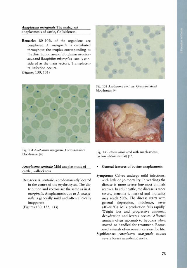
Anaplasma marginale The malignant anaplasmosis of cattle, Gallsickness
Remarks: 80-90% of the organisms are peripheral. A. marginale is distributed throughout the tropics corresponding to the distribution area of Boophilus decoloratus and Boophilus microplus usually considered as the main vectors. Transplacental infection occurs.
(Figures 130, 131)
Fig. 131 Anaplasma marginale; Giemsa-stained bloodsmear [4]
Anaplasma centrale Mild anaplasmosis of cattle, Gallsickness
Remarks: A. centrale is predominantly located in the centre of the erythrocytes. The·distribution and vectors are the same as in A. marginale. Anaplasmosis due to A. marginale is generally mild and often clinically inapparent.
(Figures 130, 132, 133)
Fig. 132 Anaplasma centrale; Giemsa-stained bloodsmear [4]
Fig. 133 Icterus associated with anaplasmosis (yellow abdominal fat) [15]
• General features of bovine anaplasmosis
Symptoms: Calves undergo mild infections, with little or 1).0 mortality. In yearlings the disease is more severe but" most animals recover: In adult cattle, the disease is more severe, aooemia is marked and mortality may reach 50%. The disease starts with general depression, indolence, fever (40-41°C). Milk production falls rapidly. Weight loss and progressive anaemia, dehydration and icterus occurs. Affected animals often succumb to hypoxia when moved or handled for treatment. Recovered animals often remain carriers for life.
Significance: Anaplasma marginale causes severe losses in endemie areas.
73
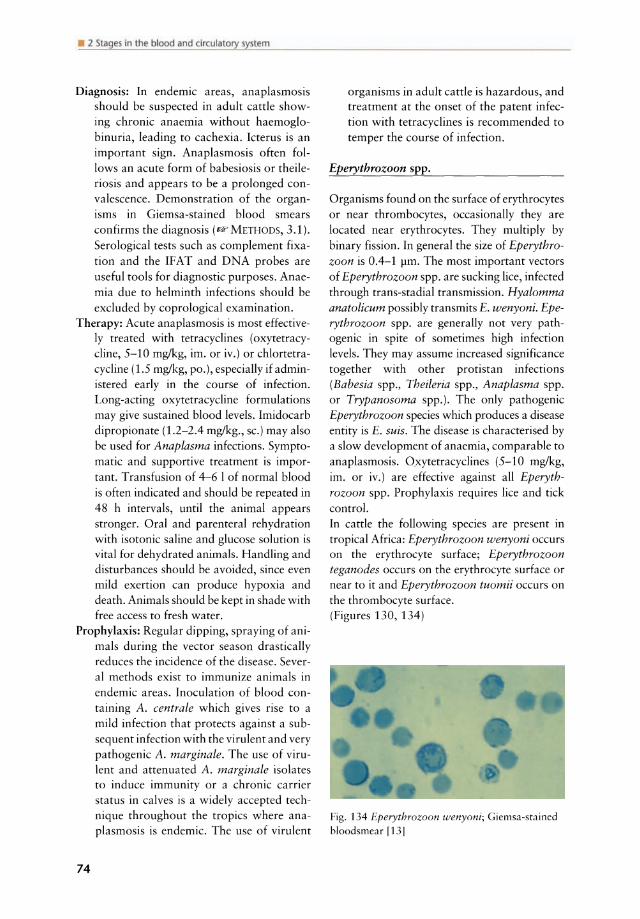
• 2 Slages in lhe blood and circulatory syslem
Diagnosis: In endemie areas, anaplasmosis should be suspected in adult cattle showing chronic anaemia without haemoglobinuria, leading to cachexia. kterus is an important sign. Anaplasmosis often follows an acute form of babesiosis or theileriosis and appears to be a prolonged convalescence. Demonstration of the organisms in Giemsa-stained blood smears confirms the diagnosis (Il? METHODS, 3.1). Serological tests such as complement fixation and the IFAT and DNA probes are useful tools for diagnostic purposes. Anaemia due to helminth infections should be excluded by coprological examination.
Therapy: Acute anaplasmosis is most effectiveIy treated with tetracyclines (oxytetracyc1ine, 5-10 mglkg, im. or iv.) or chlortetracycline (1.5 mglkg, po.), especially if administered early in the course of infection. Long-acting oxytetracycline formulations may give sustained blood levels. Imidocarb dipropionate (1.2-2.4 mglkg., sc.) mayaIso be used for Anaplasma infections. Symptomatic and supportive treatment is important. Transfusion of 4-6 I of normal blood is often indicated and should be repeated in 48 h intervals, until the anima I appears stronger. Oral and parenteral rehydration with isotonic saline and glucose solution is vital for dehydrated animals. Handling and disturbances should be avoided, since even mild exertion can produce hypoxia and death. Animals should be kept in shade with free access to fresh wa ter.
Prophylaxis: Regular dipping, spraying of an imals during the vector season drastically reduces the incidence of the disease. Several methods exist to immunize animals in endemie areas. Inoculation of blood containing A. centrale which gives rise to a mild infection that protects against a subsequent infection with the virulent and very pathogenic A. marginale. The use of virulent and attenuated A. marginale isolates to induce immunity or a chronic carrier status in calves is a widely accepted technique throughollt the tropics where anaplasmosis is endemie. The use of virulent
74
organisms in adult cattle is hazardous, and treatment at the on set of the patent infection with tetracyclines is recommended to temper the course of infection.
Eperythrozoon spp.
Organisms found on the surface of erythrocytes or near thrombocytes, occasionally they are located near erythrocytes. They multiply by binary fission. In general the size of Eperythrozoon is 0.4-1 pm. The most important vectors of Eperythrozoon spp. are sucking Iice, infected through trans-stadial transmission. Hyalomma anatolicum possibly transmits E. wenyoni. Eperythrozoon spp. are generally not very pathogenic in spite of sometimes high infection levels. They may assume increased significance together with other protistan infections (Babesia spp., Theileria spp., Anaplasma spp. or Trypanosoma spp.). The only pathogenic Eperythrozoon species which produces a disease entity is E. suis. The disease is characterised by a slow development of anaemia, comparable to anaplasmosis. üxytetracyclines (5-10 mglkg, im. or iv.) are effective against all Eperythrozoon spp. Prophylaxis requires lice and tick control. In cattle the following species are present in tropical Africa: Eperythrozoon wenyoni occurs on the erythrocyte surface; Eperythrozoon teganodes occurs on the erythrocyte surface or near to it and Eperythrozoon tuomii occurs on the thrombocyte surface. (Figures 130, 134)
Fig. 134 Eperythrozoon wenyoni; Giemsa-stained bloodsmear f13]

Haemobartonella spp.
These are organisms found on the surface of erythrocytes which form branched chains and multiply by binary fission. The organisms are coccoid (0.1-1 pm) or bacilliform (1-5 x 0.7-1.5 pm). They cause a defined disease entity in dogs and cats and rats. Haemobartonella spp. are often detected as resurgent parasites during concomitant infections or following splenectomy. Tetracyclines are the drugs of choice for treating Haemobartonella infections, but therapy is rarely indicated (IIW also above ANPLASMATACAE, Fig. 130) .
Haemobartonella bovis
Remarks: Ir occurs on the erythrocyte surface. These coccoid or bacilliform organisms occur on the erythrocyte surface of cattle in Mediterranean regions. Vectors are sucking lice and ticks (species unknown). Haemobartonella bovis is relatively nonpathogenic. In rare cases the parasite may be associated with slow anaemia and symptoms similar to anaplasmosis. Haemobartonella spp. and Eperythrozoon spp. are difficult to distinguish reliably by light microscopy (lIWabove ANPLASMATACAE, Fig. 130).
EHRLICHIACEAE
Ehrlichia bovis T ropical bovine ehrlichiosis, "Nofel" or "Nopel"
Hosts: Cattle Vector: Ticks of the genera Amblyomma, Hya
lomma and Rhipicephalus Species description: Ehrlichia bovis occurs in
mononuclear cells of cattle. The syndrome corresponds to the disease ca lied "Nofel" (Fulani/Peule in West Africa; ear = nopi in peul) or "Nopel" (Bororos in Central Africa), because of the ears being kept low at the beginning of an acute infection. The clinical signs vary greatly ranging from inapparent forms to serious fatal forms, dominated by nervous signs, ataxie gait,
stiff legs, walking in circles, phases of excitement followed by somnolence, epileptic crisis with falls, rolling eyes and salivation. Death may occur in an epileptic attack. The disease is always serious in imported cattle. The nervous signs are similar to those of cowdriosis.
Geographie distribution: North and Central Afriea, Middle East, Ceylon
Symptoms: Anorexia, fever, incoordination and enlargement of lymph nodes
Signifieanee: Ehrlichia bovis is of low signifieanee for indigenous cattle. However, it is a serious problem for imported or newly introduced cattle in many tropical areas.
Diagnosis: Microscopic examination of peripheral blood smears demonstrates E. bovis in monocytes or macrophages. For differential dia gnosis infections with Cowdria ruminantium have to be excluded. C. ruminantium is located exclusively in the endothelial cells (brain and other tissues) and not in the peripheral blood leucocytes, whereas E. bovis is found in monocytes and macrophages.
Therapy: Oxytetracyclines (5 mg/kg, im., for 4 days) or streptomycin-penicillin (3 g and 3 mio. units per day, im.); other tetracyclines may be used in emergencies.
Prophylaxis: This consists of regular tick control. There are no immunization procedures described.
(Figures 130, 135)
Fig. 135 Ehrlichia bovis; mass of elementary bodies in a monocyte [15)
75

• 2 Stages in the blood and circulatory system
Ehrlichia phagocytophila European ehrlichiosis
Remarks: E. phagocytophila occurs in the neutrophilic and eosinophilic granulocytes of cattle, sheep and other domestic and wild ruminants in Europe. Goats are not very susceptible. Ixodes ricinus transmits the disease by trans-stadial infection. The disease is characterized by fever which occurs after an incubation period of 4-11 days. The infection causes astate of immunosuppression and is therefore a predisposing factor to a variety of secondary infections. The animals usually recover after this febrile phase. However, the most characteristic problems are abortion in pregnant ewes and cattle, and progressive weight loss following infection may occur. At necropsy hypertrophy of the spleen and lymph nodes may be seen. The diagnosis is made by demonstrating the organisms in stained blood smears (Q" METHODS, 3.1). Serological tests such as IFAT (- METHODS, 5.1) may be used for epidemiological studies.
Therapy: Long acting oxytetracyclines (5 mg/kg, im.) have a certain curative effect.
Prophylaxis: Q" above Ehrlichia bovis (Figures 130, 136)
Fig. 136 Ehrlichia phagocytophila; mass of elementary bodies in a granulocyte [4J
Ehrlichia ondiri
Remarks: This parasite occurs in granulocytes of cattle in high-altitude grassland regions of East Africa. Members of the genus Hae-
76
maphysalis and other genera are suspected to be vectors of the disease. The incubation period is 1-2 weeks. The disease starts with a fever attack which is accompanied by a petechial exanthema of the mucosae of the mouth, nose, vulva, eyes (poached egg eye), digestive tract, urinary bladder, pericardium and endocardium. Extended haemorrhage in the gastrointestinal tract and the pulmonary oedema cause death. Diagnosis is made by microscopic examination of blood smears. Organisms of E. ondiri are found in granulocytes.
Therapy and Prophylaxis: Q" above Ehrlichia bovis
Cowdria ruminantium Heartwater, Tyewde
Hosts: Cattle, sheep, goat and other ruminants Vector: Several species of the tick genus
Amblyomma transmit the disease: Amblyomma variegatum in open savannas from equatorial Africa to Madagascar; Amblyomma gemma in East Africa (Massai and Somalian steppes); Amblyomma pomposum has limited geographic distribution and occurs in the Angolan plateau, Zambia, southwestern Tanzania; Amblyomma lepidum in East Africa (Massai steppes, Sahelian steppes of the Nile in Sudan) and Amblyomma tholloni in the Zambezi region. C. ruminantium is also present on the Caribbean islands.
Species description: Cowdriosis is specific to ruminants of the family Bovidae and is most severe in sheep and goats but also causes great losses in cattle. Imported ruminants are much more susceptible to cowdriosis than indigenous breeds. They may suffer heavy losses. But also indigenous ruminants are affected especially when they are in poor general condition (endoparasitism, malnutrition, chronic infections, etc.) or with weakened defence (lactation, gestation). This applies especially to the early rainy season in the SahelSudanese region when animals are weakened by the long food shortage of the dry season and when there is a drastic
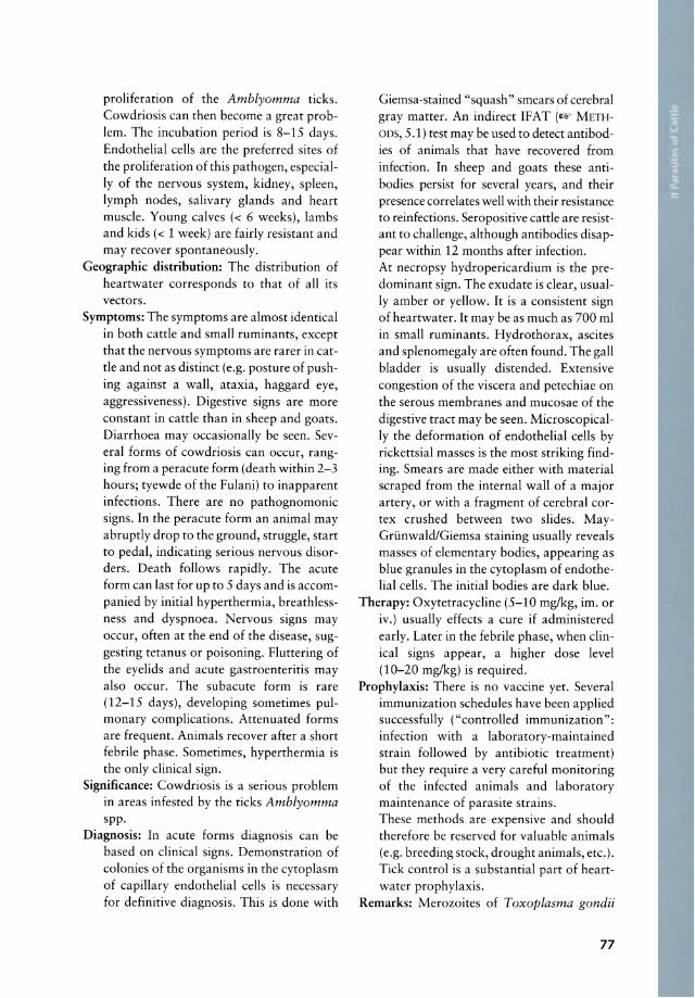
proliferation of the Amblyomma ticks. Cowdriosis can then become a great problem. The incubation period is 8-15 days. Endothelial cells are the preferred sites of the proliferation of this pathogen, especialIy of the nervous system, kidney, spleen, Iymph nodes, salivary glands and heart muscle. Young calves « 6 weeks), lambs and kids « 1 week) are fairly resistant and may recover spontaneously.
Geographie distribution: The distribution of heartwater corresponds to that of all its vectors.
Symptoms: The symptoms are almost identical in both catde and small ruminants, except that the nervous symptoms are rarer in catde and not as distinct (e.g. posture of pushing against a wall, ataxia, haggard eye, aggressiveness). Digestive signs are more constant in catde than in sheep and goats. Diarrhoea may occasionally be seen. Several forms of cowdriosis can occur, ranging from a peracute form (death within 2-3 hours; tyewde of the Fulani) to inapparent infections. There are no pathognomonic signs. In the peracute form an anima I may abrupdy drop to the ground, struggle, start to pedal, indicating serious nervous disorders. Death follows rapidly. The acute form can last for up to 5 days and is accompanied by initial hyperthermia, breathlessness and dyspnoea. Nervous signs may occur, often at the end of the disease, suggesting tetanus or poisoning. Fluttering of the eyelids and acute gastroenteritis may also occur. The subacute form is rare (12-15 days), developing sometimes pulmonary complications. Attenuated forms are frequent. Animals recover after a short febrile phase. Sometimes, hyperthermia is the only clinical sign.
Significance: Cowdriosis is a serious problem in areas infested by the ticks Amblyomma spp.
Diagnosis: In acute forms diagnosis can be based on clinical signs. Demonstration of colonies of the organisms in the cytoplasm of ca pillary endothelial cells is necessary for definitive diagnosis. This is done with
Giemsa-stained "squash" smears of cerebra I gray matter. An indirect IFAT (1& METHOD5, 5.1) test may be used to detect antibodies of animals that have recovered from infection. In sheep and goats these antibodies persist for several years, and their presence correlates weil with their resistance to reinfeetions. Seropositive catde are resistant to challenge, although antibodies disappear within 12 months after infection. At necropsy hydropericardium is the predominant sign. The exudate is clear, usualIy amber or yellow. It is a consistent sign of heartwater. It may be as much as 700 ml in sm all ruminants. Hydrothorax, ascites and splenomegaly are often found. The gall bladder is usually distended. Extensive congestion of the viscera and petechiae on the serous membranes and mucosae of the digestive tract may be seen. MicroscopicalIy the deformation of endothelial cells by rickettsial masses is the most striking finding. Smears are made either with material scraped from the internal wall of a major artery, or with a fragment of cerebra I cortex crushed between two slides. MayGrünwald/Giemsa staining usually reveals masses of elementary bodies, appearing as blue granules in the cytoplasm of endothelial cells. The initial bodies are dark blue.
Therapy: Oxytetracycline (5-10 mg/kg, im. or iv.) usually effects a eure if administered early. Later in the febrile phase, when clinical signs appear, a higher dose level (10-20 mg/kg) is required.
Prophylaxis: There is no vaccine yet. Several immunization schedules have been applied successfully ("controlled immunization": infection with a laboratory-maintained strain followed by antibiotic treatment) but they require a very careful monitoring of the infected animals and laboratory maintenance of parasite strains. These methods are expensive and should therefore be reserved for valuable animals (e.g. breeding stock, drought animals, etc.). Tick control is a substantial part of heartwater prophylaxis.
Remarks: Merozoites of Toxoplasma gondii
77
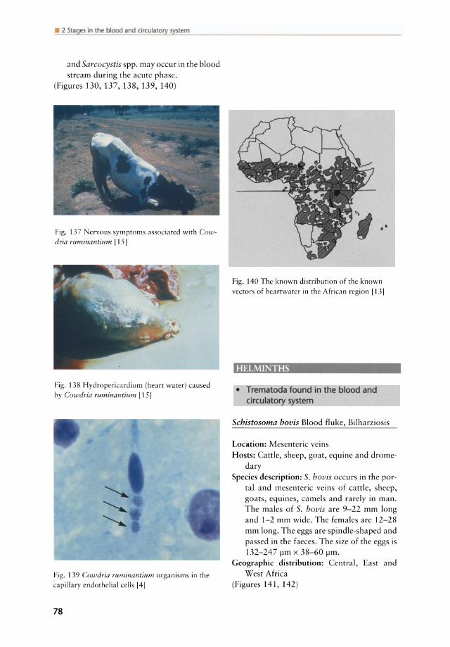
• 2 Slages in lhe blood and circulatory syslem
and Sarcocystis spp. may oeeur in the blood stream during the aeute phase.
(Figures 130, 137, 138, 139, 140)
Fig. 137 Nervous symptoms associated with Cowdria ruminantium [15 J
Fig. 138 Hydropericardium (hearr water) caused by Cowdria ruminantium [15]
Fig. 139 Cowdria ruminantium organisms in the capillary endothelial cells 141
78
Fig. 140 The known distribution of the known
vectors of hea rtwater in the African region [131
HELMINTHS
• Trematoda found in the blood and circulatory system
Schistosoma bovis Blood fluke, Bilharziosis
Loeation: Mesenterie veins Hosts: Cattle, sheep, goat, equine and drome
dary Speeies description: S. bovis oeeurs in the por
tal and mesenterie veins of eattle, sheep, goats, equines, eamels and rarely in man. The males of S. bovis are 9-22 mm long and 1-2 mm wide. The females are 12-28 mm long. The eggs are spindle-shaped and passed in the faeees. The size of the eggs is 132-247 !-Im x 38-60 !-Im.
Geographie distribution: Central, East and West Afriea
(Figures 141, 142)

Fig. 141 Schistosomabovis
Fig. 142 Schistosoma bovis; male (thick) and female (slender)
Schistosoma mattheei
Loeation: Portal and mesenterie veins and less frequently eystie and lung veins
Hosts: Cattle, sheep, goat, wild ruminants, equine and man
Speeies deseription: Females measure 18-25 mm and males 9-14 mm. The eggs measure 170-280 x 70-85 pm and have a terminal spine. The prepatent period is 7 weeks. The life eyde is similar to Schistosoma bovis. Intermediate hosts are Bulinus africanus, Bulinus globosus and other Bulinus spp.
Geographie distribution: Southern Afriea, Mozambique, Zambia, Malawi, Zimbabwe, Tanzania; Central Afriea (Tehad)
Schistosoma intercalatum
Remarks: This speeies oeeurs mainly in man and primates in Central Afriea. Sheep and goats and other domestie and wild ruminants may aeeidentally aequire infeetion. S. intercalatum is found in the portal and mesenterie vems. The eggs measure 140-240 pm x 50-85 pm.
Schistosoma curassoni
Loeation: Portal and mesenterie veins Hosts: Cattle, sheep and goat Speeies deseription: Intermediate hosts are
snails of the genus Bulinus. Eggs are similar to those of S. mattheei.
Geographie distribution: West Afriea (Senegal, Mauretania, Mali)
• General features of Schistosoma spp. infeetions
Life eyde: Aquatie snails (Bulinus spp.) aet as intermediate hosts in the life eyde of Schistosoma spp. In the ease of S. bovis, S. mattheei and S. intercalatum snails of the genus Bulinus spp. aet as intermediate hosts. The eggs hateh after eontaet with water. The miraeidia infeet aquatie snails and eereariae emerge 38-126 days after infeetion from the snail and actively penetrate the skin or the rumen of the definite host. Sehistosomosis (bilharziosis) oecurs near permanent fresh water bodies (pools, baekwaters, ponds, lakes and marshy pastures). To become infected, the final host needs the eontact with water. This might oeeure during drinking in these areas, passing through water or grazing in swamps.
Symptoms: These are rarely seen even in heavy infeetions. Symptoms are mainly caused by the eggs which are deposited by the adult flukes in the wall of the intestines. Intermittent diarrhoea which may contain specks of blood, anaemia and progressive weakness may occur. Both diarrhoea and constipation can be seen and blood and mucus are sometimes found in the faeces together
79
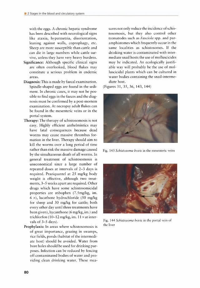
• 2 Slages in lhe blood and circulatory sySlem
with the eggs. A chronic hepatic syndrome has been described with neurological signs like ataxia, hypermetria, disorientation, leaning against walls, coprophagy, etc. Sheep are more susceptible than cattle and can die in large numbers while cattle survive, unless they have very heavy burdens.
Significance: Although specific clinical signs are often overlooked, blood flukes may constitute a serious problem in endemic areas.
Diagnosis: This is made by faecal examination. Spindle-shaped eggs are found in the sediment. In chronic ca ses, it may not be possible to find eggs in the faeces and the diagnosis must be confirmed by a post-mortem examination. At necropsy adult flukes can be found in the mesenteric veins or in the portal system.
Therapy: The therapy of schistosomosis is not easy. Highly efficient anthelmintics may have fatal consequences because dead worms may cause massive thrombus formation in the liver. Therapy should aim to kill the worms over a long period of time rather than risk the massive damage caused by the simultaneous death of all worms. In general treatment of schistosomosis is uneconomical since a large number of repeated doses at intervals of 2-3 days is required. Praziquantel at 25 mg/kg body weight is effective, although two treatments, 3-5 weeks apart are required. Other drugs wh ich have some schistosomicidal properties are stibophen (7.5mglkg, im. 6 x), lucathone hydrochloride (50 mg/kg for sheep and 30 mglkg for cattle; both every other day untit three treatments have been given), hycanthone (6 mglkg, im.) and trichlorfon (10-12 mg/kg, im. 11 x at intervals of 3-5 days).
Prophylaxis: In areas where schistosomosis is of great importance, grazing in swamps, rice fjelds, ponds (habitat of the intermediate host) should be avoided. Water from bore holes should be used for drin king purposes. Infection can be reduced by fencing off contaminated bodies of water and provi ding clean drinking water. These mea-
80
sures not only reduce the incidence of schistosomosis, but they also control other trematodes such as Fasciola spp. and paramphistomes which frequently occur in the same localities as schistosomes. If the drinking water is contaminated with intermediate snail hosts the use of molluscicides may be indicated. An ecologically justifiable way will probably be the use of molluscicidal plants which can be cultured in water bodies containing the snail-intermediate host.
(Figures 31, 35, 36, 143, 144)
Fig. 143 Schistosoma bovis in the mesenteric veins
Fig. 144 Schistosoma bovis in the portal vein of the liver
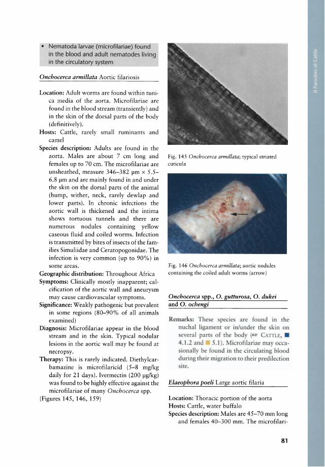
• Nematoda larvae (microfilariae) found in the blood and adult nematodes living in the circulatory system
Onchocerca armillata Aortic filariosis
Location: Adult worms are found within tunica media of the aorta. Microfilariae are found in the blood stream (transiently) and in the skin of the dorsal parts of the body (definitively) .
Hosts: Cattle, rarely small ruminants and came!
Species description: Adults are found in the aorta. Males are about 7 cm long and females up to 70 cm. The microfilariae are unsheathed, measure 346-382 rm x 5.5-6.8 rm and are mainly found in and under the skin on the dorsal parts of the animal (hump, wither, neck, rarely dewlap and lower parts). In chronic infections the aortic wall is thickened and the intima shows tortuous tunnels and there are numerous nodules containing yellow caseous fluid and coiled worms. Infection is transmitted by bites of insects of the families Simuliidae and Ceratopogonidae. The infection is very common (up to 90%) in some areas.
Geographie distribution: Throughout Africa Symptoms: Clinically mostly inapparent; cal
cification of the aortic wall and aneurysm may cause cardiovascular symptoms.
Significanee: Weakly pathogenic but prevalent in some regions (80-90% of all animals examined)
Diagnosis: Microfilariae appear in the blood stream and in the skin. Typical nodular lesions in the aortic wall may be found at necropsy.
Therapy: This is rare!y indieated. Diethylcarbamazine is microfilaricid (5-8 mg/kg daily for 21 days). Ivermectin (200 rglkg) was found to be highly effective against the microfilariae of many Onchocerca spp.
(Figures 145, 146, 159)
Fig. 145 Onchocerca armillata; typical striated cuticula
Fig. 146 Onchocerca armillata; aortic nodules containing the coiled adult worms (arrow)
Onchocerca spp., O. gutturosa, O. dukei and O. ochengi
Remark: The e peeie are found in the nuchal ligament or in/under the kin on everal part of the body ( CAlTLE, .
4.1.2 and 5.1 ). licrofilariae may QccaionalJy be found in the circulating blood
during their migration to their predilecrion ire.
Elaeophora poeli Large aortic filaria
Location: Thoracic portion of the aorta Hosts: Cattle, water buffalo Species description: Males are 45-70 mm long
and females 40-300 mm. The microfilari-
81
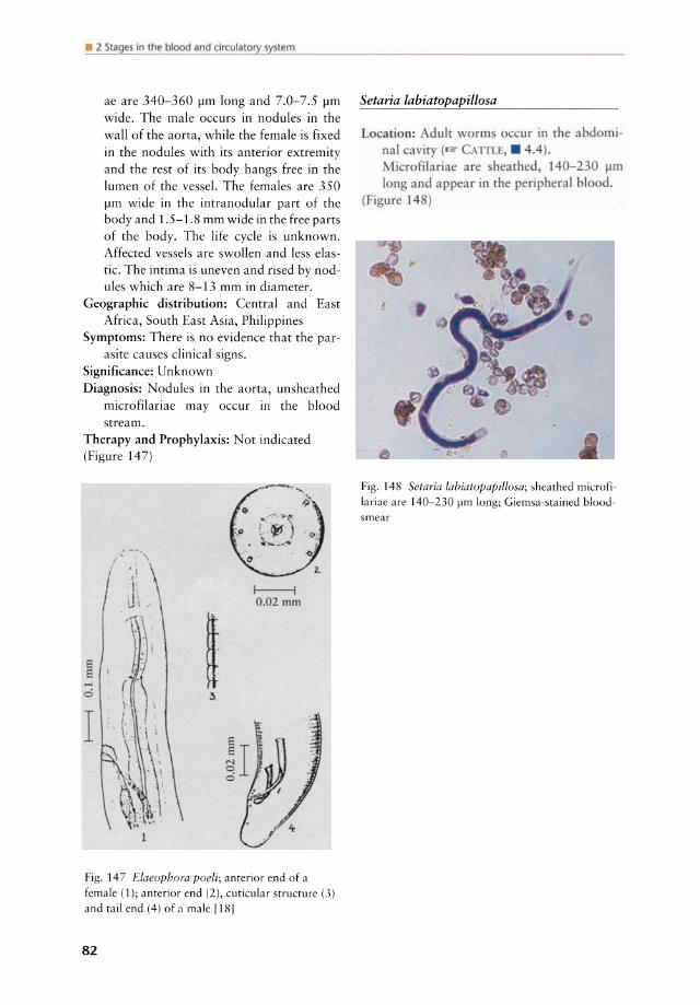
• 2 Slages in lhe blood and circulatory syslem
ae are 340-360 J.lm long and 7.0-7.5 J.lm wide. The male oeeurs in nodules in the wall of the aorta, while the female is fixed in the nodules with its anterior extremity and the rest of its body hangs free in the lumen of the vessel. The females are 350 J.lm wide in the intranodular part of the body and 1.5-1.8 mm wide in the free parts of the body. The life eyele is unknown. Affeeted vessels are swollen and less elastie. The intima is uneven and rised by nodules whieh are 8-13 mm in diameter.
Geographie distribution: Central and East Afriea, 50uth East Asia, Philippines
Symptoms: There is no evidenee that the parasite eauses elinieal signs.
Signifieanee: Unknown Diagnosis: Nodules in the aorta, unsheathed
mierofilariae may oeeur in the blood stream.
Therapy and Prophylaxis: Not indieated (Figure 147)
I------i 0.02 mm
Fig. 147 Elaeophora poeli; anterior end of a
female (1); anterior end (2), cuticular structure (3)
and tail end (4) of a male [18]
82
Setaria labiatopapillosa
Locatioo: Adult worm occur in the abdominal aviry ( CAlTl, . 4.4).
icrofilariae are hearhed, 140-230 J.lm long and appear in rhe peripheral blood.
igl.lre 148)
Fig. 148 Setaria labiatopapil/osa; sheathed microfi
lariae are 140-230 ~m long; Giemsa-stained blood
smear

3 Stages in lhe urogenital system
PROTOZOA
Tritrichomonas foetus (syn. Trichomonas foetus) Bovine trichomonad abortion
Location: In the bult the parsites are found in the preputial cavity. In cows trichomonads are found in the vagina and uterus.
Hosts: Cattle Species description: This is a venereal proto
zoal disease of cattle characterized by early foetal death and infertility, associated with greatly extended calving intervals. Ir is reported from many tropical countries. The organism is ordinarily 10-15 x 5-10 flm, but there is considerable pleomorphism. At the anterior end there are three flagelta about the same size as the parasite. An undulating membrane runs the full length of the body and is bordered by a filament wh ich continues beyond the membrane as a posterior flagellum. Multiplication is by longitudinal binary fission. Under natural conditions the infection is transmitted du ring coitus from the bull to the cow or vice versa. It mayaiso be transmitted mechanically, e.g. by gynaecological examination of cows with contaminated tools. The organisms are found only in the genital tract of the cow and bull. Most bulls remain permanently infected after a primary infection. The bull is usually the source of initial and continuous infection within a herd. Infected cows usually recover spontaneously after mean durations of 20 weeks (primary exposure) and 10 weeks (secondary exposure). Cows can remain infected throughout pregnancy and discharge trichomonads from the genital tract following calving.
Geographie distribution: World-wide Symptoms: Purulent vaginitis, metritis, placen
titis and abortion are commonly found in infected cows and can cause enormous losses. Prolonged sterility and extended calving intervals are predominant features
(cave: differential diagnosis: Brucella abortus!). There is a persistent uterine discharge containing large numbers of the parasites. Bulls are usually carrier of the parasite without c1inical symptoms.
Significance: Venereal disease of cattle characterised by abortion and infertility in cows. T. (oetus can cause considerable losses in infected herds. In many countries the incidence has greatly been reduced by the artificial insemination using a bull known to be free of the disease.
Diagnosis: History of abortions in a herd and demonstration of the organism in the vaginal or uterine discharges or in the foetus (mainly stomaeh) . In bulls the organisms are in the prepuce, frequently in small numbers. Microscopical examination of the preputial smegma for trichomonads is the most common method to confirm a herd diagnosis. Preputial fluid may be collected with a pipette (e.g. AI pipette) which is introduced into the prepuce, and smegma is collected with a combination of scraping and aspiring via an attached rubber bulb or syringe. The same procedure may be used to collect vaginal fluid. lt is important to avoid contamination with faecal material, as such material contains masses of protozoa similar in appearance to T. foetus. The organisms may not be numerous and careful, systematic examination is necessary. Diagnosis is based on the size and shape of the organisms as weil as the characteristic aimless, jerky motion on a warmed slide. Only living organisms are useful for diagnostic purposes. Confirmation depends on finding the organisms in at least one animal in a suspected herd. The organisms may be cultured in special media (e.g. Diamond's Trichomonad Medium). The diagnosis can be confirmed by the Polymerase Chain Reaction (PCR) which is the most sensitive and specific diagnostic method nowadays. Serological examinations on a herd level may be used for epidemiological purposes but are of little value for individual animals.
83
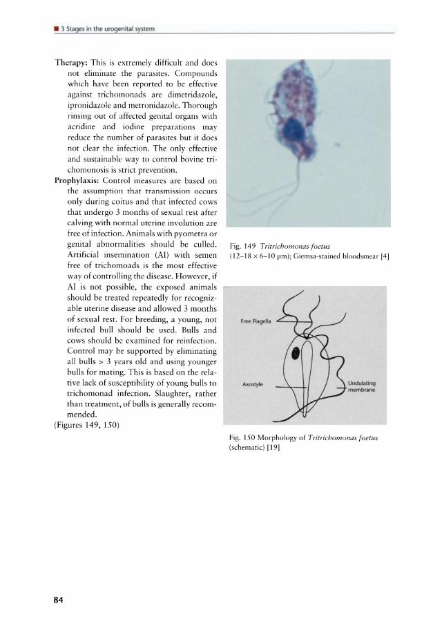
urogenital syslem
Therapy: This is extremely difficult and does not eliminate the parasites. Compounds which have been reported to be effective against trichomonads are dimetridazole, ipronidazole and metronidazole. Thorough rinsing out of affected genital organs with acridine and iodine preparations may reduce the number of parasites but it does not clear the infection. The only effective and sustainable way to control bovine trichomonosis is strict prevention.
Prophylaxis: Control measures are based on the assumption that transmission occurs only during coitus and that infected cows that undergo 3 months of sexual rest after calving with normal uterine involution are free of infection. Animals with pyometra or genital abnormalities should be cu lied. Artificial insemination (AI) with semen free of trichomoads is the most effective way of controlling the disease. However, if AI is not possible, the exposed animals should be treated repeatedly for recognizable uterine disease and allowed 3 months of sexual rest. For breeding, a young, not infected bul! should be used. Bul!s and cows should be examined for reinfection. Control may be supported by eliminating all bulls > 3 years old and using younger bulls for mating. This is based on the relative lack of susceptibility of young bulls to trichomonad infection. Slaughter, rather than treatment, of bulls is generally recommended.
(Figures 149, 150)
84
Fig. 149 Tritrichomonas foetus (12-18 x 6-10 pm); Giemsa-stained bloodsmea r 141
Free Flagella
Undulating membraM
Fig. 150 Morphology of Tritrichomonas foetus (schematic) 1191

4 Stages in interna i organs
4.1 locomoto system 4.6 Central nervous system
4.1. 7 Muse/es PROTOZOA ...................................... 103
PROTOZOA ........................................ 85 HElMINTHS • Cestoda cysts found in the central
HELMINTHS nervous system ................................ 1 04 • Cestoda larvae found in the muscles .. 88 • Nematoda larvae and adult
nematodes found in the muscles ........ 89 4.1 locomotory system
ARTHROPODS 4.1.7 Muscles • Insecta larvae found in the muscles .... 90
4.7 .2 Tendons
HELMINTHS • Nematoda larvae and adult nematodes
found in the tendons .......................... 90
4.2liver
HELMINTHS • Trematoda found in the liver .............. 90 • Cestoda found in the liver .................. 94
HELMINTHS • Cestoda found in the lungs and
trachea ............................................... 97 • Nematoda found in the lungs and
trachea ............................................... 97
4.4 Abdominal ca vi
HElMINTHS • Cestoda found in the abdominal
cavity ............................................... 101 • Nematoda found in the abdominal
cavity ............................................... 101
4.5 Pancreas
HElMINTHS • Trematoda found in the ancreas ..... 102
PROTOZOA
Sarcocystis bovicanis (syn. S. cruzi), Sarcocystis bovifelis (syn. S. hirsuta) and Sarcocystis bovihominis (syn. S. hominis) Bovine 5arcosporidiosis
Location: Cysts, containing the infective stage, ca lied bradyzoites, occur in the striated musculature of cattle.
Hosts: Cattle are intermediate hosts of three Sarcocystis species of which the dog (S. bovicanis), the cat (S. bovifelis) or man (5 . bovihominis) are final hosts.
Species description: Cysts of several Sarcocystis species are found in the museIes of cattle. Cattle always act as intermediate hosts. S. bovicanis is the most pathogenic species for cattle. Infection is acquired orally by ingestion of sporocysts from the faeces of infected hosts. Tissue cysts in cattle are infectious for at least 1 yeaL The life cyele of Sarcocystis spp. is as folIows: infective sporocysts, excreted by the final host (cat, dog, man) are ingested, sporozoites are released in the intestine and invade many tissues via blood stream. Schizogony occurs in the endothelial cells of blood vessels in most organs, preceding the development of typical cysts in the striated musculature. Cysts may occasionally also be found in the brain. Cysts vary in size, depending on the species, from a few mil-
85

• 1 Stages in internaiorgans
Table 10 Sarcocystis species found in muscles of cattle
Species Synonym Definite host Pathogenicity IH* DH**
5. boviconis
5. bovifelis
5. cruzi
5. hirsuto
5. hominis
Dog, coyote, wolf Cat
+++
o o
o o
5. bovihominis Man, rhesus monkey,
baboon, chimpanzee +
*IH = Intermediate Host; **DH = Definitive Host; 0 = non-pathogenic; + = mildly pathogenic; +++ = severely pathogenic
Iimetres to several centimetres in length. The peripheral region of a cyst contains globular forms (metrocytes), which produce two daughter cells by endodyogeny and these after further replication give rise to banana-shaped bradyzoites. Infection of the final host is by ingestion of muscle cysts, containing bradyzoites. S. bovifelis is nonpathogenic for cattle and produces microscopic cysts of 12 x 1 ~m. S. bovihominis has man and some primates as definitive hosts and the sarcocysts are compartmented, with a wall of about 6 ~m tick that a ppears radially striated by light microscopy. The pathogenicity of S. bovihominis for cattle is still in debate. The following description refers to this species. Following ingestion of sporulated oocysts or sporocysts hy cattle (excreted by dogs, wolves, coyotes, raccoons, foxes, hyenas) several generations of schizonts occur in the endothelial cells of blood vessels and metrocytes are found in the striated musculature about 1 month after infection. Fully developed muscle cysts, containing the infective bradyzoites occur 2-3 months after infection.
Geographic distribution: World-wide Symptoms: Anorexia, intermittent fever at
tacks, anaemia, weight loss were observed after experimental infection of calves. Deaths occurred about 30 days after a heavy infection with 105-106 sporocysts. Reduced milk yield, loss of condition, dyspnoea and abortion were observed in adult cattle.
Significance: Sarcocystis infection occurs
86
throughout the world. Economic losses during a fresh infection may be higher than expected. Once muscle cysts are formed the infection is inapparent.
Diagnosis: This is very difficult in the acute phase (1 month post infection). During this period schizonts may be demonstrated in vascular endothelial cells by means of stained-scrapings taken from the blood vessels or by histopathological examination. Detection of meronts in Giemsastained blood smears (1& METHODS, 3.1) or biopsy for detection of schizonts. At necropsy artificial digestion of affected muscles might reveal the muscle cysts. Serological tests (IFAT, ELISA; Il'? METHODS,
5.1 and 5.2) are indicated after the acute phase. The peR (Il'? METHODS, 6.3.2) may be used as a highly sensitive method to detect Sarcocystis infections.
Therapy: This is extremely difficult in cattle and rarely indicated. Amprolium (100 mg/kg, po., daily du ring 30 days) was effective to avoid clinical signs in cattle (endothelial schizogony) during experimental infections. Amprolium and halofuginone (0.66 mg/kg, po., 2 x on consecutive days) may be used in sheep and goats to avoid clinical disease after infection.
Prophylaxis: The contamination of cattle food with faeces of dogs, cats and man as weil as the feeding of raw cattle meat to dogs should be avoided.
(Figures 151, 152, 153, Table 10)

OOGFiNAl HOST
eyst
I 9- 22D.ys ! ... Schizonts
--~---Kidney
e.tUe - Int~j.te ho$l
Fig. 152 Sporozoites of Sarcocystis suicanis (left), Sarcocystis ovicanis (rniddle) and Sarcocystis bovicanis (right) [4)
Toxoplasma gondii
Remark : Ti ue cy [
u n in all d m ri animal
Sporocysu in f«es
Fig. 151 Life cycle of Sarcocystis bovicanis [3]
Fig. 153 Sarcocystis bovicanis (syn. S. cruzi); tissue eyst (70 firn in diameter) [10)
will thus provide an important epidemiological taol. Tissue cysts of T. gondii in cattle may constitute an infection risk of minor importance for man.
Neospora sp.
Remarks: Cysts of Neospora sp. are found in the muscles of catde. The entire life cycle is still unknown. Until1988 the parasite was diagnosed as T. gondii. N. caninum causes neuromuscular disorders in dogs and is the main cause of abortion in catde in the
87
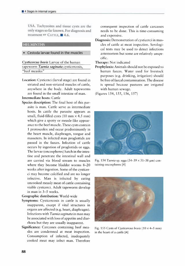
• " Stages in internat organs
USA. Tachyzoite and ti u y t are the only tag 0 far known. For diagno is and treatment A1TLE, • 4 .6.
HELMINTHS
• Cestoda larvae found in the muscles
Cysticercus bovis Larvae of the human tapeworm Taenia saginata cysticercosis, "beef measles"
Location: Cysticerci (larval stage) are found in striated and non-striated muscles of cattle, anywhere in the body. Adult tapeworms are found in the small intestine of man.
Intermediate hosts: Cattle Species description: The final host of this par
asite is man. Cattle serve as intermediate hosts. In cattle the parasite appears as smalI, fluid-filled cysts (10 mm x 4.5 mm) wh ich give a spotty or measle-like appearance to the beef muscle. These cysts contain 1 protoscolex and occur predominantly in the heart muscle, diaphragm, tongue and masseters. In infected man proglottids are passed in the faeces. Infection of cattle occurs by ingestion of proglottids or eggs. The larvae (oncospheres) hatch in the intestine and penetrate the intestinal wall and are carried via blood stream to muscles where they become bladder worms 8-20 weeks after ingestion. Some of the cysticerci may become calcified and are no longer infective. Man is infected by eating uncooked measly meat of cattle containing viable cysticerci. Adult tapeworm develop in man in 3-5 weeks.
Geographie distribution: World-wide Symptoms: Cysticercosis in cattle is usually
inapparent, except if vital structures in organs are affected (e .g. heart, diaphragm). Infeetions with Taenia saginata in man may be associated with loss of appetite and diarrhoea but they are usually inapparent.
Signifieanee: Carcasses containing beef measles are condemned at meat inspection. Consumption of infected, inadequately eooked meat may infect man. Therefore
88
eonsequent inspeetion of eattle carcasses needs to be done. This is time-consuming and expensive.
Diagnosis: Demonstration of cysticerci in muscles of catde at meat inspection. Serological tests may be used to detect infections antemortem but some are relatively unspecific.
Therapy: Not indicated Prophylaxis: Animals should not be exposed to
human faeces. Water used for livestock purposes (e.g. drinking, irrigation) should be free of faecal contamination. The disease is spread because pastures are irrigated with human sewage.
(Figures 154, 155, 156, 157)
Fig. 154 Taenia sp. eggs (34-39 x 31-38 firn) containing oncospheres [4]
Fig. 155 Cysts of Cysticercus bovis (10 x 4-5 rnrn)
in the heart of a cattle [4]

Fig. 156 Calcified tissue eyst of Cysticercus bovis in the masseter musde of a eattle
\ \. " ..
Fig. 157 Life eyde of Taenia saginata/Cysticercus bovis
Cysticercus dromedarii (syn. C. cameli)
R mar : The larva ( ysticerclIs dromedarii) of a hyena tape\ rm (Taenia hyaellae) i frequentI)' found in the mu I f the dromedary attle and g ar ( DR Mf-
DARI , . 4.l.l).lri rarel ~ und in h p . . dromedarii y t are t\ i e a larg a
bouis Olea urin l2-1 Olm in length. They are pearl \ hite and the lateral invaginated proto oie ha a d ubl r w
of hooks. Although they are not dangerous to man, their presence is repugnant, and parasitized Oleat should be condemned and not fed to dogs or other caniidae.
• Nematoda larvae and adult nematodes found in the muscles
Onchocerca dukei Muscular and subcutaneous onchocercosis of cattle
Remark: dult \ rm of chi are
non of the ho t, eil ~ r gra i h-\ hit in olour. They are o\-al ( -6 Olm in diameter) \ ith a rai d onrour. Thc c n dule are hard to rou h, and when li ed ne r everal worm appcar urround d by pu .
Thi i [he maj r differen e tO Cysticerc/ls bouis. The inrramu ular n dule an b omplet I embedded in [he mu le
only parrly v r d. The entire mu ularure ma be inv Ived e pe ially the flank, inter ral and limb mu le. ubcuraneou n dule may be fund almo t every-\ here ( .1 l.
i ure 1
Fig. 158 Sites of oeeurrenee of some Onchocerca speeies found in eattle in Afriea [20]
89
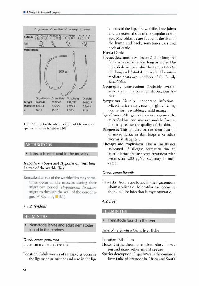
• " Stages in internat organs
O. gutturoSil O. armollata O. OChengl O. duke;
Cutlcul.~~ 'HUIfIJ! I!H"uSJ
Mkrofllari.e
0 _ gUllu,OSiI O. armlllat;J Lenght 263/249 382/346 Diameter 4.4/3.4 6.8/5.5 n 26/13 15/13
O. ochengl O. duke!
298/277 240/ 217 7.9/509 6.7/4.8
22/13 22/8
Fig. 159 Key for the identification of Onchocerca species of cattle in Africa [20J
ARTHRorODS
• Insecta larvae found in the muscles
Hypoderma bovis and Hypoderma lineatum Larvae of the warble flies
Rcmark: rimc ml rat
arvac rh \ arble Oie may m-ur in th mu le durin their
ry peri d. Hyp derma fincatllm rhrough rhe \ all f the e pha,\lTLE, -. 1).
4.1.2 Tendons
HEL.\lINTHS
• Nematoda larvae and adult nematodes found in the tendons
Onchocerca gutturosa Ligamentary onehoeereosis
Loeation: Adult worms of this speeies oeeur in the ligamentum nuehae and also in the lig-
90
aments of the hip, elbow, stifle, knee joints and the external side of the seapular eartilage. Mierofilariae are found in the skin of the hump and back, sometimes ears and neck of eatde.
Hosts: Catde Speeies deseription: Males are 2-3 em long and
females are up to 60 em long or more. The microfialriae are unsheathed and 249-263 pm long and 3.4-4.4 pm wide. The intermediate hosts are members of the family Simuliidae.
Geographie distribution: Probably worldwide, extremely eommon throughout Afflea.
Symptoms: Usually inapparent infeetions. Mierofilariae may eause a slighdy itehing dermatitis, resembling a mild mange.
Signifieanee: Allergie skin reaetions against the mierofialriae and massive nodule formation may reduee the quality of the skin.
Diagnosis: This is based on the identifieation of mierofilariae in skin biopsies or adult worms at slaughter.
Therapy and Prophylaxis: This is usually not indicated. If allergie dermatitis due to mierofilariae are suspeeted treatment with ivermeetin (200 pg/kg, Sc.) may be indieated.
Onchocerca lienalis
Remarks: Adults are found in the ligamentum abomaso-lienale. Mierofilarieae oeeur in the skin. The infeetion is asymptomatic.
4.2liver
HELMINTHS
• Trematoda found in the liver
Fasciola gigantica Giant Iiver fluke
Loeation: Bile duets Hosts: Cattle, sheep, goat, dromedary, horse,
pig and many other animal speeies Species description: F. gigantica is the common
Iiver f1uke of livestock in Afriea and South

Asian countries. F. gigantica resembles F. hepatica. Its size is larger, being 25-75 mm in length and up to 12 mm in breadth. The size of the eggs is 156-197 pm x 90-104 pm. The most frequently involved intermediate hosts are Lymnaea auricularia and L. natalensis. These are aquatic snails living in fairly large permanent bodies of water. The snail vectors can survive an amphibious existence but can only aestivate for very short periods. Metacercariae (the cyst stage of cercariae formed after the tail is cast off) encyst on plants under water such as rice plants, etc. Metacercariae may survive for up to 4 months on stored plants and thus infection may be transmitted by feeding rice straw. F. gigantica adults reach the bile ducts 12 weeks after infection. The immature stages migrate through the liver parenchyma. Infections with F. gigantica can occur extremely focal, revealing high prevalences in some regions and low prevalences in others. Infection is acquired by eating herbage wh ich is contaminated with metacercariae. The prepatent period is 9-12 weeks.
Geographie distribution: Throughout Africa and Asia
Symptoms: In sheep an acute and a chronic form can occur whereas in cattle only the chronic form seems to playa role. The pathogenesis is essentially the same as in F. hepatica.
Significance: F. gigantica causes mainly a chronic fasciolosis in cattle and an acute, often fatal, syndrome in sheep.
Diagnosis: Detection of typical operculated eggs with the sedimentation method. Cave: F. gigantica eggs are similar to the eggs of some rumen fluke species! F. gigantica eggs only a ppear 15 weeks after infection! Adult flukes are found in the bile ducts at necropsy.
Therapy: Kir· F. hepatica Prophylaxis: Ir%' F. hepatica in many parts of Afri
ca it is not possible to avoid grazing around lakes, swamps especially if these places are used for watering. The use of bore holes for watering cattle is an effective means to reduce infections with F. gigantica.
(Figure 160)
Fig. 160 Fasciola gigantica; the giant liver fluke
Fasciola hepatica Common liver fluke
Location: Bile ducts of the liver, sometimes gall bladder
Hosts: Cattle, sheep, goat, horse, donkey, pig and many other animal species
Species description: F. hepatica is occurs world-wide - except in Africa and South Asia where it is replaced by F. gigantica -and affects especially cattle and sheep. Its size is 30 by 15 mm. The eggs measure 130-150 x 65-90 pm. F. hepatica eggs hatch within 10-12 days after deposition and the miracidium, the first larval stage, penetrates actively into the intermediate host which is generally an amphibious snail of the genus Lymnaea. Cercariae leave the snail 4-7 weeks after infection and settle on plants just below water level. After casting off the tail they form a cyst about 0.2 mm in diameter (metacercariae). The metacercariae are infective. Ingested metacercariae penetrate the small intestinal wall and reach the Iiver via abdominal cavity. The duration of the migration within the Iiver parenchyma is 6-8 weeks after wh ich the young f1ukes reach maturity in the bile ducts. Infection occurs orally by ingestion of metacercariae on herbage. The incubation period is 2-4 weeks. First symptoms may be seen 5-7 days after infection in acute fasciolosis.
91
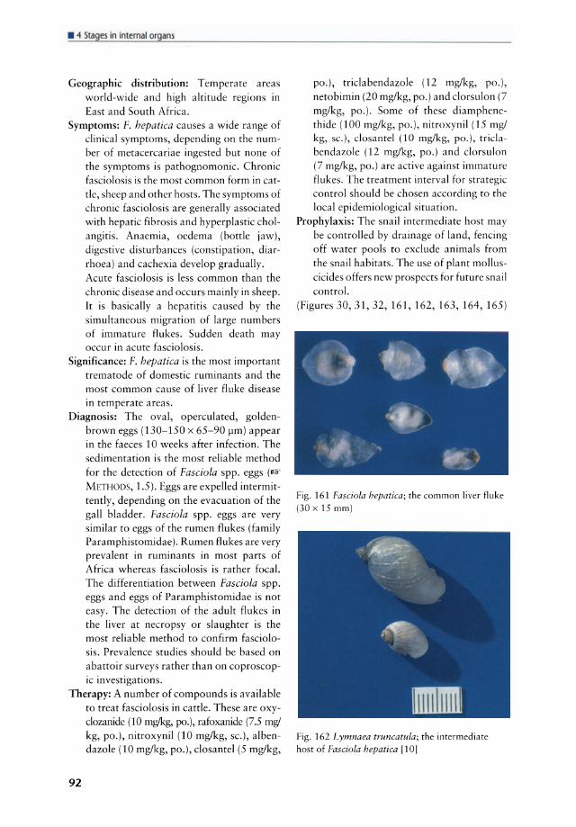
• 1 Stages in internaiorgans
Geographie distribution: Temperate areas world-wide and high altitude regions in East and South Africa.
Symptoms: F. hepatica causes a wide range of c1inical symptoms, depending on the number of metacercariae ingested but none of the symptoms is pathognomonic. Chronic fasciolosis is the most common form in catde, sheep and other hosts. The symptoms of chronic fasciolosis are generally associated with hepatic fibrosis and hyperplastic cholangitis. Anaemia, oedema (bottle jaw), digestive disturbances (constipation, diarrhoea) and cachexia develop gradually. Acute fasciolosis is less common than the chronic disease and occurs mainly in sheep. Ir is basically a hepatitis caused by the simultaneous migration of large numbers of immature f1ukes. Sudden death may occur in acute fasciolosis.
Significance: F. hepatica is the most important trematode of domestic ruminants and the most common cause of Iiver f1uke disease in temperate areas.
Diagnosis: The oval, operculated, goldenbrown eggs (130-150 x 65-90 J.Im) appear in the faeces 10 weeks after infection. The sedimentation is the most reliable method for the detection of Fasciola spp. eggs (1& METHODS, 1.5). Eggs are expelled intermittendy, depending on the evacuation of the gall bladder. Fasciola spp. eggs are very similar to eggs of the rumen flukes (family Paramphistomidae). Rumen f1ukes are very prevalent in ruminants in most parts of Africa whereas fasciolosis is rather focal. The differentiation between Fasciola spp. eggs and eggs of Paramphistomidae is not easy. The detection of the adult f1ukes in the Iiver at necropsy or slaughter is the most reliable method to confirm fasciolosis. Prevalence studies should be based on abattoir surveys rather than on coproscopic investigations.
Therapy: A number of compounds is available to treat fasciolosis in catde. These are oxyc10zanide (10 mglkg, po.), rafoxanide (7.5 mg/ kg, po.), nitroxynil (10 mg/kg, sc.), albendazole (10 mg/kg, po.), c10santel (5 mg/kg,
92
po.), triclabendazole (12 mg/kg, po.), netobimin (20 mg/kg, po.) and c1orsulon (7 mg/kg, po.). Some of these diamphenethide (100 mg/kg, po.), nitroxynil (15 mg/ kg, sc.), c10santel (10 mg/kg, po.), tricla bendazole (12 mg/kg, po.) and c1orsulon (7 mg/kg, po.) are active against immature f1ukes. The treatment interval for strategie control should be chosen according to the local epidemiological situation.
Prophylaxis: The snail intermediate host may be controlled by drainage of land, fencing off water pools to exclude animals from the snail habitats. The use of plant molluscicides offers new prospects for future snail control.
(Figures 30, 31, 32,161,162,163,164,165)
Fig. 161 Fasciola hepatica; the common liver f1uke (30 x 15 mm)
Fig. 162 Lymnaea truncatula; the intermediate host of Fasciola hepatica [10]
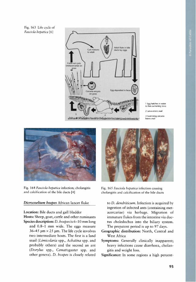
Fig. 163 Life eyde of Fasciola hepatica [6]
Fig. 164 Fasciola hepatica infeetion; eholangitis and calcifieation of the bile ducts [4]
Dicrocoelium hospes Afriean laneet f1uke
Loeation: Bile duets and gall bladder Hosts: Sheep, goat, eattle and other ruminants Speeies deseription: D. hospes is 6-10 mm long
and 0.8-1 mm wide. The eggs measure 36-45 11m x 25 11m. The Iife eyele involves two intermediate hosts. The first is aland snail (Limicolaria spp., Achatina spp. and probably others) and the seeond an ant (Dorylus spp., Cematogaster spp. and other genera). D. hospes is elosely related
1 E99 hatches in water to free-swimming larva
2 Larva enters snail
3 Swimming cercaria leaves snail
Fig. 165 Fasciola hepatica infection eausing eholangitis and calcification of the bile duets
to D. dendriticum. Infeetion is aequired by ingestion of infeeted ants (eontaining metaeereariae) via herbage. Migration of immature f1ukes from the intestine via ductus eholedoehus into the biliary system. The prepatent period is up to 97 days.
Geographie distribution: North, Central and West Afriea
Symptoms: Generally elinieally inapparent; heavy infeetions eause diarrhoea, eholangitis and weight loss.
Signifieanee: In some regions a high pereent-
93

• " Stages in internat organs
age of animals is infected. Losses occur due to poor growth and condemnation of infected livers at slaughter.
Diagnosis: Adult D. hospes in the bile at necropsy; detection of parasite eggs in the sediment of faeces.
Therapy: lfi'Y below D. dendriticum Prophylaxis: Ili)' below D. dendriticum
Dicrocoelium dendriticum (syn. D. lanceolatum) Common lancet fluke
Location: Bile ducts or gall bladder Hosts: D. dendriticum occurs mainly in sheep,
goat, cattle but also in pig, dog, donkey, rabbit and rarely in man.
Species description: The fl uke is 6-10 mm long and 1.5-2.5 mm wide. The eggs measure 36-45 x 20-30 11m. The life cyele involves two intermediate hosts, aland snail as the first (Zebrina spp., Cionella spp. and 29 other species are described) and ants of the genus Formica and Lasius as the second intermediate hosts. Ants infected with metacercariae attach to herbage overnight and are available to grazing animals in the early morning. Infection occurs by ingestion of infected ants (containing metacercariae) via herbage. Migration of immature flukes from the intestine via ductus choledochus into the biliary system. The prepatent period is 47-54 days. Cirrhosis of the liver and thickening of the bile ducts may occur in heavy infections.
Geographie distribution: Europe, Asia, the Americas, North Africa, occasionally in imported ruminants in other parts of Africa; autochthonous infections seem not to
occur in Africa. Symptoms: Often without elinical symptoms.
Heavy infections may produce weight loss, anaemia and hypoproteinaemia.
Significance: Lancet flukes may cause extensive liver damage, causing the host to "do poorly". Condemnation of livers at slaughter may cause severe economic losses among cattle herds.
Diagnosis: Demonstration of typical eggs in the sediment of faeces. The eggs are sm all
94
(40 x 25 Jlm), thick walled, yellowishbrown and contain a miracidium. Adult parasites may be found in the bile ducts at necropsy.
Therapy: Albendazole (20 mg/kg, po.), fenbendazole (50 mg!kg, po.), netobimin (20 mg /kg, po.) thiophanate (50 mg/kg, po.), thiabendazole (200 mg/kg, po.) and praziquantel (50 mg/kg, po.) are effective against the lancet fluke.
Prophylaxis: Elimination of the intermediate hosts is not feasible.
(Figures 32, 33, 166, 353)
Fig. 166 Dicrocoelium dendriticum found in the
bile ducts
Schistosoma spp.
Remark: hi r ome ma frcn tc fOllod ar p y in rhe p rtal vein of the liv r lTLF, . 2).
• Cestoda found in the liver
Echinococcus granulosus (syn. E. unilocularis) Hydatid tapeworm, hydatidosis
Location: Larvae (hydatid cysts) of E. granulosus are found in the liver and the lungs of cattle, sheep, goats, swine, horse, dromedary and other domestic animals and man (= intermediate hosts). Adult tapeworms are found in the small intestine of the dog and other related carnivores (= final hosts).
Hosts: Canids are the final hosts of E. granulosus. Intermediate hosts are ungulates and man.
Species description: E. granulosus is a small tapeworm with 3-4 proglottids and a total length of 2-6 mm. It occurs in the small intestine of the dog and other canids (hyaena, coyote, fox, etc.). Adult tapeworms in the dogs pass proglottids or eggs in the faces. When ingested by cattle or other intermediate hosts, these eggs hatch in the intestine. The oncospheres (hexacanths) penetrate the gut wall, enter the portal vein and re ach the liver, wh ich is the first capillary filter for the larvae and subsequently the lungs, wh ich are the second capillary filter. The remaining oncospheres may re ach via the arteries other organs. In all these organs hydatid cysts (bladder worms) develop. These are large cysts filled with fluid and tapeworm heads (protoscolices) . The hydatid cysts develop slowly over several months. They are commonly 5-10 cm in diameter and contain a liquid. The hydatid cyst is composed of a thick outer, concentrically lamina ted membrane. About 5 months after infection brood capsules, each containing several protoscolices develop from the inner part of the capsule (germinal membrane). The brood capsules may become detached and float free in the cyst fluid, being termed "hydatid sand". At this time the cyst is infective for the definitive host. The life cyele is completed when a dog ingests protoscolices. Hydatid cysts are usually found in the lungs of sheep and cattle, but primarily in the livers of horses. In pigs, hydatid cysts are found in both the liver and the lungs. The hydatid cysts may be multilocular (sheep, cattle, pigs) or unilocular (horse). In man hydatid cysts may be found in many organs. The significance of the hydatid cyst depends on the severity of the infection and the organ in which it is situated. In domestic animals elinical signs are not commonly seen despite heavy infections. Human hydatidosis is often associated with elinical signs and the function of the affected organ is often impaired. This is especialIy true if the heart or the brain are involved. Adult tapeworms are harmless to the dogs and seldom cause elinical symptoms.
Geographie distribution: World-wide Symptoms: Hydatid cysts do not usually cause
elinical symptoms unless the cysts are numerous or become very large.
Significance: Hydatidosis in cattle is not usually a problem, but infected organs are condemned at slaughter. In man hydatidosis is a serious problem. Infected dogs are a reservoir for both infection of domestic animals and man.
Diagnosis: Usually the diagnosis of hydatidosis is made at slaughter or necropsy. Infected dogs pass eggs in the faeces which cannot be differentiated from those of Taenia spp. Immundiagnostic tests (ELISA, Immunoblotting; 1& METHODS, 5.2 and 5.3) are widely used in medical parasitology and may ass urne significance in epidemiological studies. E. granulosus eggs, excreted by dogs are very similar to those of Taenia spp. The differentiation may be carried out by means of monoelonal antibodies.
Therapy: Treatment of hydatidosis in cattIe and other intermediate hosts is rare1y indicated. Control of hydatidosis is based on prophylaxis.
Prophylaxis: Dogs should not be fed with uncooked meat in general and infected organs in particular. In endemic areas where human hydatidosis is of great importance special attention should be paid to the safe destruction of affected organs, the reduction of stray dogs and the anthelmintic treatment of domestic dogs. Praziquantel (5 mg/kg, po. or 5.7 mg/kg, sc. or im.), bunamidine hydrochloride (25-50 mg/kg, po.) and the combination of febantellpraziquantellpyrantel are sufficiently effective against adult E. granulosus tapeworms in dogs.
(Figures 167, 168, 169, 170)
95

• 1 Stages in internaiorgans
Fig. 167 Echinococcus granulosus; hydatid eyst in the lungs of a eattle 14]
I I I I , ,
I I I I I , I I I I 1/ I I • ...1"
\\(~~S)' iifC~ ,.
Fig. 168 Life eycle of Echinococcus granulosus in sub-Saharan Afriea [21]
96
Fig. 169 Echinococcus granulosus; hydatid eyst [42] opened [41
Fig. 170 Echinococcus granulosus; protoseolex (100 x 160 in diameter) with typieal hook eollar
Stilesia hepatica
Remark : Thi para ire occur in the bile du r and mall inte rine of heep and goar (
H PA 0 AT . 4.2). In endemi area wild ruminanr and rarely carrle may al 0
be affe red. Thi para ire i very prevalent in tropical and ourhern Afri a. . hepatiC(l i non-pathogeni bur extreme\ prevalent (in 90-100% of heep) in many part of frica. Ir i non-pathogenic and doe not require treatment.
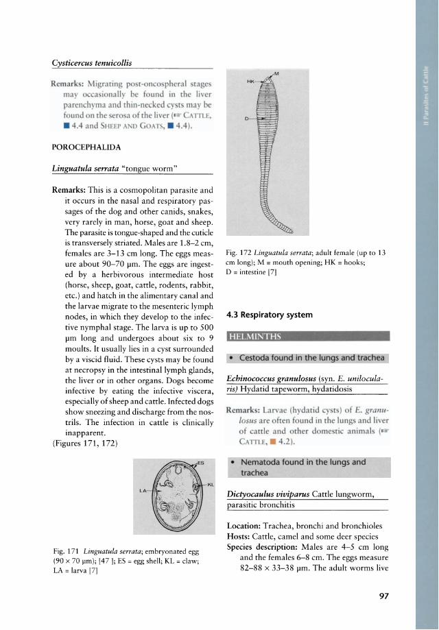
Cysticercus tenuicollis
Rcmark : ~lIgraring po r-on 0 ph ral tag ma)' 0 a ionally be f und in the liver parcnchyma and [hlO-ne kcd )' [ may bc {ound n the er a f rh liv r « .\1TU,
. 4.4 < nd 11111' \ .'1) O .\T .. , . 4.4).
POROCEPHALIDA
Linguatula serrata "tongue worm"
Remarks: This is a eosmopolitan parasite and it oeeurs in the nasal and respiratory passages of the dog and other eanids, snakes, very rarely in man, horse, goat and sheep. The parasite is tongue-shaped and the euticle is transversely striated. Males are 1.8-2 cm, females are 3-13 em long. The eggs measure about 90-70 11m. The eggs are ingested by a herbivorous intermediate host (horse, sheep, goat, cattle, rodents, rabbit, ete.) and hateh in the alimentary eanal and the larvae migrate to the mesenteric Iymph nodes, in whieh they develop to the in feetive nymphal stage. The larva is up to 500 11m long and undergoes about six to 9 moults. Ir usually lies in a eyst surrounded by a viscid fluid. These cysts may be found at neeropsy in the intestinallymph glands, the Iiver or in other organs. Dogs beeorne infeetive by eating the infeetive viscera, espeeially of sheep and eattle. Infected dogs show sneezing and diseharge frorn the nostrils. The infeetion in eattle is c1inieally inapparent.
(Figures 171, 172)
LA
Fig. 171 Linguatula serrata; embryonated egg (90 x 70 pm); [47]; ES = egg shell; KL = claw; LA = larva [7]
KL
Fig. 172 Linguatula serrata; adult female (up to 13 cm long); M = mouth opening; HK = hooks; D = intestine [7]
4.3 Respiratory system
HELMINTHS
• Cestooa found in tl1e lungs and trachea
Echinococcus granulosus (syn. E. unilocularis) Hydatid tapeworrn, hydatidosis
Remark : Lar ae (hydatid y t) f E. gral1l l
loslls are ften f lind in the lung and h er of arrle and other dome rie animal (
ATILE, • 4.2).
• Nematoda found in the lungs and trachea
Dictyocaulus viviparus Cattle lungworrn, parasitic bronchitis
Location: Trachea, bronehi and bronchioles Hosts: Cattle, earnel and some deer species Species description: Males are 4-5 cm long
and the females 6-8 cm. The eggs measure 82-88 x 33-38 11m. The adult worrns live
97
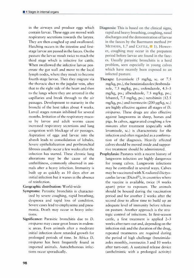
• " Stages in internat organs
in the airways and produce eggs which contain larvae. These eggs are moved with respiratory secretions towards the larynx. They are then coughed up and swallowed. Hatching occurs in the intestine and firststage larvae are passed in the faeces. On the pa sture the larvae moult twice to reach the third stage wh ich is infective for cattle. When swallowed the infective larvae penetrate the gut wall and move to the local Iymph nodes, where they moult to become fourth-stage larvae. Then they migrate via the thoracic duct to the jugular vein, after that to the right side of the heart and then to the lungs where they are arrested in the capillaries and break through to the air passages. Development to maturity in the bronchi of the host takes about 4 weeks. Larval stages remain inhibited for several months. Irritation of the respiratory mucosa by larvae and adult worms cause increased respiratory secretions and lung congestion with blockage of air passages . Aspiration of eggs and larvae into the alveoli leads to consolidation of lobules. Severe epithelialization and peribronchial fibrosis usually occur a few weeks after the infection has started. These chronic lung alterations may be the cause of the unthriftiness, commonly observed in animals after a heavy infection. Immunity is built up as quickly as 10 days after an initial infection but it wanes in the absence of reinfection.
Geographic distribution: World-wide Symptoms: Parasitic bronchitis is character
ized by severe coughing, rapid breathing, dyspnoea and rapid loss of condition. Severe ca ses lead to emphysema and pneumonia. Death may occur in heavy infections.
Significance: Parasitic bronchitis due to D. viviparus may cause great losses in endemic areas. Even animals after a moderate initial infection show retarded growth for prolonged periods of time. In Africa D. viviparus has been frequently found in imported animals. Autochthonous infections occur sporadically.
98
Diagno i : Thi i ba ed on {he linieal ign, rapid and heavy brearhing, oughing, na al diseharge and the demon rration f larvae in the fae e by the Baermann method ( MFTH 0, 1.7 and ATILE, 1). Howe -er, eoughing may eur in rhe preparem p riod before larvae are found in the fae -e . U ually para iti bron hiti i a herd problem een e pe ially in young ealve whieh have reeently b en expo cd ro an infeeted pa rure.
Therapy: Levamisole (5 mg/kg, sc. or 7.5 mg/kg, po.) , the benzimidazoles (fenbendazole, 7.5 mg/kg, po.; oxfendazole, 4.5-5 mg/kg, po.; albendazole, 7.5 mg/kg, po.; febante!, 7.5 mg/kg, po.) netobimin (12.5 mg/kg, po.) and ivermectin (200 Ilg/kg, Sc.) are highly effective against all stages of D. viviparus. These drugs are also effective against lungworms in sheep, horses and pigs. In calves, aggravated coughing a few min utes after treatment (especially with levamisole, sc.) is characteristic for the infection and often regarded as a confirmation of the diagnosis. Heavily affected calves should be moved inside and supportive treatment should be administered.
Prophylaxis: Pa stures with arecent history of lungworm infection are highly dangerous for young calves. Lungworm infections may be controlled in several ways. Calves may be vaccinated with X-radiated Dictyocaulus larvae (Dictol®) , in countries where the vaccine is available, twice (4 weeks apart) prior to exposure. The animals should be housed du ring the vaccination period and for another 2 weeks after the second dose to allow time to build up an adequate level of immunity before release on pasture. Another approach is the strategic control of infections. In first-season cattle, a first treatment is applied 2-3 weeks after turn-out and, depending on the infection risk and the duration of the drug, repeated treatments are required during the period of high challenge (benzimidazoles monthly, ivermectin 3 and 10 weeks after turn-out). A sustained release device (anthelmintic with a prolonged activity)
mayaIso be applied about 2-3 weeks after turn-out or prior to the period with the highest infection risk.
(Figures 83, 173, 174)
Fig. 174 Dictyocaulus viviparus; adult worms in trachea
Fig. 173 Life cyeIe of Dictyocaulus viviparus; the third-stage larvae penetrate the intestinal mucosa and not the mucosa of the stornach [6j
Mammomonogamus laryngeus
Location: Larynx Hosts: Cattle, water buffalo, occasionally
sheep, goat and rarely man Species description: M . laryngeus is not very
pathogenic for cattle. The worms are attached to the mucosa of the larynx. The males and females are found in permanent copulation. The life cycle is unknown.
Geographie distribution: Caribbean Islands, South America, Asia, Central Africa (Cameroon)
Symptoms: Affected animals may cough and sometimes lose condition. Calves may develop bronchitis. Pneumonia due to respiratory secretions wh ich are aspirated, causing pulmonary reactions have been observed.
99

• 1 Stages in internaiorgans
Significance: Heavy infections in endemic areas may cause losses due to massive irritation of the larynx by the parasites. Bronchitis and pneumonia may occur.
Diagnosis: This is made by finding the eggs in the faeces or adult worms at necropsy.
Therapy: Unknown. Nitroxynil (lOmg/kg, Sc.), which is used against Syngamus spp. of poultry mayaiso be used against Mammomonogamus laryngeus. Modern benzimidazoles and ivermectin (200 pg/kg. sc.) may be used in heavily infected cattle.
Prophylaxis: Unknown (Figures 175, 176, 177)
Fig. 175 Mammomonogamus laryngeus (males are 3-3.5 mm long, females are 8.5-10 mm lang) l8J
100
Fig. 176 Egg of Mammomonogamus laryngeus (323 x 50 pm) l8J
Fig. 177 Mammonogamus laryngeus; head end [8]
Mammomonogamus nasicola
Location: Nasal cavities Hosts: Cattle, sheep and goat Species description: The males and females of
this parasite are permanently joined in copulation. Males are 4-6 mm long and females 11-23 mm. Eggs are 54 x 98 pm and have only a few blastomeres. Life cyde and pathogenesis are unknown. An optional reservoir host (e.g. earthworm or snail) is being suspected. After oral ingestion of infective larvae the parasite reaches the respiratory tract through the blood stream. The parasites suck blood and are bright red in fresh sampies.
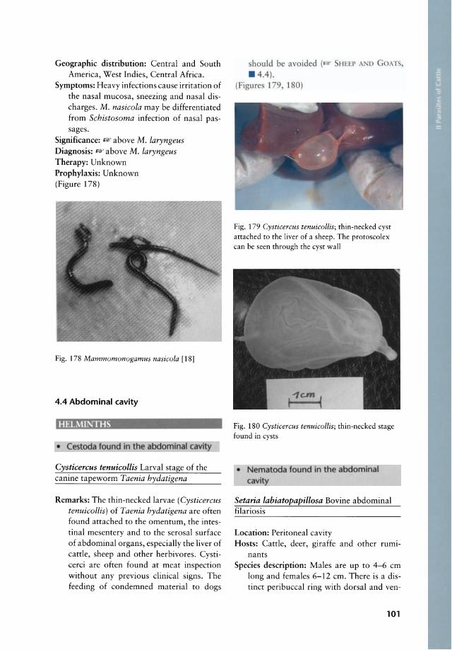
Geographie distribution: Central and South Ameriea, West Indies, Central Afriea.
Symptoms: Heavy infeetions eause irritation of the nasal mueosa, sneezing and nasal diseharges. M. nasicola may be differentiated from Schistosoma infeetion of nasal passages.
Signifieanee: "above M. laryngeus Diagnosis: " above M. laryngeus Therapy: Unknown Prophylaxis: Unknown (Figure 178)
Fig. 178 Mammomonogamus nasicola [18]
4.4 Abdominal cavity
HELMl;\;THS
• Cestoda found in the abdominal cavity
Cysticercus tenuicollis Larval stage of the eanine tapeworm Taenia hydatigena
Remarks: The thin-neeked larvae (Cysticercus tenuicollis) of Taenia hydatigena are often found attaehed to the omen turn, the intestinal mesentery and to the serosal surfaee of abdominal organs, espeeially the liver of eattle, sheep and other herbivores. Cystieerei are often found at me at inspeetion without any previous clinieal signs. The feeding of eondemned material to dogs
hould be avoided . 4.4).
HEEP Al D GOAT ,
(Figure 179 1 0)
Fig. 179 Cysticercus tenuicollis; thin-neeked eyst attached to the liver of a sheep. The protoseolex ean be seen through the eyst wall
I ~c.m (
Fig. 180 Cysticercus tenuicollis; thin-neeked stage found in eysts
• Nematoda found in the abdominal cavity
Setaria labiatopapillosa Bovine abdominal filariosis
Loeation: Peritoneal eavity Hosts: Cattle, deer, giraffe and other ruml
nants Speeies deseription: Males are up to 4-6 em
long and females 6-12 em. There is a distinet peribueeal ring with dorsal and ven-
101
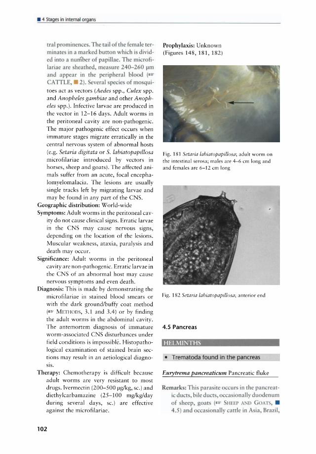
• " Stages in interna! organs
rral pr min n e . The rail f the fcmale rerminat in a marked burr n \ hi h i di ided im a num er of papillae. The mi r filariae ar hearhed mea ure 240-260 pm and appear in rhe peripheral bl cl (
TTL, 2). eral p ie of mo qui-
toes act as vectors (Aedes spp., Cu lex spp. and Anopheles gambiae and other Anopheles spp.). Infective larvae are produced in the vector in 12-16 days. Adult worms in the peritoneal eavity are non-pathogenic. The major pathogenic effeet oeeurs when immature stages migrate erratieally in the eentral nervous system of abnormal hosts (e.g. Setaria digitata or S. labiatopapillosa microfilariae introdueed by veetors in horses, sheep and goats). The affeeted animals suffer from an acute, foeal encephalomyelomalaeia. The lesions are usually single tracks left by migrating larvae and may be found in any part of the CNS.
Geographie distribution: World-wide Symptoms: Adult worms in the peritoneal ca v
ity do not eause clinieal signs. Erratic larvae in the CNS may eause nervous signs, depending on the loeation of the lesions. Muscular weakness, ataxia, paralysis and death may oeeur.
Significance: Adult worms in the peritoneal eavity are non-pathogenie. Erratie larvae in the CNS of an abnormal host may eause nervous symptoms and even death.
Diagnosis: This is made by demonstrating the mierofilariae in stained blood smears or with the dark ground/buffy eoat method (I@' METII()[)S, 3.1 and 3.4) or by finding the adult worms in the abdominal eavity. The antemortem diagnosis of immature worm-assoeiated CNS disturbances under field conditions is impossible. Histopathological examination of stained brain seetions may result in an aetiologieal diagnoSlS.
Therapy: Chemotherapy is difficult because adult worms are very resistant to most drugs. Ivermeetin (200-500 flglkg, sc.) and diethylcarbamazine (25-100 mg/kglday during several days, sc.) are effective against the mierofilariae.
102
Prophylaxis: Unknown (Figures 148, 181, 182)
Fig. 181 Setaria labiatopapillosa; adult worm on the intestinal serosa; males are 4-6 cm lang and and females are 6-12 cm long
Fig. 182 Setaria labiatopapillosa; anterior end
4.5 Pancreas
HELMINTHS
• Trematoda found in the pancreas
Eurytrema pancreaticum Panereatie fluke
Remar : Thi para ire i du r ,biledu
f h ep, oar 4. ) and 0 a i nall
cur in rhe pan reata i nally du denum
o T, . arrle in ia Brazil
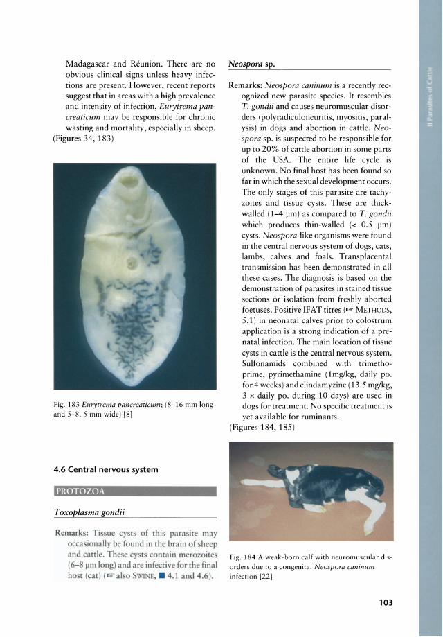
Madagasear and Reunion. There are no obvious dinieal signs unless heavy infeetions are present. However, reeent reports suggest that in areas with a high prevalenee and intensity of infeetion, Eurytrema pancreaticum may be responsible for ehronie wasting and mortality, espeeially in sheep.
(Figures 34, 183)
Fig. 183 Eurytrema pancreaticum; (8-16 mm long and 5-8. 5 mm wide) [81
4.6 Central nervous system
PROTOZOA
Toxoplasma gondii
Remark : Ti ue cy t of chi para ire may a i nally be found in ehe brain of heep
and anle. The e eyst conrain mer z i re (6- I-Im I ng) and are infeccive for the final ho r (carl ( al 0 ~ h E, . 4. 1 and 4.6) .
Neospora sp.
Remarks: Neospora caninum is a reeently reeognized new parasite speeies. Ir resembles T. gondii and eauses neuromuseular disorders (polyradieuloneuritis, myositis, paralysis) in dogs and abortion in eattle. Neospora sp. is suspeeted to be responsible for up to 20% of eattle abortion in so me parts of the USA. The entire Iife eyde is unknown. No final host has been found so far in whieh the sexual development oeeurs. The only stages of this parasite are taehyzoites and tissue eysts. These are thiekwalled (1-4 pm) as eompared to T. gondii whieh produces thin-walled « 0.5 I-Im) eysts. Neospora-Iike organisms were found in the eentral nervous system of dogs, eats, lambs, ealves and foals. T ransplaeental transmission has been demonstrated in all these eases. The diagnosis is based on the demonstration of parasites in stained tissue sections or isolation from freshly aborted foetuses. Positive IFAT titres (IJ!W' METHODS, 5.1) in neonatal ealves prior to eolostrum applieation is a strong indieation of a prenatal infeetion. The main loeation of tissue eysts in eattle is the eentral nervous system. Sulfonamids eombined with trimethoprime, pyrimethamine (Img/kg, daily po. for 4 weeks) and dindamyzine (13.5 mg/kg, 3 x daily po. during 10 days) are used in dogs for treatment. No speeifie treatment is yet available for ruminants.
(Figures 184, 185)
Fig. 184 A weak-born calf with neuromuscular disorders due to a congenital Neospora caninum infection [22)
103
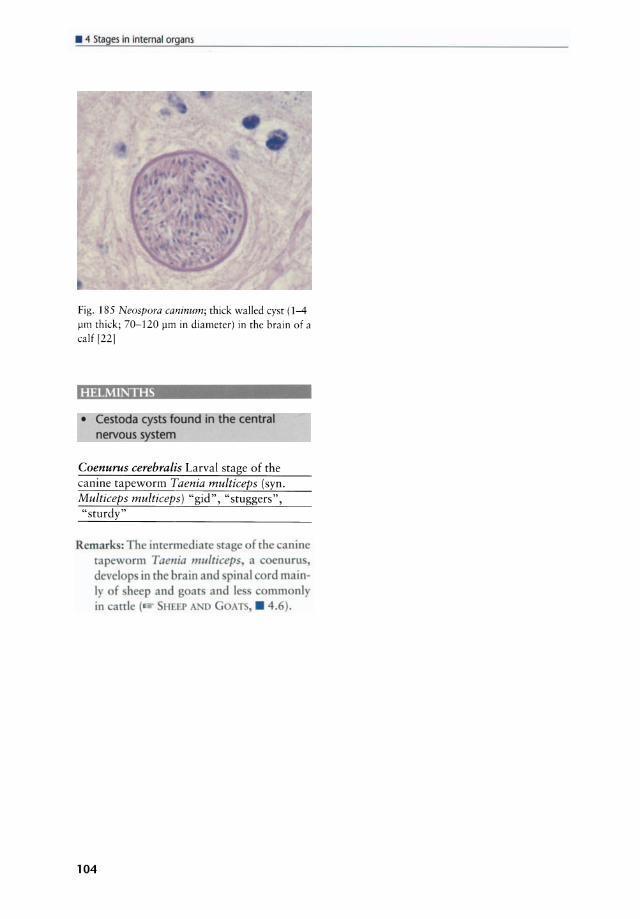
• " Stages in internat organs
Fig. 185 Neospora caninum; thick walled eyst (1-4 pm thiek; 70-120 pm in diameter) in the brain of a
ca lf [22]
HELMINTHS
• Cestoda cysts found in the centra nervous s stern
Coenurus cerebralis Larval stage of the canine tapeworm Taenia multiceps (syn. Multiceps multiceps) "gid", "stuggers", "sturdy"
Remarks: The inrermedlate ca e f rhe anine tapew rm Tael/ia fIlltltlceps a oenuru, devcl p in [he bram and pinal ord main-Iy of he p and oar and le ommonl in canlc ( HErp Al D OA . 4.6).
104

5 Stages on lhe body surface
5.1. Skin and coat
PROTOZOA ................... 105
HElMINTHS • Nematoda found in the skin ...... 106
ARTHROPODS • Arachnida found in/on the skin .... 109
- Ticks . ..................... 109 - Mites . ..................... 121
• Insecta found on the skin ........ 127 - Lice ....................... 127 - Fleas ....................... 129 - Dipterida ................... 129
5.1. Skin and coat
PROTOZOA
Besnoitia besnoiti (syn. Sarcocystis besnoiti) Elephant skin disease
Location: Besnoitia cysts are found in the dermis, subcutaneous tissue, fasciae, laryngeal, nasal and other mucosae.
Hosts: Domestic and wild cats act as definitive hosts. Intermediate hosts are cattle, goat, wilde beest, impala and kudu. Sheep, rabbit and some rodents may be experimental intermediate hosts.
Species description: This is a protozoan disease of the skin, subcutis, blood vessels and mucous membranes. These Toxoplasmalike organisms multiply in endothelia, histiocytic and other cells and produce characteristic large, thick -walled cysts filled with bradyzoites. The Besnoitia cysts may re ach up to 600 ~m in diameter. They are usually spherical and packed with crescentic trophozoites (bradyzoites) each 2-7 ~m
in length. Parenteral application of blood, taken from an infected animal may transmit the disease to other animals . Unsporulated oocysts (15 x 13 ~m) are shed in the faeces of the final host (Felidae). The mode of transmission is not completely known. In particular the transmission from infected cattle to cats remains to be substantiated. Biting flies (e.g. Glosssina palpalis and other biting flies) and ticks may transmit B. besnoiti mechanically to cats. Needle inoculation of tissues that contain cysts can transmit so me Besnoitia spp. to other hosts. Ir has also been suggested that contaminated water troughs may be an important source of infection. The mortality is usually below 10% although animals may lose condition. Pregnant animals may abort. Bulls may become sterile and hides are of little value for leather-making purposes. Animals of all ages, from 6 months upwards may be infected.
Geographie distribution: Southern Europe, Africa, Asia, South America
Symptoms: The initial stage of the disease starts with fever after an incubation period of 6-10 days. Cyst formation in the skin starts 1-4 weeks after the start of the temperature rise. Inappetence, photophobia, enlargement of the lymph nodes and oedematous swellings on the limbs and the lower parts of the body, orchitis and rhinitis may accompany the febrile stage. Animals are reluctant to move, respiration is rapid and diarrhoea and abortion may occur during this phase. In mild infections recovery may occur in this stage. In the second stage of the disease, the skin becomes hard, thick and wrinkled and develops cracks that allow secondary bacterial infections and myiasis to develop. Movement is painful. A serosanguineous fluid oozes from the cracks. The skin is scurfy and folded and the whole appearance is one of extensive mange. There is hair loss over the swollen skin. Severe infections cause emaciation and death may occur. Cysts may appear in the scleral conjunctiva and nasal mucosa. Although mortality is low, conva-
105
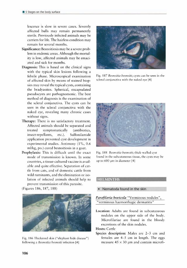
• 5 Stages on the body surface
lescence is slow in severe cases. Severely affected bulls may remain permanently sterile. Previously infected animals may be carriers for life. The hairless condition may remain for several months.
Significance: Besnoitiosis may be a severe problem in endemie areas. Although the mortality is low, affected animals may be emaciated and siek for months.
Diagnosis: This is based on the c1inical signs with the typical skin lesions following a febrile phase. Microscopical examination of affected skin by means of stained biopsies may reveal the typical cysts, containing the bradyzoites. Spherical, encapsulated pseudocysts are pathognomonic. The best method of diagnosis is the examination of the scleral conjunctiva. The cysts can be seen in the scleral conjunctiva with the naked eye, revealing many chronic ca ses without signs.
Therapy: There is no satisfactory treatment. Affected animals should be separated and treated symptomatically (antibiotics, insect-repellants, etc.). Sulfonilamide application prevented cyst development in experimental studies. Antimony (1 %, 0.6 ml/kg, po.) cured besnoitiosis in a goat.
Prophylaxis: This is difficult until the exact mode of transmission is known. In some countries, a tissue-cultured vaccine is available and quite effective. Separation of catde from cats, and of domestic catde from wild ruminants, and the elimination or isolation of infected animals should help to
prevent transmission of this parasite. (Figures 186, 187, 188)
Fig. 186 Thickened skin ("elephanr hide disease") following a Besnoitia besnoiti infection [4 J
106
Fig. 187 Besnoitia besnoiti; cysts can be seen in the scleral conjunctiva with the naked eye [4J
Fig. ] 88 Besnoitia besnoiti; thiek -walled eyst found in the subcutaneous tissue, the eysts may be
up to 600 I-Im in diameter [41
HELMINTHS
• Nematoda found in the skin
Parafilaria bovicola "Verminous nodules" , "verminous haemorrhagic dermatitis"
Location: Adults are found in subcutaneous nodules on the upper side of the body. Microfilariae are found in the bloody excretions of the skin nodules.
Hosts: Catde Species description: Males are 2-3 cm and
females are 4-5 cm in length. The eggs measure 45 x 30 ).Im and contain microfi-

lariae. Development occurs in Musca spp. which become infected when they feed on the skin lesions. When feeding on lacrimal secretions or wounds they transmit the microfilariae to the next definitive host. The verminous nodules are likely to break and ooze blood, causing a haemorrhagie dermatitis.
Geographie distribution: World-wide, espeeially many parts of Afriea
Symptoms: Subeutaneous nodules on the upper side of the body; these nodules are painful and eontain the worms. Nodules are likely to break when the gravid female starts with the exeretion of eggs. These haemorrhages and nodules are typieal signs of the infection with P. bovicola.
Signifieanee: Nodules are painful and irritating, and the carcasses at slaughter are downgraded.
Diagnosis: Verminous nodules, haemorrhage, mierofilariae in the exudate
Therapy: Ivermectin (0.2 mg/kg, Sc.) is fully effective against adults. Nitroxynil (20 mg/kg, Sc.) is also effective but must be repeated 3 days later. High doses of levamisole and fenbendazole (50 mg/kg, po.) daily for 5 days were effective.
Prophylaxis: Fly (and tick) control may reduce the entrance points of infeetive larvae.
(Figure 189)
Fig. 189 Parafilaria bovicola causing "bleeding spots" and nodules on the skin of cattle; "bleeding spots" are markedly seasonally
Onchocerca dukei Muscular onchoeereosis
Loeation: Adult worms of this speeies are found in the subeutaneous, perimuscular and museular tissue of eattle in Africa.
Hosts: Cattle Speeies deseription: The nodules are found in
the ventral thorax region, abdomen, diaphragm and thighs. These nodules may be confused with the cysts of Cysticercus bovis (Taenia saginata). Microfilariae are 240-217 11m long and 4.8-6.7 11m wide and their predilection site is the subcutis of the lower thorax and the umbilieal region. The parasite is transmitted by speeies of the genus Simulium.
Geographie distribution: Tropieal Afriea and also in sub-Saharan West Africa
Symptoms: Infeetions are usually c1inically inapparent.
Signifieanee: Losses oeeur mainly by eondemnation of carcasses when a lot of subeutaneous and intramuseular nodules are seen during meat inspeetion. Such meat is repugnant.
Diagno is: Thi is offen made ar meat in pecdon. odule are fund parri ularly in the thorax abd men and diaphra m and ma be confu ed wirh [he y f Cyslicerclis bovis ( lTlf., • 4.1). kin biop ie oaked for 12 hour in ph iol gical alin
may be inve tigated after taining with iem a ( ME11-I 0 3. 1). Typieal miero
filariae may be een. Therap: hi i not u ually indicat d. Prophylaxi : Thi i generally nor fea ible in
endemie area ( arrle, • 4. 1.J ).
Onchocerca gibsoni
Loeation: The adult worms are found in subcutaneous nodules on the brisket, shoulders and extern al surfaces of the hind-limbs. Mierofilariae are found intradermally around the nodules with a predileetion to
the brisket region . Hosts: Cattle Speeies deseription: Adult worms eause nodu
lar swellings in the skin but infected ani-
107

• 5 5tag~ on the body surface
mals are not clinieally ill. Infeeted eareasses must be trimmed to remove the nodules. The adults lie in groups or "worm nests" in the subeutaneous tissue and a nodule is formed around them. The worm nests may be up to 5 em in diameter. There is a fibrous eapsule around the nodules whieh becomes thieker as the nodule ages. This eapsule may eventually become ealcified. The males are 30-53 mm and the females 140-190 mm long (sometimes up to 500 mm). The tail of the male is eurved and it bears lateral alae and 6-9 papillae on either side. The spieules are unequal in size and measure 0.14-0.22 and 0.047-0.094 mm, respeetively. The mierofilariae are not sheathed and 240-280 11m long. The intermediate hosts are members of the genus Culicoides (e.g. Culicoides pungens and probably others).
Geographie distribution: Asia, Australia, southern Afriea
Symptoms: The infeetions do not usually cause clinical signs. Nodules may be found at the predilection sites.
Signifieance: Infected earcasses may be trimmed or even condemned at slaughter.
Diagnosis: This is based on finding the adult worms in subeutaneous nodules or microfilariae in the skin of the brisket. Microfilariae are not evenly distributed in the skin, but occur 111 irregularly distributed "nests". A number of small skin biopsies (2 mm in diameter) may be plaeed in a drop of physiologieal saline solution for 1-2 hours. MierofiIariae then migrate into the solution and can be examined.
Therapy: 1ßf' below Onchocerca ochengi Prophylaxis: Unknown; insect repellents may
reduce the attacks by the vectors.
Onchocerca ochengi (syn. Onchocerca dermata) "Intradermal onehocercosis of cattle"
Loeation: This species is found in subcutaneous and intradermal nodules on the ventral regions and flanks, primarily the udder and serotum.
Hosts: Cattle
108
Species description: In endemie areas up to 50% of the eattle are infeeted. The intermediate host has not been identified yet but a biting inseet is suspeeted. Small nodules occur deep in the hide. Mierofilariae oceur intradermally around the nodules. The mierofilariae of O. dukei and O. ochengi are coneentrated in the skin of the poster ior and anterior belly.
Geographie distribution: East and West Africa Symptoms: Intradermal nodules may be
formed mainly in the abdominal region, on the udder and serotum and sometimes in other parts of the body. Mierofilariae can eause allergie reactions and local hyperkeratosis.
Signifieance: The value of leather, whieh is often perforated by these nodules, is markedly deereased by this parasite.
Diagnosis: Intradermal nodules can be detected by palpation. At slaughter they are visible on the internal surface of the hide.
Therapy: This is generally not indicated. However, the microfilariae may be killed by diethylcarbamazine (5-8 mg/kg, po. during 3 weeks), ivermectin (200 I1g/kg, sc.), but also levamisole at increased dosage rates.
Prophylaxis: Unknown; inseet repellents may reduee the attacks by the veetors.
(Figures 159, 190)
Fig. 190 Onchocerca ochengi; skin nodules [23]
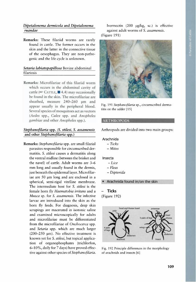
Dipetalonema dermicola and Dipetalonema ruandae
Remarks: These filariid worms are rarely found in cattle. The former occurs in the skin and the latter in the connective tissue of the oesophagus. They are non-pathogenie and the Iife cyele is unknown.
Setaria labiatopapillosa Bovine abdominal filariosis
R mark: li r filariae f [hi filariid \ rm ur in th abdominal a iry f
attl ( ATILE . 4.4) ma a i nall be found in th kin. Th mi rofilariae ar heath d, maure 240-260 J.lm and
appear u uall in th p riph ral bl d. veral pe ie ofm quir a t3 ve t r
(Aedes pp., li/ex pp. and All phe/e gambiae and rher Anopheles pp.).
Stephanofilaria spp. (S. stilesi, S. assamensis and other Stephanofilaria spp.)
Remarks: Stephanofilaria spp. are small filarial parasites responsible for circumscribed dermatitis. S. stilesi causes a dermatitis along the ventral midline (between the brisket and the navel) of cattle. Adult worms are 3-6 mm long and usually found in the dermis, just beneath the epidermallayer. Microfilariae are 50 pm long and are enelosed in a spherical, sem i-rigid vitelline membrane. The intermediate host for S. stilesi is the female horn f1y Haematobia irritans and a Musca sp. for S. assamensis. The infective larvae are introduced into the skin as the horn f1y feeds. For diagnosis, deep skin scrapings are macerated in isotonic saline and examined microscopically for adults and microfilariae must be differentiated from the microfilariae of Onchocerca spp. and Setaria spp. which are much larger (200-250 pm). No effective treatment is known yet for S. stilesi, but topical application of organophosphates (trichlorfon, 6-10%, daily for 7 days) have proved effective against other species of Stephanofilaria.
Ivermectin (200 pg/kg, sc.) is effective against adult worms of S. assamensis.
(Figure 191)
Fig. 191 Stephanofilaria sp., circumscribed dermatitis on the ud der (15)
ARTHROPODS
Arthropods are divided into two main groups:
Arachnida - Ticks - Mites
Insecta - Lice
- Fleas - Dipterida
• Arachnida found in/on the skin
Ticks (Figure 192)
Fig. 192 Principle differences in the morphology of arachnids and insects (6)
109
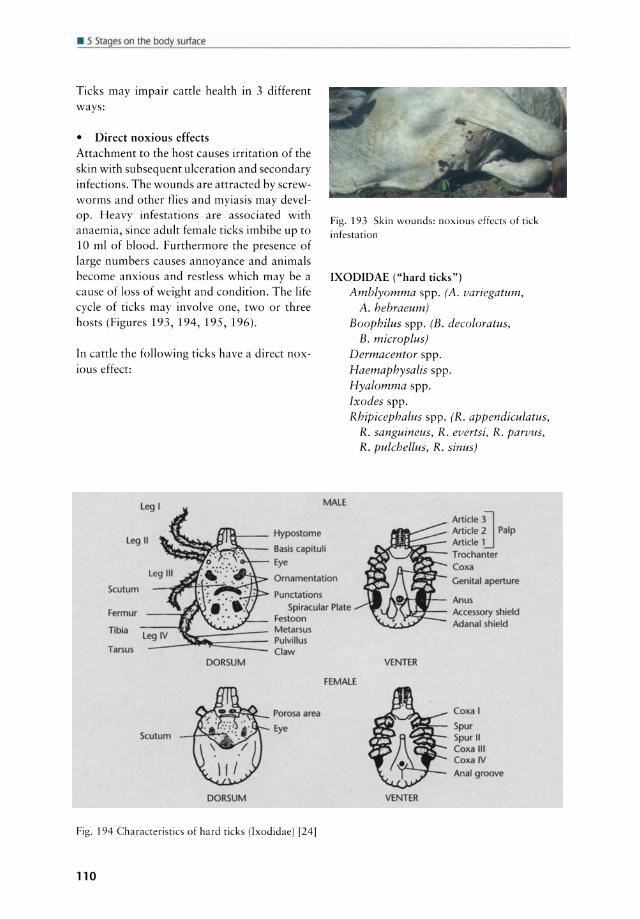
• 5 Stages on the body surface
Ticks may impair cattle health in 3 different ways:
• Direct noxious effects Attachment to the host causes irritation of the skin with subsequent ulceration and secondary infections. The wounds are attracted by screwworms and other flies and myiasis may develop. Heavy infestations are associated with anaemia, since adult female ticks imbibe up to 10 ml of blood. Furthermore the presence of large numbers causes annoyance and animals become anxious and restless which may be a cause of loss of weight and condition. The life cycle of ticks may involve one, two or three hosts (Figures 193, 194, 195, 196).
In cattle the following ticks have a direct noxious effect:
I~ _ _ Hypostome
Fig. 193 Skin wounds: noxious effects of tick infestation
IXODIDAE ("hard ticks") Amblyomma spp. (A. variegatum,
A. hebraeum)
MALE
Boophilus spp. (B. decoloratus, B. microplus)
Dermacentor spp. Haemaphysalis spp. Hyalomma spp. Ixodes spp. Rhipicephalus spp. (R. appendiculatus,
R. sanguineus, R. evertsi, R. parvus, R. pulchellus, R. sinus)
Article 2 Pa Ip
~~~r..~...-- Basis capituli Leg 11
ArticieD
Article 1 )'-..../4:.x;;.. -- Trochanter
Scutum
Fermur
Tlbia
Tarsus
Sculum
OORSUM
Eye
Omamentation
Punctations Spiracular Plate
Festoon Metarlus Pulvillus Claw
FEMALE
-'-'CW'"""'-- Porosa area Eye
OORSUM
Fig. 194 Characteristics of hard ticks (Ixodidae) [24]
110
VENTER
VENTER
Coxa Genital aperture
Anus Accessory shield Adanal shield
Coxa I
Spur Spur" Coxa 111 Coxa IV Anal groove

TICK8: KEY TO GENEaA IN \lNITED ITATE8 • ....,. D. PraU
i c:.I ...... -.-_. FAIIU AlIASlDAE - SOFT nexs
~~~ , I
H".- "'. ....... ___ .......... ......... ............. .. 1iIIdiw_ . .......
~ --..... -~
u.s. DEPAIITMfNT OP HIALTH, EDUCATlOH, AND WELP"ilE
i c:.I-.-. _.F"" .XODIDAE -MAID ncas
~.\..IC HCAL.TH ..... VIC •• c-.. ...... 0. .... c. .. , T' .............. At'.", Geertl. - •• 1
Fig. 195 Key for the identification of tick genera 1251
111

• 5 Stages on the body surface
~ I(~t
Adults
9
Moit on soil 8
Fig. 196 Life cycle of a three-host tick (Ixodes ricinus) 1.1 and 1.2: Eyeless adults on their hosts; fed females (1.2) reach a length of up to 1.5 cm. Their scutum (SC) then appears smal!. Copulation occurs while the females feed; they then drop down to the soil; 2: Over aperiod of 1 month females lay about 2000 spherical to ovoid eggs, which become attached to each other and thus appear as clusters on the soil; 3 and 4: Larvae hatch from the eggs after 3-36 weeks (depending on the temperature) and creep onto the tops of grass, from where they attach to
112
~ ~ fJJ\~ La",. 3
_ J
~ Molton soil
passing hosts (mostly small mammals, but also humans); 5 and 6: Fed larvae drop to the soil, and moult within 5-7 weeks (sometimes up to 5 months) to become eight-legged nymphs; 7 and 8: The nymphs attack larger mammals, suck blood for 2-7 days and drop to the soil, where they moult within 2-8 months and become sexually mature adults (1.1 and 1.2); 9: Mostly in spring the adults attack larger mammals including a variety of domestic animals and man, where especially females suck for 5-14 days. The whole development is temperature-dependent and in Europe needs about 3 years 1261
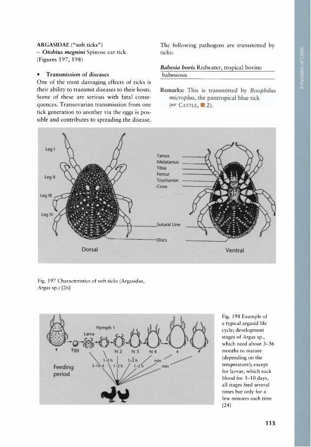
ARGASIDAE ("soft ticks") - Otobius megnini Spinose ear tick (Figures 197, 198)
• Transmission of diseases One of the most damaging effects of ticks is their ability to transmit diseases to their hosts. Some of these are serious with fatal consequences. Transovarian transmission from one tick generation to another via the eggs is possible and contributes to spreading the disease.
Leg 1
Leg 11
Leg 111
Dorsal
Fig. 197 Characteristies of soft ticks (Argasidae, Argas sp.) [26)
The following pathogens are transmitted by ticks:
Babesia bovis Redwater, tropical bovine babesiosis
Remark: Thi micr plus ( ATTl.
Tal'5U5
Metatal'5U5
nbia Femur Trochanter Coxa
I Une
i tran mirr d by 8 phillls the pantr pi al blue ri k . 2).
Ventral
i::· Nymphl ~Öö :--:
:.:.;i};; O-~-Ö.{j.ö 'Egg \ N 2 N 3 N 4 ,
'\. 1- 2h I l-h / mln./'" ~
Fig. 198 Example of a typical argasid life eyde; development stages of Argas sp., which need about 3-36 months to mature (depending on the temperature ); except for larvae, wh ich suck blood for 3-10 days, all stages feed several times but only for a few minutes each time (24)
Feeding period
~~~ .. -l~
113
• 5 51~ges on the body surface
Babesia divergens European bovine babesiosis
Remark : B. divergensis transmitred by Ixodes ricinus ( CAlTLE, • 2).
Babesia bigemina (syn. Piroplasma bigeminum) Redwater, tropical bovine piroplasmo sis, tick fever, "Texas fever"
Remarks: This para ite is transmirred by Boophilus micropills; B. deco/oratlls (the common blue tick), B. calcaratus, Rhipicephalus pp. (Rh. evertsi Rh. bursa, Rh. appendiculatus) and Haemaphysalis pp. (H. punctata) ( CAlTLE, • 2).
Babesia major (syn. Piroplasma major) European bovine piroplasmosis
Remarks: B. ma;oris tran mitred by Haemaphysalis punctata ( CAlTLE, . 2).
Cowdria ruminantium Heartwater, Tyewde
Hosts: Cattle, sheep, goat and other ruminants
Remark : Thi para ite i tran mitred by several pecies of the tick genu Amblyomma, particularly by Amblyomma variegatum (the tropical bont tick) and A. hebraeum (the Sourh African bont tick). Other pecies wh ich may be involved are A. gemma, A. pomposum, A. lepidum and A. tholloni ( C lTLE, . 2).
Theileria parva (syn. T. bovis, T. lawrencei) East Coast Fever, bovine theileriosis, Corri
dor disease, Rhodesian tick fever
Remarks: This para ite is transmitred by Rhipicephaills appendiculatus (the brown ear tick) and Rhipicephalus evertsi (the red-Iegged tick) and Rhipicephaills duttoni. Other vecrors are Hyalomma excavatum, H. dromedarii, H. tnmcatllm and some other Rhipicephalus pp. ( CAlTLE, . 2).
114
Theileria annulata Mediterranean Coast Fever, Tropical theileriosis, Egyptian Fever
Remark: Thi parasite i tran mitred by Hyalomma detritum H. truncatum and H. anatolicum ( CAlTLE, • 2).
Theileria mutans Benign bovine theileriosis, mild gallsickness
Remarks: Thi parasite i tran mitred by Amblyomma variegatum, A. hebraeum and po ibly also by Rhipicephalus appendiculatus and R. evertsi ( CAlTLE, • 2).
Anaplasma marginale Malignant anaplasmosis of cattle, gallsickness
Remark: This parasite i tran mitted by Boophilus decoloratus and B. microplus ( CAlTLE, • 2).
Anaplasma centrale Mild anaplasmosis of cattle, gallsickness
Remarks: Thi parasite is transmitted by Boophilus decolorattts ( . CAlTLE, . 2).
Eperythrozoon wenyoni
Remark: Thi parasite tran mitred by Hya/omma anatolicum. The pathogen
ccur on the erythrocyte surface ( CAlT E . 2).
Ehrlichia bovis Tropical bovine ehrlichiosis, "Nofel" or "Nopel"
Remark: Thi parasite i tran mitted by tick specie of the genera Amblyomma, Hyalomma and Rhipicephalus ( CAlTLE
. 2).
Ehrlichia phagocytophila European ehrlichiosis
Remarks: This parasite is transmitted by Ixodes ricinus ( CAlTLE, • 2).

Ehrlichia ondiri
Remar ' : Thi para ire ur '" granul )'r f atrle in hi h attitude gra t nd area f
fr! a. The 'a t v tri unkno\ n bur ti k ( the g nLl Haemaph 'salis are
LI pccted Ce nu, 2).
Borrelia theileri Tick Spirochaetosis
Remarks: This is transmitted by Boophilus decoloratus and Rhipicephalus evertsi. Tick spirochaetosis occms in catde, sheep, goats and horses.
• Tick toxicosis
Tick paralysis 43 tick species belonging to 10 genera are known to indLlce tick para lysis in man and a variety of mammals. These ticks are capable of releasing a toxin into the host which caLlses a condition associated with progressive, ascending, afebrile, symmetrical para lysis, with hind legs being affected first followed by the forelegs.
Fig. 199 Rhipicephalus evertsi; the red-Iegged tick, an important cause of tick paralysis [101
Animals may die. Paralysis is relieved if ticks are removed in time. Most domestic anima I species appear to be sLlsceptible to tick paralysis. Lambs and calves and to a lesser extent adult catde are susceptible to tick para lysis. Ixodes rubicundus (the karoo pa ra lysis tick) mainly affects sheep and goats in South Africa and Rhipicephalus evertsi causes the "spring lamb paratysis" in lambs but also in calves. (Figure 199)
Sweating sickness An acute, febrile tick-borne toxicosis characterised by a profuse, moist eczema and hyperaemia of the skin and visible mucous membranes. Watering of the eyes and nose, salivation and a extremely sensitive skin with a sour odom are other typical signs. Eventually the skin becomes cracked and predisposed to secondary infections (ind. screw-worm infections and myiasis). Often the course is acute and death may occur within a few days. In less acute ca ses recovery may occur. It is mainly a disease of young calves but also sheep in eastern, central and southern Africa. The causative agents are certain strains of Hyalomma truncatum which produce an epitheliotropic toxin. Calf mortality may reach 70%.
Symptoms: Generalized hyperaemia with subsequent desquamation of the superficial layers of the mucous membranes of the upper respiratory, gastrointestinal and external genital tracts and profuse moist dermatitis. For diagnosis it is essential to determine the presence of the vector. C1inical signs appear 4-11 days after the tick bite.
Therapy: Ticks must be removed quickly. Antibiotics and anti-inflammatory agents are useful to combat secondary infections. Immune serum may be used.
Prophylaxis: Control of tick infestations is the only effective measure. Removal of ticks, symptomatic treatment and good nmsing are indicated.
(Figures 200, 201)
115
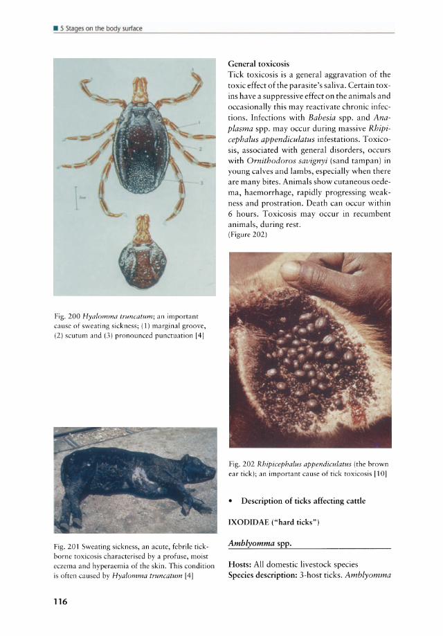
• 5 Stages on the body surface
Fig. 200 Hyalomma truncatum; an important cause of sweating sickness; (1) marginal groove, (2) scutum and (3) pronounced punctuation [4]
Fig. 201 Sweating sickness, an acute, febrile tickborne toxicosis characterised by a profuse, moist eczema and hyperaemia of the skin. This condition is often caused by Hyalomma truncatum [4]
116
General toxicosis Tick toxicosis is a general aggravation of the toxic effect of the parasite's saliva. Certain toxins have a suppressive effeet on the animals and oeeasionally this may reaetivate ehronie infeetions. Infeetions with Bahesia spp. and Anaplasma spp. may oeeur during massive Rhipicephalus appendiculatus infestations. Toxieosis, assoeiated with general disorders, oeeurs with Ornithodoros savignyi (sand tampan) in young ealves and lambs, espeeially when there are many bites. Animals show eutaneous oedema, haemorrhage, rapidly progressing weakness and prostration. Death ean oeeur within 6 hours. Toxieosis may oeeur in reeumbent animals, during rest. (Figure 202)
Fig. 202 Rhipicephalus appendiculatus (the brown ear tick); an important cause of tick toxicosis [10]
• Description of ticks affecting eattle
IXODIDAE ("hard ticks")
Amblyomma spp.
Hosts: All domestie livestock speeies Speeies deseription: 3-host ticks. Amblyomma
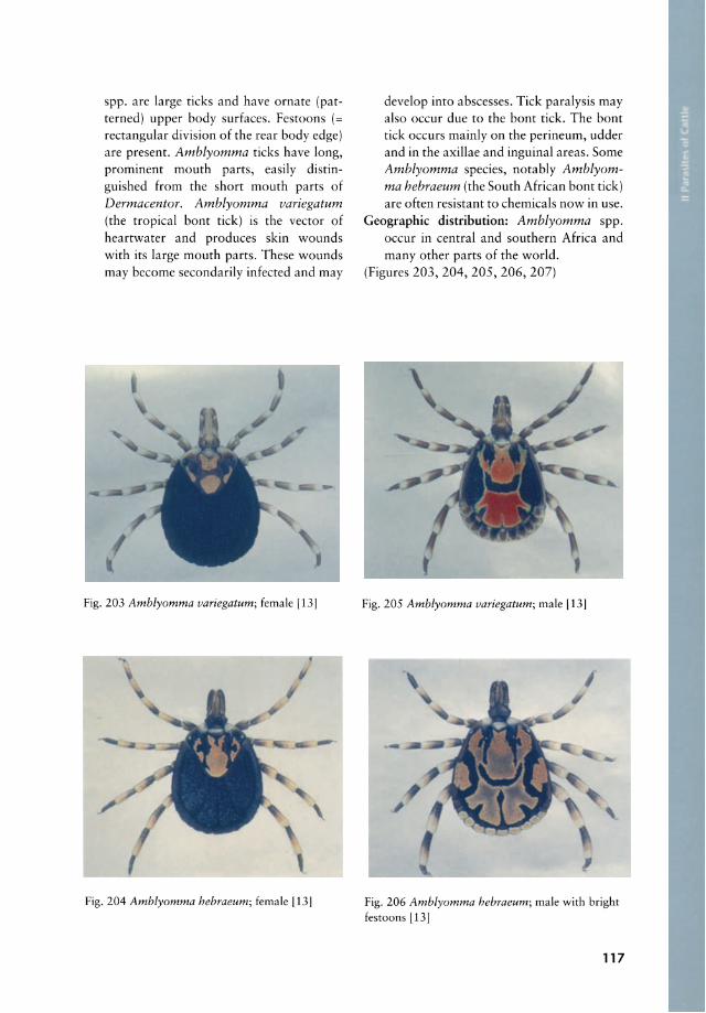
spp. are large ticks and have ornate (patterned) upper body surfaces. Festoons (= rectangular division of the rear body edge) are present. Amblyomma ticks have long, prominent mouth parts, easily distinguished from the short mouth parts of Dermacentor. Amblyomma variegatum (the tropical bont tick) is the vector of heartwater and produces skin wounds with its large mouth parts. These wounds may become secondarily infected and may
-
Fig. 203 Amblyomma variegatum; female [13]
Fig. 204 Amblyomma hebraeum; female [13]
develop into abscesses. Tick paralysis may also occur due to the bont tick. The bont tick occurs mainly on the perineum, udder and in the axillae and inguinal areas. Some Amblyomma species, notably Amblyomma hebraeum (the South African bont tick) are often resistant to chemieals now in use.
Geographie distribution: Amblyomma spp. occur in central and southern Africa and many other parts of the world.
(Figures 203, 204, 205, 206, 207)
Fig. 205 Amblyomma variegatum; male [13]
Fig. 206 Amblyomma hebraeum; male with bright festoons [13]
117

• 5 Sl~ges on the body surf~(e
Fig.207 Amblyomma variegatum; chelicera and palpes
Boophilus spp. Cattle fever tick, tropical cattle tick
Hosts: Horse and cattle Species deseription: I-host ticks. Boophilus
spp. are plain brown in colour. They are inornate with eyes. Males are small (3-4 mm) and fully engorged females may be up to 12 mm in length. Members of Boophilus transmit several diseases and cause skin irritation which leads to licking and scratching and to secondary bacterial infections. Large numbers may cause anaemia. I-host ticks such as Boophilus spp. are often the first to develop resistance to chemieals. Therefore control measures
Fig. 208 Boophilus sp.; engorged female (up to
2 cm in diameter) [10]
118
have to be changed frequently. Boophilus decoloratus (the blue tick) is one of the most important species of this genus and is found mainly on the neck, thorax, back and udder.
Geographie distribution: Throughout the world in warm climates
(Figure 208)
Dermacentor spp.
Hosts: Many wild and domestic mammals Species description: Some species are I-host
ticks while others are 3-host ticks. Dermacentor ticks have ornate (patterned) upper body surfaces. Festoons are present. The mouth parts are short.
Geographie distribution: Asia, Europe, North and South America and some parts of Africa; Dermacentor may damage their hosts by transmitting diseases (e.g. Q fever, tu laremia), causing tick worry and anaemia.
(Figure 209)
fig. 209 Dermacentor marginatus; male 141
Haemaphysalis spp.
Hosts: Dogs, birds and In Africa to a lesser extent cattle
Species description: 3-host ticks which are

smalI, inornate without eyes. Festoons are present. They may be found on the skin on all parts of the body. In addition to annoying eattle they may transmit eattle tick fever, anaplasmosis, and Q fever. They mayaIso produee paralysis and anaemia in heavy infestations.
Geographie distribution: World-wide (Figure 210)
Fig. 210 Haemaphysalis punctata; female [4J
Hyalomma spp. The bont-Iegged tick
Hosts: Horse, domestie ruminants and eamel Speeies deseription: Usually 2-host ticks,
although three hosts may be used by some speeies. Inornate (sometimes ornate) eyes are present and festoons may be absent or present. Hypostome and palps are long. Hyalomma truncatum eauses sweating siekness. Predileetion sites are the distal parts of the legs, udder, tip of the tail and perianal region.
Geographie distribution: Hyalomma spp. oeeur in many parts of Afriea, depending on the partieular speeies.
(Figure 200)
Ixodes spp. Paralysis ticks
Hosts: Cattle, horse and many other animal speCies
Species deseription: 3-host tick. Ixades ticks
are plain brown in eolour and the only eyeless speeies. The paralysis tick (Ixades rubicundus) has a 2-year Iife eyele with speeifie requirements of temperature and humidity. Ixades spp. are found anywhere on the skin of their host. I. rubicundus prefers lower parts of legs and abdomen. Ixades spp. are a primary eause of tick paralysis in sheep and goats but they mayaIso transmit diseases.
Geographie distribution: Europe, North Ameriea, South Afriea
(Figures 211, 212)
Fig. 211 Ixades ricinus; mauth parts [4]
Fig. 212 Ixades ricinus; female (large) and male (small) [4]
Rhipicephalus spp.
Hosts: Cattle, sheep and goat and many other animal speeies
Speeies deseription: 2- or 3-host ticks; festoons are present. Rhipicephalus appendicula-
119

• 5 Stages on the body surface
tus (the brown ear tick) is a 3-host tick, ARGASIDAE ("soft ticks") plain brown in colour and occurs in the ears of the host. Ir is the chief vector of Otobius megnini Spinose ear tick ECF and other diseases. Brown ear ticks are impossible to control by pasture rotation because they can survive up to 2 years on the pasture without feeding and they can feed on a large number of different speeies of wild and domestic animals. Rhipicephalus evertsi (the red-Iegged tick) is a 2-host tick and has red legs. The adults typically occur around the anus and the nymphae are found deep in the ears. The red-Iegged tick is known to develop resistance to acaricidal drugs very quickly. It transmits ECF and other diseases.
Geographie distribution: Africa south of the equator
(Figures 199,202,213)
Fig. 213 Rhipicephalus appendiculatus; male (smalI) and egg excreting female (1 cm in diameter) [8J
120
Location: Larvae and nymphae feed deep in the external ear canal. Adults are non-parasitic and live on the ground.
Hosts: Horse, donkey, mule, cattle, sheep, goat Species description: l-host tick. Free-living
adult females lay eggs on the ground which hatch within 3 weeks. Larvae and nymphs live and feed on one host for up to 7 months. Infestations of this species usually build up in kraals and stables, where the host densities are high. O. megnini can persist in empty kraals and stables for more than 2 years.
Geographie distribution: Arid and semi-arid areas of South and South West Africa
Symptoms: Infested animals shake their heads. Loss of appetite, debilitation and anaemia may be present. Irritation in the ear associated with secondary infections (incl. myiasis) may dominate the c1inical picture. Ulceration (ear canker) may occur and ticks may clog the ears, causing deafness. Waxy and oily material is discharged from the ear. Larvae and nymphs may sometimes be seen in masses. Infested animals hold their head to one side and feeding is impaired. Weight loss may occur.
Significance: Heavy infestations cause otitis externa, great annoyance and blood loss. O. megnini may cause severe problems in live stock in many parts of Africa.
Diagnosis: Ticks may be found by swabbing the ear or by direct inspection of heavily infected ears. Larvae and nymphs are found inside the ear. The adult ticks are difficult to find as they breed in hidden cracks of barns, fences and trees.
(Figures 214, 215)
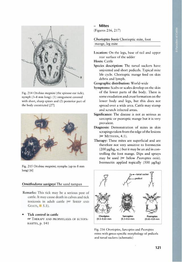
2
Fig. 214 Otobius megnini (the spinose ear tick); nymph (3-8 mm long): (1) integument covered with short, sharp spines and (2) posterior part of the body constricted [27]
Fig. 215 Otobius megnini; nymphs (up to 8 mm long) [6]
Ornithodoros savignyi The sand tampan
Remark : Thi ti k may be a attlc. lt may au e dearh in
in adulr attlc ( .1).
• Tick control in catde
f k o
llF THERAPY AND PROPHYLAXIS OF ECTOPA
RASITES, p. 141
Mites (Figures 216, 217)
Chorioptes bovis Chorioptic mite, foot mange, leg mite
Location: On the legs, base of tail and upper rear surface of the udder
Hosts: Catde Species description: The tarsal suckers have
unjointed and short pedicels. Typical mite Iife cyele. Chorioptic mange feed on skin debris and lymph.
Geographie distribution: World-wide Symptoms: Scabs or scales develop on the skin
of the lower parts of the body. There is some exudation and crust formation on the lower body and legs, but this does not spread over a wide area. Catde may stamp and scratch infected areas.
Significance: The disease is not as serious as sarcoptic or psoroptic mange but it is very prevalent.
Diagnosis: Demonstration of mites in skin scrapings taken from the edge of the lesions (1lF METHODS, 4.1).
Therapy: These mites are superficial and are therefore not very sensitive to Ivermectin (200 pglkg, sc.) but it may be an aid in controlling the foot mange. Dips and sprays may be used (1lF below Psaraptes avis). Ivermectin applied topically (500 pglkg)
Chorlptes (O.S-O.65 mm
t-ursal sucker i /ypedkel A
sarcoptes (O.3-0.6mm
~ ~
Psoroptes (O.6S-O.8 mm
Fig. 216 Chorioptes, Sarcoptes and Psoroptes mites with genus-specific morphology of pedicels and tarsal suckers (schematic)
121
• 5 Stages on the body surface
-
'0 ~ ~ et • -.::::r.
!:l "3 -0
Ǥ <
~ • ()t ~ -:::::> ~:= ....
= ~ ~ t 1 :2
ff CL
~ E >. ..... z
1 t - t '"
~ tt fJ[ J:; CL
Ct~ t z
1 1 ~ t cu
tt ~ '" ~ €:::> '" ...
..J
1 t i t ;;, 0 0 0 ~ Cl w
~ ~~ ~ --~ I~ ~ ,\ : ;; :f~ ~ , ~ E cu ...
",,~
~ Psoroptes Chorloptes c:: cu u •
Fig. 217 Developmental stages in the life cycle of important groups of mites. All stages live ontin the skin of their hosts. Larvae have only three pairs of legs. Feeding larvae and nymphs increase in size and moult. In some species there is clear sexual dimorphism; (1) Psoroptes spp. feed (as piercing
122
Sarcoptes Demodex
• • Jl
mites) on the lymph fluid and occasionally on the blood of their hosts, (2) Chorioptes spp. feed (as chewing mites) on the epidermal products, (3) Sarcoptes spp. penetrate the epidermis, forming canals and (4) Demodex spp. feed on hair follicles or on sebaceous glands [261
is effective against chorioptic mange. Crotoxyphos (0.25%) applied as a spray can also be used against the leg mite.
Prophylaxis: - below Psaraptes avis (Figures 218, 219)
Fig. 218 Chorioptes bovis, adult female (max. 650 11m long) with short, unjointed pedicels [4]
Fig. 219 Chorioptes bovis infestation [41
Sarcoptes bovis Itch mite, mange mite
Location: Neck, the back in front of the tail, inner surface of the thighs and the udder and sometimes the whole body surface
Hosts: Cattle
pread t eh r part. arcoptes mire pun rure rh kin r fe d n Iymph and km debn .
Geographie distribution: World-wide Symptoms: Intense itching is caused by irrita
tion. Animals scratch wh ich may result in dermatitis, accompanied by an exudate that coagulates and dries to form crusts. The skin is thickened, wrinkled and ha ir is lost.
Significance: Irritation to the host causes weight loss and emaciation. Skins of carcasses are of poor quality. Animals in poor condition due to mite attacks are subject to other diseases.
Diagnosis: Demonstration of mites of deep skin scrapings taken from the edge of the lesions (- METHODS, 4.1). Sarcoptes mites are usually found deep in the skin whereas Psaroptes mites are more superficial.
Therapy: This mite is very sensitive to Ivermectin (200 Ilglkg, sc.). After one treatment no Iiving mites were recovered. Dips and sprays mayaiso be used to reduce the c1inical effects (- below Psaroptes avis).
Prophylaxis: IJir' below Psoroptes avis (Figures 220, 221)
123

• 5 Stages on the body surface
Fig. 220 Sarcoptes sp.; mire wirh unjoinred, long pedicels (30-35 ~m long) 14J
fig. 221 Sarcoptes sp. infesrarion of a cattle [41
Psorpotes ovis (syn. Psoroptes communis var. bovis) Psoroptic mite
Location: Any part of the body, especially areas of dense hair such as withers, back and root of tail
Hosts: Cattle, sheep Speeies description: The mites are oval in shape
and the tarsal suckers have long, jointed pedicels. Typical mite life cycle, taking 9 days. This is a notifiable and quarantinable disease in many parts of the world.
Geographie distribution: World-wide Symptoms: Affected skin is covered with exu
date. This dries to form ascab. Massive loss of hair usually occurs. Lesions may eover the entire body. Deaths in untreated calves are not uncommon. The course may be acute in young calves and chronic nature mayaiso be found. The prognosis is usualIy favourable following early treatment.
Signifieanee: Psoroptic mange is a widespread and serious disease of cattle. Infested animals lose weight and the skins are of lower quality.
124
Diagnosis: Demonstration of mites in skin scrapings taken from the edge of the lesions (1Rf METHons, 4.1).
Therapy: Infested cattle should be dipped, not sprayed. The following compounds may be used for dips: toxaphene (0.5-0.6%), coumaphos (0.3%), phosmet (0.2-0.25%), diazinon (0.025%), flumethrine (1 I of a 6% solution in 1500 I of water) and many other compounds. Depending on the product, repeated treatments are required (label instructions should be checked). Ivermectin (200 Ilglkg, sc.) is effective against Psoroptes ovis. One treatment resulted in c1inical healing, but a second treatment is indicated in eradication programs.
Prophylaxis: Newly introduced animals are the main source of infection for a herd. These animals must be checked earefully and possibly treated before being introduced into the new herd. A quarantine may be indicated.
(Figures 222, 223, 224)
Fig.222 Psoroptes sp. female mire (max. 800 ~m long) wirh egg [4]
Fig. 223 Psoroptes sp.; mire, jointed, long pedicels (68 pm long) [4]
Fig. 224 Psoroptes sp. infesrarion of a cartle [4]
Demodex bovis Demodectic or follicular mite
Location: Neck, brisket, shoulder, face Hosts: Cattle Species description: Demodex mites are cigar
shaped, elongated mites, about 0.25 mm long. The thorax bears 4 pairs of stout, short legs and the abdomen is transversely striated. The life cyde is not dearly understood. Demodex is transmitted from the cow to the calf while nursing and may cause considerable damage to hides. Lesions are pronounced in young dairy cattle but are rarely visible or palpable on beef cattle. Rarely the lesions may appear over the entire body.
Geographie distribution: World-wide Symptoms: Small papules and nodules may be
seen. They are red and thick and a waxy, white material can be expressed from
them. This material contains numerous mites. Nodules mayaiso be filled with pus and abscesses covered with small scales may be seen. The course of bovine demodectic mite infection is usually mild but may extend over many months.
Significance: Damage to skin may affect the production of leather. Demodectic mange is not considered to be a major parasite of cattle but it may open the skin for secondary problems (bacterial and fungal infections, myiasis, etc.).
Diagnosis: Microscopical examination of the cheesy, waxy fluid from the nodules which may contain many mites. Long-standing nodules of the skin are characteristic.
Therapy: There is no satisfactory treatment. Systemic and topical applications of chemical compounds have given some relief.
Prophylaxis: Unknown (Figures 225, 226)
Fig. 225 Cigar-shaped Demodex bovis mite, (250-400 x 70 pm)
Fig. 226 Demodex bovis causing multiple skin nodules [81
125
• 5 Sl~ges on the body surf~(e
Psoregates bos Itch mites of cattle, "Australian itch"
Remarks: Itch mites occur in Canada, Australia and South Africa. These mites a re minuscule (189 x 189 11m) and difficult to collect. They are spherical and have paired c1aws and legs which are arranged radialIy. Alopecia and desquamation occur, but the lesions lack the scab formation associated with mange mite infestations. Limesulfur spray or dip applied twice, with a 2-week interval, is indicated to be the acaricide of choice.
(Figure 227)
Fig. 227 Psoregates sp.; female mite r281
Raillietia spp. (R. auris and R. caprae)
Location: Middle and inner ear Hosts: Cattle, sheep (R. auris) and goat
(R. caprae) Species description: The mites feed on epider
mal cells and wax but not on blood. Geographie distribution: North America, Aus
tralia, East Africa, Europe.
126
Symptoms: Infestations are usually inappa rent but otitis media and interna with nervous signs, including head shaking, head rotation to the affected side, circling, and general incoordination, can be found in a progressive stage of the infestation .
Signifieanee: Raillietia spp. infections can cause signs similar to those of other CNS infections and should therefore be excluded.
Diagnosis: It is almost impossible to diagnose Raillietia spp. antemortem. Demonstration of the mites in the middle or inner ear at necropsy.
Therapy: Ivermectin ma y eliminate the mites. Acaricides, applied topically mayaiso be effective.
Prophylaxis: Unknown (Figures 228, 229)
Fig. 228 Raillietia auris causing otitis media and
interna
A B
Fig. 229 Raillietia auris; dorsal view (A) and
ventral view (B) [29J

• Insecta found on the skin
lice (Figure 230)
Fig. 230 Cattle lice: Bovicola bovis (A), Haematopinus eurysternus (B) and Linognathus vituli (C) 131
MALLOPHAGA Chewing lice
Bovicola bovis (syn. Damalinia bovis) Red louse, "chewing louse"
Location: Skin surface, especially the neck, withers and root of tail
Hosts: Cattle Species description: These Iice belong to the
Mallophaga and are 1-2 mm long and wingless. They have a broad head with mouth parts adapted for chewing. The entire Iife is spent on the host. The infestations are heaviest in winter when the coats are dense.
Geographie distribution: World-wide, primarily in cooler areas
Symptoms: Intense itching associated with these Iice may be observed. Animals show typical parasite worry (not feeding, not sleeping). Scratching may produce skin wounds or bruises and the co at becomes rough.
Significance: Biting Iice are widespread and important parasites. Losses such as reduced growth and secondary skin infections due to intense itching may occur.
Diagnosis: Lice may be seen on skin. The eggs occur as white specks attached to the hairs.
Therapy: 1& below THERAPY AND PROPHYLAX-15 OF ECTOPARASITES
Prophylaxis: 1& below THERAPY AND PROPHYLAXIS OF ECTOPARASITES, p. 141
(Figure 231)
Fig. 231 Bovicola bovis (1.5-2 rnrn long); the cattle chewing lause [41
ANOPLURA Blood sucking lice of cattle
Haematopinus eurysternus Short-nosed cattle louse
Remarks: This species occurs world-wide. It is 3.4-4.8 mm long. The louse is fairly broad and the head is short.
(Figures 232, 233, 234)
Fig. 232 Haematopinus eurysternus; eggs attached to the ha ir of a cattle
127
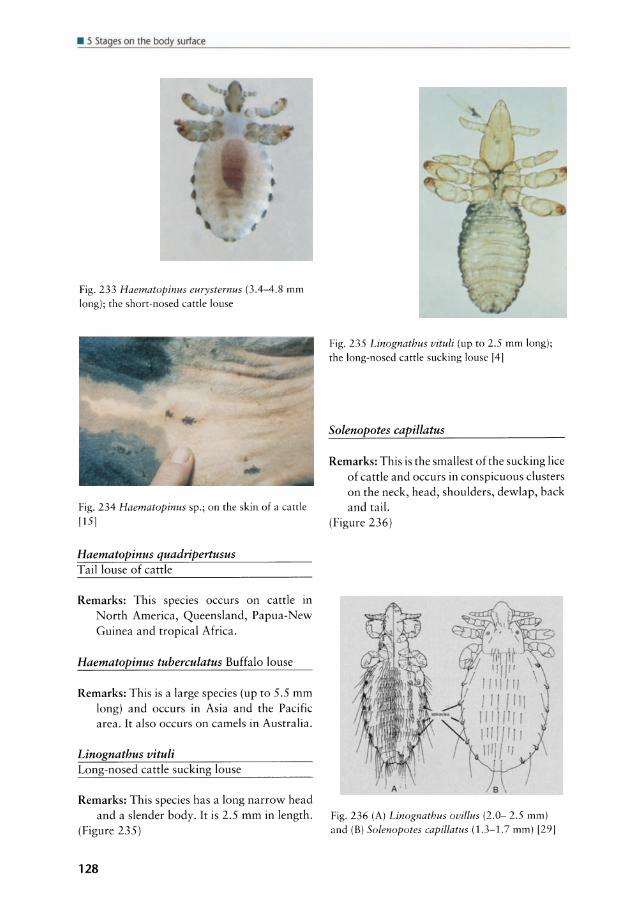
• 5 Stages on lhe body surface
Fig. 233 Haematopinus eurysternus (3.4-4.8 mm long); the short-nosed emtle louse
Fig. 234 Haematopinus sp.; on the skin of a cattl e
[151
Haematopinus quadripertusus Taillouse of cattle
Remarks: This species occurs on cattle in North America, Queensland, Papua-New Guinea and tropical Africa.
Haematopinus tubereulatus Buffalo louse
Remarks: This is a large species (up to 5.5 mm long) and occurs in Asia and the Pacific area. It also occurs on camels in Australia.
Linognathus vituli Long-nosed cattle sucking louse
Remarks: This species has a long narrow head and asIender body. It is 2.5 mm in length.
(Figure 235)
128
Fig. 235 Linognathus vituli (up to 2.5 mm long);
the long-nosed cattle sucking louse 141
Solenopotes capillatus
Remarks: This is the smallest of the sucking lice of cattle and occurs in conspicuous clusters on thc neck, head, shoulders, dewlap, back and tail.
(Figure 236)
Fig. 236 (A) Linognathus ovillus (2.0- 2.5 mm) and (B) Solenopotes capillatus (1.3-1.7 mm) [291
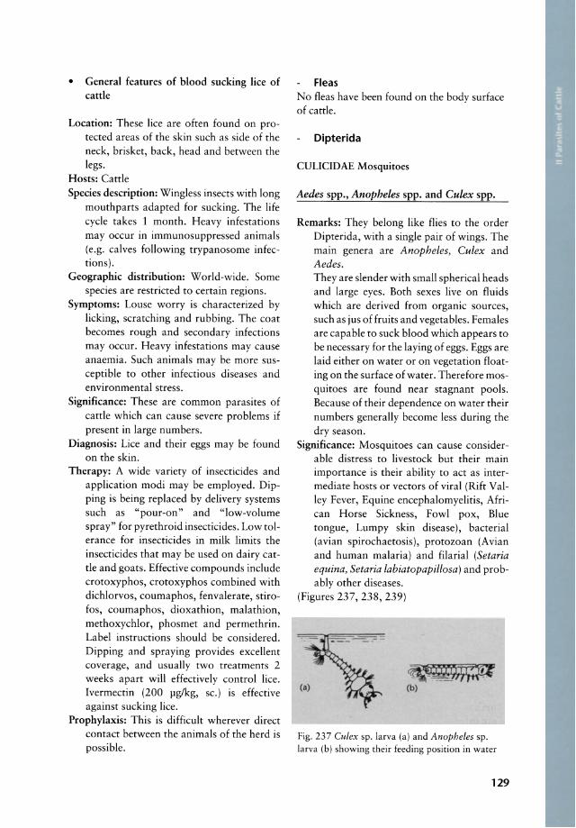
• General features of blood sucking lice of cattle
Location: These lice are often found on protected areas of the skin such as side of the neck, brisket, back, head and between the legs.
Hosts: Cattle Species description: Wingless insects with long
mouthparts adapted for sucking. The life cyele takes 1 month. Heavy infestations may occur in immunosuppressed animals (e.g. calves following trypanosome infections).
Geographie distribution: World-wide. Some species are restricted to certain regions.
Symptoms: Louse worry is characterized by licking, scratching and rubbing. The coat becomes rough and secondary infections may occur. Heavy infestations may cause anaemia. Such animals may be more susceptible to other infectious diseases and environmental stress.
Significance: These are common parasites of cattle which can cause severe problems if present in large numbers.
Diagnosis: Lice and their eggs may be found on the skin.
Therapy: A wide variety of insecticides and application modi may be employed. Dipping is being replaced by delivery systems such as "pour-on" and "low-volume spray" for pyrethroid insecticides. Low tolerance for insecticides in milk limits the insecticides that may be used on dairy cattle and goats. Effective compounds inelude crotoxyphos, crotoxyphos combined with dichlorvos, coumaphos, fenvalerate, stirofos, coumaphos, dioxathion, malathion, methoxychlor, phosmet and permethrin. Label instructions should be considered. Dipping and spraying provides excellent coverage, and usually two treatments 2 weeks apart will effectively control lice. Ivermectin (200 pg/kg, sc.) is effective against sucking lice.
Prophylaxis: This is difficult wherever direct contact between the animals of the herd is possible.
Fleas No fleas have been found on the body surface of cattle.
Dipterida
CULICIDAE Mosquitoes
Aedes spp., Anopheles spp. and Culex spp.
Remarks: They belong like flies to the order Dipterida, with a single pair of wings. The main genera are Anopheles, Culex and Aedes. They are slender with small spherical heads and large eyes. Both sexes live on fluids which are derived from organic sources, such as jus of fruits and vegetables. Females are capable to suck blood which appears to be necessary for the laying of eggs. Eggs are laid either on water or on vegetation floating on the surface of water. Therefore mosquitoes are found near stagnant pools. Because of their dependence on water their numbers generally become less during the dry season.
Significance: Mosquitoes can cause considerable distress to livestock but their main importance is their ability to act as intermediate hosts or vectors of viral (Rift Valley Fever, Equine encephalomyelitis, African Horse Sickness, Fowl pox, Blue tongue, Lumpy skin disease), bacterial (avian spirochaetosis), protozoan (Avian and human malaria) and filarial (Setaria equina, Setaria labiatopapillosa) and probably other diseases.
(Figures 237, 238, 239)
~~ (b)
Fig. 237 Cu/ex sp. larva (a) and Anopheles sp. larva (b) showing their feeding position in water
129
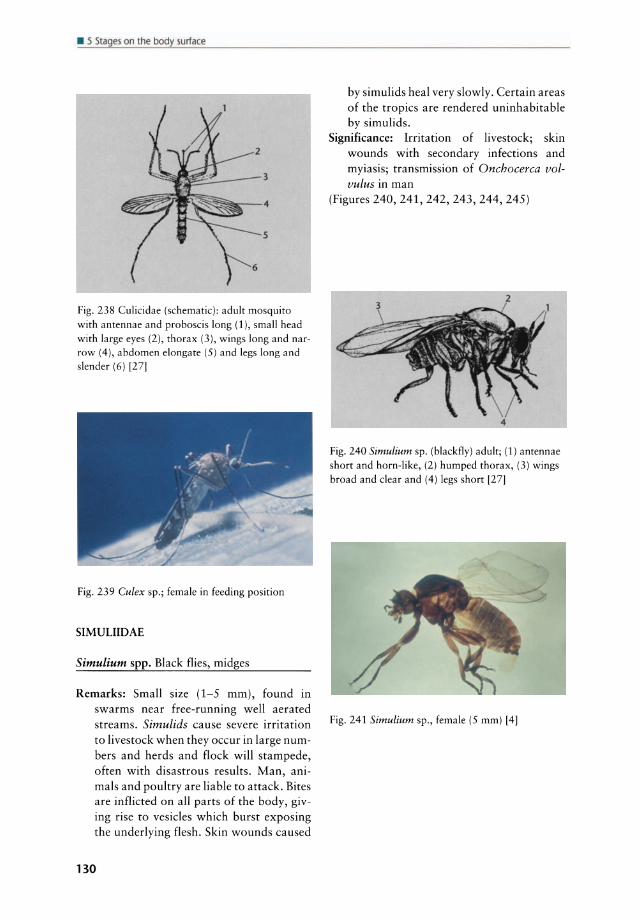
• 5 Stages on the body surface
Fig. 238 Culicidae (schematic): adult mosquito with antennae and proboscis long (1), small head with large eyes (2), thorax (3), wings lang and narrow (4), abdomen elongate (5) and legs lang and slender (6) [27]
Fig. 239 Cu lex sp.; female in feeding position
SIMULIIDAE
Simulium spp. Black flies, midges
Remarks: Small size (1-5 mm), found in swarms near free-running weil aerated streams. Simulids cause severe irritation to livestock when they occur in large numbers and herds and flock will stampede, often with disastrous results. Man, animals and poultry are liable to attack. Bites are inflicted on all parts of the body, giving rise to vesicles which burst exposing the underlying flesh. Skin wounds caused
130
by simulids heal very slowly. Certain areas of the tropics are rendered uninhabitable by simulids.
Significance: Irritation of livestock; skin wounds with secondary infections and myiasis; transmission of Onchocerca volvulus in man
(Figures 240, 241, 242, 243, 244, 245)
Fig. 240 Simulium sp. (blackfly) adult; (1) antennae short and horn-like, (2) humped thorax, (3) wings broad and clear and (4) legs short [27]
Fig. 241 Simulium sp., female (5 mm) [4]
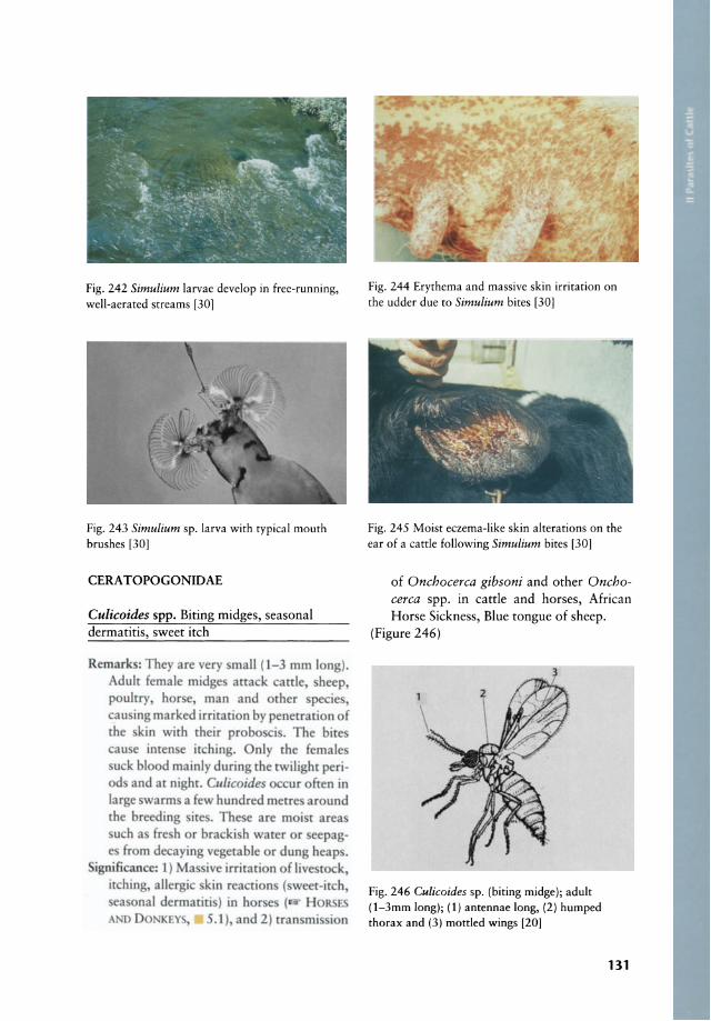
Fig. 242 Simulium larvae develop in free-running, well-aerated streams [30)
Fig. 243 Simulium sp. larva with typical mouth brushes [30)
CERATOPOGONIDAE
Culicoides spp. Biting midges, seasonal dermatitis, sweet itch
Rematk : They are very mall (1-3 mm long). Adulr female midge attack cattle heep poultry hor e man and other pecies cau ing marked irritation by penetration of the kin with rheir probe ci . The bite au e imen e itching. nly the female u k blood mainly du ring the rwilight perid and at nighr. Clllicoides occur ohen in
lar e varm a few hundred merre around the breeding ite. The e are moi t area u h a fre h or bra ki h water or eepag
fr m de aying vegetable r dung heap . ignificancc; 1) Ma ive irritati n f live rock
it hin allergie kin reacri n (weet-ireh ea nal dermatiri ) in h r ( H R
ANO 0 K Y 5.l), and 2) tran mi i n
Fig. 244 Erythema and massive skin irritation on the udder due to Simulium bites [30)
Fig. 245 Moist eczema-like skin alterations on the ear of a cattle following Simulium bites [30)
of Onchocerca gibsoni and other Onchocerca spp. in cattle and horses, African Horse Sickness, Blue tongue of sheep.
(Figure 246)
3
Fig. 246 Culicoides sp. (biting midge); adult (1-3mm long); (1) antennae long, (2) humped thorax and (3) mottled wings [20)
131
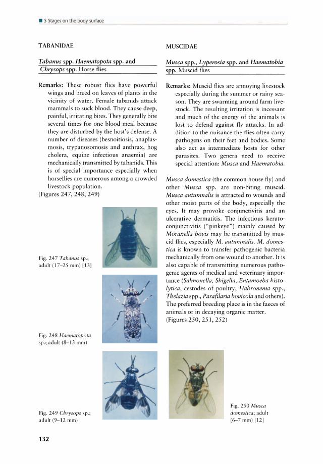
• 5 Stages on the body surface
TABANIDAE
Tabanus spp. Haematopota spp. and Chrysops spp. Horse flies
Remarks: These robust flies have powerful wings and breed on leaves of plants in the vicinity of water. Female tabanids attack mammals to suck blood. They cause deep, painful, irritating bites. They generally bite several times for one blood meal because they are disturbed by the host's defense. A number of diseases (besnoitiosis, anaplasmosis, trypanosomosis and anthrax, hog cholera, equine infectious anaemia) are mechanically transmitted by tabanids. This is of special importance especially when horseflies are numerous among a crowded livestock population.
(Figures 247, 248, 249)
Fig. 247 Tabanus sp.; adult (17-25 mm) [13]
Fig. 248 Haematopota sp.; adult (8-13 mm)
Fig. 249 Chrysops sp.; adult (9-12 mm)
132
MUSCIDAE
Musca spp., Lyperosia spp. and Haematobia spp. Muscid flies
Remarks: Muscid flies are annoying livestock especially during the summer or rainy season. They are swarming around farm livestock. The resulting irritation is incessant and much of the energy of the animals is lost to defend against fly attacks. In addition to the nuisance the flies often carry pathogens on their feet and bodies. Some also act as intermediate hosts for other parasites. Two genera need to receive special attention: Musca and Haematobia.
Musca domestica (the common house fly) and other Musca spp. are non-biting muscid. Musca autumnalis is attracted to wounds and other moist parts of the body, especially the eyes. Ir may provoke conjunctivitis and an ulcerative dermatitis. The infectious keratoconjunctivitis ("pinkeye" ) mainly caused by Moraxella bovis may be transmitted by muscid flies, especially M. autumnalis. M. domestica is known to transfer pathogenic bacteria mechanically from one wound to another. It is also capable of transmitting numerous pathogenic agents of medical and veterinary importance (Salmonella, Shigella, Entamoeba histolytica, ces todes of poultry, Habronema spp., Thelazia spp., Parafilaria bovicola and others). The preferred breeding place is in the faeces of animals or in decaying organic matter. (Figures 250, 251, 252)
Fig. 250 Musca domestica; adult (6-7 mm) l12]

Fig. 251 Musca domestica; larvae [4]
Fig. 252 Musca autumnalis; adult flies in situ
Haematobia minuta and other Haematobia spp. (the horn flies, buffalo flies) are biting museids and act as vectors of Stephanofilaria stilesi in cattle and Parafilaria multipapillosa in horses. The adults of this fly live almost permanently on cattle, buffalo, sheep and other animals. They congregate along the backs where their bites cause severe irritation so that the cattle rub themselves raw. The female only leaves her host briefly to lay her eggs in fresh cattle or buffalo dung. The larval development requires a high relative humidity of nearly 100%. The fly is therefore widely distributed in Central, East and southern Africa. The horn flies are ob liga tory parasites of cattle and a serious pest wherever they occur. Haematobia spp. cause intense worry and irritation to animals, the bite being very painful. Serious blood loss may occur when large numbers attack and loss of condition, reduced performance is a common result. The flies cause sores at the base of the horns, on the poil, ears, neck, withers and tail root. (Figures 253, 254, 255)
Fig. 253 Haematobia sp.; adult flies on the back of a cattle
Fig. 254 Haematobia irritans; adult flies (5-6 mm)
[10]
Stomoxys calcitrans and Stomoxys nigra Stable fly
Remarks: S. calcitrans also belongs to the museids. Ir attacks almost alllivestock species. Stomoxys breeds in manure (preferably horse manure) or in decaying organic matter. They require a very damp situation (heaped-up decaying vegetable matter). It is a biting museid and occurs world-wide. Both sexes of this fly are bloodsuckers and can become extremely irritating pests of man and domestic animals. On cattle the flies prefer to feed on the legs while on dogs the ears may be so bitten that they become sore and scabby. Their salivary secretions
133

• 5 51~ges on the body surface
au tO ireaction v ith an immuno uppre ive effe t rcnderin the ho r m re u eprible t di ca c .
nificance: . calcitrans a t im rmediate ho t of HabrOllema I1l1eros/oma ( H R
A 0 DONKEY, I ) Trypa1/os lila eIJal/si, Anthrax, Denn /ophilus congolel/sis, agen f rhe lumpy \ I in heep and probabi many orhcr parh gen . Ir painful bit au e imcn c \ rr and irritation to animal . Ir ma produ to i rca rion and Immuno uppre ion . BI d I ma bc marked followin ontinuou heav arrack .
Control: Sanitation is the most important control measure in stable fly contro!. Manure, straw and decaying matter should be kept away from the environment of cattle, since this provides the development medium for the flies. If good sanitation procedures are practiced, then chemical control is less likely to be needed. Without sanitation Fig. 257 Stomoxys calcitrans; adult flies in situ chemical control measures are likely to fai!.
(Figures 255, 256, 257)
Fig. 255 Stomoxys calcitrans (the stable GLOSSINIDAE
Fig. 256 Stomoxys calcitrans; adult fly (4-6 mm) with prominent biting mouthparts (arrow) 1101
134
fly); adult fly (4-6 mm) with a prominent and rigid proboscis; the maxillary palps are shorter than the proboscis and the thorax has longitudinalstripes [27]
Glossina spp. Tsetse flies, "flies that are destructive of cattle" from Sechuana
Remarks: Most tsetse flies are active during the daytime and hunt by sight and smell and feed every 2 to 3 days. Both sexes suck blood and are equally capable of transmitting trypanosomes. The female is larviparous, producing one larva at a time and an estimated total of 8-12 larvae during her life. Apart from two species in south-western Ara bia, tsetse flies are confined to the African continent, where they infest around 7.5 million square kilometres, mainly tropical regions on either side of the equator. Wherever these bloodsucking flies are, they render the area unsuitable for man and his livestock.
(Figures 258, 259)
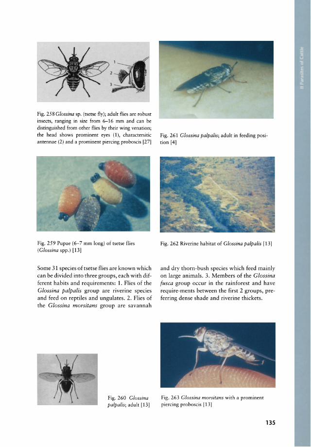
Fig. 258 Glossina sp. (tsetse fly); adult flies are robust insects, ranging in size from 6-16 mm and can be distinguished from other flies by their wing venation; the head shows prominent eyes (1), charactersitic antennae (2) and a prominent piercing proboscis [27]
Fig. 259 Pupae (6-7 mm long) of tsetse flies (Glossina spp.) [13]
Some 31 species of tsetse flies are known which can be divided into three groups, each with different habits and requirements: 1. Flies of the Glossina palpalis group are riverine species and feed on reptiles and ungulates. 2. Flies of the Glossina morsitans group are savannah
Fig. 260 Glossina palpalis; adult [13]
Fig. 261 Glossina palpalis; adult in feeding position [4]
Fig. 262 Riverine habitat of Glossina palpalis [13]
and dry thorn-bush species which feed mainly on large animals. 3. Members of the Glossina fusca group occur in the rainforest and have require-ments between the first 2 groups, preferring dense shade and riverine thickets.
Fig. 263 Glossina morsitans with a prominent piercing proboscis [13]
135
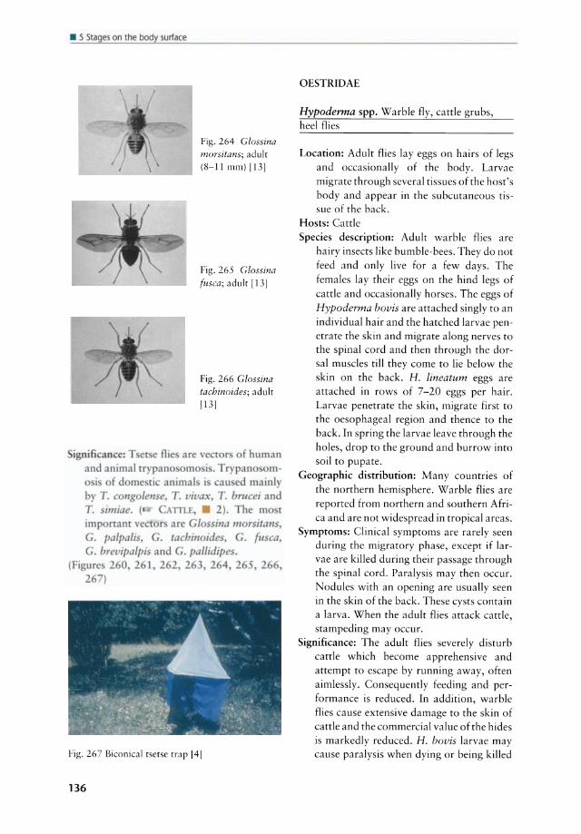
• 5 Sl~ges on the body surf~(e
Fig. 264 Glossina morsitans; adult (8-11 mm) \13]
Fig. 265 Glossina fusca; adult [131
Fig. 266 Glossina tachinoides; adult
1131
ignifi an e: T r e nie are f human and animal rr pan m i. rypano m-
i f cl me ri anima I i ,au cl mainl by T. c 1/g lellse. T. vivax. T. brucel and T. imiae. ( TILI:. . 2). he m r
,266,
Fig. 267 Biconical tsetse trap 141
136
OESTRIDAE
Hypoderma spp. Warble fly, cattle grubs, heel flies
Location: Adult flies lay eggs on hairs of legs and occasionally of the body. Larvae migrate through several tissues of the host's body and appear in the subcutaneous tissue of the back.
Hosts: Cattle Species description: Adult warble flies are
hairy insects like bumble-bees. They do not feed and only live for a few days. The females lay their eggs on the hind legs of cattle and occasionally horses. The eggs of Hypoderma bovis are attached singly to an individual hair and the hatched larvae penetrate the skin and migrate along nerves to the spinal cord and then through the dorsal muscles till they come to lie below the skin on the back. H. lineatum eggs are attached in rows of 7-20 eggs per hair. Larvae penetrate the skin, migrate first to the oesophageal region and thence to the back. In spring the larvae leave through the holes, drop to the ground and burrow into soil to pupate.
Geographie distribution: Many countries of the northern hemisphere. Warble flies are reported from northern and southern Africa and are not widespread in tropical areas.
Symptoms: Clinical symptoms are rafely seen during the migratory phase, except if larvae are killed during their passage through the spinal cord. Paralysis may then occur. Nodules with an opening are usually seen in the skin of the back. These cysts contain a larva. When the adult flies attack cattle, stampeding may occur.
Significance: The adult flies severely disturb cattle which become apprehensive and attempt to escape by running away, often aimlessly. Consequently feeding and perform ance is reduced. In addition, warble flies cause extensive damage to the skin of cattle and the commercial value of the hides is markedly reduced. H. hovis larvae may cause paralysis when dying or being killed
during their migration through the spinal canal. Severe oedema may occur when larvae of H. lineatum are killed during their migration through the oesophageal region.
Diagnosis: Larvae in cysts or lumps under the skin of the back indicate warble infestation. Eggs may be found on hair of the animal's legs.
Therapy: Cattle, especially calves, in areas where grub numbers are high, should be treated as soon as possible after the end of the warble f1y season. They should not be treated later than 8-12 weeks before the anticipated first appearance of grubs in the backs, since adverse reactions may occur when migratory larvae are killed in the spinal cord. Pour-on treatments of couma-
Fig. 268 Hyporderma bovis (warble fly); adult (15 rnrn long) [4]
Fig. 269 Life eyele of Hyporderma spp. [6]
Fig. 270 Hyporderma bovis larvae callsing nodules on the back
phos, famphur, fenthion, phosmet, trichlorfon or ivermectin may be used and poured evenly along the midline of the back. Sprays containing coumaphos or phosmet also control cattle grubs. The entire surface of the skin should be wet for sufficient absorption. Coumaphos and phosmet mayaIso be used as dips. Ivermectin (200 Ilg/kg, sc.) is highly effective against cattle grub. Oral application is also effective.
Prophylaxis: Attacks by adult warble flies are difficult to avoid and entail excessive use of insecticides. Early treatment in endemie areas is indicated.
(Figures 268, 269, 270, 271)
urv .. ,AI lD grouod ..,., PUPO" ...... Adult fty 0m<r9<' 'rom pup<
lern
Fig. 271 Hypoderma bovis; section through a skin nodllie containing a larva
137
• 5 51~ges on the body surface
CALLIPHORIDAE The blowflies and their allies
Remarks: They are highly important in many domestic animal species and man. The adults are free-living and the larvae are parasitic maggots which develop in the tissue of their host, causing a condition ca lied myiasis. The larvae may be laid into preexisting wounds. Myiasis is often a secondary skin problem.
(Figure 272)
Fig. 272 Calliphora sp.; adult (12 mm long) [4)
The family of Calliphoridae can be divided into two groups, the metallic flies and the nonmetallic flies .
The metallic flies are green, blue or purplish coloured and belong to the genera Luci/ia, Chrysomyia and Callitroga
Lucilia cuprina and other Lucilia spp. "Green-bottle" or "copper-botde" flies
R m arks: L. CIIprifla i rhe pr d minam cau e of heepbl w A trike in uth fri a and
u tralia. It cau e m ia i in heep. Ir i (he mo t imp rtant primary bl w Ay
rrike n Iiving he p ( HF P
.1 ).
Fig. 273 Lucilia sericata (the green-bottle flies); adult (8-9 mm)
138
Chrysomya bezziana Carde screwworm, the Old World screwworm, blow fly strike of catde
Remarks: C. bezziana infests catde but also horses, sheep, dogs and sometimes man. Ir occurs in tropical and southern Africa and oriental regions. C. bezziana causes severe myiasis and toxins produced by the larvae result in retarded healing of wounds. Death may occur in severe cases.
(Figure 274)
Fig. 274 Chrysomya bezziana (the ca ttle
screwworm); adult (8-10 mm) [20)
Callitroga hominivorax (syn. Coch/iomyia hominivorax) "American screwworm"
Remarks: C. hominivorax has recendy been found to occur also in North Africa. Catde, pigs and equines suffer most frequendy, but other animals, including fowls and even man mayaiso be affected. Pathology is essentially the same as in C. bezziana.
(Figures 275, 276)
Fig. 275 Callitroga hominivorax (syn . Cochliomyia hominivorax) the "screwworm"; adult (8-10 mm long)
[19]

cf' - --.... 9 COf"ULATtON
OIrV LO"'M NT FROM
t:G(; THftOUGM , LARYAI..
STAOII:' IN THC ' WOUHO
~UI..LY oeVILO~EO LJ
\ YOUNO ...... 11::.
) ItM RGC:s AB
YDUNG AOULT
~ . . --_ . OURROWa ... ~ _ .... •• ~. - - _ ••
INTO SOlL" d O.POSITIOH 0" eoo. M"5S ON WOUNO
OC\IICl.O,..
IHTO pur"
The non-metallic flies are dull grey, yellowbrown or black and belong to the genera Wohlrartia, Sarcophaga and Cordylobia
Sarcophaga haemorrhoidalis Red-tailed fleshfly
Fig. 276 Life eyele of Callitroga hominivorax [31)
Remarks: The fly is very common in Africa south of the Sahara and is frequently found around human habitations. Flies of this species are larviparous and may lay their larvae in wounds or sores although larvae mayaiso be deposited on faeces, carrion or fresh meat. Several anima I species and man may be affected. The fly may cause myiasis, especially in sheep in some parts of the world.
Fig. 277 Sarcophaga sp.; adult f1 y depositing its larvae on the skin of a eattle [S)
(Figures 277, 278) Fig. 278 Sarcophaga sp.; adult f1y (13-15mm long) (4)
139

• 5 51~ges on the body surface
Wohlfartia magnifica Old World fleshfly
Remafks: This speeies oeeurs in North Afriea. The fly attaeks man and other animals. The larvae may be deposited into the external ear of man or in SOfes around the eyes.
(figure 279)
rig.279 Wohlfartia sp.; adult Ay (8-14 mm long)
141
Cordylobia anthropophaga Tumbu fly or "skin maggot fly"
Remafks: The fly is widely distributed in Afriea south of the Sahara. It produees myiasis in man, dogs and other domestie animals. Eggs are laid around plaees where animals lie. After hatehing the larvae penetrate the skin of the host, produeing a boi I of about 1 em aeross with a hole in the eentre. When mature the larvae wriggle out of the hole and pupate on the ground. Tumbu boils are found eommonly on the ventral parts of the body but also on any other area of the body.
(Figures 280, 281)
rig. 281 Cordylobia anthropophaga; thirdstage larva (12-28 mm
lang) 1271
140
Fig. 280 Cordylobia anthropophaga (Tumbu f1y); stout fly with two black marks
on the thorax; the face and legs are yellow (8-12 mm lung) [27]
HIPPOBOSCIDAE The louse flies
Hippobosca equina Horse louse fly
Ho [ : Hor e antI -::mlc cographic di triburion: ~ orkl-wide
([ HOK'" A I> Do,,,, ''', . 1) (Figurc. 2 2,2 )
Fig. 282 Hippobasca equina (horse louse f1y; 7-9 mm long)
Fig. 283 Hippobosca sp.; adult louse flies on a
curie [41
Hippobosca variegata
Hosts: Horse and eattle Geographie distribution: Tropieal Afriea and
oriental regions
Hippobosca rufipes Cattle louse fly
Hosts: Mainly wild and domestie Bovidae, less frequently horses
Geographie distribution: Tropieal Afriea (arid and semi-arid areas of Afriea)
(Figure 284)
Hippobosca maculata
Hosts: Horse and cattle
Fig. 284 Hippobosca rufipes (eattle louse fly); this fly is very hard-bodied and diffieult to squash. It has well-developed wings and strong legs ending in well-developed claws (8-11 rnrn long) [271
Geographie distribution: Tropieal and subtropieal areas of Afriea
(Figure 285)
Fig. 285 Hippobosca maculata; adults (8 rnrn long) attaehed to the jugular vein region of a eattle [8)
Hippobosca camelina Camellouse fly
Hosts: Camel, horse and oeeasionally eattle Geographie distribution: Northern Afriea and
Middle East
• General features of Hippobosca spp.
Loeation: The adult louse flies remain for long periods on their host and cluster in the perineal region, between the hindlegs to the pubic region, but mayaIso bite on other parts of the body.
Speeies deseription: The flies live permanentlyon their host and feed on blood. MainIy eattle and horses are affeeted. They rare-
Iy fly and then usually not more than a few metres. They spend their whole time on the host and are strongly attaehed to their host and therefore diffieult to dislodge. When disturbed they ean quickly move sideways.
Symptoms: Infested animals serateh and rub, and skin-trauma are often seen as a consequenee of heavy infestations.
Signifieanee: These flies are a souree of great irritation. They transmit the non-pathogenie Trypanosoma theileri, anthrax and other bacterial infections to cattle.
Diagnosis: Identification of flies loeated under the tail and between the hindlegs.
Therapy: l& below, THERAPY AND PROPHYLAX-15 OF ECTOPARA5ITE5, p. 141
Prophylaxis: Regular grooming may be helpful to reduee louse fly infestations.
• Therapy and prophylaxis of eetoparasites (for araehnids and inseets)
A great number of inseetieides is used to control both araehnids and inseets (Table 11) . Methods of applieation include whole-body sprays, dips, dusts and topieal applieation to the dermis and ears. Dips are more effeetive for ticks. Thorough treatment of the infested area is required, espeeially when ticks infest the ears or underside of the tail or mites infest loealized patehes on the skin. Impregnated ear tags, eontaining pyrethroids or organophosphates, are of increasing importanee in fly control. Elimination of parasites or stages in the environment is diffieult beeause many eetoprasites are eapable of surviving in the environment for prolonged periods (e.g. some ticks ean live on the ground up to 300 days without feeding). In areas where many ticks exist reinfection of the host oeeurs eontinuously and treatment therefore must be repeated regularly . Some ticks are not strietly speeies-speeifie and ticks normally adapted to horses ean oeeasionally also affeet eattle or other animals and they must be treated, too, if attempts are made to reduee tick infestations. In sub humid areas the period of highest tick aetivity is the wet season and only few ticks are found on animals during the dry season. Con-
141

sequently tick contral is focussed to the wet season. In tropical areas most ticks are active
thraughout the year and must be controlled continuously.
Table 11 Some common compounds to contral extern al parasites of cattle
Compound
Organophosphates
Chlorpyrifos
Coumaphos
Crotoxyphos and dichlorvos
Dichlorvos
Dichlorvos and tetrachlorvinphos
Diazinon
Dioxathion
Fenthion
Malathion
Phoxim
Phosmet
Stirofos
Tetrachlorvinphos
Trichlorfon (metrifonat)
Carbamates
Carbaryl
Promacyl
Chlorinated Hydrocarbons*
Lindane
Methoxychlor
Toxaphen
Pyrethrins and Pyrethroids
Cyfluthrin
Cypermethrin
Deltamethrin
Fenvalerate
Flumethrin
Permethrin
Pyrethrin
Avermectins
Doramectin
Ivermectin
Pest
Lice, horn flies
Horn flies, lice, ticks, grubs, screw worms, rnites
Horn flies, face flies, stable flies, house flies,lice, ticks
Horn flies, face flies, stable flies, house flies
Horn flies, face flies, lice, ticks
Horn flies (including pyrethroid-resistant flies),
spinose ear ticks, lice, mites, Hypobosco spp.
Horn flies, lice, mites
Horn flies, lice, Hypoderma spp.
Lice, keds, mites
Mites, lice, flies, myiasis, ticks
Grubs, lice, horn flies, ticks, mites
Horn flies, face flies, house flies, stable flies
Horn flies, lice, ticks
Grubs, lice, mites
Mites, lice, ticks, flies
Mites, lice, ticks, flies
Screwworm larvae, mites, lice, ticks, flies
Mites, lice, ticks, flies
Mites, lice, ticks, flies
Horn flies, horse flies
Horn and face flies, ticks
Flies, including tsetse flies
Horn and face flies, spinose ear ticks
Flies, including tsetse flies, ticks
Horn flies, face flies, mites, ticks, lice
Horn flies, face flies, mites, lice
Mites, lice, ticks, Hypoderma spp.
Mites, lice, Hypoderma spp.
* some are no longer approved by some governments
142

5.2 Eyes
Thelazia rhodesi Cattle eyeworm
Location: Conjunctival sac Hosts: Cattle, rarely sheep and goat Species description: Milky-white worms,
males 8-12 mm long and females 12-18 mm. The cuticle is transversely striated. The spicules are 0.75-0.85 and 0.115-0.13 mm long. Intermediate hosts are Musca larvipara and Musca amica. Infected flies transmit the infective larvae via saliva to calves. Adult Thelazia appeared 20-25 days after infection. In some regions a high percentage of calves is infected but lesions are often absent.
Geographie distribution: World-wide Symptoms: Conjunctivitis, clouded cornea,
marked lacrimation. The affected eyes may be swollen and covered with exsudate and pus. Without treatment progressive keratitis and ulceration of the cornea may occur.
Significance: Infections with eyeworms may be very prevalent in some regions. Heavy infections may cause marked irritation, keratitis and weight loss of affected animals.
Diagnosis: This is made by the detection of worms in the conjunctival sacs. Examination of the lacrimal secretions may reveal eggs or first-stage larvae.
Therapy: Levamisole (5 mg/kg, sc.) and ivermectin (200 flg/kg, Sc.) are active against Thelazia spp. Concurrent use of antibiotic ointment for secondary invaders is recommended.
Prophylaxis: Fly control measures against the face fly aid in the control of thelaziosis in both cattle and horses.
(Figures 286, 287, 288)
Fig. 286 Thelazia rhodesi (eyeworm); anterior end with typical cuticular striation
Fig. 287 Thelazia rhodesi in the eyes of a cattle
Fig. 288 N'Dama cattle with lacrimation due to
Thealzia rhodesi infection
143
Copyright © 2022 FDOKUMEN