Clinical observations of cattle and buffalos with experimentally induced chronic copper poisoning
Upload
khangminh22Category
view
5download
0
Observations on Abortions in Cattle: A Comparisonof Pathological, Microbiological and Immunological
Findings in Aborted Foetuses and FoetusesCollected at Abattoirs
R. B. Miller and P. J. Quinn*
portance of immunoglobulin determinations inaborted foetuses are discussed.
Fifty nonaborted and 50 aborted bovine foe-tuses were examined utilizing histology, im-munoelectrophoresis, bacteriology and thefluorescent antibody technique.
Lesions were observed in 12 of the non-
aborted foetuses and in four of these immuno-globulins were demonstrated. In addition, twoof the nonaborted foetuses had immunoglobu-lins in the absence of observed lesions.
Lesions were observed in 48 of the abortedfoetuses and immunoglobulins were detectedin 22 of these. An etiological diagnosis was ar-
rived at in 24 of the 50 aborted foetuses. Thetissues most frequently observed to have le-sions of diagnostic significance were eyelid,intestine, liver, lung and placenta. Intestinallesions were observed in several foetuses inassociation with a variety of agents includinginfectious bovine rhinotracheitis.
Foetuses diagnosed as aborting because ofmycotic infection consistently displayed lesionsin their eyelids.The value of taking eyelid sections in cases
of suspected mycotic abortions, the signifi-cance of foetal intestinal lesions, the evaluationof abomasal aspirates and the diagnostic im-
*Department of Pathology, Ontario Veterinary College,University of Guelph, Guelph, Ontario (Miller) andDepartment of Veterinarv Medicine, Veterinary CAl-lege, Dublin 4, Ireland (Quinn).
Submitted August 9, 1974.
RESUME
Cette etude visait a examiner plusieurs tis-sus de 50 foetus bovins et d'autant d'avortons,tous ages de plus de 120 jours, par les metho-des suivantes: histologie, immuno-electropho-rese, bacteriologie et immunuofluorescence.On de&ela des lesions chez 12 foetus et on
demjnontra le presence d'immuno-globulines,chez quatre d'entre eux. De plus, deux foetusrecelaient des immuno-globulines, en l'absencede lesions visibles.Par ailleurs, on decela des lesions chez 48
avortons et des immuno-globulines, chez 22d'entre eux. On reussit a poser un diagnosticprecis dans 24 des 50 cas d'avortement. Lespaupieres, l'intestin, le foie, les poumons et leplacenta representaient les tissus dans lesquelson decela le plus souvent des lesions diagnosti-ques importantes. Plusieurs avortons presen-taient des lesions intestinales attribuables adivers agents infectieux, y compris le virus dela rhino-tracheite infectieuse bovine.Dans les cas d'avortement mycotique, les le-
sions des paupieres des avortons s'avererentconstantes.
Les auteurs commentent la valeur des cou-
pes histologiques des paupieres, lorsqu'onsoupgonne un avortement mycotique, la signi-fication des lesions intestinales, l'evaluation duliquide qu'on aspire de la caillette et l'impor-tance diagnostique de la mise en evidence d'im-muno-globulines, chez les avortons.
Can. J. comp. Med.
ABSTRACT
270

INTRODUCTION
The incidence of -bovine abortion has beenthe subject of papers by Fosgate and Smith(41), Holt (48), Mitchell (77), Withers(112), Dearborn et al (34), Amiel et al (4)and David et al (31). General factors mili-tating against the birth of a live calf havebeen reviewed by Andrews (5), Ashton andFallon (6), Bishop (12), Boyd (17), Fech-heimer (39) and Hanly (44). Discussionsregarding the most common infectiousagents associated with an unsuccessful re-
productive event have been included in textsby Blood and Henderson (16), Faulkner(38), Merchant and Packer (75), Roberts(91) and in a recent paper on -bovine abor-tion by Hubbert et al (50). The lesions com-monly associated with many of these agentsare referred to by Jubb and Kennedy (55).The immunological capability of the bovinefoetus has been examined by Butler (20),Butler et al (21), Osburn and Hoskins(83), Dunne et at (36), Hubbert et al (51),Schultz (95), Schultz et al (96) and Horneret al (49).
Correlated studies on aborted and non-aborted bovine foetuses utilizing the tech-niques of histopathology, immunology, bac-teriology and virology are not available.Frequently in foetal pathology the signifi-cance of a lesion can not be ascertained. Astudy of the differences and the similaritiesbetween the aborted and nonaborted foe-tuses therefore should aid in the diagnosesof bovine abortion. In this study samplesfrom 50 nonaborted and 50 aborted bovinefoetuses over the estimated gestational ageof 120 days were examined histologically,immunoelectrophoretically, bacteriologicallyand by fluorescent antibody tests for thepresence of two viruses.
MATERIALS AND METHODS
Two populations of foetuses were exam-ined, nonaborted and aborted. The non-aborted foetuse3 were collected at an abat-toir located in Kitchener, Ontario. Most ofthe cows were from southern Ontario, somewere from Manitoba, Saskatchewan andAlberta. Collections were made duringMarch and October in 1971. All foetuses
were removed from the uterus within 30minutes after the cow was stunned. Bloodfor immunoelectrophoretic analysis wascollected from the umbilical veins.Twenty aborted foetuses were collected
from submissions made at the Guelphdiagnostic laboratory of the Ontario Vet-erinary Services Branch and at the hospitalof the Ontario Veterinary College in Guelphbetween December 1972 and January 1973.Thirty aborted foetuses were collected fromsubmissions made to the Provincial Vet-erinary Laboratory in Edmonton, Albertaduring February 1973. The placenta wasavailable from 16 of the aborted foetuses.Blood was usually collected on cutting thebrachial vessels but when blood was notavailable at this site the serosanguinousfluid present in the thorax was used forimmunoelectrophoretic analysis. On boththe aborted and nonaborted foetuses usedin this study the gestational age was esti-mated following the descriptions of Hub-bert et al (52). Foetuses judged to be lessthan 120 days were discarded. Samples ofblood or thoracic fluid for immunoelectro-phoretic analysis and abomasal contents formicrobiological examination were collectedin syringes, transferred to sterile vials andheld at -70°C. Samples of placenta, eyelid,thyroid, thymus, lymph node, lung, myocar-dium, liver, pancreas, adrenal, kidney,spleen, intestine, skeletal muscle and brainfor histopathological examination werefixed in formalin. The sections of the brainwere taken from the cortex, medulla andcerebellar folia. Additional portions oflung, kidney and intestine were held at-70°C for examination by the fluorescentantibody technique.
IMMUNOELECTROPHORETIc ANALYSIS
Sera from the nonaborted foetuses andsera or thoracic fluid from the aborted foe-tuses were examined for immunoglobulinsby immunoelectrophoresis. Electrophoresisof the samples was done alongside a pooledsample of serum from ten mature cows.Precipitation patterns were developed withrabbit antibovine serum using the methodof Gelman (42). Immunoglobulins wereidentified using the descriptions of Butleret al (21) and the presence of IgM was veri-fied by repeating the immunoolectrophoresisusing specific antibovine IgM' in the centraltrough.
'From Miles Laboratory.
Vol. 39-July, 1975 271

MICROBIOLOGICAL EXAMINATION
Abomasal contents were examined micro-scopically on wet mount and after stainingwith Gram's stain. Two drops of abomasalcontent from each foetus were plated onsheep blood agar and incubated in an atmo-sphere of reduced oxygen tension. In addi-tion, two drops of abomasal content wereplated on McConkey's agar and incubatedat normal atmospheric pressure. All plateswere examined at 24, 48 and 72 hours.Plates containing over 100 colonies of onetype with no other colony types visible wereclassified as "Pure cultures". Those platescontaining two or more types of colonieswere classified as "Mixed cultures (MC)".The frozen samples of kidney from all
foetuses and one sample of intestine fromfoetus number 16A were examined for in-fectious bovine rhinotracheitis (IBR) viralantigen using the direct fluorescent anti-body method described by Reed et aX (90).Lung was examined for parainfluenza-3(PI-3) viral antigen using a similar tech-nique.2 Positive or questionable samp!eswere reexamined using the indirect methodwith a known positive and negative control.Virus isolation was attempted on thoracicfluid of foetus number 16A.
HISTOLOGICAL EXAMINATION
All specimens fixed in formalin were pro-cessed routinely, cut at six microns andstained with haematoxylin and eosin (H&E).
RESULTS
Nineteen nonaborted foetuses and 48aborted foetuses were observed to have apositive finding in at least one of the threecategories in which they were examined.Thirty-six of the aborted foetuses werefound to have a positive finding on histo-
2A1I fluorescent antibody conjugates used int this studywere obtained from Dr. E. J. Carbrey, National Ani-mal Disease Laboratory, Ames, Iowa.
logical examination and in at least oneother category. In 12 of the aborted foe-tuses lesions only were found with no posi-tive findings in the other categories. Thepresence of immunoglobulins and the micro-biological and histological observations in19 nonaborted foetuses are shown in TableI and in 36 aborted foetuses in Table II.
IMMUNOELECTROPHORESIS
In the foetal serum from the nonabortedfoetuses IgM was demonstrated two timesand IgG six times. Immunoglobulins werepresent without observed lesions in two ofthe nonaborted foetuses. The immunoelec-trophoretic pattern of a 120 day non-aborted foetus is compared to the patternof a pooled serum sample from mature cowsin Fig. 1.
In the fluid from the aborted foetusesIgM was demonstrated 19 times and IgG11 times. In the aborted foetuses lesionsaccompanied the demonstration of immuno-globulins in every case. Fig. 2 is an im-munoelectr.ophoretic pattern showing IgMin the thoracic fluid of a 165 day abor;edfoetus. Fig. 3 is an immunoelectrophoreticpattern demonstrating IgM and IgG in thefoetal sera of a 195 day aborted foetus.
MICROBIOLOGY
Bacteria, yeast forms or mycotic ele-ments were demonstrated in eight of thenonaborted foetuses and in 32 of the abortedfoetuses. In the stomach contents from twoof the nonaborted foetuses bacteria weredemonstrated with Gram's stain but not onculture (see Table I). Gram-positive cocciwere observed in one and gram-negativebacilli in the other. No lesions were ob-served in either of these foetuses. Mixedcultures consisting of occasional bacterialcolonies belonging variously to the generaAlcaligenes, Streptococcus, Diplococcus andEscherichia were observed four times.Large numbers of Aspergillus fumigatuswere found in the stomach contents of non-aborted foetus 4NA. A lesion was observedon microscopic examination of the eyelidbut no hyphae were detected. No lesionswere found in the one cotyledon examinedhistologically. In the gastric aspirates fromaborted foetuses pure cultures of bacteriawere observed six times. These consisted ofthree E. coli, two C. pyogenes and one Yer-
Can. J. comp. Med.272

TABLE I. Immunological, Microbiological and Histopathological Findings in 19 NonabdOhedFoetuses. Each Foetus Represented was Found Positive in at Least One Category
ImmunoglobulinsFoetusNumber Age IgM IgG Microbiology Lesions37NA...........llNA...........1NA...........
26NA...........48NA...........28NA...........32NA...........33NA5NA...........8NA...........40NA...........lONA...........20NA...........23NA...........14NA...........24NA...........35NA...........4NA...........
29NA...........Totals 19
120135150150150165180180195195195210210210225255255270270
_ +
+ +
PC& (Gm + cocci)
mC
MC
- - yeast forms
2
2 6
MCPCa (Gm- Bacilli)
MCMycotic
8
Age: in daysMC Mixed culture of bacteriaPC - Pure culture of bacteriaBacteria seen on smear but no growth on culture
sinia pse-udotuberculosis. Fourteen foetusesyielded mixed cultures of bacteria belong-ing variously to the genera Alcaligenes,Streptococcus, Bacillus, Escher ichia, Pro-teus and Mficrccoccus. Five of the foetuseshad hyphae in large numbers in theirstomach contents. In addition from one ofthese five a mixed culture of bacteria wasgrown on blood agar. The five aborted foe-tuses in which fungal hyphae were demon-trated in the stomach contents had immuno-globulins in their thoracic fluid. In two ofthe foetuses, 27A (aborted) and 5NA (non-aborted), yeast forms were observed withGram's stain. No lesions or immunoglobu-lins were demonstrated in 5NA. Foetus 27Ahad lesions but no immunoglobulins.
Fig. 1. The upper antigen well contains serum from amature cow. The lower antigen well contains serum froma nonaborted foetus collected at approximately 120 daysgestation. The immunoelectrophoretic pattern was de-veloped by placing rabbit antibovine serum in the cen-tral trough. No immunoglobulins are visible in the foetalpattern.
Fig. 2. The upper antigen well contains serum from a
foetus aborted at approximately 165 days gestation. Thelower antigen well contains serum from a mature cow.The immunoelectrophoretic patt2rn was developed byplacing rabbit antibovine serum in the central trough.An IgM line (arrow) can be distinguished in the foetalpattern.
Infectious bovine rhinotracheitis viralantigen was not found in the kidneys ofany of the nonaborted foetuses but was de-monstrated in the kidneys of seven of theaborted foetuses and in the intestine ofaborted foetus 16A. Fig. 4 is a photomicro-graph of focal fluorescence in the intestineof case 16A positive for infectious bovinerhinotracheitis. All foetuses with demon-strable IBR viral antigen in the kidney hadlesions in the liver. Aborted foetus 16A hadlesions in the intestine and lung.
In three of the IBR cases immunoglobu-lins were demonstrated in the thoracicfluid. The specificity of these immunog!o-
Vol. 39-July, 1975
+
++
+++
12
273

TABLE II. Immunological, Microbiological and Histopathological Findings in 36 AbortedFoetuses. Each Foetus Represented was Found Postivie in at Least Two Categories
FoetusNumber
17A.............2A.............
15A............27A............19A.............31A.............9A.............
38A.............39A.............42A.............6A.............1OA.............18A.............28A.............33A.............37A.............45A.............24A.............35A.............31x.............
21.x.............). .............
*34A .............5 ............
12. ............7 ............
14A.............16A.............36A.............40A.............43A.............44A.............46A.............48A.............49A.............50A.............
Totals 36
Immunoglobulins
Age
150165165165180180195195195195210210210210210210210225225240
240240240255255270270270270270270270270)270270270
IgM
+
++
++
+
IgG
+
+
+
+
+
+
+
+ +
+
+ +_
+
+ _
19 11
MicrobiologyMC
MCyeastIBR
Mycotic
MCC. pyogenesIBR
MycoticE. coli
Mycotic/MCIBRMCMycoticMCMC
Y. pseudo uber-culosisIBRMC
MC & IBRE. coliMycotic
E. coliIBRMCMCMCMCIBRIBR
C. pyogenes?MC
32
Age: in daysMC - Mixed culture of bacteria
bulins was not determined. Parainfluenza-3viral antigen was not demonstrated in anyof the foetal lungs.
IhISTOLOGICAL EXAMINATION
Lesions were observed in 12 of the non-aborted foetuses and in four of these im-munoglobulins were demonstrated in theserum. Lesions were observed in 48 of theaborted foetuses and immunoglobulins weredetected in the fluids from 22 of these. Ofthe 50 aborted foetuses exam.ned 24 weregiven a probable etiological diagnosis.Twenty-three of the 24 were diagnosed as
having been aborted because of infections.One was diagnosed as nutritional. Twentyof the 24 aborted foetuses given diagnoseshad histological lesions which were con-sidered highly suggestive of the final diag-nosis. These results are tabulated with theappropriate immunological and microbio-logical findings in Table III. In ten of the23 diagnosed as infectious the agent wasobserved histologically. In the 20 of the 23cases diagnosed as infectious the agent wasrevealed using the microbiological tech-niques. Immunoglobulins were demonstratedin the fluids of seven of the 26 abortedfoetuses in which the cause of the abortionwas not diagnosed.
Can. J. comp. Med.
Lesions
++
+±
++++++
++++
+36
274
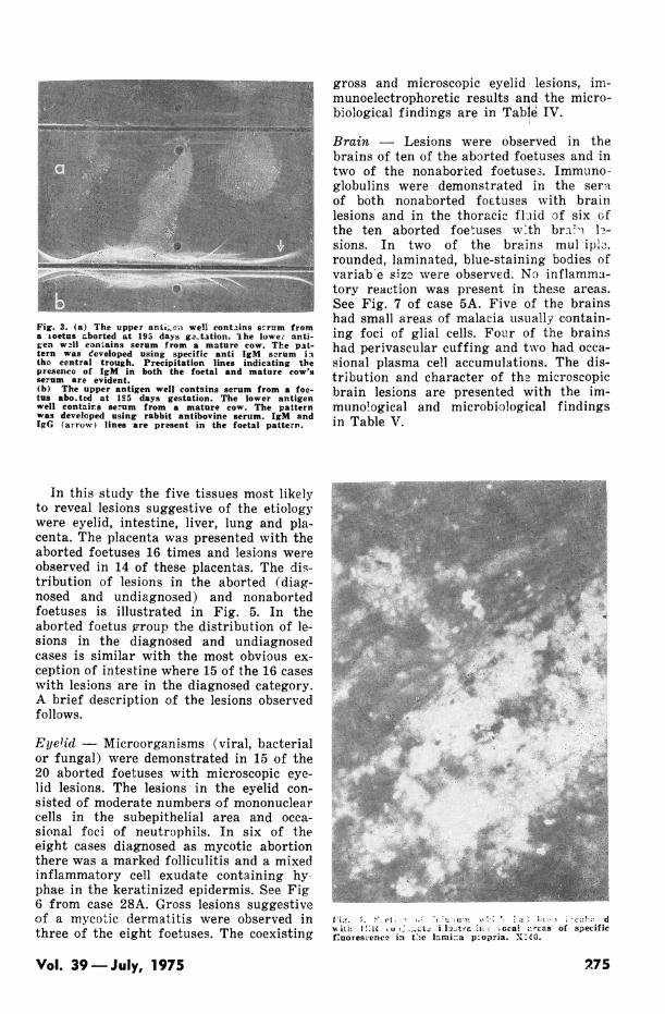
Fig. 3. (a) The upper anti;,ea well contains scrrm froma ioetus aborted at 195 days ga-ttion. 'ihe lowe; anti-ten wall contains serum from a mature cow. The pat-tern was eeveloped using specific anti IgM s2rum iatho central trough. Precipitation lines indicating thepresence of IgM in both the foetal and mature cow'sserum are evident.(b) The upper antigen well contains serum from a foe-tus abo.ted at 1°5 days gestation. The lower antigenwell contairs serum from a mature cow. The patternwas developed using rabbit antibovine serum. IgM andIgG (arrow) lines are present in the foetal pattern.
In this study the five tissues most likelyto reveal lesions suggestive of the etiologywere eyelid, intestine, liver, lung and pla-centa. The placenta was presented with theaborted foetuses 16 times and lesions wereobserved in 14 of these placentas. The dis-tribution of lesions in the aborted (diag-nosed and undiagnosed) and nonabortedfoetuses is illustrated in Fig. 5. In theaborted foetus group the distribution of le-sions in the diagnosed and undiagnosedcases is similar with the most obvious ex-ception of intestine where 15 of the 16 caseswith lesions are in the diagnosed category.A brief description of the lesions observedfollows.
Eye1id - Microorganisms (viral, bacterialor fungal) were demonstrated in 15 of the20 aborted foetuses with microscopic eye-lid lesions. The lesions in the eyelid con-sisted of moderate numbers of mononuclearcells in the subepithelial area and occa-sional foci of neutrophils. In six of theeight cases diagnosed as mycotic abortionthere was a marked folliculitis and a mixedinflammatory cell exudate containing hy-phae in the keratinized epidermis. See Fig6 from case 28A. Gross lesions suggestiveof a mycotic dermatitis were observed inthree of the eight foetuses. The coexisting
gross and microscopic eyelid lesions, im-munoelectrophoretic results and the micro-biological findings are in Table IV.
Brain - Lesions were observed in thebrains of ten of the aborted foetuses and intwo of the nonaborted foetuse3. Immuno-globulins were demonstrated in the seraof both nonaborted foetuses with brainlesions and in the thoracic fluid of six ofthe ten aborted foetuses wvth bra u 1h-sions. In two of the brains mul ip10,rounded, laminated, blue-staining bodies ofvariab-e size were observed. No inflamma-tory reaction was present in these areas.See Fig. 7 of case 5A. Five of the brainshad small areas of malacia usually contain-ing foci of glial cells. Four of the brainshad perivascular cuffing and two had occa-sional plasma cell accumulations. The dis-tribution and character of the microscopicbrain lesions are presented with the im-munological and microbiological findingsin Table V.
4p~~ ~ ~ ~ ~~
1,'i;. 'o. vIt't; (' ( 't2u- tll?.-7 i9; , .t*,'fi~ ;.I ;2 ; d
'A it" t ! .r ilt 'it ' ca! ir'cas of specificfuorescence in tCe lami a p:opria. X :40.
Vol. 39 -July, 1975 2,75

o~~~~~~~~~~~~~~~~~~~~~~~~~~~~~~~~~~~~~~~~~~~~~~~W o
b 88 00=~~~~~~~~~~~~~~~~~~~~~~~~.,Q0.UQ, Q,
. g +l+ i+i + +I ++II+I++
4_
°~~~~~~~~~o Iu 8i 8-~~~~~~~~~~~~~~~~~~~~~~~~~~~~~~~~~~~~~~~~~~~~~-
4)~~~~~~~~~~~~~~~~~~~~~~~~~~~~~~~~~~~~~~~(
o! oi,P
4.Q|0 Cu°=° e e :° =° ffi c x°S4b W U.
aqC) C) C) C) c) C) W). . cn , cn D.cnc
X a I : r .0.0 .0 0.°. z 0 0
w0 I ShD I we we W + w * 8 8 8 8 8 0 8 8 E =X~~~~S-41-414c
< 0I O S S S S (S S CS S @H > Q *Q Q ° > N
Y o0 00 _ - -_t- 00- - - t r- t- t- C):st-LOs3OO O O O : s O 10 0 0
~~o ~~~~ ~~~ 4) 4) 4) 4) 4) C) 4)~~~~~~~~4-~4- 1-- .1-
<>tpl tCu Cu Cu Cu Cu Cu Cu Cu 4 11 9 9 C s > C f 9 _
,Cu 8Cu ' ¢ ¢ CZ
Ed%.4.'0 q.- ,.- 4 Cu0
.00 7 . _. 4 .OrbbO , UCnuC 0
4 CJ 0
Cl)ci Ccol I) C) C) 0co C
CI)4) C C CZ
'0 '~~~~~~0'0'0 0 0 0
.0 Cl CII cq cq cq Cu Cu Cu c, Cu Cu 0qC] C,
4) 4) 4-l) 4) m4) t4 3) to V W o CD c t0- - Ca I - 4 --r -:'m' C) 'I CI)I,
Can. J. comp. Med.276

m No*Abortmd Foetus_
U Aborted Foetus (etiology disosed?
* Aborted Foptuses (etiologw not diwosedp
I
0~~~~~~~~~~~~~~
Eyelid Brain Thymus Lymphl Lung Heart Liver K;dney Adrenal Spleen Intestine Skeletal PlacentaNode , Muscle
Fig. 5. Distribution of lesions in 50 aborted and 50 nonaborted foetuses. *The placenta was submitted only 16 times.writh aborted foetuses.
Thymus -Lesions were observed in thethymus of six of the aborted foetuses. In p :xfour of these the lesions consisted ofmarked cortical atrophy giving rise to the ; J';
appearance of an increased medulla result-_ i*
ing in early l-oss of thymic architecture. A -S 5few of the interlobular areas contained ag- W)<
gregates of mononuclear and neutrophilic * ^cells. In the other two of the six cases with @ * ' w W i 5 '.
thyion walesions eryextensive mineralizaa-
tionm was preen in th''riclara
7~~~~~~~~~~~~~~~~~~~~~~~7
Lymph nodes Lesions were observed in f W ^ t;t-.,*the lymph nodes of 13 of the aborted andin two of the nonaborted foetuses. Focal tt ' j r ^> , '
13~ ~ ~ ~ ~
areas of necrosis were infrequently seen in Liver Kidne Ae Spleen nstn Ske Pe
conjunction with cases of infectious bovinerhinotracheitis. Other lesions in the lymipht
nodes included accumulations of neutro-I4n4phils, macrophages or plasma cells in the A$subeapsular sinuses and medullary areas.
Lung Thirty-two of the aborted foetusesand six of the nonaborted foetuses had le- i,sions in the lung. Material identified as-meconium was a frequent findinginthe.orticarea.air passages and was commonly associated* ;b 7 ; _twith squamous epithelial cells. The meco- Fig. 6. Photomlcrograph of the eyelid from a foetusnium varied in colour from pale greyish aborted with a mycotic placentitis and in which no
gross lenions were observed on the skin. A folliculitisyellow to orange on H&E sections.TF e and a mixed inflammatory exudate in the keratinizeds o n w eepithelium are demonstrated. Hyphae are present butsquamous epithelial cells were nucleatet or not easily distinguished at this magnification. X85.
Vol. 39-July, 1975 277'
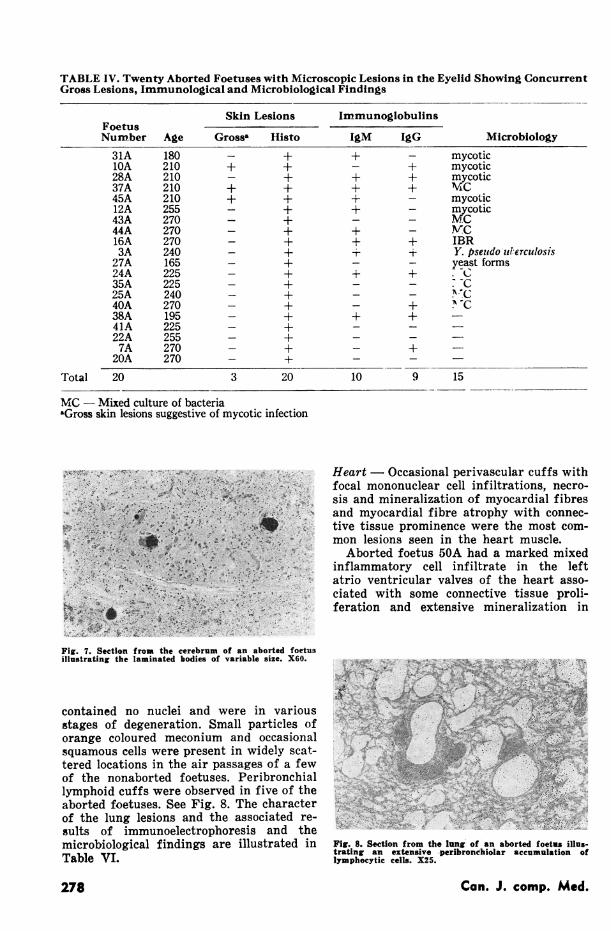
TABLE IV. Twenty Aborted Foetuses with Microscopic Lesions in the Eyelid Showing ConcurrentGross Lesions, Immunological and Microbiological Findings
Skin Lesions
Gross, Histo
+ +
_ +
+ +
+ +_ +
+
_ +
_ +
+
_ +
_ +
_ +
_ +
_ +
_ +
_ +
+
_ +
_ +
Immunoglobulins
IgM IgG
+
+
+
+
+
+
+
Microbiology
mycoticmycoticmycoticVyoicMCmycoticmycoticMCMCIBRY. pseudo zlTerculosisyeast forms
,_
a -Ca cr
+
Total 20 3 20 10 9 15
MC - Mixed culture of bacteriazGross skin lesions suggestive of mycotic infection
Heart Occasional perivascular cuffs withfocal mononuclear cell infiltrations, necro-sis and mineralization of myocardial fibresand myocardial fibre atrophy with connec-tive tissue prominence were the most com-mon lesions seen in the heart muscle.
Aborted foetus 50A had a marked mixedinflammatory cell infiltrate in the leftatrio ventricular valves of the heart asso-ciated with some connective tissue proli-feration and extensive mineralization in
V ~ ~ ~ ~M51B _ 5zt. A;Y 2g?9rii .:i-;S - .'_
Fig. 7. Section from the cerebrum of an aborted foetusillustrating the laminated bodies of variable size. X60.
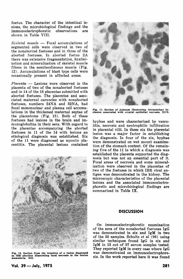
contained no nuclei and were in variousstages of degeneration. Small particles oforange coloured meconium and occasionalsquamous cells were present in widely scat-tered locations in the air passages of a fewof the nonaborted foetuses. Peribronchiallymphoid cuffs were observed in five of theaborted foetuses. See Fig. 8. The characterof the lung lesions and the associated re-sults of immunoelectrophoresis and themicrobiological findings are illustrated inTable VI.
Fig. 8. Section from the lung of an aborted foetus illus-trating an extensive peribrondhiolar accumulation oflymphocytic cells. X25.
Can. J. comp. Med.
FoetusNumber
31AlOA28A37A45A12A43A44A16A3A27A24A35A25A40A38A41A22A7A20A
Age
180210210210210255270270270240165225225240270195225255270270
278

this area. There was a coexisting chronicpericarditis and a marked interstitial ne-phritis. A minced culture was isolated fromthe stomach contents. No other agents weredemonstrated. Foci of degeneration andnecrosis in the myocardium were observedin association with the demonstration ofIBR viral antigen in a few foetuses.
Liver - Lesions were observed in the liversof 20 of the aborted foetuses and in two ofthe nonaborted foetuses. Seven of the foe-tuses had lesions of focal coagulation ne-crosis in the liver and were positive onfluorescent antibody examination of thekidney for IBR. Nine of the foetuses hadliver lesions consisting of periacinar de-generation with and without necrosis. Theliver lesions are tabulated in Table VIIwith the corresponding immunoelectropho-retic and microbiological results.
Kidney - Lesions were not observed inthe kidneys of any of the nonaborted foe-tuses but were observed in eight of theaborted foetuses. Focal areas of necrosiswere present in three of the cases diag-nosed as IBR. An interstitial nephritis wasdiagnosed in five of the aborted foetuseswith kidney lesions (one in a case in whichtoxoplasma-like cysts were observed, an-other in which large numbers of E. coliwere cultured from the gastric aspirate andthree cases in which no specific agent wasfound).
Adrenal - Focal areas of necrosis wereobserved in the cortex of the adrenals insix of the aborted foetuses, three of whichwere diagnosed as IBR.
Spleen - Lesions were observed in 18 ofthe aborted foetuses and consisted of pre-cocious development of germinal centres,focal areas of necrosis and small accumula-tions of neutrophils in the sinusoids.
Pancreas - No lesions were observed inthe pancreas in the nonaborted foetuses andin the aborted foetuses the pancreas wasalmost invariably too autolysed to detectlesions.
Intestines - Intestinal lesions were notseen in any of the nonaborted foetuses butwere observed in 16 of the aborted foetuses.
Accumulations of a mixed inflammatorycell exudate and necrotic debris in theglands of Lieberkuhn (Fig. 9-) with vari-
KA, ~~~~~ . ~
Fig. 9. Section taken from the jejunum illustrating ne-crosis of cryptal epithelial cells and an accumulation ofexudate and cellular debris in a gland of Lieberkuhn.X220.
able numbers of infiltrating neutrophils inassociated villi was the most frequently ob-served lesion in the intestinal tract and wasassociated with a variety of possible causesincluding IBR. Focal coagulation necrosiswithout cellular infiltration was observedin the muscularis and submucosa of theintestine (Fig. 10) in association with thedemonstration of IBR viral antigen in thekidney in seven foetuses.
Foetus 16A had marked necrosis of theepithelial cells in several of the glands ofLieberkuhn in the intestine and a moderateneutrophilic infiltration in the associatedlamina propria and epithelium of the villi.Amphophilic inclusions were observed inthe nuclei of a few of the cells in the af-fected area (Fig. 11) and IBR viral antigenwas demonstrated in the intestine by thesame fluorescent antibody technique util-ized on the kidney. IBR viral antigen wasnot demonstrated by the fluorescent anti-body technique in the kidney of this foetusand no lesions were observed in the liver.IBR virus was cultured on bovine embry-onic kidney from the thoracic fluid of this
Vol. 39 -July, 1975 279

U
0
U
to)-a
9
1: 1)m Q>
+ + + + +
++
+ +
+ +
I
I
I +
+
+ I + I
+ I+ I
+ I + I
Q 0 0 0 0 0 0 10 0 0
'-4 Cl Cl C1 Cl Cl Cl Cl Cl Cl
- C 1 ° A 0t°-° -4tO - 0¢ ¢CY) ¢t 10¢ ¢ Cl
' + +
10 +
m
Cq
14 +
C) + +
+I+
'Id 0 I04)
4)Pk oo Lo
wo Cl J- 0 C') Cl
za " -
P4 o n C9z
H
.Vtt
0oS
4._U0
4)
-0
I-
0_
0
.0
4)
0
0
U-
4)
.U
0
280
co
c .t
.0.Co
S =
liDico
4)_
(U0
C,-bOcn
4Q
,:O4)0
fil
0
0
Cl-
0
0
0
0gH
4)
4)0
;4)10
,0
4-.
Can. J. comp. Med.
I I
foetus. The character of the intestinal le-sions, the microbiological findings and theimmunoelectrophoretic observations areshown in Table VIII.
Skeletal mu-scle - Focal accumulations ofsegmented cells were observed in two ofthe nonaborted foetuses and in three of theaborted foetuses. In aborted foetus 2Athere was extensive fragmentation, hyalini-zation and mineralization of skeletal musclefibres in the semitendinosus muscle (Fig.12). Accumulations of blast type cells wereoccasionally present in affected areas.
Placenta - Lesions were observed in theplacenta of two of the nonaborted foetusesand in 14 of the 16 placentas submitted withaborted foetuses. The placentas and asso-ciated maternal caruncles with nonabortedfoetuses, numbers 24NA and 32NA, hadfocal mononuclear and plasma cell accumu-lations in the thickened maternal septae ofthe placentome (Fig. 13). Both of thesefoetuses had lesions in the brain and im-munoglobulins in their sera. With regard tothe placentas accompanying the abortedfoetuses in 11 of the 14 with lesions anetiological diagnosis was established. Sixof the 11 were diagnosed as mycotic pla-centitis. The placental lesions contained
Fig. 10. Section from the intestine of a foetus diagnosedas IBR abortion illustrating focal necrosis in the tunicamuscularis. X85.
Fig. 11. Section of jejunum illustrating intranuclear in-clusion associated with cryptal epithelial necrosis. X570.
hyphae and were characterized by vascu-litis, necrosis and neutrophilic infiltrationin placental villi. In these six the placentallesion was a major factor in establishingthe diagnosis. In four of the six, hyphaewere demonstrated on wet mount examina-tion of the stomach content. Of the remain-ing five of the 11 in which a diagnosis wasestablished the placenta supported the diag-nosis but was not an essential part of it.Focal areas of necrosis and some mineral-ization were observed in the placentas oftwo of the foetuses in which IBR viral an-tigen was demonstrated in the kidney. Themicroscopic characteristics of the placentallesions and the associated immunoelectro-phoretic and microbiological findings aresummarized in Table IX.
DISCUSSION
On immunoelectrophoretic examinationof the sera of the nonaborted foetuses IgGwas demonstrated in six and IgM in twoof the 50 samples. Schultz et al (96) usingsimilar techniques found IgG in six andIgM in 13 out of 57 serum samples tested.They reported IgM in every case where IgGwas demonstrated on immunoelectrophore-sis. In the work reported here it was found
Vol. 39-July, 1975 281

*XC
.SQQQ *w"*" oo'S8a MO°
4.54.54.54.54.4< >; t $> ;>I
+ +++I + + +++I +
I1 1+ 1 1+ 1
II I-
lIIII lI++I -I IIIIIIIII
. *-**+-- +I* IV
III"
1+1 1+1
-
I I I I
1+1
1+1
l+
I++I
I I
1 III + 1-- 1
++ ++
++++ E4E)
Il l l + + l l ++
I ++9+++,C- IE,+E,+++O+ l++++-14) &4
ul++ I+ I+ I++l++
oo m t- mf-O -- to".cFtsc s O O OS--v "1-v
'4Cv1_C 91 Cl Cl tC Cl Cl
I E 4,
1 ++
'-4
LO
CQP-
0.
0 V
+ co I X
CZCa Jo
;
~CNiC1Cv O
"tL
c
Can. J. comp. Med.
0-
0
04
la) coCa
a z
P
0,
4)
X~
0,
C.
0
0.0o0
1oC.
U
g.0
0
0
0
0,
4)0
*00,
4-
08
I I
I00

that of the six samples containing IgG onlytwo contained demonstrable IgM. IgM isusually considered to be the first immuno-globulin to appear in the bovine foetus asevidenced by the findings of Schultz et al(97) and Osburn and Hoskins (83) in bo-vine foetal sera. A 7S gamma globulin(IgG) was observed by Silverstein (98) asthe primary humoral response in foetalsheep immunized with Freund's adjuvantcontaining antigens to which the foetuscould not yet respond. Such a situation oc-curring naturally is impossible i.e. with ad-juvant but other factors may alter the foe-tal response. Radial diffusion is more sensi-tive than immunoelectrophoresis for the de-tection of immunoglobulins and utilizationof this method may disclose many animalswith IgM in their sera that are inapparenton immunoelectrophoretic examination(96). These samples are being examined
Fig. 12. Cross section of semitendinosis muscle illustrat-ing extensive mineralization of skeletal muscle fibres.X60.
*;i,-<W...... . ..........s 5 -.*.wz-- ---
Fig. 13. Section from the placentome of a nonabortedfoetus illustrating the accumulations of mononuclearcells and plasma cells in the heavy connective tissuestroma of a maternal septum. X60.
by this method and will be reported onlater.
Several factors must be considered indetermining the significance of immuno-globulins in foetal body fluids. The num-ber of aborted foetuses in which immuno-globulins were demonstrated was over threeand one-half times the number of non-aborted foetuses found to have immunoglo-bulins. Immunoglobulins probably do notcross the normal placenta in the cow (18).IgG does but IgM does not cross the pla-centa in women (23). Evidence suggeststhat immunoglobulin production, whetherspecific as antibody or as a nonspecificimmunoglobulin, reaches recordable levelsonly under the influence of antigenic stimu-lation (99, 100). Under natural conditions,immunoglobulins to a variety of agents in-cluding IBR virus, bovine virus diarrheavirus (BVD) and maternal red blood cells,have been reported in the sera of non-aborted and aborted bovine foetuses (19,26, 36, 49, 51, 64, 76, 94, 97). Elevated IgMlevels (20 mgm%) in umbilical cord serumof newborn babies has been associated withan increased incidence of intrauterine in-fection (3). The investigations of Sawyeret al (94) and Trainin and Meirom (108)suggest that quantitative evaluation ofIgM and IgG levels would be of consider-able value in diagnosing congenital infec-tions in cattle. This evidence indicates thatimmunoglobulins are frequently present inthe body fluids of the bovine foetus as aresult of antigenic stimulation and thatthese antigens may be infectious or nonin-fectious in nature. This evidence also de-monstrates that under experimental andnatural conditions many infectious agentsstimulate foetal antibody production withor without abortion. In these situations theexpected frequency of microbiological isola-tion would be diminished (68,105). In ourinvestigation the greater incidence of im-munoglobulins in the abortion group sup-ports the diagnostic value of specificallyidentifying the immunoglobulins. The uni-formity of lesion distribution in the diag-nosed and undiagnosed aborted foetuses in-dicates that a high percentage of the lesionsobserved in foetuses ab.orted at a given ageare similar regardless of the cause of abor-tion. Some of the lesions may occur as aresult of what happens when a foetus be-comes sick in utero and do not reflect thedirect activity of an agent in the foetus.All observations should be compared in
Vol. 39 -July, 1975 283
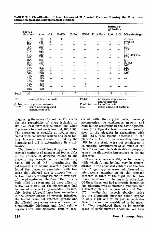
TABLE VII. Classification of Liver Lesions of 20 Aborted Foetuses Showing the ConcurrentImmunological and Microbiological Findings
Immuno-globulins
FoetusNumber Age N.S. PADN C.Nec FNM F. of Bact IgMl IgG Microbiology
31A 180 + + - - - + - mycotic12A 255 + - - - - + - mycotic19A 180 - - + - - + - IBR6A 210 - + - - - IBR33A 210 - - + - - - - IBR21A 240 - - + - - - - IBR34A 240 - - + - - + - IBR and MC46A 270 - - + - - - - IBR48A 270 - - + - - - - IBR18A 210 - - - - + - - E. coli15A 165 - + - - + - - MC24A 225 + + - - - + + MC35A 225 + + - - - - - MC50A 270 + + - - - + - MC1A 180 - + - - - - - -
32A 195 + + - - -
47A 210 - + - - - - -41A 225 - - - + - _ _22A 255 + + - - - - -20A 270 + - - - - - -
Total 20 8 9 7 1 2 6 1 14
N.S. - neutrophils in sinusoids PADN - periacinar degeneration
C. Nec - coagulation necrosisFNM - foci of neutrophils and
mononuclear cells
diagnosing the cause of abortion. For exam-ple, the probability of virus isolation inBVD or PI-3 intrauterine infections whenit proceeds to abortion is low (60, 105, 106).The detection of specific antibodies asso-ciated with consistent lesions and herd his-tory, however, would assist in making thediagnois and aid in determining its signi-ficance.The observation of fungal hyphae in the
stomach contents of nonaborted foetus 4NAin the absence of detected lesions in theplacenta may be explained on the followingbasis. Hill et al (46) investigating thepathogenesis of bovine mycotic placentitisfound the placentas associated with foe-tuses that aborted due to Aspergillus in-fection had necrotizing lesions in over 80%of the placentomes. He found that in ani-mals killed at seven and 14 days after in-fection only 24% of the placentomes hadlesions of a mycotic placentftis. Presum-ably, foetus 4A could have been somewherein the earlier stages of mycotic infection,the lesions were not detected grossly andthe affected cotyledons were not examinedhistologically. Moderate and focal cellulardegeneration and necrosis, usually asso-
and/or necrosisF. of Bact - foci of bacteriaMC - mixed culture of bacteria
ciated with the cryptal cells, normallyaccompanies the continuous growth andremodelling occurring in the bovine placen-tome (13). Specific lesions are not usuallyseen in the placenta in association withIBR (61). The lesions described in theplacenta in two of the cases diagnosed asIBR in this study were not considered tobe specific. Examination of as much of theplacenta as possible is desirable to properlyassess the diagnostic importance of lesionsobserved.
There is some variability as to the easewith which fungal hyphae may be demon-strated in the abomasal contents of the foe-tus. Fungal hyphae were not observed onmicroscopic examination of the stomachcontents in three of the eight aborted foe-tuses considered to be mycotic abortions.Of these three, one had a mycotic dermatitis(no placenta was submitted) and two hada mycotic placentitis. Austwick and Venn(7) investigating mycotic abortion usingroutine diagnostic methods found hyphaein only eight out of 18 gastric aspiratesfrom 18 abortions considered to be myco-tic. They considered these to be possiblecases of early myootic abortion. We have
Can. J. comp. Med.284

TABLE VIII. Classification of Intestinal Lesions in 16 Aborted Foetuses Showing the ConcurrentImmunological and Microbiological Findings
Immuno-Intestinal Lesions globulins
FoetusNumber Age MICE in glands F. Nec IgM IgG Microbiology37A 210 + - + + MC43A 270 + - - - MC19A 180 - + + - IBR33A 210 - + - - IBR6A 210 - + - - IBR21A 240 - + - - IBR34A 240 + + + - IBR16A 270 + - + + IBR46A 270 - + - - IBR48A 270 + + - - IBR36A 270 - - + + MC39A 195 + - - - MC24A 225 + - + + MC40A 270 + - - + MC41A 225 + - - - -13A 270 + - - - -
Total 16 10 7 6 5 14
MICE in glands - mixed inflammatory cell exudate in glands of LieberkuhnF. Nec - focal necrosisMC - mixed culture of bacteria
found that even after careful microscopicexamination as many as three samples ofabomasal aspirate may have to be examinedto disclose the presence of hyphae.There appears to be an association be-
tween the microscopic appearance of sec-tions of eyelid and the results of the micro-biological examinations conducted on thefoetuses in this study. It would seem thatunder some circumstances the amnioticflulid contains the microorganisms for suf-ficient time to allow a reaction to occur inthe skin of the foetus before death or abor-tion takes place. The proportion of mycoticabortions with gross lesions in the skinis similar to that reported by Hillman (47)who observed gross skin lesions associatedwith mycotic abortion in six out of 18cases. Lesions in the thoracic or abdominalviscera of the foetus are observed in ap-proximately one third of mycotic abortionsassociated with Aspergivllus fumigatus (7,28, 47). In contrast Cordes et al (27) havereported that eight of the 11 foetuses in-fected with Mortierella wolfii had lesionsin the viscera. In our investigation all ofthe aborted foetuses considered to be my-cotic abortions had lesions. However, ex-cept for those with lesions in the skin andplacenta or where fungal hyphae could bedemonstrated in the air passages most ofthe lesions were nonspecific. The stage ofplacental involvement at which dermatitis
begins, the proportion of mycotic abortionsin which dermatitis is visible on micro-scopic examination and the variationsbrought about by age and different fungirequire further investigation. Our resultsindicate that histological sections of eyelidwould frequently be of assistance in diag-nosing mycotic abortion especially sincerelatively few placentas are submitted withfoetuses.The significance of the bacteriological
findings in abomasal aspirates is frequent-ly difficult to assess. In our investigationE. coli was demonstrated in pure culturesfrom samples of the abomasal contents ofthree aborted foetuses and Corynebacteriumpyogenes from two. E. coli group 0.78 wereisolated by Jelev et al (54) from all of sevenaborted foetuses in one herd and thesestrains were subsequently found by him tobe capable of causing abortion in pregnantguinea pigs. Corynebacterium pyogenesabortion in ruminants is described bySmith et al (102) in sheep and by Kolar(65) and Kozlowski 2nd Kozlowska (66) incattle.Mixed cultures of bacteria were cultured
from four of the nonaborted foetuses andthirteen of the aborted foetuses in oursurvey. Mixed cultures which do not containknown pathogens are commonly regarded ascontaminants obtained during aspirationof the abomasal contents or to be the result
Vol. 39 -July, 1975 285

TABLE IX. Classification of Lesions Associated with the Placenta in 14 Aborted and 2 NonabortedFoetuses Showing the Concurrent Immunological and Microbiological Findings
Characteristics of Lesions Immuno-Associated with the Placenta globulins
FoetusNumber Age N.Acc Nec Haem Vasc S-Mono S.C.T. Min 1gM IgG Microbiology31A 180 + + - - - - + + - mycotic10A 210 + + + + - - + - + mycotic45A 210 - + - + - - + + - mycotic12A 255 + + - - - - + + - mycotic43A 270 - + - + - - + - - MC44A 270 + + - + - - + + - MC19A 180 - + - - - - + + - IBR46A 270 + -r - + - - + - - IBR5A 255 + + - - - - + + - E. coli14A 270 - + - - - - + + + E. coli2A 165 + + - - + + - + -35A 225 - + + - - - - - - MC11A 150 + + - - - - + - _ _8A 270 - - + + - -
Total 14 8 14 2 5 1 2 12 8 2 11
Nonaborted Foetuses32NA 180 - - - - + + - + + -
24NA 255 - - - - + + - - + -
Total 2 0 0 0 0 2 2 0 1 2 0
N. Acc - neutrophil accumulationNec - focal or diffuse necrosisHaem - hemorrhageVasc - vasculitis
of invasion occurring after death of thefoetus. Austwick and Venn (7) found foe-tuses diagnosed as mycotic abortions fre-quently contained a variety of bacteria inthe stomach contents when the period oftransportation from farm to the laboratorywas extended. A wide variety of micro-organisms may ibe found in the reproductivetract of otherwise healthy cows (25, 33, 37).Prevedourakis et al (88) examined amnio-tic fluid obtained by amniocentesis from140 human pregnancies and found 11 of thesamples to contain bacteria. Three of thesesamples contained several species of bacte-ria. No foetal or maternal illness was sub-sequently observed in any of these subjects.It has been demonstrated in women that therisk to the foetus of acquiring an infectionin utero is positively correlated with thelength of labor and/or the length of timefrom membrane rupture to parturition (9,14, 40). This evidence indicates the diffi-culty in interpreting observations made onstomach contents collected from abortedfoetuses and that isolation of agents alonedoes not necessarily designate the cause ofabortion. It is possible that some as yetundisclosed abortifacient has instigateddilatation of the cervix or in some other
S. mono - septal mononuclear cell infiltrationS.C.T. - septal connective tissue thickeningMin - mineralizationMC -mixed culture of bacteria
manner allowed the entry of bacteria or themultiplication of bacteria already present.Precipitation of the first stage of laborunder the influence of cortisols as proposedby Liggins and his associates (69) wouldallow entry of bacteria. Bacterial endo-toxins stimulate the production of prosta-glandins and abortion may result (101).
In order to detect IBR viral antigen bythe fluorescent antibody technique in allfoetuses containing the antigen a varietyof organs must be examined (90). Reed etal (90) investigating the use of the fluores-cent antibody technique for the detectionof IBR viral antigen in aborted foetusesfound that in three out of 31 cases whereIBR viral antigen was detected by the fluo-rescent antibody technique the histologicalfindings were negative. In those three casesIBR virus was also cultured. The numberof tissues and a description of the tissuesexamined histologically was not included inhis report. Ludwig and Storz (70) suggestthat IBR virus may be present in the la-tent form in the bovine foetus. This evi-dence indicates that on occasion the virusmay be present in the bovine foetus and notcause abortion and further that the fluo-rescent antibody test applied to kidney alone
Can. J. comp. Med.286
or histology on liver alone will fail to detecta small percentage of cases of IBR infec-tion. The demonstration of IBR virus onculture of the thoracic fluid and by fluores-cent antibody examination of the small in-testine in a foetus which did not have liverlesions is consistent with these observa-tions.
Intestinal lesions in foetuses infected withIBR virus have received little attention inthe literature (61, 63, 78, 84, 85). IBR oc-curs in young calves, usually without liverlesions, and there may manifest itself asan alimentary tract infection (8, 24, 90,107). Newborn calves experimentally ex-posed to IBR virus by Baker et al (8) de-veloped lesions confined to the upper respi-ratory tract, rumen, reticulum, omasum,liver, kidney, spleen and lymph nodes.Lymph nodes draining infected areas weredescribed as having massive areas of ne-crosis in the region of the peripheralsinuses and extending into the centres ofthe nodes along the sinuses. Chennekatuet al (24) exposed six to seven months-oldcalves to IBR virus by the oral, intranasaland intravenous routes. Lesions were de-scribed as occurring variously throughoutthe respiratory and alimentary tracts. As-sociated lymphoid tissue usually had le-sions as did the spleen and adrenal gland.The causes of many of the intestinal le-sions observed in our investigation werenot determined. Chlamydial agents havebeen associated with intestinal lesions incalves as have adenoviruses (29, 30, 35, 72,113). Several viruses have been found inthe intestinal tracts of young calves (73,74, 92, 103, 111). Studies on foetal intestineutilizing electron microscopy, light micro-scopy and fluorescent antibody tests alongwith bacteriological, virological and sero-logical investigations in the foetus are in-dicated to identify the causes of intestinallesions.
Lesions have been described in the brainsof bovine foetuses in association with sev-eral diseases including BVD, epizootic bo-vine abortion (EBA) and malignant catar-rhal fever (22, 56, 57, 58, 59, 62, 87, 109,110). The lesions observed in these condi-tions are by themselves generally consideredto be nonspecific. In our investigation avariety of inflammatory and degenerativechanges were observed in several of thefoetal brains. Fragments of mineralizedmaterial were observed in the brain and inseveral other tissues in association with
one case of IBR. Foci of mineralizationhave been associated with viral infections-in babies and animals (80). Blue staininglaminated bodies similar in appearance andstaining characteristics to corpora amyla-cea were observed in the cerebrum of oneaborted foetus which also had lesions con-sisting of a marked hydrocephalus, syringo-myelia, left front micromelia, broncho-pneumonia and placentitis. Immunoglobu-lins were present in the thoracic fluid andE. coli was isolated from the abomasal aspi-rate of this foetus. Corpora amylacea havebeen observed in the human brain in asso--ciation with several degenerative disordersof the nervous system and are most consis-tently associated with old age. They arecomplex chemically and their origin andsignificance are still controversial (53, 89,93). Their pathogenesis in this case was not-determined.
Lesions of variable specificity have beenobserved in the thymus of the aborted bo-vine foetus in association with several viralinfections. Focal thymic necrosis has beendescribed in IBR (63), thymic hemorrhage,and hyperplasia of histiocytic elements inEBA (62) and thymic hemorrhage in casesof BVD and EBA (22, 104). Several humancongenital diseases displaying thymic le-sions have been documented (43). Six ofthe foetuses that we examined had lesionsin the thymus. Two of these were diagnosedas mycotic abortion but the lesions in thethymus were nonspecific and similar to twoothers in which the cause of abortion wasnot diagnosed. Mineralization was observedin two foetal thymuses and appeared to be-part of a generalized mineralization occurr-ing in other tissues. This may representeither mineralization of necrotic foci or ametabolic imbalance. Neither of these caseswas diagnosed.The presence of meconium and epithelial
squames in the air passages of 31 of the-50 aborted foetuses appeared to be a fairlyreliable indicator of persistent foetal dis-tress and it was almost completely absentin the lungs of the nonaborted foetuses.The direction of the flow of fluid in thelung while somewhat controversial is gen-erally accepted in a healthy foetus to be outof the lung and towards the larynx (1, 11,32, 67). Aspiration of amniotic fluid or gas-tric contents can be induced naturally orartificially by a state of foetal hypoxia(2, 45, 71, 86). The duration of foetal dis-tress can to some degree be estimated by
Vol. 39 -July, 1975 287'

the freshness and volume of debris aspir-ated. Fresh meconium appears brightorange on H&E and degenerates to a tan-nish pink colour when present in the lungfor extended periods of time (45). Meco-nium alone may be irritating and incite aninflammatory response (10, 40).
Five of the 50 aborted foetuses examinedby us had lung lesions characterized by in-terstitial pneumonia and extensive peri-bronchial lymphoid accumulations. Onlyrarely was meconium or squamous epithe-lium observed in the air passages of thesefoetuses. PI-3 viral antigen was not de-tected in the lungs by the fluorescent anti-body test. However, four of these fivefoetuses had immunoglobulins in their tho-racic fluid. Swift and Kennedy (106) haveobserved interstitial pneumonia and lym-phoreticular hyperplasia in the lungs ofbovine foetuses aborting after having beenexperimentally infected with PI-3 virus.Virus was recovered in surgically removedfoetuses but was not recovered at the timeof abortion. Specific antibody to PI-3 wasdetected by them in all foetuses infectedfor more than 20 days. Serological examina-tion of aborted foetuses would appear to benecessary to determine if they have beenexposed to PI-3 virus.
Degenerative changes consisting of hya-linization, fragmentation and mineraliza-tion of muscle fibres were observed in thesemitendinosus muscle of one aborted foe-tus. Similar changes have been well docu-mented in association with nutritional mus-cular dystrophy in calves (15, 79, 81, 82).The association between the lesions of nu-tritional muscular dystrophy and dietarydeficiencies of vitamin E and selenium hasnot been well documented as a cause ofabortion in cattle. The lesions in the skele-tal muscle of this foetus appear the sameas those found in nutritional muscular dys-trophy. As the lesion in skeletal mucles isconsidered to be bilaterally symmetricalincreased confidence in the diagnosis anddifferentiation from viral causes could beattained by taking muscle samples fromthe same areas on opposite sides.A clinical history pertaining to the intra-
uterine health of the bovine foetus is sel-dom available. Unlike most animals sub-mitted for post mortem examination noclinical evidence pointing to the site orsystem most likely to be involved and there-fore to be examined most thoroughly by thepathologist is presented. A thorough search
of all tissues in a routine diagnostic situa-tion is niot practical. In our study four tis-sues revealed 17 of the 20 etiological diag-noses that could be made on histologicalexamination. This would suggest that in aroutine diagnostic situation a complete setof tissues for histological examination,stomach contents for microbiology andfluids for serology be collected but that onlythe four tissues mentioned above need beexamnined initially along with routine micro-biology on gastric aspirates. This wouldeliminate many unnecessary slide prepara-tions and would also point the direction totake if further tests were required.
If a causal agent is not disclosed on his-tological or microbiological examinationand immunoglobulins are present in thefoetal fluids identification of the antibodypresent could be most useful. Positive sero-logical findings are of considerable impor-tance in that they give information re-garding the frequency and distribution ofinfection and when interpreted in the lightof histological evidence for abortion wouldbe of immediate diagnostic value.
ACKNOWLEDGMENTS
This study was supported by the OntarioMinistry of Agriculture and Food, by grantnumber 5528134 from the Alberta Agri-cultural Research Trust and the AlbertaCattle Commission. The authors gratefullyacknowledge the technical assistance ofMary Ann Waring who performed the fluo-rescent antibody techniques and the im-munoelectrophoresis and of Marsha Kier-stead and Brenda Buchanan who did thebacteriology.The cooperation of the staff of the Al-
berta Provincial Veterinary Laboratoryand especially Dr. G. Klavano is acknowl-edged. The authors are also thankful tomembers of the Department of Pathologyand also Dr. N. C. Palmer for his assistancein preparation of the manuscript.
REFERENCES
1. ADAMS, F. H., D. T. DESILETS and B. TOWERS.Control of flow of fetal lung fluid at the laryngealoutlet. Resp. Physiol. 2: 302-309. 1967.
2. ADAMSONS, K. and R. E. MYERS. Perinatal as-phyxia: Causes, detection and neurologic sequelae.Pediat. Clins N. Am. 20: 465-480. 1963.
288 Can. J. comp. Med.

3. ALFORD, C. A., J. W. FOFT, W. J. BLANKEN-SHIP, G. CASSADY and J. W. BEINTON. Subelin-ical central nervous system disease of neonates: Aprospective study of infants born with increasedlevels of IgM. J. Pediat. (Suppl.) 75: 1167-1178. 1969.
4. AMIEL, D. K. and E. W. MOODIE. Dairy her-dwastage in Soutlheastern Queensland. Aust. vet. J.49: 69-73. 1973.
5. ANDREWS, L. G. The major non-infectious causesof reproductive wastage in beef cattle in the north-ern territory. Aust. vet. J. 48: 41-46. 1972.
6. ASHTON, G. C. and G. R. FALLON. B-globulintype, fertility and embryonic mortality in cattle. J.Reprod. Fert. 3: 93-104. 1962.
7. AUSTWICK, P. K. C. and J. A. J. VENN. Routineiinvestigations into mycotic abortion. Vet. Rec. 69:488-491. 1957.
S. BAKER, J. A., K. McENTEE and J. H. GILLESPIE.Effects of infectious bovine rhinotracheitis-infectiouspustular vulvo-vaginitis (IBR-IPV) virus on newborncalves. Cornell Vet. 50: 156-170. 1960.
9. BENIRSCHKE, K. Routes and types of infection inthe fetus and the newborn. Am. J. Dis. Child. 99:711-721. 1960.
10. BENIRSCHKE, K. and S. G. DRISCOLL. Amnionand chorion. In The Pathology of the Human Pla-centa. pp. 39-55. New York, New York: Springer-Verlag. 1967.
11. BIGGS, J. S. G., T. J. GAFFNEY and H. M. Mc-GEARY. Evidence that fetal lung fluid and phos-pholipids pass into amniotic fluid in late humanpregnancy. J. Obstet. Gynaec. Br. Commonw. 80:125-129. 1973.
12. BISHOP, M. W. H. Paternal contribution to em-bryonic death. J. Reprod. Fert. 7: 383-396. 1964.
13. BJORKMAN, N. H. Light and electron microscopicstudies on cellular alterations in the normal bovineplacentome. Anat. Rec. 163: 17-30. 1969.
14. BLANC, W. A. Pathways of fetal and early neo-natal infection. J. Pediat. 59: 473-496. 1961.
15. BLAXTER, K. L. and F. BROWN. Vitamin E in thenutrition of farm animals. Nutr. Abstr. Rev. 22:1-21. 1952.
1 6). BLOOD, D. C. and J. A. HENDERSON. Diseas?scaused by bacteria-Ill. In Veterinary Medicine. 3rdEdition. pp. 368-383. London, England: Bailliere,Tindall and Cassell Ltd. 1968
17. BOYD, H. Embryonic death in cattle, sheep andpies. Commonwv. Bur. Anim. Health 35: 251-266.1965.
18. BRAMBELL, F. W. R. The passive immunity of theyoung mammal. Biol. Rev. 33: 488-531. 1958.
19. BRAUN, R. K., B. I. OSBURN and J. W. KEN-DRICK. Immunologic response of bovine fetus tobovine vir-al diarrhea virus. Am. J. vet. Res. 31:1127-1132. 1973.
20. BUTLER, J. E. Bovine immunovlobulins: A review.J. Dairy Sci. 52: 1895-1909. 1969.
21. BUTLER, J. E., A. J. WINTER and G. G. WAGNER.Symposium: Bovine immune system. J. Dairy Sci.54: 1309-1340. 1971.
22. CASARO, A. P. E., J. W. KENDRICK and P. C.KENNEDY. Response of the bovine fetus to bo-vine viral diarrhea-mucosal disease virus. Am. J.vet. Res. 32: 1543-1562. 1971.
23. CEDERQVIST, L. L., J. T. QUEENAN, E. C. GA-DOW and S. D. LITWIN. The origin of gamma G-globulins in human amniotic fluid. Am. J. Obstet.Gynec. 113: 838-840. 1972.
-24. CHENNEKATU, P. P., J. B. GRATZEK and F. K.RAMSEY. Isolation and characterization of a strainof infectious bovine rhinotracheitis virus associatedwith enteritis in cattle: Pathogenesis studies byfluorescent antibody tracing. Am. J. vet. Res. 27:1583-1598. 1966.
25. CLARK, W. A. and W. G. STEVENSON. The bac-terial flora of the normal non-gravid and gravidbovine uterus. Can. J. comp. Med. 13: 92-93. 1949.
*26. CLASSICK, L. G. and A. L. FERNELIUS. Bovineviral diarrhea virus: Neutralizing antibodies in acalf obtained by cesarean section from cow whiehwas infected. Am. J. vet. Res. 31: 393-395. 1970.
27. CORDES, D. O., M. E. CARTER and M. E. diMENNA. Mvcotic ineumonia and placentitis causedby Mortierella wolfii. Vet. Path. 9: 190-201. 1972.
-28. CYSEWSKI, S. J. and A. C. PIER. Mycotic abor-tion in ewes produced by Aspergillus fumigatus:Pathologic changes. Am. J. vet. Res. 29: 1135-1151.1968.
29. DARBYSHIRE, J. H., A. R. JENNINGS, P. S.DAWSON, P. H. LAMONT and A. R. OMAR. Thepathogenesis and pathology of infection in calveswith a strain of bovine adenovirus type 3. Res. vet.Sci. 7: 81-93. 1966.
30. DARBYSHIRE, J. H. Bovine adenoviruses. J. Am.vet. med. Ass. 152: 786-794. 1968.
31. DAVID, J. S. E., M. W. H. BISHOP and H. J.CEMBROWICZ. Reproductive expectancy and in-fertility in cattle. Vet. Rec. 89: 181-184. 1971.
32. DAWES, G. S., H. E. FOX, B. M. LEDUC, G. C.LIGGINS and R. T. RICHARDS. Respiratory move-menits and rapid eye movement sleep in the foetallamb. J. Physiol., Lond. 220: 119-143. 1972.
33. DAWSON, F. L. M. The microbial content and mor-phological chalacter of the normal bovine uterusand oviduct. J. agric. Sci., Camb. 40: 150-157. 1950.
34. DEARBORN, D. D., R. M. KOCH, L. V. CUNDIFF,K. E. GREGORY and G. E. DICKERSON. An anal-ysis of reproductive traits in beef cattle. J. Anim.Sci. 36: 1032-1040. 1973.
35. DOUGHRI, A. M., K. P. ALTERA and J. STORZ.Host cell range of Chlamydial infection in the neo-natal bovine gut. J. comp. Path. 83: 107-114. 1973.
36. DUNNE, H. W., S. M. AJINKYA, G. R. BUBASHand L. C. GRIEL, Jr. Parainfluenza-3 and bovineenteroviruses as possible important causative factorsin bovine abortion. Am. J. vet. Res. 34: 1121-1126.1973.
37. ELLIOTT, L., K. J. McMAHON, H. T. GIER andG. B. MARION. Uterus of the cow after parturition:Bacterial content. Am. J. vet. Res. 29: 77-81. 1968.
IS. FAULKNER, L. C., editor. Abortion Diseases of Live-stock. Springfield, Illinois: Charles C. Thomas. 1968.
39. FECHHEIMER, N. S. Chromosome abnormalities andembryo death in farm animals. Vet. Rec. 90: 241-243. 1972.
40. FEDRICK, J. and N. R. BUTLER. Certain causes ofneonatal death. III. Pulmonary infection. (b) Preg-nancy and delivery. Biol. Neonate. 18: 45-57. 1971.
41. FOSGATE, 0. T. and V. R. SMITH. Prenatal mor-tality in the bovine between pregnancy diagnosis at34-50 days post-insemination and parturition. .J.Dairy Sci. 37: 1071-1073. 19.54.
42. CLINICAL ELECTROPHORESIS. In Gelman Pro-cedures for Special Electrophoresis. pp. 44-53. AnnArbor, Michigan: Gelman Instrument Co. 1970.
43. GOOD, R. A. Disorders of the immune system. InImmunobiology. R. A. Good and D. W. Fisher, edi-tors. pp. 3-16. Stanford, Connecticut: Sinaver Asso-ciates, Inc. 1973.
44. HANLY, S. Prenatal mortality in farm animals. J.Renrod. Fert. 2: 182-194. 1961.
45. HELWIG, F. C. Congenital aspiration pneumonia instillborn and newborn infants. Am. J. Obstet. Gynec.26: 849-857. 1933.
46. HILL, M. W. M., C. E. WHITEMAN, M. M. BEN-JAMIN and L. BALL. Pathogenesis of experimentalbovine mycotic placentitis produced by Aspergillusfumiratus. Vet. Path. 8: 175-192. 1971.
47. HILLMAN, R. B. Bovine mycotic placentitis in NewYork State. Cornell Vet. 59: 269-288. 1969.
48. HOLT, A. F. Analysis of conception rates and otherdata in artificial insemination. Vet. Rec. 64: 31-38. 1952.
49. HORNER, G. W., R. H. JOHNSON, D. P. DENNETTand W. R. LANE. A serological study of bovine foe-tal immunoglobulins. Aust. vet. J. 49: 325-329. 1973.
50. HUBBERT, W. T.. G. D. BOOTH. W. D. BOLTON.H. W. DUNNE, K. McENTEE, R. E. SMITH andM. E. TOURTELLOTTE. Bovine abortions in fivenortheastern states, 1960-1970: Evaluation of diagnos-tic laboratory data. Cornell Vet. 63: 291-316. 1973.
51. HUBBERT, W. T., J. H. BRYNER, A. L. FERNE-LIUS, G. H. FRANK and P. C. ESTES. Viral in-fection of the bovine fetus and its environment.Arch. wes. Virusforsch. 41: 86-98. 1973.
52. HUBBERT, W. T., Chairman. Recommendations forstandardizing bovine reproductive terms. CornellVet. 62: 216-237. 1972.
53. JASPAR, H. H. J. and J. J. G. PRICK. Morpho-logy and histochemistry of the corpora amylacea inthe brain. I. Proc. K. ned. Akad 72: 385-412. 1969.
54. JELEV, W., KARADSHOV and A. ANGELOV.Uber die pathogenese der kolibacteriose bei kalbern.Zentbl. VetMed. 16B: 725-730. 1969.
55. JUBB, K. V. F. and P. C. KENNEDY. The femalegenital system. In Pathology of Domestic Animals.2nd Edition. Vol. I. pp. 487-585. New York: Aca-demic Press. 1970.
56. KAHRS, R. F. Effects of bovine viral diarrhea onthe developing fetus. J. Am. vet. med. Ass. 163:877-878. 1973.
57. KAHRS, R. F., F. W. SCOTT and A. de LAHUNTA.Bovine viral diarrhea-mucosal disease, abortion, andcongenital cerebellar hypoplasia in a dairy herd.J. Am. vet. med. Ass. 156: 851-857. 1970.
58. KAHRS, R. F., F. W. SCOTT and A. de LAHUNTA.Congenital cerebellar hypoplasia and ocular defectsin calves following bovine viral diarrhea-mucosaldisease infection in pregnant cattle. J. Am. vet.med. Ass. 156: 1443-1450. 1970.
Vol. 39-July, 1975 289

59. KAHRS, R. F., F. W. SCOTT and A. de LAHUNTA.Epidemiological observations on bovine viral diarrhea-mucosal disease virus-induced congenital cerebellarhypoplasia and ocular defects in calves. Teratology.3: 181-184. 1970.
60. KENDRICK, J. W. Bovine viral diarrhea-mucosaldisease virus infection in pregnant cows. Am. J.vet. Res. 32: 533-544. 1971.
61. KENDRICK, J. W., L. SCHNEIDER and 0. C.SihAUhi. rlacental reaction to the infectious bo-viAe rhinotiacheiLs- infectious pustular vulvovagi-nitis virus. Am. J. vet. ktes. 32: 1u45-1051. 1971.
62. KENNEDY, P. C., H. J. OLANDER and J. A.HOW ARfh. Pathology of epizootic bovine abortI ion.Cornell Vet. 50: 41l-429. 1960.
63. KENNEDY, P. C. and W. P. C. RICHARDS. Thepachology of abortion caused by the virus of infec-tious bovine rhinotracheitis. Path. Vet. 1: 7-17. 1964.
64. KNIAZEFF, A. J. and V. RIMER. G-globulin in foe-tal bovine sera: Significance in virology. Nature,Lond. 214: 80a-806. 19b7.
65. KOLAR, J. Vyznam C. pyogenes jako puvodee abor-tu u skotu. Vet. Med. (Prague) 8: 301-310. 1963.
66. KOZLOWSKI, S. and I. KOZLOWSKA. Ronienia ubydla wywolane przez Cornyebacterium pyogenes.Medycyna wet. 25: 616-617. 1969.
67. LANMAN, J. T., A. SCHAFFER, L. HEROD, Y.OGAWA and R. CASTELLANOS. Distensibility ofthe fetal lung with fluid in sheep. Pediat. Res. 5:E86-590. 1971.
68. LENNETTE, E. H. Laborator-y diagnosis of viral in-fections: Ceneral principles. Am. J. clin. Path. 57:737-750. 1972.
69. LIGGINS, G. C., P. C. KENNEDY and L. W. HOLM.Failure of initiation of parturition after electro-coagulation of the pituitary of the fetal lamb. Am.J. Obstet. Gynec. 98: 1080-1086. 1967.
70. LUDWIG, H. and J. STORZ. Activation of herpes-virus from normal bovine fetal spleen cells afterprolonged cultivation. Med. Microbiol. and Immun.158: 2C9-217. 1973.
71. MANDELBAUM, B. Gestational meconium in thehigh-risk piregnancy. Obstet. Gynec. 42: 87-92. 1973.
72. MATTSON, D. E. Naturally occurring infection ofcalves with a bovine adenovirus. Am. J. vet. Res.34: 623-629. 1973.
73. MEBUS, C. A., E. L. STAIR, M. B. RHODES andM. J. TWIEHAUS. Neonatal calf diarrhea: Propaga-tion, attenuation, and characteristics of a corono-virus-like agent. Am. J. vet. Res. 34: 145-150. 1973.
74. MEBUS, C. A., E. L. STAIR, N. R. UNDERDAHLand M. J. TWIEHAUS. Pathology of neonatal calf(liarrhea induced by a reo-like virus. Vet. Path.8: 490-505. 1971.
75. MERCHANT, I. A. and R. A. PACKER. VeterinaryBacteriology and Virology, 7th edition. Ames, Iowa:Towa State University Press. 1967.
76. MERRIMAN, M. J. G. S. Serum immunoglobulins innewborn calves before and after colostrum feeding.Can. J. comp. Med. 35: 269-273. 1971.
77. MITCHELL, D. Bovine abortion - An analysis of227 cases. Can. vet. J. 1: 337-343. 1960.
78. MOLELLO, J. A., T. L. CHOW, N. OWEN and R.JENSEN. Placental pathology. V. Placental lesionsof cattle experimentally infected with infectious bo-vine rhinotracheitis virus. Am. J. vet. Res. 27: 907-915. 1966.
79. MUTH, 0. H. White muscle disease (myopathy) inlambs and calves. I. Occurrence and nature of thedisease under Oregon conditions. J. Am. vet. med.Ass. 126: 355-361. 1955.
80. NARATAN, 0. and R. T. JOHNSON. Effects ofviral infection on nervous system development. I.Pathogenesis of bluetongue virus infection in mice.Am. J. Path. 68: 1-14. 1972.
81. OKSANEN, H. E. Studies on nutritional musculardegeneration (NMD) in ruminants. Acta vet. scand.(Suppl. 2) 6: 1-110. 1965.
82. OKSANEN, A. and R. POUKKA. An electron micro-scopical study of nutritional muscular degeneration(NMD) of myocardium and skeletal muscle in calves.Acta path. microbiol. scand. 80: 440-448. 1972.
83. OSBURN, B. I. and R. K. HOSKINS. Infection withVibrio fetus in the immunologically immature fetalcalf. J. Infect. Dis. 123: 32-40. 1971.
84. OWEN, N. V., T. L. CHOW and J. A. MOLELLO.Bovine fetal lesions experimentally produced by in-fectious bovine rhinotracheitis virus. Am. J. vet.Res. 25: 1617-1625. 1964.
85. OWEN, N. V., T. L. CHOW and J. A. MOLELLO.Infectious bovine rhinotracheitis: Correlation offetal and placental lesions with viral isolations. Am.J. vet. Res. 29: 1959-1965. 1968.
86. PENNER, D. W. and A. C. McINNIS. Intrauterineand neonatal pneumonia. Am. J. Obstet. Gynec. 69:147-168. 1955.
87. PLOWRIGHT, W., M. KALUNDA, D. M. JESSETTand K. A. J. HERNIMAN. Congenital infeotion ofcattle with the herpesvirus causing malignant ca-tarrhal fever. Res. vet. Sci. 13: 37-45. 1972.
88. PREVEDCURAKIS, C. N., E. STRIGOU-CHARALA-BIS and D. B. KASKARELIS. Bacterial invasion ofamniotic cavity during pregnancy and labour. Obstet.Gynec. 37: 459-461. 1971.
89. RAMSEY, H. J. Ultrastlucture of corpora amylacea.J. N.uropath. Exp. Neurol. 24: 25-39. 1965.
t.0. REED, D. E., E. J. BICKNELL, C. A. LARSON, W.U. KINUDTSON and C. A. KIRKBRIDE. Infectiousbovine rhinotracheitis virus-induced abortion: Ra-pid diagnosis by fluorescent antibody technique. Am.J. vet. Res. 32: 1423-1426. 1971.
91. ROBERTS, S. J. Veterinary Obstetrics and GenitalD:seases (Theriogenology). 2nd edition. Ithaca, NewYotk. 1971.
92. ROMVARY, J. Incidence of virus diarrhea in new-born calvcs. Acta vet. hung. 15: 341-347. 1965.
93. SAKAI, M., J. AUSTIN, F. WITMER and L.TRUEB. Studi.-s of corpora amylacza. I. Isolationand preliminaly characterization by chemical andhistochemical techniques. Arch. Ncurol. 21: 626444.1 69.
94. SAWYER, M., B. I. OSBURN, H. D. KNIGHT andJ. W. KENDRICK. A quantitative serologic assayfol diagnosing congenital infeztions of cattle. Am.J. vet. Res. 34: 1281-1284. 1973.
S 5. SCHULTZ, R. D. Developmental aspects of the fetalbovine immune response: A review. Cornell Vet..(3: 507-535. 1973.
96. SCHULTZ, R. D., F. CONFER and H. W. DUNNE.Occurrence of blood cells and serum proteins in bo-vine fetuses and calves. Can. J. comp. Med. 35:93-98. 1971
97. SCHULTZ, R. D., H. W. DUNNE and C. E. HEIST.Ontogeny of the bovine immune response. Infection& Immunity. 7: 981-991. 1973.
98. SILVERSTEIN, A. M. Ontogeny of the immune re-sponse. Science 144: 1423-1428. 196 t.
99. STLVERSTEIN, A. M., G. J. THORBECKE, K. L.KRANER and R. J. LUKES. Fetal response to anti-genic stimulus. III. G-globulin production in normaland stimulated fetal lambs. J. Immun. 91: 384-395.1963.
100. SILVERSTEIN, A. M., J. W. UHR, K. L. KRANERand R. J. LUKES. Fetal response to antigenic sti-mulus. II. Antibody production by the fetal lamb.J. exp. Med. 117: 799-812. 1963.
101. SKARNES, R. C. and M. J. K. HARPER. Relationi-ship between endotoxin-induced abortion and thesynthesis of prostaglandin F. Prostaglandins 1:191-203. 1972.
102. SMITH, R. E., I. M. REYNOLDS, G. W. CLARKand J. A. MILBURY. Feto-placental effects ofCorynebacterium pyogenes in sheep. Cornell Vet.61: 573-590. 1971.
103. STAIR, E. L., M. B. RHODES, R. G. WHITE andC. A. MEBUS. Neonatal calf diarrhea: Purificationand electron microscopy of a coronavirus-like agent.Am. J. vet. Res. 33: 1147-1155. 1972.
104. STORZ, J., J. W. CALL, R. W. JONES and M. L.MINER. Epizootic bovine abortion in the inter-mountain region. Some recent clinical, epidemiologicand pathologic findings. Cornell Vet. 67: 21-37.1967.
105. SWIFT, B. L. Bovine parainfluenza-3 virus: Experi-mental fetal disease. J. Am. vet. med. Ass. 163:861-862. 1973.
106. SWIFT, B. L. and P. C. KENNEDY. Experimental-ly induced infection of in utero bovine fetuseswith bovine parainfluenza-3 virus. Am. J. vet. Res.33: 57-63. 1972.
107. THOMSON, R. G. and M. SAVAN. Letter to theeditor. Can. vet. J. 4: xiii. September, 1963.
108. TRAININ, Z. and R. MEIROM. Calf immunoglo-bulins and congenital malformation. Res. vet. Sci.15: 1-7. 1973.
109. WARD, G. M. Bovine cerebellar hypoplasia appa-rently caused by BVD-MD virus. A case report.Cornell Vet. 59: 570-576. 1969.
110. WARD, G. M. Bovine viral diarrhea-mucosal dis-ease implicated in a calf with cerebellar hypoplasiaand ocular disease. A case report. Cornell Vet. 61:224-228. 1971.
111. WELCH, A. B. Purification, morphology and partialcharacterization of a reovirus-like agent associatedwith neonatal calf diarrhea. Can. J. comp. Med.35: 195-201. 1971.
112. WITHERS, F. W. Wastage and disease incidence indairy herds. Vet. Rec. 69: 446-453. 1957.
113. YORK, C. J. and J. A. BAKER. A new member ofthe psittacosis-lymphogranuloma group of virusesthat cause infection in calves. J. exp. Med. 93:587-603. 1951.
290 Can. J. comp. Med.
Copyright © 2022 FDOKUMEN