CYTOPLASMIC I. Hydra - NCBI
22
CYTOPLASMIC I. Hydra MICROTUBULES DAVID B. SLAUTTERBACK, Ph.D. From the Anatomy Department, The University of Wisconsin Medical College, Madison ABSTRACT Small cytoplasmic tubules are present in the interstitial cells and cnidoblasts of hydra. They are referred to here as "microtubules." These tubular elements have an outside diameter of 180 A and an inside diameter of 80 A. By difference, the membranous wall is estimated to be 50 A thick. The maximum length of the microtubules cannot be determined from thin sections but is known to exceed 1.5/z. In the interstitial cells the microtubules are found in the intercellular bridges, free in the cytoplasm and in association with the centrioles. In the cnidoblast they form a framework around the developing nematocyst and in late stages are related to the cnidocil forming a tight skein in the basal part of the cell. Especially in this cell, confluence of microtubules with small spherical vesicles of the Golgi complex has been observed. It is proposed that these tubules function in the transport of water, ions, or small molecules. It is the purpose of this paper to present some ob- servations on a fine tubular component of the cyto- plasm as seen in the cnidoblasts and interstitial cells of hydra. Later papers will deal with the presence of similar structures in other hydra cells, and in a secretory cell and a photoreceptor found in the annelid Enchytraeusfragmentosus. In addition, some suggestions as to the probable function of these tubular elements will be made. They will be designated throughout this paper by the descrip- tive term "microtubules." They should not be con- fused with the agranular reticulum of such cells as the gastric parietal (34, 74), lutein (21), or tes- ticular interstitial (14), nor with the so-called "irregular tubulo-membranous component" re- ported to be present in the cytoplasm following certain unusual preparative procedures (2). Observations of similar structures in a wide variety of cell types have been reported in the last 5 or 6 years. In some cases they were reported as filamentous elements, and in others as tubules or canaliculi, although all these elements appear in the published electron micrographs to have a tubular form. The most frequently reported occur- rence of such tubules has been in the achromatic spindle of mitotic cells, from which a number of authors (33, 42, 54, 63) have concluded that the spindle fibers are in reality 120 to 200 A filaments or tubules. These elements have also been re- corded frequently in the dendrites of neurons (7, 30, 49, 69, 85) and in the cytoplasm of numerous protozoa (45, 57, 60), and have sometimes been recorded in association with the centrioles of a variety of cell types (15, 25). In fact, one of the most striking features of these microtubules is their ubiquity. More than a century of light microscopic inves- tigations have. recorded the presence of delicate filaments or striations in the cytoplasm of cnido- Masts and musculo-epithelial cells (in addition to the myofilaments of the muscular foot) which were most commonly interpreted as supporting fibrils ("Statzfibrillen"). Will (86), Toppe (83), and yon Gelei (27) concluded that the filaments in cnido- blasts which run from the cnidocil, surround the nematocyst and coil in the basal part of the cyto- 367
-
Upload
khangminh22 -
Category
Documents
-
view
0 -
download
0
Transcript of CYTOPLASMIC I. Hydra - NCBI
CYTOPLASMIC
I. Hydra
M I C R O T U B U L E S
D A V I D B. S L A U T T E R B A C K , Ph.D.
From the Anatomy Department, The University of Wisconsin Medical College, Madison
A B S T R A C T
Small cytoplasmic tubules are present in the interstitial cells and cnidoblasts of hydra. They are referred to here as "microtubules." These tubular elements have an outside diameter of 180 A and an inside diameter of 80 A. By difference, the membranous wall is estimated to be 50 A thick. The maximum length of the microtubules cannot be determined from thin sections but is known to exceed 1.5/z. In the interstitial cells the microtubules are found in the intercellular bridges, free in the cytoplasm and in association with the centrioles. In the cnidoblast they form a framework around the developing nematocyst and in late stages are related to the cnidocil forming a tight skein in the basal part of the cell. Especially in this cell, confluence of microtubules with small spherical vesicles of the Golgi complex has been observed. It is proposed that these tubules function in the transport of water, ions, or small molecules.
It is the purpose of this paper to present some ob- servations on a fine tubular component of the cyto- plasm as seen in the cnidoblasts and interstitial cells of hydra. Later papers will deal with the presence of similar structures in other hydra cells, and in a secretory cell and a photoreceptor found in the annelid Enchytraeusfragmentosus. In addition, some suggestions as to the probable function of these tubular elements will be made. They will be designated throughout this paper by the descrip- tive term "microtubules." They should not be con- fused with the agranular reticulum of such cells as the gastric parietal (34, 74), lutein (21), or tes- ticular interstitial (14), nor with the so-called "irregular tubulo-membranous component" re- ported to be present in the cytoplasm following certain unusual preparative procedures (2).
Observations of similar structures in a wide variety of cell types have been reported in the last 5 or 6 years. In some cases they were reported as filamentous elements, and in others as tubules or canaliculi, although all these elements appear in the published electron micrographs to have a
tubular form. The most frequently reported occur- rence of such tubules has been in the achromatic spindle of mitotic cells, from which a number of authors (33, 42, 54, 63) have concluded that the spindle fibers are in reality 120 to 200 A filaments or tubules. These elements have also been re- corded frequently in the dendrites of neurons (7, 30, 49, 69, 85) and in the cytoplasm of numerous protozoa (45, 57, 60), and have sometimes been recorded in association with the centrioles of a variety of cell types (15, 25). In fact, one of the most striking features of these microtubules is their ubiquity.
More than a century of light microscopic inves- tigations have. recorded the presence of delicate filaments or striations in the cytoplasm of cnido- Masts and musculo-epithelial cells (in addition to the myofilaments of the muscular foot) which were most commonly interpreted as supporting fibrils ("Statzfibrillen"). Will (86), Toppe (83), and yon Gelei (27) concluded that the filaments in cnido- blasts which run from the cnidocil, surround the nematocyst and coil in the basal part of the cyto-
367
plasm, were muscular , and funct ioned in some way in nematocyst firing. In the musculo-epithelial cells, however, similar fi laments were in terpre ted as support ing in function. Muel ler (43), on the other hand , rejects these interpretat ions and, as a result of a special staining procedure devised by him, finds the cnidocil fi laments to be elastic and the vertical fi laments of the musculo-epithelial cell to be contracti le myonemes. However contro- versial the funct ion of these fibers has been, there is general agreement as to their presence. The i r dis t r ibut ion among the cells and disposition wi th in them is consistent with thei r identification as bundles of the microtubules described here.
M A T E R I A L S A N D M E T H O D S
The animals used in these experiments were of the species Hydra (Pdrnatohydra) oligactis and Hydra littoralis. These animals are kept under continuous cultivation in this laboratory according to the methods devised by W. F. Loomis (39). The hydras were fixed in solutions of osmium tetroxide buffered with veronal acetate (Palade, 47) or collidine (Bennett, 4), although the latter provided a somewhat less satis- factory delineation of the objects of this study. The pH of the solution was controlled at 7.5, or occasion- ally 7.7. Most of the fixatives contained calcium chlo- ride at a concentration of l0 -3 ~, or calcium chloride and sucrose, the latter at about 0.6 M. The tissues were rapidly dehydrated in a graded series of alco- hols, and embedded in Epon 812 according to the procedure recommended by Luft (40). The sections were then stained with lead hydroxide as described by Watson (84) or Karnovsky (36). Thin sections were cut on either a Porter-Blum or a Huxley-Cam- bridge ultramicrotome (equipped with glass or dia- mond knives). In some cases the sections were then coated with a thin film of amorphous carbon. The
material was examined in an RCA E M U 3E electron microscope, and micrographs were made on Cramer high demarcation photographic plates at initial mag- nifications of from 8 to 30,000 and subsequently en- larged photographically.
O B S E R V A T I O N S
The description which follows will deal with the appearance and relationships of the microtubules dur ing differentiat ion from interstit ial cell to mature cnidoblast. Because the morphological manifestat ions of interstit ial cell differentiat ion into cnidoblast as well as the coincident development of the highly s t ructured secretory product, the nema- tocyst, have been described previously (76, 78, 79), only those aspects of the process which are per t inent to the subject a t hand will be described here.
The intersti t ial cell is a small, relatively un- differentiated cell as i l lustrated in Fig. 1. Char - acteristic of this cell is the paucity of cytoplasmic m e m b r a n o u s elements, part icular ly those identified with the endoplasmic ret iculum. The Golgi com- plex is present but is very small and has few small spherical vesicles associated with it. Most charac- teristic of the interstit ial cell is the a b u n d a n c e of ribosomes found in its cytoplasm. This cell under- goes rapid proliferation and the daugh te r cells remain bound together by intercel lular bridges, i l lustrated in Fig. 1. There may be fourteen or more such cells bound together by these pa ten t cytoplasmic communicat ions. W h e n the prolifera- t ion of the intersti t ial cells ceases, the first indica- tions of differentiat ion into a cnidoblast can be seen in the appearance of small scattered vesicles of g ranu la r endoplasmic ret iculum. These vesicles
Abbreviations for Figures
B, intercellular bridge Ca, nematocyst capsule Ce, centriole Ci, cilium of cnidocil G, Golgi vesicles Hg, heterogeneous granular material Li, lipid droplet Mb, midbody
ME, musculo-epithelial cell Mr, microtubule N, nucleus Nn, neuron No, nuc]eolus 0, operculum R, stiff rods of cnidocil
Ftom~E 1 An interstitial cell from the ectoderm. This cell is bound to at least one other like it by an intercellular bridge (B) and has already begun to increase slightly the nnmber of membranous vesicles in its cytoplasm. × 25,500.
368 THE JOURNAL OF CELL BIOLOGY • VOLUME 18, 1963

DAVID B. SI~rTERBACK Cytoplasmic Microtubules. I 369

continue to increase in size and number, and at the same time the vesicles of the Golgi complex become more numerous (Fig. 7). Soon the secretory prod- uct can be recognized in a single large Golgi vesicle, and some of its elements can be distin- guished (Fig. 8). In these early stages of develop- ment there appears a less dense peripheral material which is the capsule (Fig. 8, Ca) of the nemato- cyst. The capsule is flask-shaped and open at one end. Inside the capsule there is a heterogeneous aggregation of particles, some relatively electron transparent, others electron-opaque (Fig. 8, Hg). This granular material extends to the open end of the capsule and, as it continues to accumulate, exceeds the volume of the latter and coils about in the cytoplasm of the developing cnidoblast. Ulti- mately this material will be withdrawn into the capsule, as the inverted tube, and a variety of spines and thorns (complex enough to permit distinction of at least seventeen types of weapon) will develop upon it. From the beginning of nematocyte development the Golgi complex is very prominent, with numerous small spherical vesicles and large expanded sacs. Furthermore, it is always closely applied to the tip of the elongat- ing tube, no matter how tortuous its extracapsular course. In Fig. 8, this condition can be seen as the tube has just begun to exceed the volume of the shell and to project into the cytoplasm. When the endoplasmic reticulum attains its greatest elabo- ration it nearly fills the cytoplasm forming large flat sacs concentrically arrayed, and the Golgi complex also becomes very extensive. (This condi- tion probably signifies maximum synthetic rate.) When the synthesis is complete, the endoplasmic reticulum breaks up into small vesicles and gradu- ally disappears, and finally the Golgi complex returns to a much less prominent condition (Fig. 20). The tube, coiled through the cytoplasm, is withdrawn into the capsule which is then closed by an operculum. Having thus been separated
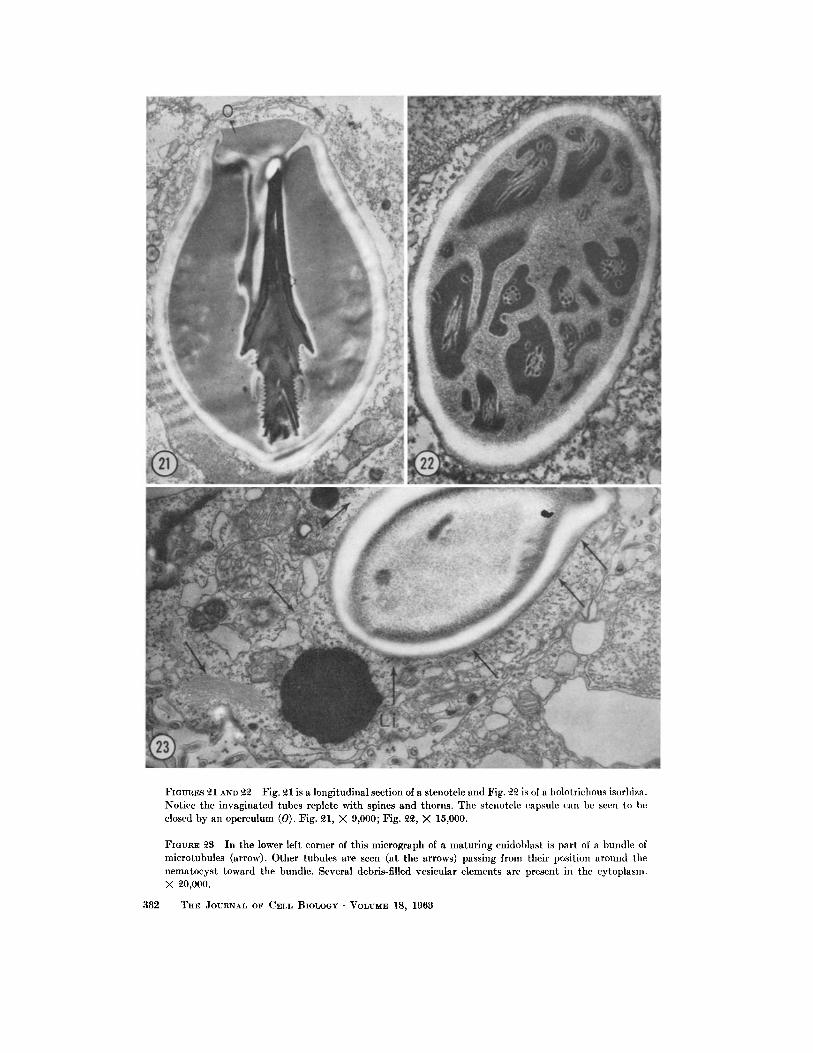
from the cytoplasm, the granular material begins to form the elaborate structure of the nematocyst, two kinds of which can be seen in Figs. 21 and 22. Fig. 21 is of a stenotele with three different varie- ties of thorns on its tube, and Fig. 22 is of a holo- trichous isorhiza.
In Fig. 2, a small part of the cytoplasm of an interstitial cell has been considerably enlarged. In addition to ribosomes, mitochondria, and small scattered vesicles of the Golgi complex or endo- plasmic reticulum, another cytoplasmic element can be seen. It is composed of small tubules, having an outside diameter of approximately 180 A, an inside diameter of approximately 80 A, and a mem- branous wall 50 A thick. As seen in this illustra- tion (Fig. 2, Mt), they are found sometimes dis- posed in packets at right angles to each other, but elsewhere they are scattered throughout the cyto- plasm. Their relationship to the centrioles is sug- gested, even in this micrograph, by the oblique section of the centriole (Fig. 2, Ce) from which tubules radiate, but it should be stressed that these tubules may be seen in parts of the cytoplasm very distant from the centrioles. Fig. 6 illustrates more clearly the relationship between the microtubules and centrioles. One of the centrioles in this micro- graph has been cut at right angles to its long axis and shows the typically cylindrical disposition of nine triple-compartmented tubular structures. Numerous microtubules (Mt) appear to radiate from these centrioles. In other illustrations it can be seen that the microtubules rise from both the base of the centriole and the sides of the tubules of the centrioles. Fig. 26 shows another centriole cut in such a way as to illustrate this radiating pattern again.
Figs. 3-5 show three intercellular bridges con necting interstitial cells. The microtubules can be seen running in these bridges but they exhibit no preferred orientation relative to either axis of the bridge. In Fig. 5 there is a dense body making an
FIGURE ~ A small piece of interstitial cell cytoplasm is enlarged to show numerous microtubules (Mt) in its cytoplasm. Some are cut in cross-section, others in longitudinal section. In the upper left corner of the micrograph, just beneath the plasmalemma is an oblique section of the base of a centriole from which microtubules evidently emanate. X 5~,500.
FIGURES 3, 4, AND 5 Intercellular bridges connecting interstitial cells and illustrating the numerous disoriented microtubulcs within them. These tubules are remnants of the mitotic spindle. The bridge in Fig. 5 is imperfectly obstructed by a midbody. Fig. 3, X ~7,000; Fig. 4, )< 64,500; Fig. 5, )< 5~,000.
370 THE JO~nN~L OF CELL BIOLOGY • VOLUME 18, 1963

DAVID B. SI..,AUTTERBACK Cytoplasmic Microtubules. I 371
incomplete septum between the two adjacent cells at the level of the annular expansion of the bridge. This dense body is interpreted as the so called "intermediate body" (3). It will be remembered that the intercellular bridge is the last remaining area of the achromatic spindle, and indeed these microtubules resemble closely structures which have been interpreted by other authors as spindle remnants (9, 10).
Among the most striking relationships shown by these small tubules is that which they bear to the nematocyst. Fig. 9 illustrates a cross-section of a developing nematocyst near the open end of the capsule and in a plane similar to that indicated by line A-A' in Fig. 8. The developing nematocyst tube is encased in its Golgi membrane, and many vesicles of the Golgi complex are seen surrounding it completely. Between the Golgi membrane of the nematocyst and the outer Golgi vesicles are numer- ous short oblique sections of microtubules, encom- passing the developing nematocyst like the staves of a barrel. Between these sections of microtubules and the Golgi vesicles are many small spherical vesicles of the Golgi complex. In cross-sections such as in Figs. 10-12, and in longitudinal sec- tions tangential to the nematocyst capsule as in Figs. 16 and 17, it can be seen that these tubular structures are in fact microtubules. Their wall is invariably 50 A thick and they pursue a relatively straight unbranched course set at regular intervals around the nematocyst. In many instances, such as those illustrated in Figs. 13-15, a connection can be seen between the microtubules and the small spherical vesicles of the Golgi complex lying im- mediately adjacent to them. In sone cases it is difficult to tell whether the tubules end in the small vesicles or whether, as illustrated in Fig. 18, the tubules have short branches which in turn connect with the small spherical vesicles. The former are more often seen.
Fig. 19 illustrates another activity of the Golgi membrane which encloses the developing nemato- cyst tube. It appears that small spherical vesicles
are being formed from this membrane, although it is possible that they are being added to it. The significance of this observation is not yet under stood.
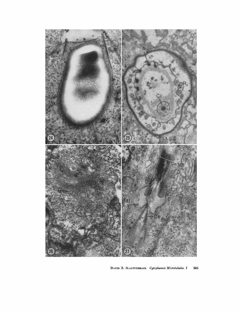
As the nematocyst matures, the centrosome moves to the opercular end where it participates in the formation of a complex structure known as the cnidocil. The latter is associated with the firing mechanism of the nematocyst and is composed in part of a large, slightly modified cilium with basal granules, surrounded by an eccentric circle of twenty-one (-4-3) highly modified cilia with equally modified basal granules (Figs. 24 and 25). It can be seen in favorable sections that the microtubules arise near all twenty-two of these basal granules (Figs. 24, 26 and 27), although some seem to arise near the plasmalemma (Fig. 27). Particu- larly striking is the way they radiate from the circumference of the upright centriole of the main cilium. Although it is difficult to obtain sections illustrating this condition, it may be faintly dis- cerned in Fig. 26. The microtubules pass fi'om the area of the cnidocil to surround the nematocyst beneath it (Fig. 24). Their number seems to in- crease gradually during nematocyst enlargement (compare Figs. 10-12 and 20) but not enough to keep pace with the increasing girth of the secretory product since, as illustrated in Fig. 20, the micro- tubules are more widely spaced around the mature nematocyst than around the immature one. They continue along the length of the nematocyst and beyond, where they coil tightly in the vicinity of the nucleus and the reduced Golgi complex to form a skein of tubules (Figs. 23 and 24).
D I S C U S S I O N
One of the earliest observations of microtubules was reported by Palay (48), who called attention to their presence in both dendrites and axon terminals in the neuropil of the abducens nucleus of rats. He described them as "long, tubular elements of the endoplasmic reticulum, about 180 A wide and re- markably straight." Though present in axons, they
FIGURE 6 A pair of centrioles (Ce) in an interstitial cell which has begun to differentiate into a cnitloblast. Notice the numerous microtubules (Mr), most of which appear to be radiating from the diplosome region. The plasmalemmas of this and the adjacent cell are in the upper right and Golgi vesicles are prominent in the lower left. X 75,500
FmvaE 7 Golgi complex of an interstitial cell differentiated to about the same extent as that in Fig. 6. Compare this Golgi complex with the one in Fig. 1. Notice especially the prominence of the small spherical vesicles. X ~7,500.
372 THE JOURNAL OF CELL BIOLOGY • VOLU~E 18, 1963

DAVID B. SLAUTTERBACK Cytoplasmic Mierotubules. 1 373

were found to be most prominent in the dendrite and distinct from the smaller neurofilaments which he described elsewhere (50). Their identification as " tubular or canalicular, membranous elements of the endoplasmic ret iculum" was supported in the following way (48). "At irregular intervals they are dilated into chains of vesicles of various di- ameters. Occasional clusters of small, dense granules, 50 to 60 A in diameter, lie among these canaliculi, especially at the bases of branches. These granules represent small clusters of basophil material corresponding to the small clumps of Nissl substance seen particularly at sites of den- dritic branching in stained sections examined with the light microscope." Subsequently, numerous authors (7, 17, 20, 30, 49, 72, 85) have reported mlcrotubules in the dendrites of a wide variety of neurons. However, further evidence for the inter- pretation of microtubules of neurons as specializa- tions of the endoplasmic reticulum has not been adduced.
Though the presence of nerve cells in Hydra was recorded many years ago (70), the subject has remained controversial. However, it is evident from recent investigations in this laboratory that such cell types do exist in Hydra, and that they, like other neurons, contain 180 A microtubules in their cell processes. More details of this occur- rence will be published in another paper.
As mentioned above, perhaps the most con- sistent reports of microtubules have been in the motitic apparatus of dividing cells. Early in the application of electron microscopy to cytological problems, a search was undertaken for evidence of the fibrous nature of the achromatic figure. The earliest observations were severely hampered by the inadequacy of techniques for cell preservation
and sectioning (1, 32, 75), but by 1954 Porter (53) reported a tubular component associated with the achromatic figure. Since that t ime it has been established by a number of authors (42) that spindle fibers and chromsome fibers and probably fibers of the astral rays, in such diverse genera as crayfish (68, 87), sea urchin (33, 37), amoeba (63, 64), chicken (5), mouse (73), and man (5), are in fact fine tubular elements having an outside diameter of from 140 A as reported for Pelomyxa carolinensis (62, 63) to perhaps 210 A as reported for some mammal ian cells (5). In Hydra, mitotic figures have been observed only rarely since the introduction of techniques which favor adequate preservation of the microtubules. However, the intercellular bridges which bind together the differentiating cnidoblasts contain spindle rem- nants (as shown earlier by Fawcett, Ito, and Slaut- terback, 24), and these are tubular structures, with an outside diameter of 180 A as seen else- where in the same cells. Nagano (44) has also re- ported the appearance of fine tubular structures which he interpreted as spindle remnants in and about the bridges binding together spermatocytes of the rooster. Buck and Tisdale (9, 10) have pub- lished recently a description of the spindle remnant and midbody in the intercellular bridges of pro- liferating rat erythroblasts. These authors did not comment on the tubular nature or dimensions of the spindle fibers; some of their published micro- graphs are too low in contrast or magnification to illustrate these properties, but others of their micrographs and the originals which were demon- strated at a recent meeting of the American Asso- ciation of Anatomists (8) clearly support the tubule concept and exhibit a dimension of approxi- mately 160 A. Particularly difficult to explain is
FIGURE 8 This micrograph illustrates a cnidoblast containing a partly developed nemato- cyst which has been cut in longitudinal section. The faintly reticular material with a light inner margin and a smooth membrane confining it and C-shaped in profile is the flask-shaped nematocyst capsule (Ca). The heterogeneous granular materia] (Hg) which will ultimately form tile tube has begun to exceed the capacity of the capsule. Note the conspicuous cap of Golgi vesicles (G) over the growing tip. The line A-A' indicates the plane of section of Fig. 9. )< ~,000.
FIGUnE 9 In this transverse section of the upper part of a developing nematocyst the smooth Golgi membrane encasing the nematocyst can be seen. Immediately peripheral to it are numerous oblique sections of microtubules. External to these is an incomplete layer of small spherical vesicles, a few of which are closely approached by microtubules. A layer of large flattened Golgi sacs, followed by more small spherical vesicles, completes the circumferential laminations. )< 41,000.
374 THE JOURNAL OF CELL BIOLOGY • VOLUME t~, 1963

DAVID B. SI, AUTTERBACK Cytoplasmic Microtubules. I 375

the random orientation of the mlcrotubules in the intercellular bridges of Hydra. This evident disori- entation of the microtubules relative to their con- dition in the spindle may be related to the transient appearance of the midbody of B~lfir (3). This latter structure appears after the intercellular bridge has been formed, and later disappears, though the bridge itself persists. For a time the midbody seems to interrupt the intercellular bridge and may in this way disorganize the tubules.
Other observations of cytoplasmic tubules have been reported from the protozoa, particularly the ciliates and flagellates. Roth (59, 60, 61) has shown them to be present beneath the plasma lemma of Euglena gracilis, Peranema trichophorum, Euplotes patella, and Paramecium aurelia. The list of investigators who have observed fine filamentous structures in the cytoplasm of protozoa is too long to include here, but every observation of which this author is aware records a structure having a more dense periphery and less dense center, which would seem to be tubular. Most of these elements measure from 140 to 200 A in diameter. In addi- tion to being found in the ciliates mentioned above, they have been seen both beneath the plasmalemma and in association with the para- basal body in such animals as Carchesium (56), CoIeps (66), Ephelota (65), Tokophrya (67), Opalina (45, 46), and in Sp#ostomum ambiguum (57). In the last named animal a 200 A tubular element was associated by Randall (57) with the myonemes, a presumably contractile element of the cytoplasm. Also, Steinert (81), Steinert and Novikoff (82), and Schulz and MacClure (71) have observed tubular elements beneath the plasmalemma of Trypano- somes. Not all of these tubular structures have been reported to have the same size. However, if allow- ance is made for the probability of some inac- curacy in the reported dimensions resulting from such things as microscope calibration and differing methods of preservation, it would seem that there are two size categories of such tubules in protozoa, These are represented by those tubules measuring about 120 to 200 A and those which have an out- side diameter of about 270 A. The latter are usually
found in association with the plasmalemma, whereas the former are more often seen near the centrioles or basal granules. Though usually found separately, at times both types of tubules are present in the same cell. In Hydra, only those tubules having a 180 A diameter have been seen. It is believed that these tubules have different functional capacities. Support for this belief will be cited below and in a subsequent paper.
Microtubules have also been reported to be found in association with centrioles. Although the techniques of preservation militated against accu- rate preservation of detail, Bessis, Breton-Gorius and Thi~ry (6) in 1958 observed fine tubular struc- tures 200 A in diameter projecting, in two tiers along radii, outward from the circumference of the cylindrical centrioles in blood leucocytes. They concluded that these tubules terminated, 650 to 900 A from the centriole, in small spherical masses or vesicles. These observations are reminis- cent of the situation in Hydra, where the micro- tubules can be seen to emanate from the circum- ference of the centrioles, and elsewhere continuity with small spherical vesicles has been demon- strated, de Harven and Bernhard (15), Bernhard and de Harven (5), Gibbons (28), Gibbons and Grimstone (29), and Gall (25) have noted a simi- lar relationship between structures (which their micrographs confirm to be microtubules) and the centrioles in mitotic and interphase cells. It is in- teresting to note that the ciliary filaments which emanate from the distal ends of centrioles are similar in dimensions and general appearance to the microtubules which arise from the circumfer- ence and proximal ends of the same centrioles. And further, in considering the relation to eentrioles and the continuity of microtubules with Golgi vesicles demonstrated here, it may be significant that other investigators ( 1, 31 ) have suggested that the centrioles regulate the amount of Golgi mem- branes present in a cell.
In 1955, Burgos and Fawcett (11) reported the presence of tubular elements of the cytoplasm in the manchette (caudal sheath) of the developing spermatids of the cat Felis domestica. A year later
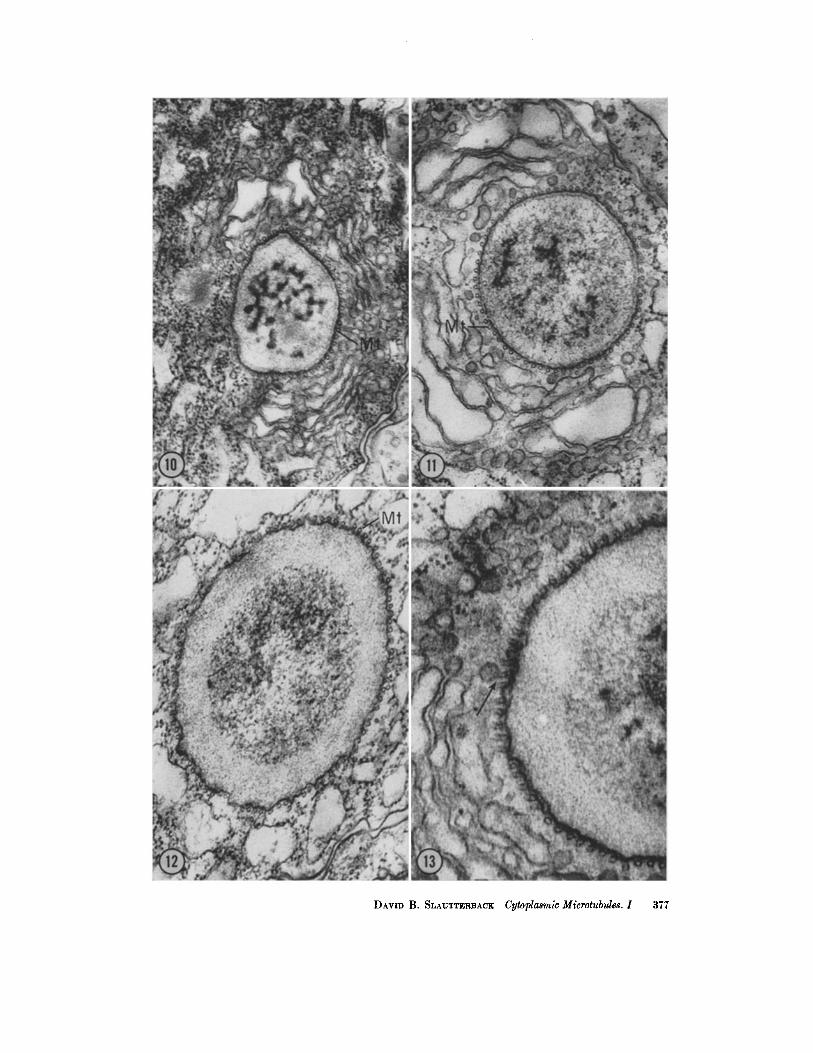
FmuaEs 10, 11, ANn ]~ Transverse sections of the microtubules. The latter are clearly seen surrounding the growing tip of the nematocyst tube distal to the capsule. Figs. 10 and 11, X 50,000; Fig. 1~, X 74,000.
FIGURE 13 For legend see page 378.
376 THE JOURNAL OF CELL BIOLOGY • VOLUME 18, 1963
DAVID B. SLXUTTv.aBACK Cytoplasmic Microtubules. I 377

the same authors (12) reported a similar observa- tion in spermatids of the toad, Bufo. These tubules were said to have an outside diameter on the order of 250 A. Careful scrutiny of these rnicrographs, however, suggests that 150 to 200 A would be a better approximation. Later, Gatenby and Dalton (26) observed 150 A tubules around the nucleus of Lumbricus spermatids and believed them to cor- respond to the tubular elements of the caudal sheath of vertebrate spermatids. And Manton (41) found 200 A "fibers" (probably tubules) in the ciliated band and nuclear sling of fern spermato- zoids.
Other reports have similarly extended the ob- servation of microtubules in sperm cells beyond the spindle remnant regularly found in the inter- cellular bridges. Studying Otala lactea, Rebhun (58) saw "200 A tubules" set around the nucleus at 300 A intervals and concluded, "This component of the cell continues over the centriole and forms the outermost layer of the tail sheath of the mature sperm." He considered this structure to be homologous with the cytoplasmic tubular ele- ments in the manchette described by Burgos and Fawcett. Rebhun ' s observations were confirmed in the sperm of another snail, Helix aspera, by Roth (60). Sotelo and Trujil lo-Cenoz (80) found similar tubules in sperm of the avian Passer said to be 240 A in diameter, and the micrographs of De Robertis and Ruffo (16) show microtubules in the sperma- tocytes of Laplatacris dispar, a grasshopper.
In addition to these findings of tubules in male germ cells, a recent observation has shown a 180 A tubule to be present in the cytoplasm of spermato- cytes from the flatworm Plagiostomum. These ob- servations were reported by Christensen (13), who saw them beneath the plasmalemma where "they pursue a spiral course up the body of the sperm." " T h e cytoplasm around the middle portion of the nucleus contains whorls of flattened cisternae, resembling endoplasmic reticulum (without ribo-
somes), which extend out to insert along every other or every few" tubules. Christensen, to- gether with Fawcett (14), has also published an elegant paper concerning the fine structure of the testicular interstitial cell of the opossum, in which a 180 A tubule occurring in bundles in the cyto- plasm of that cell is described. Although open communicat ion between these tubules and the endoplasmic reticulum was described, Christensen was unable to demonstrate a specific involvement of the microtubules in the secretory function of this cell. It is significant to note that these tubules of the interstitial cell were shown by the authors to have continuity with a small spherical vesicle, the membrane of which was free of ribosomes. They interpreted this observation as indicative of direct continuity between the tubules and the agranular reticulum.
Another report of microtubules in a secretory cell was made recently by Philpott (51, 52), who found 180 A microtubules in the socalled "chlo- r ide" cells of the fish, Fundulus. Although the micro- tubules in this " ion secreting" cell are found scat- tered throughout the cytoplasm, they are generally oriented parallel to the long axis of the cell and seem to be directed toward the apical vesicle, into which the secretory products are liberated.
In a report by Fawcett (23) on the erythrocytes of the toadfish (Opsanus tau) it is evident that the cytoplasm of some fish cells also contains a small tubular component. Fawcett showed that the striae bordante are composed of small tubules, about 270 A in diameter, and attributed to them the elastic properties typical of these cells. This con- clusion followed the observation that erythrocytes, when deformed in the plane of the circle made by these tubules, quickly and elastically recover their original shape, whereas cells distorted along any other part of their surface do not recover in this manner.
In a paper read before the American Associa-
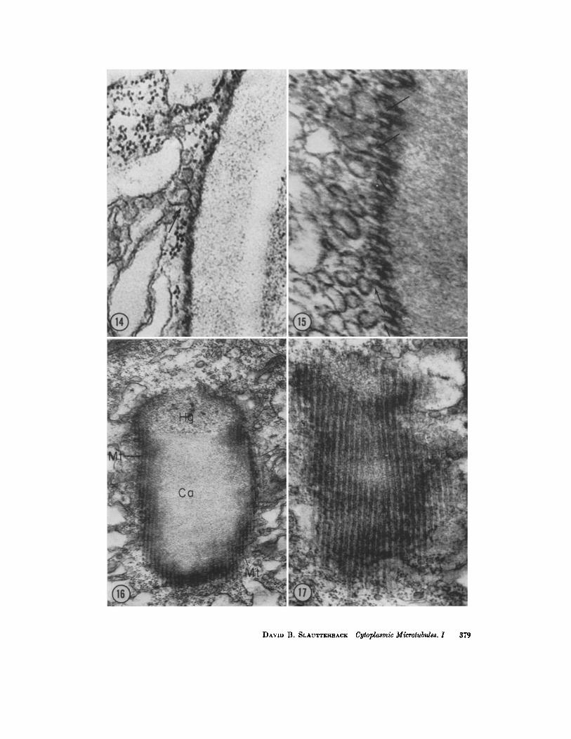
FIGURES 18, 14, AND 15 These sections are similar to the section illustrated in Fig. 9 but exhibit (arrows) confluence between microtubules and small spherical vesicles of the Golgi complex. Fig. 13, X 81,000; Fig. 14, X 116,000; and Fig. 15, X 158,000.
FmURES 16 AND 17 These micrographs illustrate longitudinal sections of microtubules (Mr) disposed around the circumference of the nematocyst. The section has passed tan- gential to the surface of the capsule in Fig. 17, and in Fig. 16 it has cut a little deeper, exposing a small amount of nematocyst material. The sections are serial but not consecu- tive. They have been printed at different magnifications to illustrate, on one hand, the relation of the tubules to the nematocyst and, on the other hand, the characteristics of the tubules themselves. Fig. 16, X 47,000; Fig. 17, X 71,000.
378 THE JOURNAL OF CELL BIOLOGY • VOLUME ]8, 1963
DAVID B. SLAUTTERBACK Cytoplasmic Mieeotubules. I 379
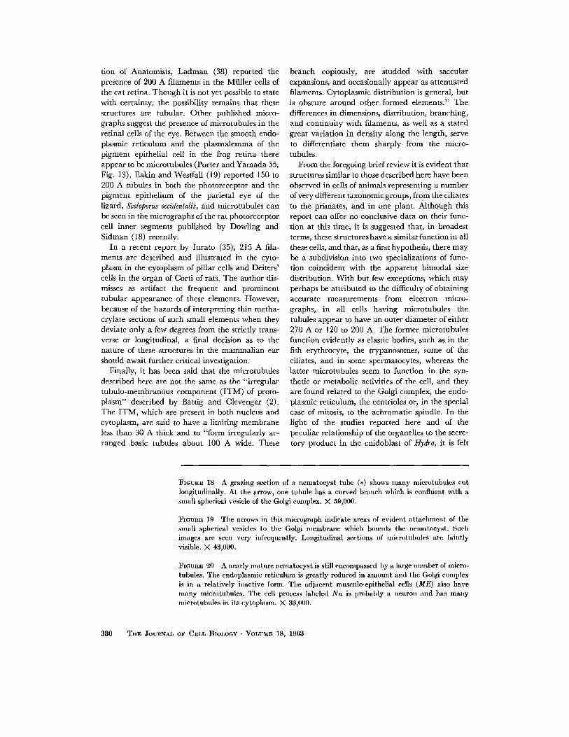
tion of Anatomists, Ladman (38) reported the presence of 200 A filaments in the Mtiller cells of the cat retina. Though it is not yet possible to state with certainty, the possibility remains that these structures are tubular. Other published micro- graphs suggest the presence of microtubules in the retinal cells of the eye. Between the smooth endo- plasmic reticulum and the plasmalemma of the pigment epithelial cell in the frog retina there appear to be microtubules (Porter and Yamada 55, Fig. 13). Eakin and Westfall (19) reported 150 to 200 A tubules in both the photoreceptor and the pigment epithelium of the parietal eye of the lizard, Sceloporus occidentalis, and microtubules can be seen in the micrographs of the rat photoreceptor cell inner segments published by Dowling and Sidman (18) recently.
In a recent report by Iurato (35), 215 A fila- ments are described and illustrated in the cyto- plasm in the cytoplasm of pillar cells and Deiters' cells in the organ of Corti of rats. The author dis- misses as artifact the frequent and prominent tubular appearance of these elements. However, because of the hazards of interpreting thin metha- crylate sections of such small elements when they deviate only a few degrees from the strictly trans- verse or longitudinal, a final decision as to the nature of these structures in the mammal ian ear should await further critical investigation.
Finally, it has been said that the microtubules described here are not the same as the "irregular tubulo-membranous component ( ITM) of proto- plasm" described by Battig and Clevenger (2). The I T M , which are present in both nucleus and cytoplasm, are said to have a limiting membrane less than 30 A thick and to "form irregularly ar- ranged basic tubules about 100 A wide. These
branch copiously, are studded with saccular expansions, and occasionally appear as attenuated filaments. Cytoplasmic distribution is general, but is obscure around other formed elements." The differences in dimensions, distribution, branching, and continuity with filaments, as well as a stated great variation in density along the length, serve to differentiate them sharply from the micro- tubules.
From the foregoing brief review it is evident that structures similar to those described here have been observed in cells of animals representing a number of very different taxonomic groups, from the ciliates to the primates, and in one plant. Although this report can offer no conclusive data on their func- tion at this time, it is suggested that, in broadest terms, these structures have a similar function in all these cells, and that, as a first hypothesis, there may be a subdivision into two specializations of func- tion coincident with the apparent bimodal size distribution. With but few exceptions, which may perhaps be attributed to the difficulty of obtaining accurate measurements from electron micro- graphs, in all cells having microtubules the tubules appear to have an outer diameter of either 270 A or 120 to 200 A. The former microtubules function evidently as elastic bodies, such as in the fish erythrocyte, the trypanosomes, some of the ciliates, and in some spermatocytes, whereas the latter microtubules seem to function in the syn- thetic or metabolic activities of the cell, and they are found related to the Golgi complex, the endo- plasmic reticulum, the centrioles or, in the special case of mitosis, to the achromatic spindle. In the light of the studies reported here and of the peculiar relationship of the organelles to the secre- tory product in the cnidoblast of Hydra, it is felt
FmURE 18 A grazing section of a nematocyst tube (*) shows many microtubules cut longitudinally. At the arrow, one tubule has a curved branch which is confluent with a small spherical vesicle of the Golgi complex. )< 59,000.
FIGURE 19 The arrows in this micrograph indicate areas of evident attachment of the small spherical vesicles to the Golgi membrane which bounds the nematocyst. Such images are seen very infrequently. Longitudinal sections of microtubules are faintly visible. X 43,000.
FIGURE g0 A nearly mature nematocyst is still encompassed by a large number of micro- tubules. The endoplasmic reticulmn is greatly reduced in amount and the Golgi complex is in a relatively inactive form. The adjacent musculo-epithelial cells (ME) also have many microtubules. The cell process labeled Nn is probably a neuron and has many microtubules in its cytoplasm. X 38,000.
380 THE JOURNAL OF CELL BIOLOGY " VOLUME 18, 1963

DAWD B. S~-VTTF~BACK Cytoplasmic Microtubules. 1 381
382
FmvaEs o-1 AND ~ Fig. ~1 is a longitudinal section of a stenotele and Fig. ~ is of a holotrichous isorhiza. Notice the invaginated tubes replete with spines and thorns. The stenotele capsule can be seen to be closed by an operculum (0). Fig. ~1, X 9,000; Fig. ~ , X 15,000.
FIaURE £$ In the lower left corner of this micrograph of a matur ing cnidoblast is par t of a bundle of microtubules (arrow). Other tubules are seen (at the arrows) passing from their position around the nematocys t toward the bundle. Several debris-filled vesicular elements are present in the cytoplasm. X ~0,000.
THE JOVRNAL OF CELL BIOLOGY • VOLUME lg, 1963

that certain other speculations as to function may be justified.
It has been shown (79) that, during the process of secretion of the components of the nematocyst, the newly synthesized material is apparently de- hydrated and condensed in the Golgi complex before it is added to the partly formed nematocyst. The intimate relationship of the microtubules to the Golgi complex suggests that they may also be involved in this process. Furthermore, when the tube of the nematocyst is completed it is coiled about loosely in the cytoplasm and must be with- drawn into the nematocyst shell and the operculum closed, before more structure appears in this tube. It is difficult to understand how this large volume of material can be withdrawn into a shell whose volume changes little, without a considerable de- gree of dehydration of that material. The very close association of the microtubules with the mem- branes derived from the Golgi complex surround- ing the nematocyst at this time is again suggestive of a function related to water or electrolyte trans- port. One of the enigmas of nematocyst develop- ment has been that the elaborate structure formed in the tube of this secretory product does not begin to develop until the tube is withdrawn into the shell and thus completely isolated from the cytoplasmic organelles, which might be found responsible for the imposition of structure. Since it is not uncommon to produce structured systems out of unstructured ones by relatively minor varia- tion of the ionic strength of the medium, it is tempting to infer from the events in the cnidoblast that the microtubules are involved in the produc- tion of a change in ionic strength within the nematocyst shell. The fact that small vesicles can be seen forming on the membrane bounding the nematocyst and that similar small vesicles can be seen in continuity with the microtubules lends support to this speculation. The presence of these microtubules in ion secreting cells and neurons is also consistent with this notion of their function. And in this regard, it should be noted that in published micrographs of the glomerular epi- thelium of mammalian kidney (22) there appear to be numerous microtubules in the cytoplasm near the plasmalemma.
In our present state of understanding of mem- brane function and construction it is difficult to interpret the significance of the thinness of the membranous wall of the microtubules, and no positive proposal as to the molecular organization can be made at this time. However, the structure
of the thin membrane reported here may not be accurately represented by the conventional inter- pretation of a bimolecular phospholipid leaflet with expanded polypeptide chains adsorbed on both surfaces. Furthermore, the ubiquity of the so called "unit membrane" and the support which this concept has given to the conventional inter- pretation may be cast in some doubt for micro- tubules, since the three-layer profile, which is a defining property of the "unit membrane," is not seen in them. Whether this is indicative of a fundamentally different organization of the pro- tein and phospholipid of the membrane, or whether the relatively fluid phospholipid layer has been compressed below the limit of resolution by osmotic or other pressures within the tubule can only be guessed at, although it may be noted that the width of this membrane is about equal to two of the lines of the "unit membrane." Whatever may be the correct interpretation of this and other thin membranes, their presence in cells necessitates a reevaluation of the requirements for stability and of the concepts of molecular orientation in proteolipid membranes.
Regardless of the above considerations, it seems reasonable to assume that the membrane bound- ing the microtubules has properties similar to those of other complex phospholipid-protein mem- branes with which it is continuous. One of the best established properties of such membranes is their ability to concentrate ions at their surfaces. Such a situation would greatly favor the ability to transport ions in the tubule parallel to its long dimension, assuming that this membrane, like others, bears negative charges on its inner and outer surfaces. In addition, it should be pointed out here that a high degree of selectivity of this membrane may be inferred from its continuity with the membranes of other organelles having demonstrably selective properties. The transport of positive ions through the tubules would be dependent upon the ability to remove them at one end of the tubule. It is also tempting to speculate further that, given this function for the micro- tubules, their association with the firing mech- anism of the nematocyst might be more easily explained. Those ions or small molecules being removed from the nematocyst by the microtubules, were they to be stored in the extensively coiled portion of the microtubules at the base of the cnidoblast, could be returned to the nematocyst upon stimulation of the cnidocil, thus allowing for its sudden expansion and eruption through the
DAVID B. SLAUTTERBACK Cytoplasmic Microtubules. I 383

operculum. I t is interesting to note in this regard tha t the firing of the nematocyst is extremely sensitive to the relative concent ra t ion of divalent metal ions, such as calcium and magnesium. As ment ioned above, this funct ion also facilitates explanat ion of the relatively sudden appearance of complex structure in the previously formless contents of the nematocyst capsule.
I t should be stated tha t the au thor is aware and respectful of the a rguments tha t structures which appear to be tubules may in fact be filaments whose inhomogeneous density is an artifact result- ing from such p h e n o m e n a as preferential deposi- t ion of osmium or stains at the surface. While every effort has been made to preclude such mis- in terpre ta t ion, and the constant thickness of the m e m b r a n e militates strongly against it, two counte ra rguments must be borne in mind. The one is that , in the absence of incont rover table evidence, the choice of the te rm f i lament r a the r t han tubule is ne i ther more cautious nor less com- mit ted since the different sets of properties denoted by the two terms are approximate ly equivalent . In fact, when one is confronted wi th the sort of evidence adduced here, the preferred designation iS clearly " t ubu l e . " The other is tha t a s tructure need not be hollow to be a tubule. Such trivial examples as domestic p lumbing in which the pipes, filled with water, would not be regarded as rods by any observer, or ch roma tog raph columns,
in which the funct ional properties are m u c h more akin to tubules t h a n rods, will serve to i l lustrate this point. In fact, we might say tha t minor differ- ences between the core and per iphery of an elon- gate structure, in such properties as the ra te of diffusion of a given ion, consti tute a funct ional ly accurate definition of a tubule. Indeed, from such considerations one might suggest tha t the conven- t ional designation of the nine plus two elements of cilia, flagella, and sperm tails, and the nine triple- compar tmen ted elements of centrioles as ciliary or centr iolar " f i laments" is an imped iment to the accurate unders tand ing of their function. Indeed, wha t is hoped for here is tha t some of these specu- lations, r a ther than closing the issues, will engender renewed interest in the numerous peculiar proper- ties of tubules and tha t it be general ly recognized tha t cells possess a "mic ro tubu la r system" whose unique s t ructural and funct ional properties and widespread occurrence justify its considerat ion as a discrete cytoplasmic organelle.
The author wishes to apologize to the few investigators who have reported the presence of tubules in such cells as neurons and protozoa but go unmentioned here owing to the limitations of space.
Indebtedness is acknowledged for the expert tech- nical assistance of Marjorie A. Johnson and Gunvor Sommerhaug.
The studies here were supported by research
FIGURE 24 "Parasa~ttal" section of a late nematocyst showing many longitudinal sections of microtubules. Some of them appear to arise in the vicinity of the "stiff rods" (R) of the cnidocil apparatus. X 36,000.
FIGURE ~5 The figure illustrates a cross section of a cnidocil apparatus. To one side of the center is the slightly modified cilium (Ci) surrounded by cross-striated filamentous material connecting it to the "stiff rods" (R) and operculum. The number of the stiff rods varies slightly; in this case there are 18. Some of them appear more dense (R1) because the section has passed through the rootlets, and others appear less dense (R2) because the modified basal granules or cilium itself has been sectioned. X 37,000.
FIGURE ~6 This is a cross-section through the centriole at the base of the cnidocil cilium of a stage earlier than that of Fig. ~5, before the formation of the "stiff rods." There are many longitudinal sections of microtubules emanating from the circumference of the centriole. )< 35,000.
FIGURE ~7 This is the same stage as the cell in Fig. ~6, but the cilium is seen in longi- tudinal section. Because the cilium lies flat against the surface of the cell, the same cy- toplasm is seen above and below. The section has missed most of the microtubules which arise from the circumference of the centriole, but other microtubules can be seen to arise at or near the plasmalemma. Note the cross-striated filaments constituting the rootlets. X 28,000.
384 TRE JOURNAL OF CELL BIOLOGY • VOLUME 18, 1963
Dxvm B. SLAUTTERBACK Cytoplasmic Microtubules. I 385

grant RG 6934 from the National Institutes of Health, United States Public Health Service.
Some of these observations were presented before
B I B L I O G R A P H Y
1. AMANO, S., The structure of the centrioles and spindle body as observed under the electron and phase contrast microscopes-- a new ex- tension-fiber theory concerning mitotic mecha- nism in animal cells, Cytologia, 1957, 22, 193.
2. BATTIO, C. G., and CLEVENOER, M. R. An irregu- lar tubulo-membranous component (ITM) of protoplasm, Anat. Rec., 1961, 139, 328.
3. B~L£R, K., Beitr~ige zur Kenntnis des IV[echa- nismus der indirekten Kernteilung, Natur- wissenschaften, 1927, 15, 725.
4. B~NNETT, H. S., and LUFT, J. H., s-Collidine as a basis for buffering fixatives, J. Biophysic. and Biochem. Cytol., 1959, 6, 113.
5. BERNHARD, W., and DE HARVEN, E., L'ultra- structure du centriole et d'autres 616ments de l 'appareil aehromatique, Proc. 4th Internat. Conf. Electron Micr., Berlin, 1958, 2, 217.
6. BESSIS, M., BRETON-GoRIUS, J., and THII~RY, J.-P., Centriole, corps de Golgi et aster des leuco- cytes, l~tude au microscope 61ectronique, Rev. Hematol., 1958, 13, 363.
7. BOYCOTT: B. B., GRAY, E. G., and GUILLERY, R. W., Synaptic structure and its alteration with environmental temperature: a study by light and electron microscopy of the central nervous system of lizards, Proc. Royal Soc. London, Series B, 1961, 154, 151.
8. BucK, R. C., Some observations on cytokinesis in mammalian cells studied with the electron microscope, Anat. Rec., 1962, 142, 336.
9. BUCK, R. C., and TISDALE, J. M., The fine struc- ture of the mid-body of the rat erythroblast, J. Cell Biol., 1962, 13, 109.
10. BUCK, R. C., and TISDALE, J. M., An electron microscopic study of the development of the cleavage furrow in mammalian cells, o r . Cell Biol., 1962, 13, 117.
11. BURGOS, M. H., and FAWCETT, D. W. Studies on the fine structure of the mammalian testis. I. Differentiation of the spermatids in the cat (Fells domestica), J. Biophysic. and Biochem. Cytol., 1955, 1, 287.
12. BURGOS, M. H., and FAWCETT, D. W., An elec- tron microscope study of spermatid differen- tiation in the toad, Bufo arenarum Hensel, J . Biophysic. and Biochem. Cytol., 1956, 2, 223.
13. CHRISTENSEN, A. K., Fine structure of an unusual spermatozoon in the flatworm Plagiostomum, Biol. Bull., 1961, 121, 416.
14. CHRISTENSEN, A. K., and FAWCETT, D. W., The
the American Society for Cell Biology at its first annual meeting (77). Received for publication, October 29, 1962
normal fine structure of opossum testicular interstitial cells, J. Biophysic. and Biochem. Cytol., 1961, 9, 653.
15. DE HARVEN, E., and BERNHARD, W., Etudie au microscope 61ectronique de l 'ultrastructure du centriole chez les vert6br~s, Z. Zellforsch. u. Mikr. Anat., 1956, 45, 378.
16. DE ROBERTIS, E., and RUFFO, H. F., Submicro- scopic organization of the mitochondrial body and other cytoplasmic structures of insect testis, Exp. Cell Research, 1957, 12, 66.
17. DE ROBERTIS, E., PELLIGRINO DE IRALDI, A., De LORES ARNAXZ, G. R., and SALOANmOFF, L., Electron microscope observations on nerve endings isolated from rat brain, Anat. Rec., 1961, 139, 220.
18. DOWLmG, J. E., AND SXDMAN, R. L., Inherited retinal dystrophy in the rat, or. Cell Biol., 1962, 14, 73.
19. EAKIN, R. M., and WESTFALL, J. A., Fine struc- ture of the retina in the reptilian third eye, 3". Biophysic. and Biochem. Cytol., 1959, 6, 133.
20. ELFVXN, L.-G., Electron-microscopic investiga- tion of filament structures in unmyelinated fibers of cat splenic nerve, J. Ultrastruct. Research, 1961, 5, 51.
21. ENDERS, A. C., Observations on the fine struc- ture of lutein cells, J. Cell Biol., 1962, 12, 101.
22. FARQUHAR, M G , and PALADE, G. E., Glomeru- lar permeability. II. Ferritin transfer across the glomerular wall in nephrotic rats, J. Exp, Med., 1961, 114, 699.
23. FAWCETT, D. W., Electron microscopic observa- tion on the marginal band of nucleated eryth- rocytes, Anat. Rec., 1959, 133, 379.
24. FAWCETT, D. W., ITO, S., and SLAUTTERBAGK, D. B., The occurrence of intercellular bridges in groups of cells exhibiting synchronous differ- entiation, J. Biophysic. and Biochem. Cytol., 1959, 5, 453.
25. GALL, J. B., A study of spermatogenesis in the snail Viviparus, J. Biophysic. and Bioehem. Cytol., 1961, 10, 163.
26 GATENBY, J. B., and DALTON, A. J. , Spermio- genesis in Lumbricus herculeus. An electron microscope study, J. Biophysie. and Bioehem, Cytol., 1959, 6, 45.
27. GELnI, YON, J. , Beitr~ige zur Cytologie der Hydra grisea nebst einigen biologischen Bemerkungen, Z. Zellforseh. u. Mikr. Anat., Abt. Histochem., 1924, 1,471.
386 THE JOURNAL OF CELL BIOLOGY • VOLUME 18, 1968

28. GIBBONS, I. R., The relationship between the fine structure and direction of beat in the gill cilia of a lamellibranch mollusk, J. Biophysic. and Biochem. Cytol., 1961, 11, 179.
29. GIBV, ONS, I. R., and GRI~STONE, A. V., On flagellar structure in certain flagellates, J . Biophysic. and Biochem. Cytol., 1960, 7, 697.
30. GRAY, E. G., Axo-somatic and axo-dendritic synapses of the cerebral cortex: an electron microscope study, J. Anat., 1959, 93, 420.
31. GRIMSTONE, A. V., Cytoplasmic membranes and the nuclear membrane in the flagellate Trichonympha, J. Biophysic. and Biochem. Cytol., 1959, 6, 369.
32. Gitoss, P. R., PHILPOTT, D. E., and NAss, S., The fine-structure of the mitotic spindle in sea urchin eggs, J. UItrastruct. Research, 1958, 2, 55.
33. HARRIS, P., Electron microscope study of mitosis in sea urchin blastomeres, J. Biophysic. and Biochem. Cytol., 1961, 11,419.
34. ITO, S., The endoplasmic reticulum of gastric parietal cells, J. Biophvsic. and Biochem. Cytol., 1961, l l , 333.
35. IURATO, S., Submicroscopic structure of the membranous labyrinth. 2. The epithelium of Corti's organ, Z. Zellforsch. u. Mikr. Anat., 1961, 53, 259.
36. KARNOVSK¥, M. J., Simple methods for "staining with lead" at high pH in electron microscopy, J. Biophysic. and Biochem. Cytol., 1961, l l , 729
37. KUROSUMI, K., Electron microscope studies on mitosis in sea urchin blastomeres, Protoplasma, 1958, 49, I 16.
38. LADMAN, A. J., Electron microscopic observa- tions on the fine structure of Mfiller cells in the retina of the cat, Anat. Rec., 1961, 139, 247.
39. LooMIs, W. F., and LENHOFF, H. M., Growth and sexual differentiation of Hydra in mass culture, J. Exp. Zool., 1956, 132, 555.
40. LVFT, J. H., Improvements in epoxy resin em- bedding methods, J. Biophysic. and Biochem. Cytol., 1961, 9, 409.
41. MANTON, I., Observations on the microanatomy of the spermatozoid of the bracken fern (Pteridium aquilinum), J. Biophysic. and Biochem. Cytol., 1959, 6, 413.
42. MAzIA, D., Mitosis and the physiology of cell division, in The Cell, (J. Brachet and A. E. Mirsky, editors), Academic Press, Inc., New York, 1961, 3, 77.
43. MUELLER, J. F., Some observations on the struc- ture of Hydra, with particular reference to the muscular system, Tr. Am. Micr. Soc., 1950, 69, 133.
44. NAOANO, T., The structure of cytoplasmic bridges in dividing spermatoeytes of the rooster, Anat. Rec., 1961, 141, 73.
45. NGmOT-T1MoTH~E, C., Etude au microscope
filectronique des fibres retrociliares des Ophryo- scolecidae: leur ultrastructure, leur insertion, leur r61e possible, Compt. rend. Acad. sc., 1958, 246, 1286.
46. NOmOT-TI~aOTH~E, C., Recherches sur l 'ultra- structure d 'Opalina ranarum, Ann. Sc. Nat., Zool., 1959, 1, 265.
47. PALADE, G. E., A study of fixation for electron microscopy, J. Exp. Med., 1952, 95, 285.
48. PALAY, S. L., Synapses in the central nervous system, J. Biophysic. and Biochem. Cytol., 1956, 2, No. 4, suppl., 193.
49. PALAY, S. L., The fine structure of secretory neurons in the preoptic nucleus of the goldfish (Carassius auratus), Anat. Rec., 1960, 138, 417.
50. PALAY, S. L., and PALADE, G. E., The fine struc- ture of neurons, J. Biophysic. and Biochem. Cytol., 1955, 1, 69.
51. PmLPOTT, C. W., The adaptive morphology of the chloride secreting cells of Fundulus as re- vealed by the electron microscope, American Society for Cell Biology, Abstracts, 1961, 167.
52. PHILPOTT, C. W., The comparative morphology of the chloride secreting cells of three species of Fundulus as revealed by the electron micro- scope, Anat. Rec., 1962, 142, 267.
53. PORTER, K. R., Changes in cell fine structure accompanying mitosis in Fine Structure of Cells, Symposium of the 8th Congress on Cell Biology, Leiden, Gronigen, Voordhoff, Ltd., 1954, 236.
54. PORTER, K. R., The submicroscopic morphology of protoplasm, Harvey Lectures, 1957, 51, 175.
55. PORTER, K. R., and YAMADA, E., Studies on the endoplasmic reticulum. V. Its form and differ- entiation in pigment epithelial cells of the frog retina, J. Biophysic. and Biochem. Cytol., 1960, 8, 181.
56. RANDALL, J. T., Fine structure of some ciliate protozoa, Nature, 1956, 178, 9.
57. RANDALL, J. T., The fine structure of the proto- zoan Spirostomum ambiguum, Syrup. Soc. Exp. Biol., 1957, 10, 185.
58. REBttUN, L. I., Nuclear changes during spermio- genesis in a pulmonate snail, J. Biophysic. and Biochem. Cytol., 1957, 3, 509.
5% ROTH, L. E., An E-M study of the cytology of the protozoan Euplotes patella, J. Biophysic. and Biochem. Cytol., 1957, 3, 985.
60. ROTH, L. E., A filamentous component of proto- zoan fibrillar systems, J. Ultrastruct. Research, 1958, 1, 223.
61. ROTH, L. E., An electron-microscope study of the cytology of the protozoan Peranema tricho- phorurn, J. Protozool., 1959, 6, 107.
62. ROTI4, L. E., and DANIELS, E. W., The structure
DAVID B. SLAUTTERBACK Cytoplasmic Microtubules. 1 387

of spindle fibrils in a giant amoeba, American Society for Cell Biology, Abstracts, 1961, 183.
63. ROTH, L. E., and DANmLS, E. W., Electron micro- scopic studies of mitosis in amebae. II. The giant ameba Pelomyxa carolinensis, J. Cell Biol., 1962, 12, 57.
64. ROTH, L. E., OBETZ, S. W., and DANIELS, E. W., Electron microscope studies of mitosis in amebae. I. Amoeba proteus, d. Biophysic. and Biochem. Cytol., 1960, 8, 207.
65. ROUILLER, CH., FAUR~-FREMIET, E., and GAUCH- ERY, M., Les tentacules d'Ephelota; etude au microscope electronique, J. Protozool., 1956, 3, 194.
66. ROUILLER, CH., FAUR~-FREMIET, E., and GAUCH- ERY, M., The pharyngeal protein fibres of the ciliates, Proceedings of the Stockholm Confer- ence on Electron Microscopy, Stockholm, Almqvist and Wiksell, 1956, 216.
67. RUDZINSKA, M. A., Mechanisms involved in the function of the contractile vacuole in Toko- phrya infusionum as revealed by electron micros- copy, J. Protozool., 1957, 4 suppl., 9.
68. RUTHMANN, A., The fine structure of the meiotic spindle of the crayfish, J. Biophysic. and BiD- chem. Cytol., 1959, 5, 177.
69. SCHMITT, F. O., and GEREN, B. B., The fibrous structure of the nerve axon in relation to the localization of "neurotubules," J. Exp. Med., 1950, 91,499.
70. SCHNEIDER, K. C., Histologie yon Hydra fusca mit besonderer Berficksichtigung des Nerven- systems der Hydropolypen, Arch. mikr. Anat., 1890, 35, 321.
71. SCHULZ, H. and MAcCLURE, E., Elektronen- mikroskopische Untersuchung des Trypano- soma cruzi mit Besonderer Berficksichtigung des Periplasten und des Blepharoplasten, Z. Zellforsch. u. Mikr. Anat., 1961, 55, 389.
72. SCHULTZ, R. L., MAYNARD, E. A., and PEASE, D. C., Electron microscopy of neurons and neuroglia of cerebral cortex and corpus callo- sum, Am. J. Anat., 1957, 100, 369.
73. SCHULTZ-LARSEN, J., On the structure of the nuclear spindle, Acta Path. et Microbiol. Scand., 1953, 32, 567.
74. SEDAR, A. W., Electron microscopy of the oxyntic cell in the gastric glands of the bullfrog, Rana catesbiana. II. The acid-secreting gastric
mucosa, J. Biophysic. and Biochem. Cytol., 1961, 10, 47.
75. SELBY, C. C., Electron micrographs of mitotic ceils of the Ehrlich's mouse ascites tumor in thin sections, Exp. Cell Research, 1953, 5, 386.
76. SLAUTTERBACK, D. B., An electron microscopic study of the gastroderm cells of Hydra, Anat. Rec., 1957, 127, 368.
77. SLAUTTERBACK, D. B., A fine tubular component of secretory cells, American Society for Cell Biology, Abstracts, 1961, 199.
78. SLAUTTERBACK, D. B., Nematocyst development in The Biology of Hydra, Miami, University of Miami Press, 1961, 77.
79. SLAUTTERBACK, D. B., and FAWCETT, D. W., The development of the cnidoblasts of Hydra. An electron microscope study of cell differen- tiation, J. Biophysic. and Biochem. Cytol., 1959, 5, 441.
80. SOTELO, J. R., and TRUJILLO-CENOZ, 0., Elec- tron microscope study of the kinetic apparatus in animal sperm ceils, Z. Zellforsch, u. M#r. Anat., 1958, 48, 565.
81. STEINERT, M., Mitochondria associated with the kinetonucleus of Trypanosoma mega, J. Bio- phys#, and Bioehem. Cytol., 1960, 8, 542.
82. STEmERT, M., and NOVIKOFF, A. B., The exist- ence of a cytostome and the occurrence of pino- cytosis in the trypanosome, Ttypanosoma mega, J. Biophysic. and Biochem. Cytol., 1960, 8, 563.
83. TOPEE, O., Untersuchnngen uber Bau und Funktion der Nesselzellen der Cnidarier, Zool. Jahrb., Abt. Anat. u. Ontog. Tiere, 1910, 29, 191.
84. WATSON, M. L., Staining of tissue sections for electron microscopy with heavy metals. II. Application of solutions containing lead and barium, J. Biophysic. and Biochem. Cytol., 1958, 4, 727.
85. WHITEAR, M., An electron microscope study of the cornea in mice, with special reference to the innervation, J. Anat., 1960, 94, 387.
86. WILL, L., Ueber das Vorkommen kontraktiler Elemente in den Nesselzellen der Coelentera- ten. Sitzun~sber. u. Abhandl. naturf. Ges. Rostock. Neue Folge, 1909, 1, 33, 52.
87. YASAZUMI, G., KAYE, G. I., PAPPA$, D. G., Yamamoto, H., and TSUBO, I., Nuclear and cytoplasmic differentiation in developing sperm of the crayfish, Cambaroides japonica, Z. Zell- forsch, u. Mikr. Anat., 1961, 53, 141.
388 THE JOUtt~XL OF CELL BIOLOGY • VOL~YME 18, 1963