
Endothelial cells express normal cellular prion protein
10
Correspondence CONCOMITANT MYELODYSPLASTIC SYNDROME AND CHRONIC MYELOID LEUKAEMIA: TREATMENT OUTCOMES WITH IMATINIB MESYLATE A 74-year-old man with slowly progressive leucopenia, macrocytic anaemia and thrombocytopenia over a 10-year period presented at our institution in September 2001 with a haemoglobin 9Æ8g ⁄ dl (transfused), leucocyte count of 1Æ4 · 10 9 ⁄ l (18% neutrophils, 82% lymphocytes) and a platelet count of 42 · 10 9 ⁄ l without evidence of spleno- megaly by examination. A bone marrow biopsy was hypercellular (80%) and demonstrated trilineage dysplasia with no increase in myeloblasts. Conventional G-banded chromosome analysis revealed a karyotype of 46,XY,del (20)(q13.1) in all 20 metaphases examined. The patient was given a diagnosis of refractory cytopenias with multi- lineage dysplasia (RCMD; International Prognostic Scoring System stage of intermediate-1; Greenberg et al, 1997), began a course of palliative erythropoietin for his anaemia and was followed. In July 2002, he developed an increased leucocyte count with marked circulating immature myeloid cells (leucocyte count 9Æ1 · 10 9 ⁄ l; 36% myelocytes, 12% metamyelocytes, 6% undifferentiated blasts, 9% progra- nulocytes, 16% segmented neutrophils, 26% lymphocytes and 1% monocytes), progressive splenomegaly, lymphaden- opathy and new cutaneous lesions (erythematous plaques). Biopsy of the lymph nodes and skin lesions revealed infiltrating myeloid progenitors consistent with extramed- ullary haematopoiesis. A repeat bone marrow biopsy was markedly hypercellular (100%) with an increase in blasts (10%), suggesting progression of his myelodysplastic syn- drome (MDS) to refractory anaemia with excess blasts. In comparing the two recent bone marrows, the morphological features were similar. Both were hypercellular on account of panhyperplasia with trilineage dysplasia. The most prom- inent changes were the development of neutrophilia and an increase in bone marrow blasts. Although frequent, small, hypolobated megakaryocytes were present in the second specimen, as often seen in chronic myeloid leukaemia (CML), they were also seen in the first specimen. Cytogenetic analysis again showed a deletion of chromosome 20q in each metaphase, but also the interval development of a subclone of cells (three out of 20 metaphases) with the karyotype 46,XY,del (20)(q13.1),t(9;22)(q34;q11.2). Simi- larly, fluorescent in situ hybridization (FISH) studies on peripheral blood showed 38Æ4% of 500 nuclei with BCR ⁄ ABL fusion [double fusion FISH (D-FISH) strategy, normal < 1%]. Owing to the marked activity of imatinib mesylate (Gleevec; Novartis Pharmaceuticals, Hanover, NJ, USA) therapy in patients with CML and t(9;22)(q34;q11.2) (Druker et al, 2001), the patient was begun on imatinib mesylate therapy at 400 mg ⁄ d. The patient remained on imatinib for 6 months. During this time, the percentage of cells in his blood with BCR ⁄ ABL fusion by FISH fell from 38Æ4% to a minimum of 9Æ8%. However, the patient had no improvement in his anaemia or thrombocytopenia. Indeed, additional attempts to esca- late the dose of imatinib further, to gain a greater response, were hindered by dose-limiting thrombocytopenia. Secon- dary to the persistent thrombocytopenia, attempts were made to discontinue the imatinib mesylate, which resulted in marked leucocytosis (felt to be secondary to his CML phenotype) (130 · 10 9 ⁄ l) that resolved with reinstitution of therapy (see Fig 1), demonstrating that he did indeed manifest a CML phenotype concurrent with his MDS. In January 2003, the patient developed pneumonia and died. Post-mortem examination demonstrated an infiltration of myeloid progenitors in all major organs. The development of a Philadelphia chromosome associ- ated with the typical t(9;22)(q34;q11.2) in patients with MDS is a very rare, but previously reported, event and is possibly a consequence of the acquired genetic instability associated with MDS (Verhoef et al, 1992). Goldberg et al (2003) recently reported the progression of an MDS subclone in a patient with CML treated with imatinib mesylate. Suppression of the Ph + clone by imatinib mesylate led to a complete haematological response with respect to CML and normalization of marrow FISH-based studies for the BCR- ABL translocation, yet karyotyping revealed evolution of the MDS subclone in the marrow. In order to assess the frequency of patients with a t(9;22) subclone, a retrospective review of the Mayo Cytogenetics Laboratory database was performed (included both Mayo Clinic patients and Fig 1. Leucocyte response to imatinib mesylate therapy in a patient with MDS with a secondary t(9;22)(q34;q11.2) subclone. British Journal of Haematology, 2003, 123, 366–375 366 Ó 2003 Blackwell Publishing Ltd
Transcript of Endothelial cells express normal cellular prion protein
Correspondence
CONCOMITANT MYELODYSPLASTIC SYNDROME AND CHRONIC MYELOID LEUKAEMIA:
TREATMENT OUTCOMES WITH IMATINIB MESYLATE
A 74-year-old man with slowly progressive leucopenia,macrocytic anaemia and thrombocytopenia over a 10-yearperiod presented at our institution in September 2001 witha haemoglobin 9Æ8 g ⁄ dl (transfused), leucocyte count of1Æ4 · 109 ⁄ l (18% neutrophils, 82% lymphocytes) and aplatelet count of 42 · 109 ⁄ l without evidence of spleno-megaly by examination. A bone marrow biopsy washypercellular (80%) and demonstrated trilineage dysplasiawith no increase in myeloblasts. Conventional G-bandedchromosome analysis revealed a karyotype of 46,XY,del(20)(q13.1) in all 20 metaphases examined. The patientwas given a diagnosis of refractory cytopenias with multi-lineage dysplasia (RCMD; International Prognostic ScoringSystem stage of intermediate-1; Greenberg et al, 1997),began a course of palliative erythropoietin for his anaemiaand was followed. In July 2002, he developed an increasedleucocyte count with marked circulating immature myeloidcells (leucocyte count 9Æ1 · 109 ⁄ l; 36% myelocytes, 12%metamyelocytes, 6% undifferentiated blasts, 9% progra-nulocytes, 16% segmented neutrophils, 26% lymphocytesand 1% monocytes), progressive splenomegaly, lymphaden-opathy and new cutaneous lesions (erythematous plaques).Biopsy of the lymph nodes and skin lesions revealedinfiltrating myeloid progenitors consistent with extramed-ullary haematopoiesis. A repeat bone marrow biopsy wasmarkedly hypercellular (100%) with an increase in blasts(10%), suggesting progression of his myelodysplastic syn-drome (MDS) to refractory anaemia with excess blasts. Incomparing the two recent bone marrows, the morphologicalfeatures were similar. Both were hypercellular on account ofpanhyperplasia with trilineage dysplasia. The most prom-inent changes were the development of neutrophilia and anincrease in bone marrow blasts. Although frequent, small,hypolobated megakaryocytes were present in the secondspecimen, as often seen in chronic myeloid leukaemia(CML), they were also seen in the first specimen. Cytogeneticanalysis again showed a deletion of chromosome 20q ineach metaphase, but also the interval development of asubclone of cells (three out of 20 metaphases) with thekaryotype 46,XY,del (20)(q13.1),t(9;22)(q34;q11.2). Simi-larly, fluorescent in situ hybridization (FISH) studies onperipheral blood showed 38Æ4% of 500 nuclei withBCR ⁄ABL fusion [double fusion FISH (D-FISH) strategy,normal < 1%]. Owing to the marked activity of imatinibmesylate (Gleevec; Novartis Pharmaceuticals, Hanover, NJ,USA) therapy in patients with CML and t(9;22)(q34;q11.2)(Druker et al, 2001), the patient was begun on imatinibmesylate therapy at 400 mg ⁄ d.
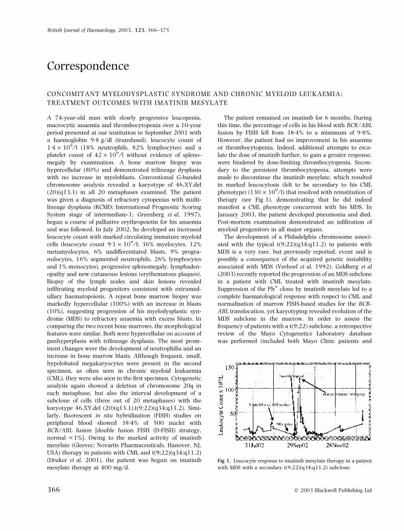
The patient remained on imatinib for 6 months. Duringthis time, the percentage of cells in his blood with BCR ⁄ABLfusion by FISH fell from 38Æ4% to a minimum of 9Æ8%.However, the patient had no improvement in his anaemiaor thrombocytopenia. Indeed, additional attempts to esca-late the dose of imatinib further, to gain a greater response,were hindered by dose-limiting thrombocytopenia. Secon-dary to the persistent thrombocytopenia, attempts weremade to discontinue the imatinib mesylate, which resultedin marked leucocytosis (felt to be secondary to his CMLphenotype) (130 · 109 ⁄ l) that resolved with reinstitution oftherapy (see Fig 1), demonstrating that he did indeedmanifest a CML phenotype concurrent with his MDS. InJanuary 2003, the patient developed pneumonia and died.Post-mortem examination demonstrated an infiltration ofmyeloid progenitors in all major organs.
The development of a Philadelphia chromosome associ-ated with the typical t(9;22)(q34;q11.2) in patients withMDS is a very rare, but previously reported, event and ispossibly a consequence of the acquired genetic instabilityassociated with MDS (Verhoef et al, 1992). Goldberg et al(2003) recently reported the progression of an MDS subclonein a patient with CML treated with imatinib mesylate.Suppression of the Ph+ clone by imatinib mesylate led to acomplete haematological response with respect to CML andnormalization of marrow FISH-based studies for the BCR-ABL translocation, yet karyotyping revealed evolution of theMDS subclone in the marrow. In order to assess thefrequency of patients with a t(9;22) subclone, a retrospectivereview of the Mayo Cytogenetics Laboratory databasewas performed (included both Mayo Clinic patients and
Fig 1. Leucocyte response to imatinib mesylate therapy in a patient
with MDS with a secondary t(9;22)(q34;q11.2) subclone.
British Journal of Haematology, 2003, 123, 366–375
366 � 2003 Blackwell Publishing Ltd

specimens referred to Mayo Medical Laboratories). A t(9;22)was identified by conventional karyotype analysis in 659different patients over a 3-year period. Of the 659 patients(including the patient described above), only one otherpatient had a t(9;22) as a subclonal event, indicating afrequency of 0Æ3% (two out of 659) for the acquisition of thissecondary anomaly. This second patient’s primary karyo-typic anomaly was t(3;21)(q26.2;q22), a translocationpreviously associated with genotoxic chemotherapy, whichmay represent a medication-related event (Zent et al, 1997).
The findings in the present case and the patient describedby Goldberg et al (2003) suggest several conclusions whenmultiple clonal myeloid processes exist in the marrow. First,a Ph+ subclone appears to result in phenotypic featurestypical of CML, including organomegaly, leucocytosis andextramedullary haematopoiesis, even within a backgroundof MDS. Secondly, imatinib mesylate is able to suppress at(9;22)-driven clonal process even when this process issecondary in origin, although this suppression may beincomplete. Finally, suppression of a t(9;22) clonal processin a patient with MDS does not necessarily alleviate themanifestations of MDS, such as other cytopenias, and theMDS may progress clinically.
Ruben A. Mesa1
David P. Steensma1
James Hoyer2
Rhett P. Ketterling2
1Division of Hematology and2Department of LaboratoryMedicine and Pathology, MayoClinic, Rochester, MN, USA.E-mail: [email protected]
REFERENCES
Druker, B.J., Talpaz, M., Resta, D.J., Peng, B., Buchdunger, E., Ford,
J.M., Lydon, N.B., Kantarjian, H., Capdeville, R., Ohno-Jones, S. &
Sawyers, C.L. (2001) Efficacy and safety of a specific inhibitor of
the BCR-ABL tyrosine kinase in chronic myeloid leukemia. New
England Journal of Medicine, 344, 1031–1037.
Goldberg, S.L., Madan, R.A., Rowley, S.D., Pecora, A.L., Hsu, J.W. &
Tantravahi, R. (2003) Myelodysplastic subclones in chronic
myeloid leukemia: implications for imatinib mesylate therapy.
Blood, 101, 781.
Greenberg, P., Cox, C., LeBeau, M.M., Fenaux, P., Morel, P., Sanz,
G., Sanz, M., Vallespi, T., Hamblin, T., Oscier, D., Ohyashiki, K.,
Toyama, K., Aul, C., Mufti, G. & Bennett, J. (1997) International
scoring system for evaluating prognosis in myelodysplastic syn-
dromes. Blood, 89, 2079–2088.
Verhoef, G., Meeus, P., Stul, M., Mecucci, C., Cassiman, J.J., Van
Den Berghe, H. & Boogaerts, M. (1992) Cytogenetic and mole-
cular studies of the Philadelphia translocation in myelodysplastic
syndromes. Report of two cases and review of the literature.
Cancer Genetics and Cytogenetics, 59, 161–166.
Zent, C., Rowley, J.D. & Nucifora, G. (1997) Rearrangements of the
AML1 ⁄CBFA2 gene in myeloid leukemia with the 3;21 translo-
cation: in vitro and in vivo studies. Leukemia, 11, 273–278.
Keywords: myelodysplastic syndrome, chronic myeloidleukaemia, imatinib mesylate, t(9;22) subclone.
SEVERE ACUTE RESPIRATORY SYNDROME AND LUPUS ANTICOAGULANTS IN CHILDREN
Severe acute respiratory syndrome (SARS) caused by thenovel corona virus is a new emerging disease that hasaffected countries worldwide, with more than 8461 casesreported (WHO, 2003a; World Health Organization Mult-icentre Collaborative Network for Severe Acute RespiratorySyndrome (SARS) Diagnosis, 2003). Between 24 March and31 March 2003, the United Christian Hospital admitted atotal of 21 paediatric patients, age range from 11 months to17 years, who fulfilled the WHO definition of SARS (WHO,2003b). There were 10 boys and 11 girls. Eighteen patientswere from the major outbreak in Amoy Gardens, a high-risehousing estate in Hong Kong.
In our group of 21 paediatric patients, we detected anunexpected, isolated, prolonged activated partial thrombo-plastin time (APTT) on admission in nearly half the cases.Eight patients (38Æ1%) showed a prolonged APTT of> 38Æ6 s, which is 1 standard deviation (SD) above theupper normal limit (reference range 24Æ1–35Æ7 s). Theprothrombin time was normal in all patients. The fibrinogenlevel was within the normal range (2Æ0–4Æ0 g ⁄ l) in all 16patients tested. D-Dimer was negative in 18 patients,positive (0Æ5–1Æ0 lg ⁄ml) in two patients and not done inone patient. Further investigations were performed in five ofthe eight patients, and the results are given in Table I.Factor VIII levels ranged between 0Æ84 and 1Æ10 IU ⁄ml(mean 0Æ96 IU ⁄ml). Factor IX levels ranged between 0Æ60
and 0Æ74 IU ⁄ml (mean 0Æ68 IU ⁄ml). All factor assaysshowed non-parallelism, indicating the presence of a non-specific inhibitor. A weak lupus anticoagulant was identifiedin three patients. The prolonged APTT, found in eight cases,had normalized by d 8 of admission. There were nothrombotic complications, bleeding events or deaths recor-ded in our group of patients.
Table I. Results of investigation of patients with a prolonged APTT.
Patient
Sex ⁄age
(years)
APTT
(s)
Lupus
anticoagulant
test* (ratio)�
Factor
VIII (IU ⁄ml)
Factor
IX (IU ⁄ml)
1 M ⁄13 54Æ9 Negative (1Æ09) 0Æ94 0Æ74
2 M ⁄15 51Æ9 Borderline (1Æ19) 1Æ10 0Æ62
3 M ⁄15 44Æ9 Weakly positive (1Æ35) 0Æ84 0Æ60
4 F ⁄16 42 Weakly positive (1Æ35) 0Æ93 0Æ72
5 M ⁄13 39Æ2 Weakly positive (1Æ46) 0Æ97 0Æ71
*Lupus anticoagulant (LA) measured with the LA1 screening
reagent and LA2 confirmation reagent (Dade Behring, Marburg,
Germany).
�Ratio of LA1 ⁄ LA2. Ratio between 1Æ1 and 1Æ3, results are bor-
derline; ratio between 1Æ2 and 1Æ5, LA is weakly positive.
Correspondence 367
� 2003 Blackwell Publishing Ltd, British Journal of Haematology 123: 366–375

So far, only one study has reported a prolonged APTT inSARS patients. (Lee et al, 2003) Our investigations showedthat prolonged APTT in paediatric SARS patients is not dueto disseminated intravascular coagulation, but is rathercaused by the presence of lupus anticoagulants. Lupusanticoagulants are members of a heterogeneous group ofantibodies directed against phospholipid protein complexesand can arise after events triggering the immune system.These antiphospholipid antibodies have been found to beassociated with many infections, although the pathogenicrole for these antibodies has not usually been obvious(Asherson & Cervera, 2003). We have not repeated thelupus anticoagulant testing to determine whether thephenomenon is transient or permanent. As SARS is a newdisease, the cause and pathogenic role of lupus anticoag-ulants or other antiphospholipid antibodies remain to bestudied.
Eudora Y. Chow1
Wa K. Chiu2
Departments of 1Pathology and2Paediatrics and AdolescentMedicine, United ChristianHospital, Kwun Tong, Kowloon,Hong Kong.E-mail: [email protected]
REFERENCES
Asherson, R.A. & Cervera, R. (2003) Antiphospholipid antibodies
and infections. Annals of Rheumatic Disease, 2003, 388–393.
Lee, N., Hui, D., Wu, A., Chan, P., Cameron, P., Joynt, G.M., Ahuja,
A., Yung, M.Y., Leung, C.B., To, K.F., Lui, S.F., Szeto, C.C.,
Chung, S. & Sung, J.J.Y. (2003) A major outbreak of severe acute
respiratory syndrome in Hong Kong. New England Journal of
Medicine, 348, 1986–1994.
WHO (2003a) Cumulative number of reported probable cases of
severe acute respiratory syndrome (SARS). [WWW document]
URL http://www.who.int/csr/sars/country/2003-06-20/en/
(accessed 23 June 2003).
WHO (2003b) Case definition for surveillance of severe acute
respiratory syndrome SARS. [WWW document] URL http://
www.who.int/csr/sars/casedefinition/en/ (accessed 16 May 2003).
World Health Organization Multicentre Collaborative Network for
Severe Acute Respiratory Syndrome (SARS) Diagnosis (2003) A
multicentre collaboration to investigate the cause of severe acute
respiratory syndrome. The Lancet, 361, 1730–1733.
Keywords: severe acute respiratory syndrome (SARS), lupusanticoagulants, prolonged activated partial thromboplastintime.
MALIGNANCIES IN SICKLE CELL DISEASE PATIENTS TREATED WITH HYDROXYUREA
We read with great interest the recent review on hydroxy-urea (HU) in sickle cell disease (SCD) (Halsey & Roberts,2003). As a minor point, we would like to correct the factthat the patient who developed a lymphoproliferativedisease did not belong to the Belgian study, but wasreported by the French group (De Montalembert & Doures,2001).
However, the first case of a SCD patient who developedmalignancy has just been reported to the Belgian Registry ofSickle Cell Disease patients treated with hydroxyurea. Thepatient is a 21-year-old woman who has been treated withHU for the last 8 years for severe clinical disease (recurrentvaso-occlusive crises, osteonecrosis, acute chest syndrome).Treatment with HU was discontinued 2 years ago becausethe patient was pregnant and was restarted in July 2002after she had given birth to a healthy daughter. In March2003, the patient complained of diffuse bone pain; a bloodexamination showed leucopenia and thrombocytopenia,which persisted after discontinuation of HU. Bone marrowexamination confirmed the diagnosis of acute myeloblasticleukaemia M3v.
This is the first case of HU-associated malignancy in theSCD Belgian Registry. To date, 141 patients have beenincluded, and 598 ‘patient–years’ have been evaluated witha median follow-up of 4Æ5 years.
Several reports suggest that SCD patients may be ateither lower or higher risk for malignancy (Jackan, 1972;Stricker et al, 1986; Paydas, 2002). The contribution ofHU in the emergence of neoplasia in SCD patients is
currently unknown. However, our case, together withthose reported by Raunch et al (1999) and Wilson (2000),emphasizes the need for close monitoring, data collectionand follow-up of SCD patients in order to determine therisks and benefits of any treatment in this particularpopulation.
Alina Ferster
Eric Sariban
Nathalie Meuleman
for the Belgian
Registry of Sickle
Cell Disease
patients treated
with Hydroxyurea
Hopital Universitaire desEnfants Reine Fabiola,Brussels, Belgium.E-mail: [email protected]
REFERENCES
De Montalembert, M. & Doures, S.C. (2001) Is hydroxyurea leu-
kemogenic in children with sickle cell disease ? Blood, 98, 2878–
2879.
Halsey, C. & Roberts, I.A.G. (2003) The role of hydroxyurea in
sickle cell disease. British Journal of Haematology, 120, 177–186.
Jackan, R.E. (1972) Frequency and prognosis of coexisting sickle
cell disease and acute leukemia in children. Clinical Pediatrics,
183, 183.
Paydas, S. (2002) Sickle cell anemia and hematological neoplasis.
Leukemia and Lymphoma, 43, 1431–1334.
368 Correspondence
� 2003 Blackwell Publishing Ltd, British Journal of Haematology 123: 366–375
Raunch, A., Borromeo, M., Ghafoor, A., Khoyratty, B. & Mahesh-
wari, J. (1999) Leukemogenesis of hydroxyurea in the treatment
of sickle cell anemia. Blood, 94, 415a.
Stricker, R.B., Linker, C.A., Crowley, T.J. & Embury, S.H. (1986)
Hematology malignancy in sickle cell disease: report of four cases
and review of the literature. American Journal of Hematology, 21,
223–230.
Wilson, S. (2000) Acute leukemia in a patient with sickle cell an-
emia treated with hydroxyurea. Annals of Internal Medicine, 133,
925–926.
Keywords: sickle cell disease, hydroxyurea, malignancy.
LITHIUM EFFECTS ON NEUTROPHIL MOTILITY IN SHWACHMAN–DIAMOND SYNDROME:
EVALUATION BY COMPUTER-ASSISTED IMAGE ANALYSIS
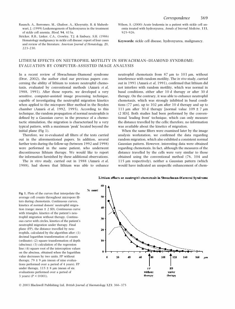
In a recent review of Shwachman–Diamond syndrome(Dror, 2002), the author cited our previous papers con-cerning the ability of lithium to restore neutrophil chemo-taxis, evaluated by conventional methods (Azzara et al,1988, 1991). After those reports, we developed a verysensitive, computer-assisted image processing technique,capable of investigating the neutrophil migration kineticswhen applied to the micropore filter method in the Boydenchamber (Azzara et al, 1992, 1995). According to thistechnique, the random propagation of normal neutrophils isdefined by a Gaussian curve; in the presence of a chemo-tactic stimulation, the migration is characterized by a verytypical pattern, with a maximum ‘peak’ located beyond theinitial plane (Fig 1).
Therefore, we re-evaluated all filters of the tests carriedout in the aforementioned papers. In addition, severalfurther tests during the follow-up (between 1992 and 1998)were performed in the same patient, who underwentdiscontinuous lithium therapy. We would like to reportthe information furnished by these additional observations.
The in vitro study, carried out in 1988 (Azzara et al,1988), had shown that lithium was able to enhance
neutrophil chemotaxis from 87 lm to 103 lm, withoutinterference with random motility. The in vivo study, carriedout in 1991 (Azzara et al, 1991), confirmed that lithium didnot interfere with random motility, which was normal inbasal conditions, either after 10 d therapy or after 30 dtherapy. On the contrary, it was able to enhance neutrophilchemotaxis, which was strongly inhibited in basal condi-tions (77 lm), up to 102 lm after 10 d therapy and up to111 lm after 30 d therapy [normal value 109 ± 7 lm(2 SD)]. Both studies had been performed by the conven-tional ‘leading front’ technique, which can only measurethe distance travelled by the cells; therefore, no informationwas available about the kinetics of migration.
When the same filters were examined later by the imageanalysis workstation, we confirmed the data regardingrandom migration, which also exhibited a consistent normalGaussian pattern. However, interesting data were obtainedregarding chemotaxis. In fact, although the measures of thedistance travelled by the cells were very similar to thoseobtained using the conventional method (76, 104 and115 lm respectively), neither a Gaussian pattern (whichwould have indicated an unspecific enhancement of chem-
Fig 1. Plots of the curves that interpolate the
average cell counts throughout micropore fil-
ters during chemotaxis. Continuous curves,
kinetics of normal donors’ neutrophil migra-
tion (range: mean ± 2 SD). Continuous curve
with triangles, kinetics of the patient’s neu-
trophil migration without therapy. Continu-
ous curve with circles, kinetics of the patient’s
neutrophil migration under therapy. Final
plane (FP), the distance travelled by neu-
trophils, calculated by the algorithm after: (1)
decimal logarithm transformation of counts
(ordinate); (2) square transformation of depth
(abscissa); (3) calculation of the regression
line; (4) square root of the interception values
on the abscissa, obtained when the logarithm
value decreases by two units. FP without
therapy, 79 ± 9 lm (mean of nine evalua-
tions performed over a period of 4 years). FP
under therapy, 115 ± 8 lm (mean of six
evaluations performed over a period of
3 years) (P < 0Æ001).
Correspondence 369
� 2003 Blackwell Publishing Ltd, British Journal of Haematology 123: 366–375

okinesis) nor a typical chemotactic pattern was obtained;this indicates that the improvement was probably associatedwith a neutrophil subpopulation, whereas the majority ofcells remained inhibited on the initial plane. The patientthen cyclically discontinued and restarted therapy (fourperiods and three periods respectively). During the off-therapy periods, neutrophil chemotaxis was always inhib-ited: 79 ± 9 lm (mean of nine evaluations performed over aperiod of 4 years). During lithium therapy, it was alwaysnormal: 115 ± 8 lm (mean of six evaluations performedover a period of 3 years) (P < 0Æ001). In addition, thekinetics of the neutrophils was always identical to thatfound in the basal condition. In particular, it can be seenthat the typical peak of maximum cell density beyond thefirst plane was never detectable (Fig 1).
Our data confirm a clear activity of lithium on neutrophilmotility in this syndrome. The drug did not affect chemo-kinesis, so it is not able to enhance random motility. Underchemotactic conditions, it probably acts by the recruitmentof a subset of neutrophils unaffected by the cytoskele-tal ⁄microtubular abnormality, which is presumed to play aprominent role in the causation of defective chemotaxis(Rothbaum et al, 1982). Of interest, most neutrophilsremained insensitive. So it can be stated that, even if thefunction is ameliorated, the cytoskeletal defect represents aspecific stable marker of the syndrome.
Antonio Azzara
Giovanni Carulli
Mario Petrini
Unit of Haematology, Departmentof Oncology, Transplantsand Advanced Technologiesin Medicine, University of Pisa,Via Roma 67, 56100 Pisa, Italy.E-mail: [email protected]
REFERENCES
Azzara, A., Carulli, G., Polidori, R., Ceccarelli, M., Simoni, F. &
Ambrogi, F. (1988) In vitro restoration by lithium of defective
chemotaxis in Shwachman–Diamond syndrome. British Journal
of Haematology, 70, 502.
Azzara, A., Carulli, G., Ceccarelli, M., Pucci, C., Raggio, R. &
Ambrogi, F. (1991) In vivo effectiveness of lithium on impaired
chemotaxis in Shwachman–Diamond syndrome. Acta Haematol-
ogica, 85, 100–102.
Azzara, A., Chimenti, M., Azzarelli, L., Fantini, E., Carulli, G. &
Ambrogi, F. (1992) An image processing workstation for auto-
matic evaluation of human granulocyte motility. Journal of
Immunological Methods, 148, 29–40.
Azzara, A., Chimenti, M., Carulli, G., Rizzuti-Gullaci, A. & Ambrogi.
F. (1995) An image processing procedure for the assessment of
normality curves of motility of human granulocytes in micropore
filters. Scandinavian Journal of Clinical and Laboratory Investigation,
55, 399–408.
Dror, Y. (2002) Shwachman–Diamond syndrome. British Journal of
Haematology, 118, 701–713.
Rothbaum, R.J., Williams, D.A. & Daugherty, C.C. (1982) Unusual
surface distribution of concanavalin A reflects a cytoskeletal
defect in neutrophils in Shwachman’s syndrome. Lancet, II,
800–801.
Keywords: lithium, neutrophil motility, image analysis.
PROGNOSTIC FEATURES OF SPLENIC LYMPHOMA WITH VILLOUS LYMPHOCYTES
The very interesting paper by Parry-Jones et al (2003) onthe prognostic features of splenic lymphoma with villouslymphocytes (SLVL) adds to our knowledge of this intrigu-ing disease. However, in our opinion several points deservefurther comments.
While 240 patients were identified, complete data wereavailable for only 53% of them. This suggests a note ofcaution about the reliability of the prognostic evaluationbecause selection bias cannot be ruled out.
In addition, we were surprised to note that none of thecases showed an intrasinusoidal infiltrate in the bonemarrow, which is highly characteristic and rather frequentin SLVL (Franco et al, 1996; Costes et al, 2002; Iannittoet al, 2002).
Another point we would like to comment on is how thecauses of death were reported. The 22 patients who died ofprogressive disease or infection certainly represent 17% ofall the series, but it would have been more informative tounderscore the fact that they made up 43% of all deaths.Unfortunately, the authors reported the occurrence ofsecondary cancer in their series, but did not specify thenumber or histotype of the cases involved.
The predictive value for survival of the covariates atdiagnosis was reported, considering both lymphoma-rela-ted deaths and all other deaths (Table III in Parry-Joneset al, 2003), but the causes of death were unavailable inas many as of 29% of cases (15 of 51). We believe that itcannot be assumed that none of these patients died oflymphoma-related causes. Therefore, the results of theCox regression analysis indicating that, for lymphoma-related deaths, only haemoglobin level and lymphocytecount were independent prognostic indicators of survival,in our opinion should be taken cautiously. It could bemisleading.
The last point is that of therapy. Parry-Jones et al(2003, Table II) reported the presenting features of treatedand untreated patients. Patients who received chemother-apy as first-line treatment had a significantly higherlymphocyte count than those who were splenectomized.This means that they were at higher risk of lymphoma-related death according to the Cox analysis. Conversely,patients who received splenectomy as first-line therapy hada trend for a less-aggressive disease (younger age, lowertotal WBC count and lower degree of leucocytosis). It
370 Correspondence
� 2003 Blackwell Publishing Ltd, British Journal of Haematology 123: 366–375

would have been more informative if the data had beenreported not as a median but as a percentage of patientswith a high-risk covariate, using the cut-off described forthe univariate analysis. Moreover, it is not clear howmany patients that were not showing signs of activedisease underwent splenectomy merely for diagnosticpurposes.
We agree that patients at high risk at diagnosis becauseof lymphocytosis and anaemia do not seem to benefit fromchemotherapy (mainly chlorambucil), but in our opinionParry-Jones et al (2003) do not provide enough data tosupport the hypothesis that these patients may benefit fromsplenectomy.
We think that the issues of prognosis and the besttherapeutic approach of SLVL deserve to be settled in aproperly designed prospective study (Franco et al, 2003).
Emilio Iannitto1
Emanuele Ammatuna1
Ada Maria Florena2
Vito Franco2
1Department of Oncology,and 2Department of SurgicalPathology, Universityof Palermo, Palermo, Italy
REFERENCES
Costes, V., Duchayne, E., Taib, J., Delfour, C., Rousset, T., Baldet, P.,
Delsol, G. & Brousset, P. (2002) Intrasinusoidal bone marrow
infiltration: a common growth pattern for different lymphoma
subtypes. British Journal of Haematology, 119, 916–922.
Franco, V., Florena, A.M. & Campesi, G. (1996) Intrasinusoidal
bone marrow infiltration: a possible hallmark of splenic lym-
phoma. Histopathology, 29, 571–575.
Franco, V., Florena, A.M. & Iannitto, E. (2003) Splenic marginal
zone lymphoma. Blood, 101, 2464–2472.
Iannitto, E., Ambrosetti, A., Ammatuna, E., Cirrincione, S., Colosi,
M., Florena, A.M., Mariani, G. & Pizzolo G. (2002) Splenic
Marginal Zone Lymphoma with or without villous lymphocytes.
Haematological findings and outcome in a series of 57 patients.
Blood, 100(Suppl. 1), 602a(abstract).
Parry-Jones, N., Matutes, E., Gruszka-Westwood, A.M., Swansbury,
G.J., Wotherspoon, A.C. & Catovsky, D. (2003) Prognostic fea-
tures of splenic lymphoma with villous lymphocytes: a report on
129 patients. British Journal of Haematology, 120, 759–764.
Keywords: splenic narginal zone lymphoma, spleniclymphoma with villous lymphocytes, intrasinusoidal,splenectomy, bone marrow.
ERYTHROCYTOSIS AND THE CHUVASH VON HIPPEL-LINDAU MUTATION
Congenital and familial polycythaemias are rare conditionsand the molecular basis of these disorders is largelyunknown. The analysis of patients with familial erythrocy-tosis associated with low plasma erythropoietin (Epo) hasdetected mutations in the Epo receptor (EpoR) and thus hasprovided insights into the Epo signal transduction pathway.
The Chuvash form of polycythaemia describes a secon-dary familial and congenital polycythaemia characterized bynormal or elevated levels of Epo. The molecular pathophys-iology underlying the Chuvash polycythaemia has recentlybeen elucidated and is ascribed to dysregulation of oxygenhomeostasis caused by mutations in the von Hippel-Lindau(VHL) gene, a key player in the hypoxia-sensing pathway(Ang et al, 2002). Arg200Trp in VHL affects its associationwith a component of the hypoxia-inducible factor-1 (HIF-1)transcription complex and impedes the proteasomal degra-dation of this complex leading to increased expression of HIF-1 target genes, including EPO (Ang et al, 2002).
The patient, whose parents were Pakistani, was born inthe UK in 1982. She required surgery at the age of3 months with a haemoglobin (Hb) content of 12Æ6 g/dlthen, and 18Æ3 g/dl at the age of 9 years. Mean cell volume(MCV), white blood cells and platelets were normal. Red cellmass was 52 ml/kg, plasma Epo level 18 mIU/ml (normalrange 8–28), and both p50 and oxygen saturation werenormal. The explanation for the polycythaemia was notobvious and she was treated by venesection. Over the next9 years venesection was carried out with difficulty becauseof poor venous access. She was reviewed in 2000 and atthat time she was well with a Hb of 15Æ0 mg/dl and MCV of60. Her parents were not polycythaemic. Analysis of herclinical data suggested similarities to patients with the
Chuvash form of polycythaemia and sequencing of the VHLgene was performed. This showed that she was homozygousfor the Arg200Trp mutation in the VHL gene. Both parentswere heterozygous for this mutation.
Chuvash polycythaemia is not limited to Chuvashians.More recently, it has been shown that other mutations inVHL may lead to polycythaemia (Pastore et al, 2003) andthat people of Asian and other ethnic origins, may alsopossess the same Arg200Trp VHL mutation described inChuvash patients (Pastore et al, 2003; Percy et al, 2003).Analysis of the six previously described di-allelic single-nucleotide polymorphisms (Percy et al, 2003) revealed thatthe Arg200Trp mutation in our patient was associated withthe same GCTACA haplotype as in the other Asian families(Percy et al, 2003), suggesting a founder mutation in theAsian population. Studies underway will establish whetherthe Arg200Trp mutation in the Chuvash and other ethnicgroups is of a common origin.
Mutations in the VHL gene are better known in thecontext of the VHL disease, a dominantly inherited familialcancer syndrome. These patients have an increased inci-dence of renal cell carcinoma, central nervous systemhaemangioblastoma, pheochromocytoma and renal, pan-creatic and endolymph cysts. The familial inheritance isrelated to a germline mutation in the VHL tumoursuppressor gene followed by a second hit in VHL as asomatic mutation (Maher & Kaelin, 1997). Although morethan 700 mutations have been characterized in VHL, noindividual with two germline mutations of the type foundin Chuvash polycythaemia (i.e. Arg200Trp) have beenreported. Polycythaemia is not common but can occur as aparaneoplastic syndrome.
Correspondence 371
� 2003 Blackwell Publishing Ltd, British Journal of Haematology 123: 366–375

It is not clear whether patients with Chuvash poly-cythaemia or their heterozygous relatives are predisposed toneoplasms. There is some evidence to suggest that Chuvashpolycythaemic patients may have increased morbidity andmortality from vascular events (Sergeyeva et al, 1997).Keeping the Hb to near normal levels is thus a priority andmeticulous attention should be paid to maintaining goodvenous access from an early age. Eventually central venouslines, Hickman catheters, and portocaths may have to beconsidered, combined with some form of anticoagulation toreduce the risk of thrombosis in major veins. Arterio-venousshunts are another option. All such procedures are invasiveand increase the risk of vascular complications. Finally, if allthese methods fail bone marrow suppression by cytotoxicagents might be justified.
Melanie J. Percy1
Michael E. J. Beard2
Chris Carter3
Swee Lay Thein4
1Department of Haematology,Belfast City Hospital,Belfast, UK, 2Department ofHaematology, ChristchurchHospital, Christchurch, NewZealand, 3Department ofHaematology, Huddersfield RoyalInfirmary, Huddersfield, and4Department of HaematologicalMedicine, Kings College Hospital,London, UK
REFERENCES
Ang, S.O., Chen, H., Hirota, K., Gordeuk, V.R., Jelinke, J., Guan, Y.,
Liu, E., Sergueeva, A.I., Miasnikova, G.Y., Mole, D., Maxwell,
P.H., Stockton, D.W., Semenza, G.L. & Prchal, J.T. (2002)
Disruption of oxygen homeostasis underlies congenital Chuvash
polycythaemia. Nature Genetics, 32, 614–621.
Maher, E.R. & Kaelin, W.G. (1997) Von Hippel-Lindau disease.
Medicine, 76, 381–391.
Pastore, Y.D., Jelinek, J., Ang, S., Guan, Y., Liu, E., Jedlickova, K.,
Krishnamurti, L. & Prchal, J.T. (2003) Mutations in the VHL
gene in sporadic apparently congenital polycythaemia. Blood,
101, 1591–1595.
Percy, M.J., McMullin, M.F., Jowitt, S.N., Potter, M., Treacy, M.,
Watson, W.H. & Lappin, T.R.J. (2003) Chuvash-type congenital
polycythemia in 4 families of Asian and Western European
Ancestry. Blood, 102, 1097–1099.
Sergeyeva, A., Gordeuk, V.R., Tokarev, Y.N., Sokol, L., Prchal, J.F. &
Prchal, J.T. (1997) Congenital polycythaemia in Chuvashia.
Blood, 89, 2148–2154.
Keywords: erythrocytosis, von Hippel-Lindau mutation,Chuvash polycythaemia.
ENDOTHELIAL CELLS EXPRESS NORMAL CELLULAR PRION PROTEIN
We read with interest the recent letter (Sivakumaran,2003) in response to our article (Starke et al, 2002) inwhich the expression of normal prion protein (PrPC) byendothelial cells was questioned. Sivakumaran’s argumentwas that endothelial cells in vitro might not have the samephenotype in vivo. Specifically Sivakumaran believes thatendothelial cells in vivo do not express PrPC and that theexpression previously reported could be the result ofartefactual activation generated by in vitro manipulation.The reasoning behind this hypothesis was that paraffinsections of umbilical cord and adult blood vessels showed‘minimal or no expression of PrPC’ by immunocytochemis-try using an antibody to prion protein (3F4).
We have demonstrated the expression of PrPC in culturedhuman umbilical vein endothelial cells (HUVECs) andhuman microvascular endothelial cells (HMEC-1) by reversetranscription polymerase chain reaction (RT-PCR), flowcytometry and immunofluorescence microscopy using anantibody to prion protein (6H4). A time-resolved fluoroim-munoassay (DELFIA�, Perkin Elmer, Seer Green, Beacons-field, UK) was used to demonstrate that endothelial cellsreleased soluble PrPC and human blood plasma alsocontained soluble PrPC. By studying various groups ofpatients we demonstrated that plasma levels of PrPC did notcorrelate with blood cell counts, leading to the conclusion
that the source of the plasma form of PrPC may be thevascular endothelium.
Semi-quantitative RT-PCR was used to compare theexpression of PrPC mRNA in endothelial cells with thehousekeeping gene iduronate-2-sulphatase (IDS). RT-PCRwas performed using radioactively labelled primers and theamount of product quantified by densitometry. The meanexpression of PrPC relative to IDS was found to be1.87 ± 0.75 for HUVECs and 2.97 ± 0.27 for HMEC-1s(Fig 1) (n ¼ 2).
Simak et al (2002) demonstrated that cultured endothel-ial cells expressed PrPC using several anti-prion antibodies.They also showed that endothelial cell microparticlesexpressed PrPC. The expression of PrPC by endothelial cellshas been described in several other investigations (seeStarke et al, 2002 and Simak et al, 2002 for reviews).
Sivakumaran used monoclonal antibody 3F4. This anti-body is more restricted in the species of PrPC that it canbind, compared with 6H4 (Barclay et al, 2002), and wefound that it did not give as strong a signal with endothelialcells as 6H4 by flow cytometry (unpublished observations).Simak et al (2003) stated that the negative results obtainedby Sivakumaran could be the result of a truncation of PrPC
in the tissue samples studied. We believe that a negativeresult should be confirmed with more than one antibody.
372 Correspondence
� 2003 Blackwell Publishing Ltd, British Journal of Haematology 123: 366–375

Erythrocytes were originally thought to be negative for PrPC
using antibody 3F4. It was later shown using antibody 6H4that they do express low levels of PrPC (Holada and Vostal,2000). Immunocytochemistry is not a particularly sensitivetechnique and using neuronal tissue as a positive control forPrPC expression may not reflect the sensitivity required forthe study of tissues with lower prion levels. Regarding theartefactual expression as a result of in vitro activation, wehave demonstrated that activation of endothelial cells withhuman thrombin and tumour necrosis factor-a did notincrease the expression of PrPC on endothelial cells andsimilar results were obtained by (Simak et al, 2002).
In conclusion, we agree that there are limitations withregards to the study of endothelial cells in vitro, but thatthere is a great deal of data to indicate that vascularendothelial cells do express PrPC. These cells remain apossible source of plasma prion protein and, as there is thepossibility that PrPSC is transmitted via the haematogenicroute, further research should be performed to determinethe role of endothelial cells in patients with transmissiblespongiform encephalopathies.
Richard Starke1
,
PaulHarrison2
,
RosemaryGale1
,
Ian Mackie1
,
OliveDrummond3
,
IanMacGregor3
and Samuel Machin1
1Haemostasis Research Unit,Haematology Department,University College London,London, 2Oxford HaemophiliaCentre, Churchill Hospital,Headington, Oxford, and3Products and ComponentsR & D Group, National ScienceLaboratory, Scottish NationalBlood Transfusion Service,Edinburgh, UK.E-mail: [email protected]
REFERENCES
Barclay, G.R., Houston, E.F., Halliday, S.I., Farquhar, C.F. & Turner,
M.L. (2002) Comparative analysis of normal prion protein ex-
pression on human, rodent, and ruminant blood cells by using a
panel of prion antibodies. Transfusion, 42, 517–526.
Holada, K. & Vostal, J.G. (2000) Different levels of prion protein
(PrPC) expression on hamster, mouse and human blood cells.
British Journal of Haematology, 110, 472–480.
Simak, J., Holada, K., D’Agnillo, F., Janota, J. & Vostal, J.G. (2002)
Cellular prion protein is expressed on endothelial cells and is
released during apoptosis on membrane microparticles found in
human plasma. Transfusion, 42, 334–342.
Simak, J., Holada, K. & Vostal, J.G. (2003) Expression of cellular
prion protein on vascular endothelial cells: more evidence than
controversies. Transfusion, 43, 680–681.
Sivakumaran, M. (2003) The expression of prion protein (PrPc) by
endothelial cells: an in vitro culture-induced artefactual phe-
nomenon? British Journal of Haematology, 121, 673–674.
Starke, R., Drummond, O., MacGregor, I., Biggerstaff, J., Gale, R.,
Camilleri, R., Mackie, I., Machin, S. & Harrison, P. (2002) The
expression of prion protein by endothelial cells: a source of the
plasma form of prion protein? British Journal of Haematology, 119,
863–873.
Keywords: endothelium, prion protein, blood plasma, PrPC.
HEPARIN-INDUCED THROMBOCYTOPENIA: PATHOGENESIS AND MANAGEMENT
I read with much interest the recent review on thepathogenesis and management of heparin-induced thromb-ocytopenia (Warkentin, 2003).
Heparin-induced thrombocytopenia type II (HIT II) is awell-recognized complication of heparin therapy that canhave serious effects. Several anticoagulation strategies havebeen developed for the management of HIT II patients whomay require anticoagulation therapy.
Hirudin is natural peptide that was originally extractedfrom the salivary glands of leeches. Nowadays, it is made byrecombinant DNA technology in yeasts. Recombinanthirudin (r-hirudin) has been used as a heparin substitutein a wide variety of clinical settings. It acts as specific directinhibitor of thrombin. Despite its short half-life, its wider usehas been limited because of the absence of a specificneutralizing agent.
In the section on lepirudin (Warkentin, 2003), theanticoagulation profile was monitored by the measurement
of the activated partial thromboplastin time (aPTT). Severalstudies have shown aPTT assay to be of inadequatespecificity to provide an accurate measurement of theanticoagulation effect of r-hirudin.
Potzsch et al (1997a) concluded, from a series of in vitroand in vivo experiments, that both the aPTT and theactivated clotting time were not sufficiently sensitive tomonitor the concentration of r-hirudin. However, there wasa good linear relationship between r-hirudin concentrationand the ecarin clotting time (ECT). Moser et al (2001)arrived at the same conclusion from their experiments. TheECT is a parameter based on the conversion of prothrombin.
Ecarin is a prothrombin-activating enzyme that is derivedfrom the venom of the snake Echis carinaatus. It activates theprothrombin to meizothrombin, which, in r-hirudin con-taining plasma, forms a stable 1:1 complex with r-hirudin.This results in a dose-dependent prolongation of the clottingtime.
Fig 1. Semi-quantitative RT-PCR on endothelial cells. RT-PCR was
performed on RNA extracted from HUVEC and HMEC-1 cells using
radioactive (32P) primers specific for iduronate-2-sulphatase (IDS)
and PrPC (prion). The products were then electrophoresed on a
denaturing polyacrylamide gel, dried and exposed to hyperfilm.
Lanes 1 and 2 are from HMEC-1s and lanes 3 and 4 are from
HUVECs. Both bands labelled prion were found to be specific for
prion mRNA (n ¼ 2).
Correspondence 373
� 2003 Blackwell Publishing Ltd, British Journal of Haematology 123: 366–375

The ECT is a sensitive, easily performable assay for theprecise measurement of the plasma r-hirudin level. Itsprecision is not affected by treatment with heparin, apro-tinin or oral anticoagulants (Potzsch et al, 1997b).
There are numerous reports of the ECT assay providingrapid and accurate monitoring of the plasma r-hirudinconcentration in patients with HIT II who underwentsuccessful cardiopulmonary bypass operations usingr-hirudin as the anticoagulant (Potzsch et al, 1997a;Fabrizio, 2002; Saad et al, 2002).
Rasheed A SaadDepartment of Thoracic Surgery,Hairmyres Hospital, East Kilbride,UK.E-mail: [email protected]
REFERENCES
Fabrizio, M.C. (2002) Use of ecarin clotting time (ECT) with
lepirudin therapy in heparin-induced thrombocytopenia and
cardiopulmonary bypass. Journal of Extracorporeal Technology, 33,
117–125.
Moser, M., Ruef, J., Peter, K., Kohler, B., Gulba, D.C., Paterna, N.,
Nordt, T., Kubler, W. & Bode, C. (2001) Ecarin clotting time but
not aPTT correlates with PEG-Hirudin plasma activity. Journal of
Thrombosis and Thrombolysis, 12, 165–169.
Potzsch, B., Madlener, K., Seeling, C., Reiss, C.F., Greinacher, A. &
Muller-Berghaus, G. (1997a) Monitoring of r-hirudin anticoag-
ulation during cardiopulmonary bypass – assessment of the
whole bloo ecarin clotting time. Thrombosis and Haemostatis, 77,
920–925.
Potzsch, B., Hund, S., Madlener, K., Unkrig, C. & Muller-Berghaus,
G. (1997b) Monitoring of recombinant hirudin: assessment of a
plama-based ecarin clotting time assay. Thrombosis Research, 86,
373–383.
Saad, R.A., Horn, I. & Mankad, P.S. (2002) Management dilemma
of cardiopulmonary bypass in patients with type II heparin in-
duced thrombocytopenia. British Journal of Haematology, 119,
880.
Warkentin, T.E. (2003) Heparin-induced thrombocytopenia: path-
ogenesis and management. British Journal of Haematology, 121,
535–555.
Keywords: heparin-induced thrombocytopenia, recombin-ant hirudin, anticoagulant therapy, ecarin clotting time.
HEPARIN-INDUCED THROMBOCYTOPENIA: PATHOGENESIS AND MANAGEMENT – RESPONSE TO
RASHEED SAAD
Dr Saad correctly states that the activated partial throm-boplastin time (aPTT) does not measure the anticoagulanteffect of lepirudin as accurately as the ecarin clotting time(ECT). However, this advantage of ECT over aPTT isknown to be relevant only in situations of very highlepirudin dosing, such as surgery requiring cardiopulmon-ary bypass (Warkentin & Greinacher, 2003). This isbecause the dose–response curve of the aPTT to increasinglepirudin concentrations flattens at high lepirudin concen-trations (Greinacher, 2001). However, for most clinicalsituations, such as prophylaxis against or the treatment ofthrombosis in acute heparin-induced thrombocytopenia(HIT), use of aPTT is believed to provide acceptablemonitoring. Indeed, the pivotal clinical trials of lepirudinfor the treatment of thrombosis complicating HIT usedaPTT monitoring, with a target aPTT of 1.5–2.5 · baselinefor most aPTT reagents (Greinacher et al, 2000). Laborat-ories should assess their aPTT responsiveness of assay toincreasing concentrations of plasma lepirudin: if the curvebegins to flatten during the high therapeutic lepirudinconcentrations (about 750–1000 ng/ml), then the physi-cian should either aim at the low end of the aPTT targetrange (to avoid the potential for significant overdosing thatmight not be apparent if the aPTT lies within the high-therapeutic aPTT range) or utilize the ECT, if available.Whether the ECT might generally provide superior anti-coagulation (i.e. greater therapeutic efficacy with lessbleeding) than the aPTT in non-cardiac surgery situations
is unknown. This hypothesis would need testing in clinicaltrials comparing monitoring by aPTT versus ECT. How-ever, even if the ECT was shown to be better, the aPTTmight still remain the preferred monitoring method.Indeed, this is the current status for the monitoring ofunfractionated heparin therapy in many clinical situations,although heparin levels can be measured more accuratelyby protamine neutralization assay or anti-factor Xa assay(Hirsh et al, 2001).
Theodore E.
Warkentin
Department of Pathology andMolecular Medicine, McMasterUniversity, Hamilton RegionalLaboratory Medicine Program,Hamilton General Hospital, EastHamilton, Ontario, Canada.E-mail: [email protected]
REFERENCES
Greinacher, A. (2001) Recombinant hirudin for the treatment of
heparin-induced thrombocytopenia. In: Heparin-induced Thromb-
ocytopenia 2nd edn (ed. by T.E. Warkentin & A. Greinacher), pp.
349–380. Marcel Dekker, Inc., New York, NY.
Greinacher, A., Eichler, P., Lubenow, N., Kwasny, H. & Luz, M.
(2000) Heparin-induced thrombocytopenia with thromboem-
bolic complications: meta-analysis of two prospective trials to
374 Correspondence
� 2003 Blackwell Publishing Ltd, British Journal of Haematology 123: 366–375

assess the value of parenteral treatment with lepirudin and its
therapeutic aPTT range. Blood, 96, 846–851.
Hirsh, J., Warkentin, T.E., Shaughnessy, S.G., Anand, S.S.,
Halperin, J.L., Raschke, R., Granger, C., Ohman, E.M. & Dalen,
J.E. (2001) Heparin and low-molecular-weight heparin: mecha-
nisms of action, pharmacokinetics, dosing, monitoring, efficacy,
and safety. Chest, 119(Suppl.), 64S–94S.
Warkentin, T.E. & Greinacher, A. (2003) Heparin-induced
thrombocytopenia and cardiac surgery. Annals of Thoracic Sur-
gery, in press.
Keywords: activated partial thromboplastin time, ecarinclotting time, heparin-induced thrombocytopenia.
Correspondence 375
� 2003 Blackwell Publishing Ltd, British Journal of Haematology 123: 366–375