Neutron Reflectometry Studies Define Prion Protein N ...
18
Article Neutron Reflectometry Studies Define Prion Protein N-terminal Peptide Membrane Binding Anton P. Le Brun, 1 Cathryn L. Haigh, 2 Simon C. Drew, 3 Michael James, 1,4 Martin P. Boland, 2 and Steven J. Collins 2, * 1 Bragg Institute, Australian Nuclear Science and Technology Organisation, New Illawarra Road, Lucas Heights, New South Wales, 2234, Australia; 2 Department of Pathology, Kenneth Myer Building, The University of Melbourne, Victoria, 3010, Australia; 3 Florey Department of Neuroscience and Mental Health, The University of Melbourne, Victoria, 3010, Australia; and 4 School of Chemistry, University of New South Wales, Kensington, New South Wales, 2052, Australia ABSTRACT The prion protein (PrP), widely recognized to misfold into the causative agent of the transmissible spongiform encephalopathies, has previously been shown to bind to lipid membranes with binding influenced by both membrane composi- tion and pH. Aside from the misfolding events associated with prion pathogenesis, PrP can undergo various posttranslational modifications, including internal cleavage events. Alpha- and beta-cleavage of PrP produces two N-terminal fragments, N1 and N2, respectively, which interact specifically with negatively charged phospholipids at low pH. Our previous work probing N1 and N2 interactions with supported bilayers raised the possibility that the peptides could insert deeply with minimal disruption. In the current study we aimed to refine the binding parameters of these peptides with lipid bilayers. To this end, we used neutron reflectometry to define the structural details of this interaction in combination with quartz crystal microbalance interrogation. Neutron reflectometry confirmed that peptides equivalent to N1 and N2 insert into the interstitial space between the phospholipid headgroups but do not penetrate into the acyl tail region. In accord with our previous studies, interaction was stronger for the N1 fragment than for the N2, with more peptide bound per lipid. Neutron reflectometry analysis also detected lengthening of the lipid acyl tails, with a concurrent decrease in lipid area. This was most evident for the N1 peptide and suggests an induction of increased lipid order in the absence of phase transition. These observations stand in clear contrast to the findings of analogous studies of Ab and a-synuclein and thereby support the possibility of a functional role for such N-terminal fragment-membrane interactions. INTRODUCTION Transmissible spongiform encephalopathies (TSEs) or prion diseases are fatal neurodegenerative diseases affecting hu- mans and animals. Misfolded conformers of the prion protein (PrP Sc ) are widely recognized as the causative agent of TSEs with expression of wildtype or cellular prion protein (PrP C ) indispensable for disease transmission and pathogenesis (1,2). During disease the predominantly alpha-helical PrP C becomes misfolded into beta-sheet-rich conformers that can template further misfolding of PrP C re- sulting in propagation of pathogenic PrP Sc . The misfolding process has been replicated in vitro utiliz- ing recombinant PrP (rPrP), and the development of a serial protein misfolding amplification assay (PMCA) has made production of misfolded rPrP highly efficient (3). However, initial experiments with misfolded rPrP could only generate a transmissible prion species if the PMCA reaction was ‘‘seeded’’ with infectious PrP Sc taken from a disease source (4). Such observations raised questions over the potential role for cellular cofactors in PrP Sc propagation. Various anionic species have been suggested as cofactors in the pro- duction of infectious prions; these have included nucleic acids, glycosaminoglycans, and lipids (reviewed in (5,6)). In the first study to report use of the PMCA to produce genu- inely infectious (transmissible in animal bioassay) rPrP spe- cies in the absence of an infectious disease-derived seed, a combination of 1-palmitoyl-2-oleoyl-sn-glycero-3-[phos- pho-rac-(1-glycerol)] (POPG) and nucleic acid cofactors were required (7). In addition to the inclusion of pure phos- pholipid species in misfolding reactions, synthetic mem- branes of mixed lipid composition have also been employed to mimic rPrP binding to cellular structures. These studies have demonstrated the affinity of rPrP for lipids, with binding invariably influenced by lipid composi- tion, metal ion binding, and pH (8–10) and often resulting in structural changes to rPrP that might predispose it to or initiate misfolding (9–12). Phospholipid-PrP binding interactions may have a wider significance beyond protein misfolding. Although a ‘‘normal’’ PrP C function is still undetermined, suggestions Submitted July 7, 2014, and accepted for publication September 19, 2014. *Correspondence: [email protected] Anton P. Le Brun and Cathryn L. Haigh contributed equally to this work. Martin P. Boland’s present address is School of Psychological and Clinical Sciences, Charles Darwin University, Darwin, Northern Territory, 0815, Australia. Michael James’ present address is Australian Synchrotron, 800 Blackburn Road, Clayton, Victoria, 3168, Australia. Editor: Francesca Marassi. Ó 2014 by the Biophysical Society 0006-3495/14/11/2313/12 $2.00 http://dx.doi.org/10.1016/j.bpj.2014.09.027 Biophysical Journal Volume 107 November 2014 2313–2324 2313
-
Upload
khangminh22 -
Category
Documents
-
view
2 -
download
0
Transcript of Neutron Reflectometry Studies Define Prion Protein N ...

Biophysical Journal Volume 107 November 2014 2313–2324 2313
Article
Neutron Reflectometry Studies Define Prion Protein N-terminal PeptideMembrane Binding
Anton P. Le Brun,1 Cathryn L. Haigh,2 Simon C. Drew,3 Michael James,1,4 Martin P. Boland,2
and Steven J. Collins2,*1Bragg Institute, Australian Nuclear Science and Technology Organisation, New Illawarra Road, Lucas Heights, New South Wales, 2234,Australia; 2Department of Pathology, Kenneth Myer Building, The University of Melbourne, Victoria, 3010, Australia; 3Florey Department ofNeuroscience and Mental Health, The University of Melbourne, Victoria, 3010, Australia; and 4School of Chemistry, University of New SouthWales, Kensington, New South Wales, 2052, Australia
ABSTRACT The prion protein (PrP), widely recognized to misfold into the causative agent of the transmissible spongiformencephalopathies, has previously been shown to bind to lipid membranes with binding influenced by both membrane composi-tion and pH. Aside from the misfolding events associated with prion pathogenesis, PrP can undergo various posttranslationalmodifications, including internal cleavage events. Alpha- and beta-cleavage of PrP produces two N-terminal fragments, N1and N2, respectively, which interact specifically with negatively charged phospholipids at low pH. Our previous work probingN1 and N2 interactions with supported bilayers raised the possibility that the peptides could insert deeply with minimal disruption.In the current study we aimed to refine the binding parameters of these peptides with lipid bilayers. To this end, we used neutronreflectometry to define the structural details of this interaction in combination with quartz crystal microbalance interrogation.Neutron reflectometry confirmed that peptides equivalent to N1 and N2 insert into the interstitial space between the phospholipidheadgroups but do not penetrate into the acyl tail region. In accord with our previous studies, interaction was stronger for the N1fragment than for the N2, with more peptide bound per lipid. Neutron reflectometry analysis also detected lengthening of the lipidacyl tails, with a concurrent decrease in lipid area. This was most evident for the N1 peptide and suggests an induction ofincreased lipid order in the absence of phase transition. These observations stand in clear contrast to the findings of analogousstudies of Ab and a-synuclein and thereby support the possibility of a functional role for such N-terminal fragment-membraneinteractions.
INTRODUCTION
Transmissible spongiform encephalopathies (TSEs) or priondiseases are fatal neurodegenerative diseases affecting hu-mans and animals. Misfolded conformers of the prionprotein (PrPSc) are widely recognized as the causative agentof TSEs with expression of wildtype or cellular prionprotein (PrPC) indispensable for disease transmissionand pathogenesis (1,2). During disease the predominantlyalpha-helical PrPC becomes misfolded into beta-sheet-richconformers that can template further misfolding of PrPC re-sulting in propagation of pathogenic PrPSc.
The misfolding process has been replicated in vitro utiliz-ing recombinant PrP (rPrP), and the development of a serialprotein misfolding amplification assay (PMCA) has madeproduction of misfolded rPrP highly efficient (3). However,initial experiments with misfolded rPrP could only generate
Submitted July 7, 2014, and accepted for publication September 19, 2014.
*Correspondence: [email protected]
Anton P. Le Brun and Cathryn L. Haigh contributed equally to this work.
Martin P. Boland’s present address is School of Psychological and Clinical
Sciences, Charles Darwin University, Darwin, Northern Territory, 0815,
Australia.
Michael James’ present address is Australian Synchrotron, 800 Blackburn
Road, Clayton, Victoria, 3168, Australia.
Editor: Francesca Marassi.
� 2014 by the Biophysical Society
0006-3495/14/11/2313/12 $2.00
a transmissible prion species if the PMCA reaction was‘‘seeded’’ with infectious PrPSc taken from a disease source(4). Such observations raised questions over the potentialrole for cellular cofactors in PrPSc propagation. Variousanionic species have been suggested as cofactors in the pro-duction of infectious prions; these have included nucleicacids, glycosaminoglycans, and lipids (reviewed in (5,6)).In the first study to report use of the PMCA to produce genu-inely infectious (transmissible in animal bioassay) rPrP spe-cies in the absence of an infectious disease-derived seed, acombination of 1-palmitoyl-2-oleoyl-sn-glycero-3-[phos-pho-rac-(1-glycerol)] (POPG) and nucleic acid cofactorswere required (7). In addition to the inclusion of pure phos-pholipid species in misfolding reactions, synthetic mem-branes of mixed lipid composition have also beenemployed to mimic rPrP binding to cellular structures.These studies have demonstrated the affinity of rPrP forlipids, with binding invariably influenced by lipid composi-tion, metal ion binding, and pH (8–10) and often resulting instructural changes to rPrP that might predispose it to orinitiate misfolding (9–12).
Phospholipid-PrP binding interactions may have awider significance beyond protein misfolding. Although a‘‘normal’’ PrPC function is still undetermined, suggestions
http://dx.doi.org/10.1016/j.bpj.2014.09.027

2314 Le Brun et al.
have included trafficking and signal transduction (reviewedin (13)), functions intimately linked with membranes.Mature PrPC is a glycosylphosphatidylinositol (GPI)-anchored membrane protein directed to cholesterol-richmembrane domains on the outer leaflet of the cell plasma-membrane (14). These membrane domains are often associ-ated with cellular signaling reactions and the control ofsignal protein activation; such a localization may support arole for PrPC in these functions. Part of the reason that eluci-dating one individual function of PrPC has been challengingis likely to be the influence of its posttranslational modifica-tions. Protein cleavage is a cellular mechanism of functionalactivation and deactivation. PrPC is known to undergo well-characterized alpha- and beta-cleavage events as well assecretory cleavage from the cell surface (14–17). Alpha-cleavage occurs either side of residue 111 (15) producingN1 and C1 fragments and the b-cleavage site is locatedaround residue 90 (18) producing N2 and C2 fragments.The N1 and N2 fragments contain a far N-terminal polybasicregion and an octameric repeat, metal ion binding domain.The fragments differ at their C terminus by one further poly-basic region containing an additional metal ion binding sitethat is only present in theN1 fragment. PrPC, therefore, existsas a minimum of six different species before other posttrans-lational modifications (such as complex glycosylation) areconsidered, with the potential for differing functions ofdifferent fragments depending on cellular environment. Asstated, posttranslational cleavage may be used for proteinactivation or deactivation and, therefore, the fragments pro-duced by these cleavages could be functional and may alterthe functional capacity of full-length PrP. Both the N1 andN2 cleavage fragments have been demonstrated to haveneuroprotective functions (19,20), but the cleavage eventsthat produce them are thought to occur at different cellularlocations, with N1 produced in the Golgi (21) and N2 atthe cell surface (16), indicating their production may bestimulated by different signals.
Our previous studies using synthetic N1 and N2 haveshown that the peptides have a propensity to interact withanionic lipid membranes independent of the PrP globularC terminus region (22). These studies suggested that theliberated peptides could interact with the acyl chain coreof the bilayer membrane without substantially disruptingthe homogeneity of the lipid system and thus our previouswork raised the possibility that the peptides may insertbenignly through the bilayer. Our recent work aimed toexpand these findings by directly probing the peptide-lipidinteractions using neutron reflectometry (NR). NR directsa beam of neutrons into a sample, measuring the intensityof reflected neutrons as a function of momentum transfer.The pattern of reflection in the context of membranes pro-vides information on the thickness and composition of thesurface and from these the fluidity can be inferred byderiving the area per lipid molecule. This technique was em-ployed specifically for its capacity to accurately probe the
Biophysical Journal 107(10) 2313–2324
depth that a peptide penetrates into the bilayer, which canbe measured using isotopic labeling. Furthermore, NR isnondestructive and nonperturbing, therefore giving a morevalid insight into the interactions within the sample. Ourfindings show that both N1 and N2 interact with anionicmembranes at pH 5 as originally proposed without pore-forming or lytic capacity but neither peptide penetratesdeeply into the membrane.
MATERIALS AND METHODS
Vesicle and peptide preparation
1-palmitoyl-2-oleoyl-sn-glycero-3-phosphocholine (POPC), 1-palmitoyl-2-
oleoyl-sn-glycero-3-[phospho-rac-(1-glycerol)] (POPG), 1-palmitoyl-2-oleoyl-
sn-glycero-3-[phospho-L-serine] (POPS), 1,2-dimyristoyl-sn-glycero-3-phos-
phocholine (DMPC), 1,2-dimyristoyl-sn-glycero-3-[phospho-rac-(1-glycerol)]
(DMPG), 1-palmitoyl(d31)-2-oleoyl-sn-glycero-3-phosphocholine (d31-POPC),
1-palmitoyl(d31)-2-oleoyl-sn-glycero-3-[phospho-rac-(1-glycerol)] (d31-POPG),
and 1-palmitoyl(d31)-2-oleoyl-sn-glycero-3-[phospho-L-serine] (d31-POPS)
were purchased from Avanti Polar Lipids (Alabaster, AL) and used without
further purification. Lipids were dissolved in chloroform/methanol (4:1 by
volume) in glass vials to give POPC only, POPC/POPG (2:1 molar ratio),
POPC/POPS (2:1 molar ratio), and DMPC/DMPG (2:1 molar ratio) lipid
mixtures. The same lipid ratios were used for preparing vesicles with
lipids containing the d31-palmitoyl chain. The solvent was evaporated
first by blowing nitrogen over the lipid solutions and then by storing under
vacuum overnight to remove any residual solvent. The dried lipid thin films
were hydrated with a buffered solution of 10 mM MOPS, 150 mM NaCl
pH 7.0 (in 100% D2O for NR experiments) with vortex mixing to a
concentration of 0.5 mg/mL. The hydrated lipid solutions were incubated
at 25�C for PO-containing lipids and 30�C for DM-containing lipids
for 1 h before small unilamellar vesicleswere prepared by sonicating the lipid
solution until clear.
Fluorescent large unilamellar vesicles (LUVs) for calcein release assays
were prepared as follows. Chloroform solutions (10 mg/mL) of 1-palmi-
toyl-2-oleoyl-sn-Glycero-3-phosphcholine (POPC) and 1-palmitoyl-2-
oleoyl-sn-Glycero-3-[phospho-rac-(1-glycerol)] (POPG) were combined
(2:1 w/w) in a round-bottom flask and the solvent removed by rotary
evaporation. The lipid film was hydrated in 10 mM Tris-HCl pH 7.4
(150 mM NaCl) containing 70 mM calcein (Sigma-Aldrich Pty Ltd,
Sydney, Australia) for 1 h at 40�C with gentle agitation. The multilamellar
dispersion was freeze-thawed in liquid nitrogen five times, then extruded
through a 100 nm polycarbonate membrane 11 times. The resultant cal-
cein-loaded LUVs were passed through a column of Sephadex G25 resin
(Sigma) preequilibrated with 50 mM Na/K acetate pH 5.2 (130 mM
NaCl) to remove free calcein from the solution. Calcein-loaded LUVs
were diluted to a concentration of ca. 50 mM in acetate buffer immediately
before use.
The mouse PrP amino acid sequence was used to generate peptide frag-
ments corresponding to N1 (23-111) and N2 (23-90) cleavage fragments.
The Peptide Technology Laboratory (Bio21 Institute, University of
Melbourne) used microwave synthesis to prepare the peptides as previously
described (23). Verification of the peptide sequence and purity was per-
formed by HPLC and mass spectrometry. Freeze-dried peptides were dis-
solved in buffer (17 mM sodium acetate, 150 mM NaCl pH 5.0) to a
concentration of 10 mM immediately before use.
Neutron reflectometry experiments and dataanalysis
Polished n-type circular silicon wafers 100 mm in diam. and 10 mm thick
(El-Cat Inc., Ridgefield Park, NJ) were cleaned for 1 h at 85�C in a strongly

Neutron Reflectometry of PrP Peptides 2315
acidic ‘‘piranha’’ solution of H2O/H2SO4/H2O2 (4:3:1 by volume) to re-
move surface impurities. After cleaning in the corrosive acid solution, the
silicon wafers were rinsed in Milli-Q water and dried before being UV-
ozone cleaned for 20 min. A final rinse with Milli-Q water and propan-
2-ol was carried out before drying under a stream of nitrogen. The silicon
wafers were assembled in aluminum cells with a silicon backing plate,
which had a 50 mm deep solvent reservoir and inlet and outlet tubes to allow
for solvent/sample exchange.
Neutron reflectivity data were measured using the Platypus time-of-
flight neutron reflectometer and a cold neutron spectrum (2.8 A % l %18.0 A) at the OPAL 20 MW research reactor (Sydney, Australia)
(24,25). Neutron pulses of 20 Hz were generated using a disc chopper sys-
tem (EADS Astrium GmbH, Munich, Germany) in the low resolution
mode (Dl/l ¼ 8%), and recorded on a two-dimensional 3He neutron de-
tector (Denex GmbH, Luneburg, Germany). Reflected beam spectra were
collected for each of the surfaces at 0.45� for 15 min (0.72 mm slits), 1.6�
for 45 min (2.56 mm slits), and 4.5� for 2 h (7.2 mm slits), respectively.
Direct beam measurements were collected under the same collimation
conditions for 1 h each. The data were reduced using the Slim reduc-
tion package, which stitches the three data sets together at the appro-
priate overlap region, re-bins the data at instrument resolution and
corrects for background and detector efficiency (26). The final scaled re-
flectivity, R, is presented as a function of momentum transfer, Q, defined
as follows:
Q ¼ 4p sinq
l
where q is the angle of incidence and l is the neutron wavelength. The re-
flectivity data was multiplied by Q4 to remove the Fresnel reflectivity and
thus final reflectivity data presented as RQ4 versus Q.
Structural parameters for the native oxide layer on the silicon blocks,
lipid bilayer, and peptide layers were refined using the MOTOFIT reflectiv-
ity analysis software (27). In the fitting routines, the genetic algorithm was
selected to minimize c2 values by varying the thickness (t), roughness (s),
and neutron scattering length density (nSLD) of each layer. Model fitting of
the resulting reflectivity profiles yields information on the real space
neutron scattering length density profile normal to the surface, from which
the structure of a lipid bilayer may be deduced. The nSLD, r, can be consid-
ered as a neutron refractive index and is a function of the chemical compo-
sition of the material according to the following:
r ¼ NA
X
i
piAi
bi
where NA is Avogadro’s number, pi is the mass density, Ai is the atomic
mass, and bi is the nuclear scattering length of component i. The errors
generated in the analysis software are51 standard deviation of the param-
eter value from which the percentage error was calculated. The advantage
of using neutrons, particularly for soft matter and biological systems, is the
difference in scattering length between hydrogen (bH ¼ -3.74 �10�5 A)
and its isotope deuterium (bD ¼ þ6.67 �10�5 A). By selective deuteration
of molecules (in this case the palmitoyl tails) different segments of the lipid
bilayer can be probed by choosing a suitable solvent contrast. We used
either a pure D2O solvent contrast (r ¼ 6.35 �10�6 A-2) to highlight the
protonated peptide or pure H2O (r ¼ -0.56 �10�6 A-2) to highlight the
partially deuterated lipid chains. When changing between different sub-
phase contrasts, the physical structure of the system is assumed not to
change. This means that the thickness and roughness parameters in the
models are constrained to be the same between each subphase while
only letting the scattering length density vary. This form of simultaneous
fitting provides for a unique solution to the model. The nSLD on each
component in a layer contributes to the final nSLD of the layer, rlayer,
such that
rlayer ¼ �rlipid4lipid
�þ ðrsolvent4solventÞ þ�rpeptide4peptide
�
where 4 is the volume fraction of each component. Once the volume
fraction has been determined the surface excess, G, of each component
(in mol m�2) can be calculated from the following:
G ¼ 4t
VNA
where V is the molecular volume. From the surface of excess the lipid to
peptide ratio can be deduced.
For consideration of changes in lipid order, the area per lipid molecule,
Alipid, is determined from the following:
Alipid ¼ V
4t
where V is the molecular volume of the lipid (1230 A3 for POPC/POPG
bilayers and 1226 A3 for POPC/POPS bilayers (28–30), 4 is the volume
fraction, and t is half the total bilayer thickness. The values used to calcu-
late parameters can be found in Table S1 in the Supporting Material.
Quartz crystal microbalance (QCM) experimentsand analysis
The Q-senseE4 instrument (Q-Sense, Gothenburg, Sweden) fitted with a
peristaltic pump (Ismatec SA, Glattbrugg, Switzerland) used a flow rate
of 100 mL min�1, a constant temperature of 22�C, and silicon dioxide-
coated sensor crystals (QSX-303, Q-sense). The sensor crystals were
cleaned by UV-ozone for 20 min, rinsing with 2% (v/v) Hellmanex fol-
lowed by copious rinsing with > 18 MU water and drying under nitrogen.
The sensors underwent a final clean using UV-ozone for 20 min immedi-
ately before use. The piezoelectric quartz crystal was excited at its
fundamental frequency (5 MHz) and the change in frequency (Df) was
observed for the third, fifth, seventh, ninth, eleventh, and thirteenth over-
tones. Data was collected using QSoft401 software and processed into fre-
quency and dissipation versus time data using Q-Tools 301 v2.1. A decrease
in frequency corresponds to an increased mass on the surface of the sensor.
For rigid films with little water content that have minimal changes in dissi-
pation (< 1), the Sauerbrey equation can be used to relate mass (Dm) and
frequency (Df) as follows:
Dm ¼ �Df rqvq
2ffiffiffiF
pn
where rq is the density of quartz (2648 kg m�3), vq is the speed of sound
through quartz (3340 m s�1), F is the fundamental frequency (5 MHz),
and n is the overtone number. For each overtone the change in dissipation
(DD) was also measured. The dissipation is the proportion of energy dissi-
pated during one cycle of the frequency oscillation and provides informa-
tion on the viscoelastic properties of the materials deposited on the
sensor surface. For films that are nonrigid and have a dissipation that is
large (> 1), the Sauerbrey equation is no longer valid.
Vesicles prepared in buffer (50 mM sodium acetate/acetic acid pH 5.2,
130 mM NaCl) were deposited onto the silicon dioxide surface. Once the
vesicles had ruptured and the bilayer formed, the excess lipid was removed
with a buffer wash. Peptide at 10 mM prepared in the same buffer was added
and incubated on the bilayer for 60 min after which the excess peptide was
removed with a buffer wash.
Calcein Release Measurements
For each condition, 95 mL of calcein-loaded LUVs was added to a black
96-narrow-well plate and the initial fluorescence (f0) monitored using a
POLARstar OPTIMA microplate reader (BMG LABTECH, Ortenberg,
Germany) using 480 5 10 nm excitation and 520 5 20 nm emission fil-
ters. Following addition of 5 mL peptide (100 mM N1, N2, or melittin
Biophysical Journal 107(10) 2313–2324

2316 Le Brun et al.
dissolved in MQ grade water), the solution briefly mixed by pipetting up
and down the fluorescence signal was further monitored as a function of
time. Finally, 5 mL Triton X-100 (10% v/v) was added to determine the
maximum fluorescence response (f1). The normalized fluorescence (f’)
was obtained from the raw fluorescence signal (f) using the following
expression:
f 0ðtÞ ¼ ½f ðtÞ � f0�=½f1 � f0�:
RESULTS
NR is an established technique for studying the structureof solid-supported phospholipid bilayers along the axisperpendicular to the plane of the bilayer (the z-axis)(31,32). The bilayers were generated using the vesicle depo-sition technique (33) onto silicon oxide surfaces typically15 5 1 A thick and with an interfacial roughness of 3 A.In these experiments we used mono-unsaturated diacylphospholipids where the palmitoyl chain was deuteratedso that there was contrast against the hydrogenous peptides.For modeling the bilayer data, the bilayers were separatedinto three discrete layers: headgroup one (closest to thesilicon oxide surface), acyl tails, and headgroup two (closestto the bulk solvent). A 3 to 7 A solvent layer between thesilicon oxide surface and headgroup one was included inthe model for each bilayer as previously described(34,35). The experiments were conducted at room tempera-ture, which is above the phase transition temperature ofPOPC, POPG, and POPS (Tm of -2�C, -2�C, and 14�C,respectively) and thus the bilayers were in the fluid (La)phase. The thickness of the bilayers ranged from 38 53 A to 56 5 3 A (Table 1 and Tables S2 and S3) and the
TABLE 1 Thickness and derived properties of the lipid bilayers wi
neutron reflectometry data
Layer
Thickness
(no peptide) / A 4lipid (no peptide)
Thickness (peptide
bound) / A
d31-POPC/d31-POPG (2:1) bilayer þ N1 peptide
Headgroup 1 15 5 1 0.94 5 0.02 18 5 1
Tails 20 5 1 1.00 5 0.02 23 5 1
Headgroup 2 10 5 1 0.41 5 0.02 7 5 1
Peptide - - 21 5 2
d31-POPC/d31-POPS (2:1) bilayer þ N1 peptide
Headgroup 1 7 5 1 0.94 5 0.06 8 5 1
Tails 20 5 1 1.00 5 0.02 29 5 1
Headgroup 2 11 5 1 0.94 5 0.03 9 5 1
Peptide - - 21 5 1
d31-POPC/d31-POPG (2:1) bilayer þ N2 peptide
Headgroup 1 7 5 1 0.56 5 0.01 7 5 1
Tails 27 5 1 1.00 5 0.02 31 5 1
Headgroup 2 11 5 1 0.86 5 0.03 12 5 1
Peptide - - 27 5 3
d31-POPC/d31-POPS (2:1) bilayer þ N2 peptide
Headgroup 1 13 5 1 0.89 5 0.02 12 5 1
Tails 26 5 1 0.98 5 0.01 27 5 1
Headgroup 2 18 5 1 0.24 5 0.02 16 5 1
Peptide - - 20 5 2
Biophysical Journal 107(10) 2313–2324
area per lipid molecule ranged from 35 to 47 A2 in the tailregion. These observations are consistent with moleculardynamics simulation data of POPC bilayers in the fluidphase (28).
For all NR experiments a peptide concentration of10 mM in a pH 5.0 buffer was employed, which has beenpreviously shown to be effective for binding to anionic bi-layers (18). The approach used was bilayer formation andcharacterization after which peptide was incubated on thesurface and then the excess removed with a pH 5.0buffer wash in either D2O or H2O. When both the N1and N2 peptides were added to zwitterionic d31-POPC bi-layers, no change in reflectivity was observed (Fig. S1)and no presence of peptide is observed in the correspond-ing real space nSLD profile. This corresponds with previ-ous data that the N1 and N2 peptides do not bind tozwitterionic bilayers (22).
When the N1 peptide was added to the negativelycharged d31-POPC/d31-POPG (2:1 molar ratio) bilayer, areduction in the intensity of the fringe at Q ¼ 0.101 A-1
was observed along with a slight shift in fringe positionto a lower value of Q by 0.01 A-1 (compare black andred reflectivity profiles in Fig. 1 A). These changes indicatethat the N1 peptide bound to the d31-POPC/d31-POPGbilayer without dramatic structural changes to the bilayer.A similar effect was observed when the N1 peptide wasadded to a d31-POPC/d31-POPS (2:1 molar ratio) bilayer;however, the reduction in the intensity was less thanfor d31-POPG-containing bilayers suggesting that lesspeptide was bound to the bilayer (Fig. 1 B). The modelingof the data to produce the real-space nSLD profilesprovided more details on the membrane-bound peptide
th and without N1 and N2 peptide bound from the fitting of the
4lipid (peptide
bound) 4peptide Glipid / mmol m�2 Gpeptide / mmol m�2
0.75 5 0.01 - 7.46 5 0.12 -
1.00 5 0.01 - 4.11 5 0.01 -
0.10 5 0.01 0.47 5 0.01 0.39 5 0.01 0.07 5 0.01
- 0.15 5 0.01 - 0.08 5 0.01
0.99 5 0.03 - 4.58 5 0.16 -
0.97 5 0.01 - 4.85 5 0.05 -
0.08 5 0.01 0.47 5 0.01 0.42 5 0.01 0.07 5 0.01
- 0.25 5 0.01 - 0.08 5 0.01
0.56 5 0.02 - 2.30 5 0.09 -
1.00 5 0.02 - 5.38 5 0.12 -
0.14 5 0.05 0.33 5 0.01 0.90 5 0.03 0.08 5 0.01
- 0.20 5 0.03 - 0.12 5 0.01
0.78 5 0.02 - 5.44 5 0.11 -
0.89 5 0.01 - 4.17 5 0.05 -
0.20 5 0.01 0.53 5 0.01 1.90 5 0.03 0.18 5 0.01
0 0.28 5 0.01 - 0.12 5 0.01

FIGURE 1 Neutron reflectometry of the N1
peptide binding to solid-supported phospholipid
bilayers on silicon: (a) d31-POPC/d31-POPG (2:1)
bilayer, (b) d31-POPC/d31-POPS (2:1) bilayer, the
symbols with error bars are the data and the solid
lines the fit. The corresponding real-space nSLD
profile from the fits is also shown: (c) d31-POPC/
d31-POPG (2:1) bilayer, (d) d31-POPC/d31-POPS
(2:1) bilayer. The dashed vertical lines delineate
the different layers in the peptide bound state. In
all panels black (B) is bilayer before N1 peptide
addition in D2O, red (,) is bilayer with N1 pep-
tide bound in D2O, and green (6) is bilayer with
peptide bound in H2O. To see this figure in color,
go online.
Neutron Reflectometry of PrP Peptides 2317
(Fig. 1 C and D). There was no change in the nSLD of theacyl tails region before peptide addition or when peptidewas bound to both the d31-POPG- and d31-POPS-contain-ing bilayers in the D2O contrast. This shows that the N1peptide does not penetrate into the bilayer acyl tail layer.Additionally, changing the isotopic contrast to H2O in thepresence of the N1 peptide also showed no isotopic-depen-dent change in the nSLD of the bilayer tails region, whichfurther confirms the acyl tails layer is devoid of peptideand that there is no detectable pore-formation by the pep-tide. The calculated volume fractions of the lipid tailsbefore and after peptide addition showed that there wasno loss of lipid from the surface (Table 1), also suggestingno detectable peptide action through the membrane lysisprocess known as the ‘‘carpet mechanism’’ (36). Themost profound effect that the N1 peptide had on bothbilayers was in the outer headgroups of the phospholipids.The N1 peptide was present in this region along withsolvent. Using the volume fractions of lipid and peptide,the surface excess and lipid-to-peptide ratio for the outerheadgroup was calculated. From the lipid-to-peptide ratiosin Fig. 2 it can be seen that a higher amount of peptide isbound to the d31-POPG-containing bilayer than the d31-POPS-containing bilayer, indicating a stronger interaction
between d31-POPG and the N1 peptide than for d31-POPS. Both bilayers have a 21 A layer of peptide abovemembrane. The peptide layer is not dense; having apeptide volume fraction of 0.15 and therefore a high sol-vent content (Table 1). A further observation was that thethickness of the acyl tail zone was increased on peptidebinding, indicating a potential increase in lipid ordering(Table 1). This was also evident as a contraction of thearea per lipid molecule of the membrane following peptideaddition (Table 2), but no phase transition of the lipids wasseen.
NR showed the N2 peptide displayed similar behavior tothe N1 peptide when binding to negatively charged phos-pholipid bilayers. As with the N1 peptide, when N2 wasbound to solid-supported bilayers the intensity of the fringeat Q ~ 0.1 A-1 decreased suggesting that the N2 peptidebound to the bilayer (Fig. 3 A and B). For the H2O contrastin the d31-POPC/d31-POPS bilayer with N2 bound (Fig.3B) the background of the data points did not match thefitted curve; however, the fit is still within a reasonablec2 value of 1.24. When the background was constrained,a poorer fit that was not biologically credible resulted.The real-space nSLD profile showed no presence of pep-tide in the acyl tail region for both d31-POPC/d31-POPG
Biophysical Journal 107(10) 2313–2324

FIGURE 2 The lipid-to-peptide ratio of N1 and N2 peptides in the outer
headgroups in anionic phospholipid bilayers as determined from neutron
reflectometry data.
2318 Le Brun et al.
bilayer and d31-POPC/d31-POPS bilayers (Fig. 3 C and D).The d31-POPS-containing bilayer showed an isotopicdependent change in the tail layer when changing betweenthe D2O and H2O contrasts, highlighting the presence ofsolvent in the tail region (Fig. 3 D). The volume fractionof the solvent in the tail region of the d31-POPC/d31-POPS bilayer was 0.11 with the lipid volume fractionreducing down to 0.89 indicting some loss of lipid materialfrom the surface. This marginal loss of lipid material overthe period that the peptide was bound to the bilayer (~ 7 h)makes it unlikely that the N2 peptide has any lytic or pore-forming mechanism. The predominant binding of the N2peptide was at the outer headgroups forming a peptidelayer above the bilayer in both negatively charged mem-branes. The lipid-to-peptide ratio of the N2 peptide in theouter headgroup region for each bilayer was calculated.Fig. 2 shows that there was essentially no difference inthe lipid-to-peptide ratio between d31-POPG- or d31-POPS-containing bilayers. However, the lipid-to-peptideratio of N1 was higher than N2 in both cases indicatingless N2 bound to the bilayer than N1.
The NR data clearly indicates that the N1 and N2 pep-tides do not insert into the hydrophobic tail region of aphospholipid bilayer. To further confirm the absence oftail insertion by the N1 and N2 peptides (and to ensurethe current results were not because of batch variation inpeptides), we conducted QCM-D experiments comparing
TABLE 2 The change in area per lipid molecule upon binding
N1 and N2 peptides
Bilayer Peptide
Alipid before peptide
addition / A2Alipid after peptide
addition / A2
POPC / POPG (2:1) N1 55 5 4 51 5 4
POPC / POPS (2:1) N1 65 5 7 54 5 4
POPC / POPG (2:1) N2 55 5 5 49 5 4
POPC / POPS (2:1) N2 44 5 3 50 5 3
Biophysical Journal 107(10) 2313–2324
the N1 and N2 fragments with melittin from bee venom, apeptide known to insert into the hydrophobic tails of anionicphospholipid bilayers (37). Fig. S2 shows that when melittinis added to a bilayer of POPC/POPG (2:1) or POPC/POPS(2:1) the dissipation increases, the overtones no longer over-lap, and the Sauerbrey equation is no longer valid as isconsistent with recent publications (38). This is indicativeof the vibration of the bilayer no longer being in concertwith the vibrations of the oscillating quartz crystal becauseof the increased hydration of the bilayer. Although themass of the bound melittin cannot be determined throughthe use of the Sauerbrey equation, the frequency of eachovertone remains more negative than the frequency of thebilayer. This shows that there is an overall mass gain sug-gesting the increased hydration of the bilayer rather thanloss of lipid material from the surface (which would resultin a less negative frequency than the initial bilayer). Ascan be seen, the QCM-D trace for the addition of melittinwas very different from that of either the N1 or N2 peptidesbinding to anionic bilayers (Fig. 4). The QCM-D traces forthe N1 and N2 peptides (N2 trace presented in Fig. 4 andtabulated results shown in Table S4) show an overall massgain on the bilayer indicating no lytic action by the peptidesand all the overtones overlap with a low dissipation indi-cating that the bilayer was intact and had not changed itshydration state. Control QCM-D experiments were alsoconducted with DM lipids with no differences found fromthe PO lipids (Table S4). By plotting Df versus DD, timeis excluded as a parameter and the plot shows how the lipidbilayer structure changes per unit of peptide mass added.This provides a ‘‘finger print’’ of the mechanistic action ofpeptides with lipid bilayers (39). Fig. 5 shows the Df-DDplots for N1 and N2 binding to both the POPG andPOPS-containing bilayers. The origin of the each graph inFig. 5 is the point of peptide addition (the point markedPA in Fig. 4 A). In all cases there is a linear increase ofDD with increasing mass (i.e., a more negative Df) as thepeptide is binding to the bilayer until the lipid bilayers aresaturated with peptide and there is no further change inDf or DD resulting in the large cluster of data points onthe right-hand of each graph. The shape of data in Fig. 5is indicative of peptides that have minimal structural impacton lipid bilayers and is similar to the nonlytic peptides api-daecin (40) and oncocin (41). Finally, to further ensure min-imal membrane perturbation and no lytic activity of thepeptides calcein release from LUVs in response to the N1and N2 peptides was compared with the action of melittinin POPC and POPC/POPG (2:1) vesicles (Fig. 6). Nochange from baseline fluorescence was seen for the POPCLUVs and minimal fluctuation was measured for thePOPC/POPG LUVs when exposed to the N1 and N2 pep-tides compared with melittin, which demonstrated rapidand almost complete release. These results further excludepore-formation by N1 or N2 and confirm our previousfindings.

FIGURE 3 Neutron reflectometry of the N2 pep-
tide binding to solid-supported phospholipid bila-
yers on silicon: (a) d31-POPC/d31-POPG (2:1)
bilayer, (b) d31-POPC/d31-POPS (2:1) bilayer, the
symbols with error bars are the data and the solid
lines the fit. The corresponding real-space nSLD
profile from the fits is also shown: (c) d31-POPC/
d31-POPG (2:1) bilayer, (d) d31-POPC/d31-POPS
(2:1) bilayer. The dashed vertical lines delineate
the different layers in the peptide bound state. In
all panels black (B) is bilayer before peptide addi-
tion in D2O, red (,) is bilayer with N2 peptide
bound in D2O, and green (6) is bilayer with N2
peptide bound in H2O. To see this figure in color,
go online.
Neutron Reflectometry of PrP Peptides 2319
DISCUSSION
The NR data presented in the current study confirm the bind-ing capabilities of the N1 and N2 endogenous cleavage frag-ments to phospholipid model membranes and further definesthe specific engagement and changes to the lipid environ-ment upon peptide binding. Our previous work showed in-teractions of the N1 and N2 peptides with anionic lipidsthat were consistent with either benign uniform insertionof the peptide across the bilayer or no insertion with peptidebound to the surface of the bilayer causing lipid ordering(22). Previous studies using 2H-solid-state NMR with onlythe palmitoyl chain of the POPC lipid labeled with deute-rium in a 2:1 mixture of POPC:POPS were only able toreport on the ordering effect on 33% of the tails in the lipidmixture. The 2H chemical shift anisotropy increased from23.2 kHz for lipid only to 25.6 and 24.4 kHz in the presenceof N1 and N2, respectively, indicating an ordering of thepalmitoyl tails in POPC. This modest increase in orderingcould reflect peptides inserting into the lipid tails; however,the increases did not account for a substantial perturbationin the freedom of movement of the palmitoyl tails associatedwith bilayer disruption and could suggest that the N1 and N2peptides do not interact with the bulk lipid bilayer (22). Inthe study all parts of the lipid bilayer and the peptide were
observed providing a complete picture and strong supportthat the latter situation is true, finding that binding was pri-marily in the outer headgroup region with no penetrationinto the acyl tails, and also confirmed that the binding affin-ity is greater for N1 than N2 on both the POPS/POPC andPOPG/POPC bilayers, most likely because of the additionalpolybasic region at the C terminus of the N1 peptide (15).Therefore, these data continue to support a nonperturbingPrP N1/N2-lipid interaction and are not consistent withthe peptide causing bilayer damage by other lipid disrup-tions such as the carpet mechanism (36).
Our findings contrast with NR studies of the neurotoxicpeptide amyloid-beta (Ab) found in Alzheimer’s diseasethat, in combination with atomic force microscopy, showsglobular aggregates of freshly prepared Ab1-40, Ab1-42,or both are adsorbed to the membrane and disruptively pene-trate into the core increasing hydration (42). Further, oncethe Ab has initially inserted into the hydrophobic core ofnegatively charged lipids, causing disordering of the lipids,an induction of folding and template assembly of Ab fibrilsoccurs (43). As with the N1 and N2 peptides, Ab forms apeptide layer on the surface of the lipid bilayer. However,with Ab, a thicker (~ 40 A), more crystalline layer is formedthan observed with N1 and N2 (43). The binding parametersof the N1 and N2 peptides also differ from those of
Biophysical Journal 107(10) 2313–2324

FIGURE 4 The QCM-D of 10 mM N2 peptide
binding to a phospholipid bilayer of POPC/POPS
(2:1). The upper panel shows the Df trace on the
left (black trace) and the DD trace is shown on
the right (red trace). The 3rd, 5th, 7th, 9th, 11th,
and 13th overtones are shown but are indistin-
guishable as all overtones overlap. The lower panel
shows the corresponding Sauerbrey mass at
each step of the measurement with final masses
deposited indicated. VD—vesicle deposition,
VR—vesicle rupture, BF—bilayer formation,
BW—buffer wash, PA—peptide addition. To see
this figure in color, go online.
2320 Le Brun et al.
a-synuclein, which forms intracellular aggregates calledLewy bodies in Parkinson’s disease. Similar to N1 andN2, a-synuclein mainly embeds in the outer headgroupsof mixed lipid bilayers containing anionic lipids, with agreater binding affinity seen at lower pH (44), but its mem-brane interaction causes thinning of the bilayer rather thanthe thickening effect seen for the PrP fragments. Also, bind-ing to lipid membranes is known to induce a helical second-
Biophysical Journal 107(10) 2313–2324
ary structure of a-synuclein (45), although we previouslyobserved the formation of increased beta-sheet secondarystructure on binding of N2 to PC/PS (22). The structuralchange of a-synuclein and its lipid interactions is thoughtto be involved in disease pathogenesis (46). A similar situ-ation is unlikely to be true for N1 and N2 given these frag-ments do not contain the amyloidogenic region of PrP andthe data presented indicate that the membrane binding

FIGURE 5 Df-DD plots from the QCM-D data
of (a) 10 mM N1 or (b) N2 binding to POPC/
POPS (2:1) bilayers, or binding to (c and d)
POPC/POPG (2:1) bilayers. The data shown is
from the 3rd (black), 5th (red), 7th (green), 9th
(blue), 11th (magenta), and 13th (cyan) overtones.
To see this figure in color, go online.
Neutron Reflectometry of PrP Peptides 2321
events of these N-terminal fragments of PrP appear mini-mally perturbing and would not be expected to be directlytoxic to cells. Indeed viability studies in cells have shownthat these peptides do not cause lysis and are not toxicbut instead demonstrate cytoprotective properties (19,20).Considering these properties, and that the N1/N2 bindingevents show no evidence of being acutely damaging tomembranes, these interactions may be relevant to thecellular functions of these fragments and/or full-length PrP.
Analysis of the NR data of N1 and N2 peptide binding toPOPG and N1 binding to POPS containing membranesshowed that there was a thickening of the lipid bilayer,reducing the area per lipid. Such changes are indicative ofan ordering of the lipids in the absence of a phase transition.Changes in lipid order are required for the formation of scaf-folds for protein binding and the assembly of multimericprotein complexes, such as those associated with AnnexinA6 (47), which through structuring membrane domainsand linking with actin were found to be involved in mem-brane repair. Interestingly, PrPC has been reported to bindto a number of proteins, with the N-terminal region (resi-dues 23–32) also containing a tubulin binding site (48), indi-cating that full length PrPC and the N1 and N2 fragmentscould link a protein scaffold to lipid membrane domainsand result in membrane ordering events under specific cir-cumstances. The membrane ordering changes were notobserved for the N2 peptide with the POPS-containing
membrane. This may have a technical basis, most likelybecause of the small loss of lipid from the bilayer over thetime of the experiment. We do not believe that this neces-sarily represents a biologically relevant event as the NR ex-periments were run over a period of hours but we havepreviously shown that the half-life of this peptide in cell sys-tems is very short, approximately minutes (20), and there-fore it is unlikely to persist for sufficiently long times tocause such an effect in vivo.
The N-terminal domain of PrPC has been shown to con-trol PrPC membrane relocalization from lipid rafts andsubsequent clathrin mediated internalization (49). The for-mation of protein scaffolds supporting tubule formationfor budding of trafficking vesicles requires organization ofthe lipid membrane domain to which the protein scaffoldattaches. The ability to engage a lipid scaffold may behighly important for the trafficking of full-length PrPC andpotentially the N-terminal cleavage fragments. However,the absolute dependence on low pH for the observed mem-brane interactions suggests that any physiological role of N1or N2 interaction with anionic lipids is likely to occur insidethe cell in organelles of increased acidity. N-terminal inter-actions may also direct trafficking once inside the cell. A pHof 5 is representative of the pH within late endosomes, fromwhich PrPC might undergo retrograde transportation to theGolgi (50), be transferred to recycling endosomes or multi-vesicular bodies, or it may be targeted to lysosomes for
Biophysical Journal 107(10) 2313–2324
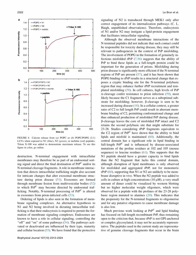
FIGURE 6 Calcein release from (a) POPC or (b) POPC/POPG (2:1)
LUVs when exposed to N1 (blue), N2 (green), or melittin (red) peptides.
Triton X-100 was added to demonstrate maximum release. To see this
figure in color, go online.
2322 Le Brun et al.
destruction. N-terminal engagement with intracellularmembranes may therefore be as part of an endosomal sort-ing signal and direct the final destination of PrPC and/or itsN-terminal cleavage fragments. A role in membrane interac-tion that directs intracellular trafficking might also accountfor intricate changes that alter exosomal membrane struc-ture during prion disease (51). Exosomes are formedthrough membrane fission from multivesicular bodies (52)to which PrPC may become directed by endosomal traf-ficking. Notably, N-terminal processing of PrPC is alteredin exosomes from prion-infected cells (53).
Ordering of lipids is also seen in the formation of mem-brane signaling complexes. An alternative hypothesis toN1 and N2 being involved in dictating intracellular traf-ficking is that their endocytosis is required to permit the for-mation of membrane signaling complexes. Endosomes areknown to have a role in cellular signaling, controlling the‘‘off’’ and ‘‘on’’ of some pathways (54). The pathways acti-vated or deactivated are influenced by their type, maturityand cellular location (55). We have found that the protective
Biophysical Journal 107(10) 2313–2324
signaling of N2 is transduced through MEK1 only aftercorrect engagement of its internalization pathways (C. L.Haigh, unpublished observations). Therefore, endocytosisof N1 and/or N2 may instigate a lipid-protein engagementthat facilitates intracellular signaling.
Although the observed membrane interactions of theN-terminal peptides did not indicate that such contact couldbe responsible for toxicity during disease, they may still berelevant to pathogenesis in the context of PrP misfolding.The involvement of POPG in the formation of genuinely in-fectious misfolded rPrP (7,56) suggests that the ability ofPrP to bind these lipids as a full-length protein could beimportant for the generation of prions. Misfolding duringprion disease is significantly more efficient if the N-terminalregions of PrP are present (57), and it has been shown thatPOPG binding to rPrP results in a structural change that ex-poses a cryptic binding site for the N-terminal polybasicregion that may enhance further rPrP recruitment and tem-plated misfolding (58). In cell cultures, high levels of PrPa-cleavage confer resistance to prion infection (59), mostlikely because the C1 fragment serves as a suboptimal sub-strate for misfolding; however, b-cleavage is seen to beincreased during disease (18). In a cellular context, a greaterratio of C2 to full length PrP could result in aberrant mem-brane binding of C2, permitting conformational change andthus enhanced production of misfolded PrP during disease.b-cleavage leaves the core of misfolded PrP intact and C2retains the second polybasic site that might substitute for23-28. Studies considering rPrP fragments equivalent tothe C2 region of PrPC have shown that the ability to bindlipids and misfold is maintained (60). Additionally, thecentral domain has a significant role in lipid binding infull-length PrPC and is influenced by disease-associatedmutations of the proline residues at 102 and 105 (mousesequence) to leucine residues (61). This supports that theN1 peptide should have a greater capacity to bind lipidsthan the N2 fragment that lacks this central domain,although disruption of lipid membranes is only observedfor misfolded and aggregated rPrP, not for monomericrPrP (60), supporting that N1 or N2 are unlikely to be mem-brane disruptive in vivo. When the N2 peptide was added tocells in culture at high concentrations (10 mM), a very smallamount of dimer could be visualized by western blottingbut no higher molecular weight oligomers, which wereobserved for a peptide with the prolines of the 23-28 poly-basic region mutated to alanines (20). This indicates thatthe propensity for the N-terminal fragments to oligomeriseand for any putative oligomers to cause membrane damageis also low.
Much previous work looking at PrPC and lipid bindinghas focused on full-length recombinant PrP, thus remainingopen to the criticism that, because rPrP is not GPI-anchoredor complex glycosylated, it may not be accurately represen-tative. The peptides used in the current study are representa-tive of genuine cleavage fragments that occur in the brain

Neutron Reflectometry of PrP Peptides 2323
(18), separating the N-terminus from the structured, glyco-sylated, and membrane-anchored C terminus, and the datatherefore present a stronger argument for how thesecleavage fragments interact with cell membranes. BothN-terminal cleavage fragments have been shown to haveneuroprotective functions and therefore lipid bindinginteractions could be crucial for these peptides to effectprotection. The binding parameters of full length PrPC,especially in the context of membrane anchoring, remainto be determined; however, the data presented herein sug-gest that specific lipid binding interactions could potentiallybe corrupted during prion disease, when the cleavage eventsshift toward greater b-cleavage.
CONCLUSION
The new data generated by NR support our previous findingsthat endogenous PrP N-terminal cleavage fragments caninteract with negatively charged supported phospholipid bi-layers at low physiological pH. We further show that thisinteraction lies predominantly at the level of the phospho-lipid headgroups with no clear penetration into the acyltail plane and without damage to the structural integrity ofthe membrane but with induction of a domain orderingeffect. These observations stand in clear contrast to the find-ings of analogous studies of Ab and a-synuclein and therebysupport the possibility of a functional role for such N-termi-nal fragment-membrane interactions.
SUPPORTING MATERIAL
Two figures and four tables are available at http://www.biophysj.org/
biophysj/supplemental/S0006-3495(14)01001-7.
This work was supported by the Australian Nuclear Science and Technol-
ogy Organisation (ANSTO), proposal ID2211, Australian Institute for
Nuclear Science and Engineering (AINSE Ltd.) for travel support, and
a National Health and Medical Research Council (NHMRC) program
grant (No. 628946). S. J. C. is funded by an NHMRC Practitioner
Fellowship (No. APP100581). S. C. D. is funded by an Australian Research
Council Future Fellowship (FT110100199). A. P. L. B. is funded by an
Australian Research Council Discovery Early Career Research Award
(DE140101788). The authors declare that they have no conflicts of interest.
REFERENCES
1. Prusiner, S. B. 1982. Novel proteinaceous infectious particles causescrapie. Science. 216:136–144.
2. Weissmann, C., H. Bueler, ., M. Aguet. 1994. PrP-deficient mice areresistant to scapie. In Slow Infections of the Central Nervous System:The Legacy of Dr Bjorn Sigurdsson. J. Bjornsson, R. I. Carp, A. Love,and H. M. Wisniewski, editors. New York Academic Sciences, NewYork, pp. 235–240.
3. Saborio, G. P., B. Permanne, and C. Soto. 2001. Sensitive detection ofpathological prion protein by cyclic amplification of protein misfold-ing. Nature. 411:810–813.
4. Kim, J. I., I. Cali,., W. K. Surewicz. 2010. Mammalian prions gener-ated from bacterially expressed prion protein in the absence of anymammalian cofactors. J. Biol. Chem. 285:14083–14087.
5. Welton, J. M., and V. A. Lawson. 2011. The site and host dependentrequirements for prion propagation. In The Cellular and MolecularBiology of Prion Disease. S. J. Collins and V. A. Lawson, editors.Research Signpost, Scarborough, Canada, pp. 189–206.
6. Zhou, Z., and G. Xiao. 2013. Conformational conversion of prionprotein in prion diseases. Acta Biochim. Biophys. Sin. (Shanghai).45:465–476.
7. Wang, F., X. Wang,., J. Ma. 2010. Generating a prion with bacteriallyexpressed recombinant prion protein. Science. 327:1132–1135.
8. Critchley, P., J. Kazlauskaite, ., T. J. T. Pinheiro. 2004. Binding ofprion proteins to lipid membranes. Biochem. Biophys. Res. Commun.313:559–567.
9. Re, F., S. Sesana, ., M. Masserini. 2008. Prion protein structure isaffected by pH-dependent interaction with membranes: a study in amodel system. FEBS Lett. 582:215–220.
10. Dong, S. L., S. A. Cadamuro,., C. Renner. 2007. Copper binding andconformation of the N-terminal octarepeats of the prion protein inthe presence of DPC micelles as membrane mimetic. Biopolymers.88:840–847.
11. Robinson, P. J., and T. J. T. Pinheiro. 2010. Phospholipid compositionof membranes directs prions down alternative aggregation pathways.Biophys. J. 98:1520–1528.
12. Steunou, S., J. F. Chich, ., J. Vidic. 2010. Biosensing of lipid-prioninteractions: insights on charge effect, Cu(II)-ions binding and prionoligomerization. Biosens. Bioelectron. 26:1399–1406.
13. Haigh, C. L., S. Y. Marom, and S. J. Collins. 2010. Copper, endopro-teolytic processing of the prion protein and cell signalling. Front.Biosci. 15:1086–1104, (Landmark ed.).
14. Borchelt, D. R., M. Rogers, ., S. B. Prusiner. 1993. Release of thecellular prion protein from cultured cells after loss of its glycoinositolphospholipid anchor. Glycobiology. 3:319–329.
15. Harris, D. A., M. T. Huber,., R. Wang. 1993. Processing of a cellularprion protein: identification of N- and C-terminal cleavage sites.Biochemistry. 32:1009–1016.
16. McMahon, H. E. M., A. Mange,., S. Lehmann. 2001. Cleavage of theamino terminus of the prion protein by reactive oxygen species. J. Biol.Chem. 276:2286–2291.
17. Mange, A., F. Beranger,., S. Lehmann. 2004. Alpha- and beta-cleav-ages of the amino-terminus of the cellular prion protein. Biol. Cell.96:125–132.
18. Chen, S. G., D. B. Teplow, ., L. Autilio-Gambetti. 1995. Truncatedforms of the human prion protein in normal brain and in prion diseases.J. Biol. Chem. 270:19173–19180.
19. Guillot-Sestier, M. V., C. Sunyach, ., F. Checler. 2009. The alpha-secretase-derived N-terminal product of cellular prion, N1, displaysneuroprotective function in vitro and in vivo. J. Biol. Chem.284:35973–35986.
20. Haigh, C. L., S. C. Drew,., S. J. Collins. 2009. Dominant roles of thepolybasic proline motif and copper in the PrP23-89-mediated stressprotection response. J. Cell Sci. 122:1518–1528.
21. Walmsley, A. R., N. T. Watt, ., N. M. Hooper. 2009. Alpha-cleavageof the prion protein occurs in a late compartment of the secretorypathway and is independent of lipid rafts. Mol. Cell. Neurosci.40:242–248.
22. Boland, M. P., C. R. Hatty, ., S. J. Collins. 2010. Anionic phospho-lipid interactions of the prion protein N terminus are minimally per-turbing and not driven solely by the octapeptide repeat domain.J. Biol. Chem. 285:32282–32292.
23. Karas, J. A., M. Boland,., D. Scanlon. 2012. Microwave synthesis ofprion protein fragments up to 111 amino acids in length generates bio-logically active peptides. Int. J. Pept. Res. Ther. 18:21–29.
24. James, M., A. Nelson, ., F. Klose. 2011. The multipurpose time-of-flight neutron reflectometer ‘‘Platypus’’ at Australia’s OPAL reactor.Nucl. Instrum. Methods Phys. Res. A. 632:112–123.
Biophysical Journal 107(10) 2313–2324

2324 Le Brun et al.
25. Saerbeck, T., F. Klose,., M. James. 2012. Invited article: polarization‘‘down under’’: the polarized time-of-flight neutron reflectometerPLATYPUS. Rev. Sci. Instrum. 83:081301–081312.
26. Nelson, A. 2010. Motofit—integrating neutron reflectometry acquisi-tion, reduction and analysis into one, easy to use, package. J. Phys.Conf. Ser. 251:012094.
27. Nelson, A. 2006. Co-refinement of multiple-contrast neutron/x-ray re-flectivity data using MOTOFIT. J. Appl. Crystallogr. 39:273–276.
28. Chiu, S. W., E. Jakobsson, ., H. L. Scott. 1999. Combined MonteCarlo and molecular dynamics simulation of fully hydrated dioleyland palmitoyl-oleyl phosphatidylcholine lipid bilayers. Biophys. J.77:2462–2469.
29. Pabst, G., S. Danner, ., V. A. Raghunathan. 2007. On the propensityof phosphatidylglycerols to form interdigitated phases. Biophys. J.93:513–525.
30. Petrache, H. I., S. Tristram-Nagle, ., J. F. Nagle. 2004. Structure andfluctuations of charged phosphatidylserine bilayers in the absence ofsalt. Biophys. J. 86:1574–1586.
31. Wacklin, H. P. 2010. Neutron reflection from supported lipid mem-branes. Curr. Opin. Colloid Interface Sci. 15:445–454.
32. Le Brun, A. P., T. A. Darwish, and M. James. 2013. Studies of biomi-metic cellular membranes using neutron reflection. J. Chem. Biol.Interfaces. 1:3–24.
33. Kalb, E., S. Frey, and L. K. Tamm. 1992. Formation of supported planarbilayers by fusion of vesicles to supported phospholipid monolayers.Biochim. Biophys. Acta—Biomembr. 1103:307–316.
34. Johnson, S. J., T. M. Bayerl, ., E. Sackmann. 1991. Structure of anadsorbed dimyristoylphosphatidylcholine bilayer measured with spec-ular reflection of neutrons. Biophys. J. 59:289–294.
35. Kiessling, V., and L. K. Tamm. 2003. Measuring distances in supportedbilayers by fluorescence interference-contrast microscopy: polymersupports and SNARE proteins. Biophys. J. 84:408–418.
36. Fernandez, D. I., A. P. Le Brun,., F. Separovic. 2012. The antimicro-bial peptide aurein 1.2 disrupts model membranes via the carpet mech-anism. Phys. Chem. Chem. Phys. 14:15739–15751.
37. Krueger, S., C. W. Meuse,., A. L. Plant. 2001. Investigation of hybridbilayer membranes with neutron reflectometry: probing the interactionsof melittin. Langmuir. 17:511–521.
38. Lu, N. Y., K. Yang, J. L. Li, B. Yuan, and Y. Q. Ma. 2013. Vesicle depo-sition and subsequent membrane-melittin interactions on different sub-strates: a QCM-D experiment. Biochim. Biophys. Acta—Biomembr.1828:1918–1925.
39. McCubbin, G. A., S. Praporski,., L. L. Martin. 2011. QCM-D finger-printing of membrane-active peptides. Eur. Biophys. J. 40:437–446.
40. Piantavigna, S., P. Czihal,., L. L. Martin. 2009. Cell penetrating api-daecin peptide interactions with biomimetic phospholipid membranes.Int. J. Pept. Res. Ther. 15:139–146.
41. Knappe, D., S. Piantavigna, ., R. Hoffmann. 2010. Oncocin(VDKPPYLPRPRPPRRIYNR-NH2): a novel antibacterial peptideoptimized against gram-negative human pathogens. J. Med. Chem.53:5240–5247.
42. Dante, S., T. Hauss,., N. A. Dencher. 2011. Nanoscale structural andmechanical effects of beta-amyloid (1–42) on polymer cushionedmembranes: a combined study by neutron reflectometry and AFMForce Spectroscopy. Biochim. Biophys. Acta—Biomembr. 1808:2646–2655.
43. Chi, E. Y., C. Ege, ., K. Y. C. Lee. 2008. Lipid membrane templatesthe ordering and induces the fibrillogenesis of Alzheimer’s diseaseamyloid-beta peptide. Proteins. 72:1–24.
Biophysical Journal 107(10) 2313–2324
44. Hellstrand, E., M. Grey, ., E. Sparr. 2013. Adsorption of a-synucleinto supported lipid bilayers: positioning and role of electrostatics. ACSChem. Neurosci. 4:1339–1351.
45. Jao, C. C., B. G. Hegde, ., R. Langen. 2008. Structure of membrane-bound alpha-synuclein from site-directed spin labeling and computa-tional refinement. Proc. Natl. Acad. Sci. USA. 105:19666–19671.
46. Bisaglia, M., S. Mammi, and L. Bubacco. 2009. Structural insights onphysiological functions and pathological effects of alpha-synuclein.FASEB J. 23:329–340.
47. Cornely, R., C. Rentero,., K. Gaus. 2011. Annexin A6 is an organizerof membrane microdomains to regulate receptor localization and sig-nalling. IUBMB Life. 63:1009–1017.
48. Osiecka, K. M., H. Nieznanska,., K. Nieznanski. 2009. Prion proteinregion 23-32 interacts with tubulin and inhibits microtubule assembly.Proteins. 77:279–296.
49. Taylor, D. R., N. T. Watt,., N. M. Hooper. 2005. Assigning functionsto distinct regions of the N-terminus of the prion protein that areinvolved in its copper-stimulated, clathrin-dependent endocytosis.J. Cell Sci. 118:5141–5153.
50. Lu, L., and W. Hong. 2014. From endosomes to the trans-Golginetwork. Semin. Cell Dev. Biol. 31:30–39.
51. Coleman, B. M., E. Hanssen, ., A. F. Hill. 2012. Prion-infected cellsregulate the release of exosomes with distinct ultrastructural features.FASEB J. 26:4160–4173.
52. Von Bartheld, C. S., and A. L. Altick. 2011. Multivesicular bodies inneurons: distribution, protein content, and trafficking functions. Prog.Neurobiol. 93:313–340.
53. Vella, L. J., R. A. Sharples, ., A. F. Hill. 2007. Packaging of prionsinto exosomes is associated with a novel pathway of PrP processing.J. Pathol. 211:582–590.
54. von Zastrow, M., and A. Sorkin. 2007. Signaling on the endocyticpathway. Curr. Opin. Cell Biol. 19:436–445.
55. Taub, N., D. Teis, ., L. A. Huber. 2007. Late endosomal traffic ofthe epidermal growth factor receptor ensures spatial and temporal fidel-ity of mitogen-activated protein kinase signaling. Mol. Biol. Cell.18:4698–4710.
56. Zhang, Z., Y. Zhang,., J. Ma. 2013. De novo generation of infectiousprions with bacterially expressed recombinant prion protein. FASEB J.27:4768–4775.
57. Lawson, V. A., S. A. Priola,., B. Chesebro. 2004. Flexible N-terminalregion of prion protein influences conformation of protease-resistantprion protein isoforms associated with cross-species scrapie infectionin vivo and in vitro. J. Biol. Chem. 279:13689–13695.
58. Zurawel, A. A., D. J. Walsh,., S. Supattapone. 2014. Prion nucleationsite unmasked by transient interaction with phospholipid cofactor.Biochemistry. 53:68–76.
59. Lewis, V., A. F. Hill, ., S. J. Collins. 2009. Increased proportions ofC1 truncated prion protein protect against cellular M1000 prion infec-tion. J. Neuropathol. Exp. Neurol. 68:1125–1135.
60. Chich, J. F., C. Chapuis, ., S. Noinville. 2010. Vesicle permeabiliza-tion by purified soluble oligomers of prion protein: a comparative studyof the interaction of oligomers and monomers with lipid membranes.J. Mol. Biol. 397:1017–1030.
61. Wang, F., S. Yin,., J. Ma. 2010. Role of the highly conserved middleregion of prion protein (PrP) in PrP-lipid interaction. Biochemistry.49:8169–8176.

SUPPORTING INFORMATION
Neutron reflectometry studies define prion protein N-terminal peptide membrane binding
Anton P. Le Brun, Cathryn L. Haigh, Simon C. Drew, Michael James, Martin P. Boland and Steven J. Collins
Supplementary Figure 1. Neutron reflectometry of the N2 peptide binding to solid-supported d31-POPC membranes on silicon. The symbols with error bars are the data and the solid lines the fit. Black () is the bilayer before peptide addition in D2O and red () is the bilayer in the presence of N2 peptide in D2O.

Supplementary Figure S2: QCM-D trace of melittin binding to a) POPC/POPS (2:1) bilayer and b) POPC/POPG (2:1) bilayer. The left axis is Δf (black traces) and the right axis is ΔD (red traces). The 3rd (), 5th (), 7th (), 9th (), 11th () and 13th (+) overtones are shown. The arrows denote the point of peptide addition (PA) and buffer wash (BW).

Supplementary Table 1. Peptide and lipid properties.
Molecular weight /
Da
Volume / Å3
nSLD in D2O / ×10-6 Å-2
nSLD in H2O / ×10-6 Å-2
Number of exchangeable
protons N1 peptide
9149 10669 3.88 2.30 162
N2 peptide
6808 7873 3.90 2.38 115
dOR peptide
5297 3494 3.89 2.12 106
d31-palmitoyl-oleoyl tails
482 944 3.19 3.19 0
POPC headgroups
311 319 1.88 1.88 0
POPG headgroups
299 257 3.59 2.78 2
POPS headgroups
244 312 3.89 3.47 1

Supplementary Table 2. The fitted parameters for N1 peptide binding to d31-POPC/d31-POPG (2:1) d31-POPC/d31-POPS (2:1) bilayers.
Before N1 peptide addition In the presence of N1 peptide Layer Thicknes
s / Å nSLD
in D2O / ×10-6
Å-2
Roughness / Å
Thickness / Å
nSLD in
D2O / ×10-6 Å-2
nSLD in H2O / ×10-6
Å-2
Roughness / Å
d31-POPC/d31-POPG (2:1) bilayer Head
group one 15 ± 1 2.65 ±
0.07 3 18 ± 1 3.87 ±
0.06 2.14 ± 0.03
4
Tails 20 ± 1 3.19 ± 0.05
3 23 ± 1 3.20 ± 0.03
3.20 ± 0.03
4
Head group two (plus N1 peptide)
10 ± 1 4.73 ± 0.12
3 7 ± 1 4.15 ± 0.05
1.18 ± 0.22
4
N1 peptide only
- - - 21 ± 2 5.86 ± 0.04
0.01 ± 0.07
5
d31-POPC/d31-POPS (2:1) bilayer Head
group one 7 ± 1 2.71 ±
0.17 3 8 ± 1 2.60 ±
0.09 2.52 ± 0.04
4
Tails 20 ± 1 3.19 ± 0.06
3 29 ± 1 3.19 ± 0.03
2.97 ± 0.03
4
Head group two (plus N1 peptide)
11 ± 1 2.73 ± 0.14
3 9 ± 1 4.92 ± 0.09
1.71 ± 0.07
4
N1 peptide only
- - - 21 ± 1 5.61 ± 0.05
0.43 ± 0.13
5
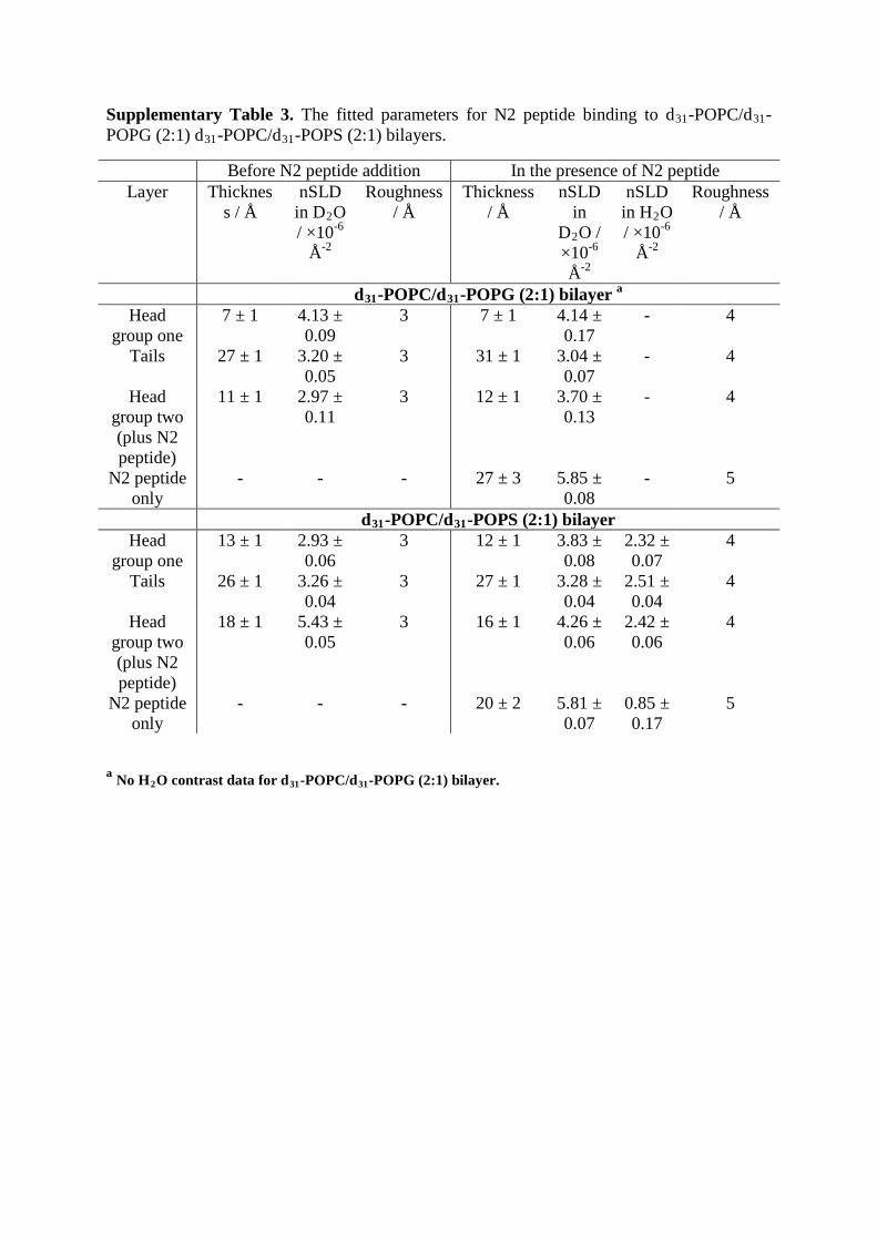
Supplementary Table 3. The fitted parameters for N2 peptide binding to d31-POPC/d31-POPG (2:1) d31-POPC/d31-POPS (2:1) bilayers.
Before N2 peptide addition In the presence of N2 peptide Layer Thicknes
s / Å nSLD
in D2O / ×10-6
Å-2
Roughness / Å
Thickness / Å
nSLD in
D2O / ×10-6 Å-2
nSLD in H2O / ×10-6
Å-2
Roughness / Å
d31-POPC/d31-POPG (2:1) bilayer a Head
group one 7 ± 1 4.13 ±
0.09 3 7 ± 1 4.14 ±
0.17 - 4
Tails 27 ± 1 3.20 ± 0.05
3 31 ± 1 3.04 ± 0.07
- 4
Head group two (plus N2 peptide)
11 ± 1 2.97 ± 0.11
3 12 ± 1 3.70 ± 0.13
- 4
N2 peptide only
- - - 27 ± 3 5.85 ± 0.08
- 5
d31-POPC/d31-POPS (2:1) bilayer Head
group one 13 ± 1 2.93 ±
0.06 3 12 ± 1 3.83 ±
0.08 2.32 ± 0.07
4
Tails 26 ± 1 3.26 ± 0.04
3 27 ± 1 3.28 ± 0.04
2.51 ± 0.04
4
Head group two (plus N2 peptide)
18 ± 1 5.43 ± 0.05
3 16 ± 1 4.26 ± 0.06
2.42 ± 0.06
4
N2 peptide only
- - - 20 ± 2 5.81 ± 0.07
0.85 ± 0.17
5
a No H2O contrast data for d31-POPC/d31-POPG (2:1) bilayer.

Supplementary Table 4: Results from QCM-D experiments. Numbers in brackets are standard deviation from n = 4.
Bilayer and peptide
Mass of bilayer / mg
m-2
Mass of peptide / mg
m-2
Moles of bilayer / µmol m-2
Moles of peptide / µmol m-2
Lipid to peptide ratio
POPC/POPG + N1
5.26 (0.19) 0.83 (0.06) 6.73 (0.25) 0.09 (0.01) 0.014 (0.001)
POPC/POPS + N1
4.54 (0.06) 3.19 (1.39) 5.92 (0.07) 0.35 (0.15) 0.059 (0.026)
DMPC/DMPG +N1
5.14 (0.10) 3.08 (1.12) 7.62 (0.15) 0.34 (0.12) 0.044 (0.016)
POPC/POPG +N2
5.17 (0.03) 0.63 (0.25) 6.62 (0.04) 0.09 (0.03) 0.014 (0.006)
POPC/POPS +N2
4.62 (0.09) 1.82 (0.16) 6.02 (0.12) 0.27 (0.02) 0.044 (0.005)
DMPC/DMPG +N2
5.10 (0.13) 0.83 (0.18) 7.56 (0.20) 0.12 (0.03) 0.016 (0.003)