The Mechanism of Prion Inhibition by HET-S
11
Molecular Cell Article The Mechanism of Prion Inhibition by HET-S Jason Greenwald, 1 Carolin Buhtz, 1 Christiane Ritter, 2 Witek Kwiatkowski, 3 Senyon Choe, 3 Marie-Lise Maddelein, 4 Frederique Ness, 4 Sandra Cescau, 4 Alice Soragni, 1 Dominik Leitz, 1 Sven J. Saupe, 4 and Roland Riek 1,3, * 1 Laboratory of Physical Chemistry, ETH Zu ¨ rich, Wolfgang-Pauli-Strasse 10, 8093 Zu ¨ rich, Switzerland 2 Helmholtz Center for Infection Research, Inhoffenstraße 7, 38124 Braunschweig, Germany 3 Structural Biology Laboratory, The Salk Institute for Biological Studies, 10010 North Torrey Pines Road, La Jolla, CA 92037, USA 4 Laboratoire de Ge ´ ne ´ tique Mole ´ culaire des Champignons, Institut de Biochimie et Ge ´ ne ´ tique Cellulaires, UMR-5095 CNRS/Universite ´ de Bordeaux 2, 1 rue Camille St Sae ¨ ns, 33077 Bordeaux, France *Correspondence: [email protected] DOI 10.1016/j.molcel.2010.05.019 SUMMARY HET-S (97% identical to HET-s) has an N-terminal globular domain that exerts a prion-inhibitory effect in cis on its own prion-forming domain (PFD) and in trans on HET-s prion propagation. We show that HET-S fails to form fibrils in vitro and that it inhibits HET-s PFD fibrillization in trans. In vivo analyses indi- cate that b-structuring of the HET-S PFD is required for HET-S activity. The crystal structures of the glob- ular domains of HET-s and HET-S are highly similar, comprising a helical fold, while NMR-based charac- terizations revealed no differences in the conforma- tions of the PFDs. We conclude that prion inhibition is not encoded by structure but rather in stability and oligomerization properties: when HET-S forms a prion seed or is incorporated into a HET-s fibril via its PFD, the b-structuring in this domain induces a change in its globular domain, generating a molec- ular species that is incompetent for fibril growth. INTRODUCTION Prions are self-propagating, usually amyloid-like protein aggre- gates (Aguzzi et al., 2008). In mammals, prions cause fatal neurodegenerative disease, while in fungi they are detected as protein-based genetic elements (Wickner et al., 2008). The fungal prion proteins have a modular organization comprising a so-called prion-forming domain (PFD) that is natively unfolded in the soluble form and that is appended to a globular domain. While the PFD of these proteins is both necessary and sufficient for prion propagation, the in vivo prion-forming abilities and aggregate morphology of a given protein can be strongly affected by mutations or polymorphism in the appended globular domain (Balguerie et al., 2003, 2004; Fernandez-Bellot et al., 2000; Liu et al., 2002; Maddelein and Wickner, 1999; Masison and Wickner, 1995). This is apparently true for amyloidogenic proteins in general, as it has also been shown that amyloid toxicity of polyQ aggregates is strongly dependent on flanking domains (Dehay and Bertolotti, 2006; Duennwald et al., 2006). While considerable efforts have been devoted to the structural and functional characterization of the PFDs of fungal prions, the mechanistic basis of this cis-regulatory effect has been only scarcely studied despite its importance in the prion propa- gation mechanism. The het-s/het-S fungal prion may be the most blatant example of such a cis-acting prion-inhibitory domain (Balguerie et al., 2003, 2004). HET-s and HET-S are natural polymorphic variants of the same protein that share a functional C-terminal PFD. Yet, in contrast to HET-s, full-length HET-S totally lacks prion-forming ability in vivo. In the filamentous fungus Podospora anserina, the prion state of the HET-s protein is involved in a programmed cell death (PCD) reaction termed heterokaryon incompatibility. Filamen- tous fungi have several incompatibility loci that regulate the fusion of mycelium between genetically distinct individuals (Glass et al., 2000; Saupe, 2000). The het-s locus has two antag- onistic alleles: het-s and het-S. Their encoded proteins, HET-s and HET-S, give rise to the compatibility phenotypes [Het-s] and [Het-S]. Although they differ by only 13 out of 289 residues (Turcq et al., 1991), only HET-s undergoes a transition to an infectious prion state that is correlated in vivo with the conver- sion from the [Het-s*] to the [Het-s] phenotype (Coustou et al., 1997). When a [Het-s] strain fuses with a [Het-S] strain, the fusion cell dies, whereas the fusion of [Het-s*] with [Het-S] leads to a viable mixed cell (heterokaryon). Colonies with the [Het-s*] phenotype convert spontaneously to [Het-s] at a very low frequency, but contact between [Het-s] and [Het-s*] strains leads to infection of the latter and conversion of its phenotype to [Het-s]. Importantly, in vivo interactions between [Het-s] and [Het-S] strains can also lead to elimination of the [Het-s] prion state (Beisson-Schecroun, 1962; Dalstra et al., 2003; Rizet, 1952). This occurs, for instance, in a [Het-s] 3 [Het-S] sexual cross in which [Het-S] leads to complete curing of the [Het-s] prion in the meiotic progeny (daughter cells with the het-s allele are [Het-s*]). Using microsurgical approaches, it was also shown that [Het-S] strongly inhibits [Het-s] propagation in vegetative hyphae (Beisson-Schecroun, 1962). Therefore, [Het-S] not only triggers cell death upon interaction with [Het-s] but also exerts an inhibitory effect on its propagation. HET-s is a two-domain protein. It comprises a C-terminal PFD (residues 218–289) that is both necessary and sufficient for amyloid formation and prion propagation and an N-terminal globular domain (residues 1 to 227) that specifies the incom- patibility type ([Het-s] or [Het-S]) (Balguerie et al., 2003). A chimeric protein construct with the HET-S globular domain appended to the HET-s PFD results in a protein of the [Het-S] Molecular Cell 38, 889–899, June 25, 2010 ª2010 Elsevier Inc. 889
Transcript of The Mechanism of Prion Inhibition by HET-S

Molecular Cell
Article
The Mechanism of Prion Inhibition by HET-SJason Greenwald,1 Carolin Buhtz,1 Christiane Ritter,2 Witek Kwiatkowski,3 Senyon Choe,3 Marie-Lise Maddelein,4
Frederique Ness,4 Sandra Cescau,4 Alice Soragni,1 Dominik Leitz,1 Sven J. Saupe,4 and Roland Riek1,3,*1Laboratory of Physical Chemistry, ETH Zurich, Wolfgang-Pauli-Strasse 10, 8093 Zurich, Switzerland2Helmholtz Center for Infection Research, Inhoffenstraße 7, 38124 Braunschweig, Germany3Structural Biology Laboratory, The Salk Institute for Biological Studies, 10010 North Torrey Pines Road, La Jolla, CA 92037, USA4Laboratoire de Genetique Moleculaire des Champignons, Institut de Biochimie et Genetique Cellulaires, UMR-5095 CNRS/Universite de
Bordeaux 2, 1 rue Camille St Saens, 33077 Bordeaux, France
*Correspondence: [email protected] 10.1016/j.molcel.2010.05.019
SUMMARY
HET-S (97% identical to HET-s) has an N-terminalglobular domain that exerts a prion-inhibitory effectin cis on its own prion-forming domain (PFD) and intrans on HET-s prion propagation. We show thatHET-S fails to form fibrils in vitro and that it inhibitsHET-s PFD fibrillization in trans. In vivo analyses indi-cate that b-structuring of the HET-S PFD is requiredfor HET-S activity. The crystal structures of the glob-ular domains of HET-s and HET-S are highly similar,comprising a helical fold, while NMR-based charac-terizations revealed no differences in the conforma-tions of the PFDs. We conclude that prion inhibitionis not encoded by structure but rather in stabilityand oligomerization properties: when HET-S formsa prion seed or is incorporated into a HET-s fibrilvia its PFD, the b-structuring in this domain inducesa change in its globular domain, generating a molec-ular species that is incompetent for fibril growth.
INTRODUCTION
Prions are self-propagating, usually amyloid-like protein aggre-
gates (Aguzzi et al., 2008). In mammals, prions cause fatal
neurodegenerative disease, while in fungi they are detected as
protein-based genetic elements (Wickner et al., 2008). The
fungal prion proteins have a modular organization comprising
a so-called prion-forming domain (PFD) that is natively unfolded
in the soluble form and that is appended to a globular domain.
While the PFD of these proteins is both necessary and sufficient
for prion propagation, the in vivo prion-forming abilities and
aggregate morphology of a given protein can be strongly
affected by mutations or polymorphism in the appended globular
domain (Balguerie et al., 2003, 2004; Fernandez-Bellot et al.,
2000; Liu et al., 2002; Maddelein and Wickner, 1999; Masison
and Wickner, 1995). This is apparently true for amyloidogenic
proteins in general, as it has also been shown that amyloid
toxicity of polyQ aggregates is strongly dependent on flanking
domains (Dehay and Bertolotti, 2006; Duennwald et al., 2006).
While considerable efforts have been devoted to the structural
and functional characterization of the PFDs of fungal prions,
M
the mechanistic basis of this cis-regulatory effect has been
only scarcely studied despite its importance in the prion propa-
gation mechanism. The het-s/het-S fungal prion may be the most
blatant example of such a cis-acting prion-inhibitory domain
(Balguerie et al., 2003, 2004). HET-s and HET-S are natural
polymorphic variants of the same protein that share a functional
C-terminal PFD. Yet, in contrast to HET-s, full-length HET-S
totally lacks prion-forming ability in vivo.
In the filamentous fungus Podospora anserina, the prion state
of the HET-s protein is involved in a programmed cell death
(PCD) reaction termed heterokaryon incompatibility. Filamen-
tous fungi have several incompatibility loci that regulate the
fusion of mycelium between genetically distinct individuals
(Glass et al., 2000; Saupe, 2000). The het-s locus has two antag-
onistic alleles: het-s and het-S. Their encoded proteins, HET-s
and HET-S, give rise to the compatibility phenotypes [Het-s]
and [Het-S]. Although they differ by only 13 out of 289 residues
(Turcq et al., 1991), only HET-s undergoes a transition to an
infectious prion state that is correlated in vivo with the conver-
sion from the [Het-s*] to the [Het-s] phenotype (Coustou et al.,
1997). When a [Het-s] strain fuses with a [Het-S] strain, the fusion
cell dies, whereas the fusion of [Het-s*] with [Het-S] leads to
a viable mixed cell (heterokaryon). Colonies with the [Het-s*]
phenotype convert spontaneously to [Het-s] at a very low
frequency, but contact between [Het-s] and [Het-s*] strains leads
to infection of the latter and conversion of its phenotype to
[Het-s]. Importantly, in vivo interactions between [Het-s] and
[Het-S] strains can also lead to elimination of the [Het-s] prion
state (Beisson-Schecroun, 1962; Dalstra et al., 2003; Rizet,
1952). This occurs, for instance, in a [Het-s] 3 [Het-S] sexual
cross in which [Het-S] leads to complete curing of the [Het-s]
prion in the meiotic progeny (daughter cells with the het-s allele
are [Het-s*]). Using microsurgical approaches, it was also shown
that [Het-S] strongly inhibits [Het-s] propagation in vegetative
hyphae (Beisson-Schecroun, 1962). Therefore, [Het-S] not only
triggers cell death upon interaction with [Het-s] but also exerts
an inhibitory effect on its propagation.
HET-s is a two-domain protein. It comprises a C-terminal PFD
(residues 218–289) that is both necessary and sufficient for
amyloid formation and prion propagation and an N-terminal
globular domain (residues 1 to �227) that specifies the incom-
patibility type ([Het-s] or [Het-S]) (Balguerie et al., 2003). A
chimeric protein construct with the HET-S globular domain
appended to the HET-s PFD results in a protein of the [Het-S]
olecular Cell 38, 889–899, June 25, 2010 ª2010 Elsevier Inc. 889

Figure 1. HET-S Does Not Form Fibrils and Can Inhibit Aggregation
of HET-s PFD
(A) Electron micrographs of negatively stained aggregates of HET-s (left) and
HET-S (right), both at the same magnification (scale bar in the lower right of
image is 500 nm). Proteins at 15 mM were incubated 24 hr at 37�C with light
agitation in 100 mM Tris-HCl (pH 8), 150 mM NaCl.
(B) Coaggregation kinetics of HET-s PFD in the presence of HET-S. The 15N-
filtered 1D NMR signal of the 15N-labeled PFD was integrated between 6.5
and 9.0 ppm for each time point, the data normalized to the first time point,
and represented as the fraction soluble. All measurements included 52 mM
PFD and the plot symbols are as follows: open circles, control PFD alone;
gray diamonds, 75 nM HET-S; gray squares, 750 nM HET-S; gray triangles,
7.5 mM HET-S; black diamonds, 750 nM HET-S[E86K]. The arrows represent
the 95% confidence intervals for the 50% aggregation time for the measure-
ments of 0, 75, and 750 nM HET-S (three or four measurements each with
the closest to the average shown in plot). The 7.5 mM HET-S sample retained
more than 70% of the PFD in solution after 65 hr, and complete aggregation
occurred sometime between 65 and 110 hr.
Molecular Cell
The Structure of HET-S
type, and conversely a chimera associating the HET-s globular
domain to the HET-S PFD displays the [Het-s] specificity. In
other words, HET-S has a functional PFD, but the HET-S globular
domain exerts a prion-inhibitory effect (in cis) on its own
C-terminal PFD. A detailed analysis of the amino acid differences
between HET-s and HET-S revealed that a single amino acid
substitution in HET-S (H33P) converted its specificity to [Het-s]
in vivo, whereas the conversion of [Het-s] to [Het-S] requires
minimally two amino acid substitutions (D23A/P33H) (Deleu
et al., 1993).
The molecular mechanism of HET-s-mediated heterokaryon
incompatibility is not known, nor has the toxic entity that leads
to cell death been identified. The reaction that occurs when
incompatible strains fuse is a form of PCD that is spatially
restricted to the fusion cell or one or two cells to each side. In
all fungal incompatibility systems except het-s, one of the het
genes involved encodes for a protein with a HET domain (no rela-
890 Molecular Cell 38, 889–899, June 25, 2010 ª2010 Elsevier Inc.
tion to the HET-s protein). It has been shown that overexpression
of the HET domain alone is sufficient to induce PCD, and that it
depends on the presence of two other genes (Paoletti and Clave,
2007). In contrast, no other genes have been found that are
essential for het-s-mediated PCD, so it is possible that HET-S
and HET-s form a toxic species that directly initiates PCD.
Deletions in the globular domain of HET-S not only alleviate the
prion-inhibitory effect of the domain but also abolish HET-S
activity in incompatibility. Thus, the HET-S globular domain
(but not the HET-s globular domain) is essential for PCD (Balgu-
erie et al., 2004).
The recently reported structures of the C-terminal PFD of HET-
s in its fibril form have shed light on the mechanism of prion
formation and propagation (Wasmer et al., 2008; Wasmer
et al., 2009). These reports revealed that the PFD aggregates
into a highly ordered b-solenoid fold. However, the question
remains as to how the N-terminal domain of HET-S can exert
a prion-inhibitory effect on the PFD both in cis and in trans. To
probe this question, we studied the functional properties of the
HET-S protein in vitro and in vivo and solved the structure of
the HET-s and HET-S N-terminal domains by X-ray crystallog-
raphy.
RESULTS
HET-s, but Not HET-S, Forms Well-Ordered FibrilsIn VitroWe expressed and purified a soluble full-length HET-S protein
(see the Experimental Procedures) allowing the comparison of
the in vitro fibrillization of HET-s and HET-S. Both proteins tend
to aggregate in vitro, and this can be partially suppressed by
storage at 4�C. However, as revealed by negatively stained elec-
tron micrographs (Figure 1A), the aggregation of HET-s leads to
long (>1 mm), well-ordered, single fibrils, while HET-S aggregates
are amorphous and range in size from 10 to 100 nm. The HET-s
fibril growth can be seeded by fibrils of the isolated PFD, greatly
shortening the nucleation time, while the addition of PFD seeds
did not lead to HET-S fibrils (data not shown). The finding that
HET-S does not readily form fibrils in vitro explains the fact
that [Het-S] strains do not have a prion-associated phenotype
and directly illustrates the cis-acting prion inhibition of the
HET-S globular domain.
HET-S Can Inhibit the Aggregation of the HET-s PFDIn trans
Since the above experiments suggest that the prion inhibition
mechanism of HET-S in cis can be recapitulated in vitro, we
wondered whether the same was true for the trans-acting
prion-inhibitory effect of HET-S on [Het-s] propagation. Thus,
we looked at the ability of HET-S to inhibit fibril formation in trans.
The aggregation kinetics of the PFD from HET-s were monitored
by solution NMR in the presence of unlabeled HET-S. In this
experiment, only the monomeric PFD is detected, so that the
decay of the signal is proportional to the amount of PFD that
has been recruited into large, presumably fibrillar aggregates.
In order to minimize the stochastic nature of self-seeded aggre-
gation kinetics, the experiments were started with the addition of
30 nM PFD fibril seeds (see the Experimental Procedures). We

Table 1. Crystallographic Data and Refinement Statistics
Model (PDB ID) HET-s(1–227) (2wvn) HET-S(1–227) (2wvo) HET-s[D23A,P33H](13–221) (2wvq)
Space group p432 p3221 p21
Unit cell dimensions
a, b, c (A) 122.6 95.1, 95.1, 170.4 49.3, 83.2, 67.4
a, b, g (�) 90 90, 90, 120 90.0, 104.5, 90.0
Resolution range (outer shell) (A) 40–2.62 (2.77–2.62) 40–2.3 (2.44–2.3) 65–2.0 (2.12–2)
Number of unique reflections 10,044 40,556 34,078
Redundancy 10.1 (10.2) 7.0 (6.2) 3.2 (3.0)
I/sigma 24.1 (3.9) 23.8 (3.6) 10.5 (3.7)
Rmeasa 0.060 (0.463) 0.048 (0.569) 0.088 (0.459)
Completeness % 99.7 (99.0) 99.3 (99.1) 95.2 (93.3)
Number of protein monomers in A.U. 1 2 2
Number of nonhydrogen atoms in refinement
Protein 1696 3533 3363
DTT – – 16
Chloride ion – 4 –
Water – 135 198
R factor/free R factorb 0.216/0.270 0.225/0.257 0.225/0.269
Rmsd bond lengths/anglesc 0.018/1.843 0.010/1.411 0.016/1.739a Rmeas =
Ph (nh/(nh�1))1/2 P
i jIi(h) � < I(h) > j /P
h
Pi Ii(h), where Ii(h) and < I(h) > are the ith and mean intensity, and nh is the multiplicity over all
symmetry-equivalent reflections h (Diederichs and Karplus, 1997).b R =
PkFC j� jFOk /
PjFOj, where jFCjis the calculated structure factor amplitude of the model, and jFOjis the observed structure factor amplitude; the
free R factor was calculated against a random 5% test set of reflections that was not used during refinement. The same test set was used for all seven
data sets.c Rmsd, root-mean-square deviation from the parameter set for ideal stereochemistry (Engh and Huber, 1991).
Molecular Cell
The Structure of HET-S
found that HET-S is able to delay the onset of fibrillization in sub-
stoichiometric ratios as low as 1:700 (HET-S: PFD). The results
plotted in Figure 1B show that at a ratio of 1:70, the onset of
aggregation is delayed by several hours yet still proceeds rela-
tively quickly once it begins. At the 1:7 ratio, the aggregation is
delayed by more than 65 hr but eventually occurs sometime
before 110 hr (data collection was not continuous after 24 hr).
As expected, the addition of HET-s at substoichiometric ratios
(up to 1:7) does not have an inhibitory affect. Also, HET-S
(1–227) does not significantly inhibit at a 1:7 ratio, thus demon-
strating that the PFD of HET-S is required for the in vitro inhibition
of PFD aggregation in trans (data summarized in Table S1, avail-
able online).
Structural Composition of HET-s and HET-SHaving demonstrated that the HET-S prion-inhibitory effect is an
intrinsic property of the protein, we set out to determine the
structural basis of this functional difference between the HET-s
and HET-S proteins. We have previously shown that the HET-s
in its soluble form is composed of an N-terminal folded domain
comprising residues �1–227 followed by a flexible and highly
dynamic C-terminal tail (Balguerie et al., 2003). Since HET-S
differs from HET-s in only 13 out of its 289 residues, it is not
surprising that we find the same domain composition for
HET-S. As in the case of HET-s, the [15N,1H]-TROSY spectrum
of HET-S has two qualitatively different kinds of peaks: a first
group of about 220 broad cross peaks with full line widths at
half height along 1H of �25 Hz and a second group of about 70
M
sharp, more intense cross peaks with line widths of�14 Hz indic-
ative of amino acid residues in a flexible conformation. Compar-
ison with the [15N,1H]-TROSY spectrum of HET-S(1–227)
(Figure S1) shows that the first group of peaks has their counter-
part in HET-S(1–227). We established the sequential assignment
of HET-S, and the 13Ca chemical shifts are consistent with these
domain boundaries: the per-residue chemical shift deviation of
HET-S (Figure S2) shows that it is composed of an N-terminal
mostly helical structured domain comprising at least residues
13–222 followed by an unstructured and flexible C-terminal tail.
In addition to having near-random coil 13Ca chemical shifts,
the C-terminal tails of both HET-S and HET-s are highly dynamic
as evidenced by low 15N{1H}-NOE values (Figure S3).
Structure of the HET-s Globular DomainThe structure of HET-s(1–227) was solved by heavy atom
phasing, and the refined coordinates were used as a starting
model for the structure determination by molecular replacement
of HET-S(1–227) and of HET-s[D23A,P33H](13–221), the glob-
ular domain of a mutant that exhibits the [Het-S] phenotype
in vivo (structure factors and coordinates were deposited in the
Protein Data Bank, see Table 1). All three proteins contain the
same a-helical fold of 8–9 helices with a short two-stranded
b sheet (Figure 2). The first helix, a1 (residues 2–8) is only visible
in the HET-S crystals. Its absence in the HET-s structure may be
a crystallographic artifact, and the HET-s[D23A,P33H](13–221)
construct lacks the residues that make up this helix. This shorter
construct was designed based on the defined regions in the
olecular Cell 38, 889–899, June 25, 2010 ª2010 Elsevier Inc. 891

Figure 2. The Structure of the HET-S N-Terminal Domain
(A) Ribbon diagram of HET-S(1–227). The ribbon color is a rainbow gradient
from N (red) to C terminus (purple). Regions of the molecule are highlighted
as follows: flexible loop L5-6 in black, residues A23 and H33 as black spheres,
location of point mutants that can convert HET-S to [HET-s] phenotype as red
spheres, and residue E86 as yellow sphere. The helices are numbered 1–9 and
b1–b2 for the strands.
(B) Perpendicular views of the crystallographically observed HET-S(1–227)
dimer with consensus residues from the b-aggregation prediction algorithms
(see the Experimental Procedures) as black tubes.
Molecular Cell
The Structure of HET-S
initial structure determination of HET-s(1-227), in an effort to
improve the diffraction by crystallizing a protein without the
flexible ends. Indeed, it did yield the best diffracting crystals
(Table 1), although the construct itself is significantly less stable
(see below). The first five helices of HET-S pack in a regular anti-
parallel bundle followed by a loop and short b strand that
connects a5 and a6. This loop (L5-6) comprising residues 137–
147 and strand b2 thread back through the molecule (between
a2 and a4) so that a5 and a6 are parallel but on opposite sides
of the molecule. The last three helices (a7–a9) form a three-helix
bundle substructure that packs approximately perpendicular to
the first six helices. The overall shape of the fold is roughly an
equilateral triangle of �55 A on a side and a thickness of �25 A.
There are to date more than 30 homologs of the HET-s
N-terminal globular domain that can be identified with a PSI-
BLAST search, all of which come from filamentous fungi and
none of which have a known function (Figure S4). One of the
homologs was identified in a screen for mutants that affect
892 Molecular Cell 38, 889–899, June 25, 2010 ª2010 Elsevier Inc.
the pathogenicity of Leptosphaeria maculans, the fungus that
causes blackleg disease of Brassica napus (rapeseed). This
loss-of-pathogenicity (LOP-B) protein has 529 amino acids,
and its N terminus is 31% similar to the HET-s globular domain.
Henceforth, we refer to this particular fungal domain with its
helical fold as the HeLo domain (HET-s/LOP-B). The conserved
residues of the HeLo domain correspond primarily to buried resi-
dues in the HET-S(1–227) structure. The lack of surface-exposed
residue conservation suggests that the fold is well conserved but
that the function or target of the domain may differ among the
HeLo domain-containing proteins. The degree of conservation
is highest in the first �60 residues, with little or no conservation
in the loops between secondary structures. The sequence of the
last �50 residues, comprising the three terminal helices, is less
well conserved (Figure S4).
Comparison of the HeLo Domain Structures of HET-sand HET-SDespite the fact that all three constructs were crystallized in
diverse conditions with different numbers of molecules in their
asymmetric units (Table 1), they exhibit only minor differences.
An overlay of HET-s and HET-S gives a root-mean-square devi-
ation (rmsd) of 0.82 A (0.81 A for chain B) for the main-chain
atoms in residues 11–223. For comparison, the overlay of chain
A and B of HET-S within the same crystal gives an rmsd of 0.31 A.
Overlays of HET-s[D23A,P33H] with HET-s and HET-S give simi-
larly low numbers. Hence, the structures are so similar that local
differences need to be interpreted carefully, taking into account
possible crystal packing effects. The first major difference
between HET-s and HET-S is the N-terminal helix a1 that is
present only in HET-S. However, the solution NMR spectra of
neither HET-S nor HET-s indicate the presence of helix a1 in solu-
tion: the 13Ca and 13Cb chemical shifts for the residues in this
region do not exhibit helical deviations from their random coil
values (Figure S2). Furthermore, we could not detect a difference
in the helical content of the two proteins by CD spectroscopy
(data not shown). There are two factors that may explain the
discrepancy between the crystal structures and solution struc-
tures: (1) Since helix a1 resides at the dimer interface, it may
not be stable in the monomeric form. If so, the 0.5–1 mM protein
used for the NMR measurements may have led to an insufficient
dimer population to support the helix (note that the crystals
contain 15–20 mM protein). (2) The crystallization conditions of
HET-S (4.0 M NaCl) may have stabilized the hydrophobic interac-
tion that the helix a1 makes with itself at the interface, while the
low salt conditions for HET-s (30% PEG 4000, �100 mM NaCl)
may have had less of a stabilizing influence. The helix is neces-
sarily absent from the mutant structure because it is not in the
construct that was crystallized (residues 13–221). Therefore, we
cannot conclude from the crystal structures that helix a1 is unique
to HET-S, but it appears to be a quasistable structure that is
absent from both HET-S and HET-s in solution.
The second major structural difference occurs in the loop L5-6,
the residues of which have higher B factors and are generally
less well ordered than the other loops (Figure 2A and Figure S5).
In the case of chain B from the mutant, its electron density is too
weak to be confidently modeled. In HET-s there is a salt bridge
between K145 of the loop L5-6 and D23 of helix a2 that stabilizes

0
0.02
0.04
0.06
0.08
0.1
0.12
0 50 100 150 2000
0.05
0.1
0.15
0.2
0.25
0.3 (0.5 mM - 0.025 mM) (2.5 mM - 0.025 mM)
wildtype - E86KHetS/E86K
HetS
(both 0.5 mM)
0.14
ΔδH
,N [
ppm
]
ΔδH
,N [
ppm
] ( w
ildty
pe -
E86K
)
residue number
( hig
h - l
ow c
once
ntra
tion
)
0
10
20
30
40 1.15 mM
0.14 mM
0.03 mM
HET-S(1-227)
0
10
20
30
40
Mas
s (k
Da) 1.30 mM
0.40 mM
0.13 mM
HET-s(1-227)
0
10
20
30
40
16 17 18 19
Elution Volume (ml)
1.25 mM
0.30 mM
0.08 mM
HET-S[E86K](1-227)
E86E85
K81R100
K96
A B
C
Figure 3. The HET-S Solution Dimer Shares
the Same Interface as the Crystallographic
Dimer
(A) Gel filtration and MALS profiles reveal the pres-
ence of a dimer of HET-S in solution. The proteins
were injected onto the sizing column at the
three concentrations indicated, and the eluted
proteins were simultaneously analyzed for con-
centration (solid lines) and molecular weight
(dotted lines). The vertical dashed lines indicate
the elution volume. The concentration-dependent
peak broadening, elution time, and calculated
mass of HET-S compared to HET-S[E86K] all indi-
cate that the former self-associates in solution.
(B) The weighted chemical shift differences (DdH,N)
between the highest and lowest concentrations
(see Figure S6) were mapped to the 3D structure
of HET-S(1–227) as a gradient of white (DdH,N <
0.02ppm) to red (DdH,N > 0.08ppm)withunassigned
residues in dark gray. The circle inlay highlights the
charged residues at the interface near E86.
(C) Per residue plot of the concentration-dependent
DdH,N (2.5 mM–25 mM) for HET-S[E86K](1–227)
and (500 mM–25 mM) for HET-S(1–227) and the
isoconcentration DdH,N for HET-S(1-227) wild-type
versus the mutant (500 mM). The isoconcentration
DdH,N is plotted on a separate y scale (�40%) to
make a better comparison of the per residue
shift pattern. The missing assignments appear as
gaps in the plot. The dashed line represents the
distance of each Ca to the interface with shortest
distances nearest the top, showing the inverse
correlation between distance from interface and
DdH,N.
Molecular Cell
The Structure of HET-S
the loop conformation, while for HET-S and the mutant this inter-
action is absent (Figure S5B). HET-S and the mutant have an
alanine at position 23, and L5-6 in their structures adopts a slightly
more open conformation, moving 2–4 A away from a2.
In the HET-S(1–227) and HET-s[D23A,P33H](13–221) crystal
structures, the residual B factors for a7-a9 are higher than those
for a1-a6 (Figure S5C). In the HET-s structure these three helices
do not have heightened B factors, but their lower sequence
conservation is indicative of a somewhat independent structural
unit. Although none of the structures was solved at atomic reso-
lution, precluding an anisotropic refinement of the B factors,
there was evidence of anisotropy in the molecule. We refined
the models with the inclusion of four TLS (translation, libration,
screw) domains, which led to a significant lowering of the crystal-
lographic R factor Rfree (0.7%, 1.8%, and 2.4% for HET-s[1–227],
HET-s[D23A,P33H][13–221] and HET-S[1–227], respectively).
The Crystallographic Dimer Is Also Present in SolutionIn the three structures reported here as well as every other
crystal form of the different HET-s constructs that we obtained
(total of 12 asymmetric molecules in 5 crystal forms), the crystal
packing incorporated a common 2-fold symmetric dimer that
was related by either crystallographic symmetry or by proper
noncrystallographic symmetry (Figure 2B). Due to their poorer
data quality, the other crystal forms were not pursued once
they were solved by molecular replacement and are not dis-
M
cussed further here. The HET-S dimer interface consists of an
equal amount of charged and nonpolar residues (nine residues
or 39%) and has a moderate surface area of 944 A2 (863 A2 for
HET-s; smaller due to lack of a1). During purification of these
molecules on a Superdex 75 gel filtration column, their elution
volumes were more consistent with their being monomeric
based on calibration with globular proteins. However, the ubiqui-
tousness of the dimer in the crystals led us to investigate whether
it also existed in solution.
To test the oligomerization state of the different HET-s con-
structs, we measured the masses of the solution species by
multiangle light scattering (MALS) coupled to the flow from a
G2000SWXL gel filtration column. By injecting protein at concen-
trations ranging from 25 to 2300 mM, we observed varying
degrees of partitioning between a monomer and dimer (Fig-
ure 3A). The association constant of the dimer is low enough
that there is significant dissociation during the dilution and sepa-
ration that occurs on the column, resulting in a single peak.
However, at higher protein concentrations there is a clear
decrease in mass from the start to the end of the peak with
a concomitant broadening and decrease in retention time.
Although such a low-affinity interaction does not at first appear
to be biologically relevant, the fact that HET-s and possibly
HET-S can oligomerize via their C-terminal PFDs implies that
the N-terminal domain might also oligomerize at the high local
concentration that exists in a fibrillar state (see below).
olecular Cell 38, 889–899, June 25, 2010 ª2010 Elsevier Inc. 893

Molecular Cell
The Structure of HET-S
An interesting mutant of HET-S (E86K) that was found in
a genetic screen (Coustou et al., 1999) unexpectedly relates to
the dimer. When expressed in P. anserina, HET-S[E86K] gives
rise to a neutral phenotype that is reactive with neither [Het-s]
nor [Het-S] phenotypes yet retains the ability to transmit the
infectious prion state of [HET-s]. This means that a HET-S[E86K]-
expressing strain that is confronted with a [Het-s] strain can
subsequently convert a colony from a [Het-s*] to a [Het-s] pheno-
type. The E86K mutation occurs at the center of the dimer inter-
face (Figure 3B), disrupting a salt bridge, several hydrogen
bonds, and much of the charge complementarity of the interface.
Indeed, GFC-MALS measurements of HET-S[E86K](1–227)
show that up to the highest concentration injected (2.3 mM) no
dimers are detected. Furthermore, HET-S[E86K] gives rise to
significantly sharper lines in [15N,1H]-TROSY spectra than
HET-S (see below). However, we wanted to confirm the impor-
tance of the dimer for HET-S activity, and so we attempted to
restore the dimerization of HET-S[E86K] via a complementary
mutation. Although we failed in the attempt, we found that
another charge-altering mutant at the interface (R100E) also
destroys the solution dimer (data not shown). Transformation
of P. anserina with this mutant yielded fungi lacking the [Het-S]
phenotype but able to propagate the infectivity as observed
with the original E86K mutant. In addition, a fraction of the trans-
formants even gave an incompatibility reaction when confronted
with [Het-S] strains and thus acquired a full [Het-s] phenotype.
Thus, these mutations that lie in the middle of the crystallograph-
ically observed dimer interface disrupt the dimer formation in
solution and inactivate the [Het-S] phenotype, yielding strains
that are able to propagate the [Het-s] prion form. Although this
is very suggestive of a biological role for the dimer, it is still indi-
rect evidence, as the loss of the dimer could be peripheral to the
loss of function.
To further verify that the dimer interface in solution is the same
as that seen in the crystal structures, we measured the concen-
tration dependence of the chemical shifts in the [15N,1H]-TROSY
spectra of the protein constructs. The backbone chemical shift
assignments for the N-terminal domains (>95% complete) were
made by standard approaches (see the Experimental Proce-
dures). Using the lowest and highest concentrations measured
for HET-S(1–227), we mapped the weighted chemical shift differ-
ences (DdH,N) for each residue onto the structure. The results
depicted in Figures 3B and 3C indicate a strong correlation
betweenchemical shiftperturbationandtheproximityofa residue
to the crystallographically observed interface. Thus, the dimer in
solution has the same interface as in crystallo. The concentration
dependence of the chemical shifts (Figure S6) indicates that the
monomer-dimer equilibrium is in the fast chemical exchange
regime. In agreement with the MALS data, the spectra collected
for HET-S[E86K](1–227) do not exhibit any significant concentra-
tion-dependent chemical shifts up to 2.5 mM protein, whereas
those for HET-s(1–227) do. The concentration-dependent DdH,N
for HET-S and the isoconcentration (0.5 mM) DdH,N between the
E86K mutant and the wild-type protein exhibit a similar residue-
specific pattern (Figure 3C). Thus the E86K mutant appears to
represent the pure monomer state of the protein. We fit the
concentration-dependent DdH,N values to a monomer-dimer
equilibrium model (see the Supplemental Experimental Proce-
894 Molecular Cell 38, 889–899, June 25, 2010 ª2010 Elsevier Inc.
dures) to obtain an estimate of the dimer affinity. For HET-S, the
fit of 20 peaks at 4 concentrations (25, 100, 270, 540 mM) gave
a KD of 78 ± 15 mM and for HET-s using 10 peaks at 5 concentra-
tions (25, 125, 500, 1000, 2500 mM) gave a KD of 320 ± 37 mM.
HeLo Domain and PFD of Neither HET-s nor HET-SInteract in Their Soluble FormsOne hypothesis as to why HET-s but not HET-S can form a prion
would be that there is an inhibitory interaction between the tail
(PFD) and the head (HeLo domain) in HET-S, which precludes
incorporation of the PFD into a fibril. We searched for evidence
of such an interaction using the following NMR-based experi-
ments: (1) Chemical shifts very sensitively probe the structural
environment. Hence, a comparison between the chemical shifts
of HET-S(1–227), HET-S(218–289), and HET-S, as well as the
corresponding HET-s constructs, should give insights into the
possible interactions. For a residue-resolved analysis, the back-
bone resonances of HET-S and HET-s, as well as those of their
isolated HeLo domains, were assigned. This analysis revealed
that no 15N-1H moieties exhibit a significant difference in chem-
ical shifts between the full-length constructs and their respective
1–227 or 218–289 constructs (data not shown). (2) The chemical
shifts of 15N-labeled HET-s(218–289) were monitored upon
addition of several-fold excess of unlabeled HET-s(1–227) or
HET-s[D23A,P33H](1–227), the mutant that mimics the HET-S
phenotype. Despite adding an excess of the HeLo, no chemical
shift changes on the PFD domain were observed (data not
shown). (3) Since an interaction of the flexible PFD would
decrease its highly dynamic nature, we measured the 1H-15N
heteronuclear NOEs (15N{1H}-NOE) (Kay et al., 1989) for HET-s
and HET-S. Similar 15N{1H}-NOE profiles were obtained
(Figure S3) indicating that both proteins have the same degree
of flexibility in their C-terminal domains, consistent with the
high degree of similarity in the chemical shifts for the PFD resi-
dues of HET-s and HET-S (Figure S7). The three-dimensional
structure of the isolated HET-s PFD in its fibril form (Wasmer
et al., 2008) suggests that the terminal tryptophan residue plays
an important role in the growth of the fibrils. So, despite the
previous negative results, we hypothesized that this aromatic
residue may bind back onto a region of the globular domain of
HET-S in order to inhibit spontaneous fibril growth. (4) Thus
a saturation transfer difference NMR experiment (Mayer and
Meyer, 2001) was measured to detect a potential interaction
by 1H-1H cross-relaxation from W287 to the HeLo domain of
the protein, yet no magnetization transfer was observed (data
not shown). (5) To exclude the possibility that interference from
the C-terminal histidine tag was masking a ‘‘head-tail’’ interac-
tion in the above experiments, the chemical shifts of a nontagged
HET-S were compared with the HET-S construct measured
above. In addition, a 15N-resolved [15N,1H]-HSQC-NOESY ex-
periment with long mixing time was executed on this nontagged
HET-S protein. Neither considerable chemical shift differences
nor NOEs between the flexible PFD and the structured HeLo
domain were observed (data not shown), further supporting the
lack of transient interactions between these domains. Our failure
to detect an interaction is not likely an artifact of the buffer used,
since they were close to physiological conditions, and experi-
ments (1) and (2) were measured in various buffers (phosphate,
-20
25
nm
[m
de
g]
Molecular Cell
The Structure of HET-S
Tris-HCl) within a pH range of 6.5–8 and by varying the NaCl
concentration between 50 and 200 mM. In summary, we failed
to show (transient) interactions between the C-terminal PFD
and the N-terminal HeLo domain of HET-S and therefore reject
the hypothesis that such an interaction could account for the
prion-inhibitory activity of HET-S.
20 40 60
-40
-30 123
Temperature [°C]
Ell
ipti
city
at
2
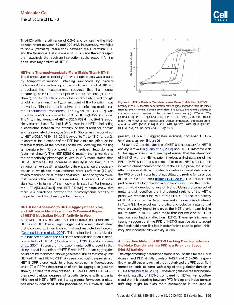
Figure 4. HET-s Protein Constructs Are More Stable than HET-S
Overlay of the CD thermal denaturation profiles (gray lines) and their fits (black
lines) for the N-terminal domain constructs. The arrows indicate the effects of
the mutations or changes in the domain boundaries: (1) HET-s/HET-s
[D23A,P33H]; (2) HET-s[D23A,P33H] (1–227)/(13–221); (3) HET-S/HET-S
[E86K]. From low to high thermal denaturation temperature, the traces corre-
spond to HET-s[D23A,P33H](13-221), HET-S(1-227), HET-S[E86K](1-227),
HET-s[D23A,P33H](1-227), and HET-s(1-227).
HET-s Is Thermodynamically More Stable Than HET-SThe thermodynamic stability of several constructs was probed
by temperature-induced unfolding monitored by circular
dichroism (CD) spectroscopy. The isodichroic point at 201 nm
throughout the measurements suggests that the thermal
denaturing of HET-s is a simple two-state process (data not
shown), and for all of the constructs tested, we observed a single
unfolding transition. The Tm, or midpoint of the transition, was
derived by fitting the data to a two-state unfolding model (see
the Experimental Procedures). The Tm for HET-S(1–227) was
found to be 48�C compared to 57�C for HET-s(1–227) (Figure 4).
The N-terminal domain of HET-s[D23A,P33H], the [Het-S] spec-
ificity mutant, has a Tm that is 5�C lower than HET-s, indicating
a correlation between the stability of the N-terminal domain
and its associated phenotype (arrow 1). Shortening the construct
to HET-s[D23A,P33H](13–221) lowered its Tm to 43�C (arrow 2).
In contrast, the presence of the PFD has a minimal effect on the
thermal stability of the protein constructs, lowering the melting
temperature by 1�C compared to the isolated HeLo domains
(data not shown). The HET-S[E86K] mutant that gives rise to
the compatibility phenotype in vivo is 3�C more stable than
HET-S (arrow 3). This increase in stability is not likely due to
a monomer versus dimer stability difference, since the concen-
tration at which the measurements were performed (10 mM)
favors monomer for all of the constructs. These analyses reveal
that in spite of their structural similarities, HET-s and HET-S differ
significantly in terms of thermodynamic stability. Furthermore,
the HET-s[D23A,P33H] and HET-S[E86K] mutants show that
there is a correlation between the thermodynamic stability of
the protein and the phenotype that it exerts.
HET-S Can Associate to HET-s Aggregates In Vivo,and b-Breaker Mutations in the C-Terminal Regionof HET-S Neutralize [Het-S] Activity In VivoA previous study showed that constitutive coexpression of
HET-s and HET-S in a single fungus led to a metastable strain
that displayed at times both normal and restricted cell growth
(Coustou-Linares et al., 2001). This instability is probably due
to a balance between the cell death reaction and prion elimina-
tion activity of HET-S (Coustou et al., 1999; Coustou-Linares
et al., 2001). Because of the experimental setting used in that
study, direct interaction of HET-S with HET-s prion aggregates
could not be monitored, so we generated strains that coexpress
HET-s-RFP and HET-S-GFP. As seen previously, expression of
HET-S-GFP alone leads to diffuse cytoplasmic fluorescence
and HET-s-RFP to the formation of dot-like aggregates (data not
shown). Strains that coexpressed HET-s-RFP and HET-S-GFP
displayed various degrees of growth defects with a partial
inhibition of HET-s-RFP dot-like aggregate formation, a situa-
tion already described in the previous study. However, where
M
present, HET-s-RFP aggregates invariably contained HET-S-
GFP signal as well (Figure 5).
Since the C-terminal domain of HET-S is necessary for HET-S
activity in vivo (Balguerie et al., 2003) and HET-S interacts with
HET-s aggregates in vivo, we hypothesized that the interaction
of HET-S with the HET-s prion involves a b-structuring of the
PFD of HET-S into the b-solenoid fold of the HET-s fibril. In the
initial structural characterization of the HET-s prion, the in vivo
effect of several HET-s constructs containing small deletions in
the PFD or point mutants that substituted a proline for a residue
of the PFD were tested (Ritter et al., 2005). The deletion and
proline mutants that resided in and hence disrupted the b-sole-
noid amyloid core led to loss of [Het-s]. Using the same set of
mutants that identified the b-structured regions of the HET-s
prion, we examined the role of the HET-S PFD on the activity
of HET-S in P. anserina. As summarized in Figure S8 and detailed
in Table S2, the exact same proline and deletion mutants that
were previously found to disrupt the HET-s prion also led to
null mutants in HET-S while those that did not disrupt HET-s
function also had no affect on HET-S. These genetic results
strongly suggest that the PFD of HET-S must be able to adopt
the b-solenoid prion-like fold in order for it to exert its prion-inhib-
itory and incompatibility activity in vivo.
An Insertion Mutant of HET-S Lacking Overlap betweenthe HeLo Domain and the PFD Is a Prion and Loses[Het-S] ActivityThe experimentally determined domain boundaries for the HeLo
domain and PFD slightly overlap (1–227 and 218–289, respec-
tively), and it was shown that the structuring of the PFD upon fibril
formation leads to some unfolding of the globular domain in
HET-s (Wasmer et al., 2009). Considering the decreased thermo-
dynamic stability of HET-S compared to HET-s, we hypothe-
sized that this coupling between PFD folding and HeLo domain
unfolding might be even more pronounced in the case of
olecular Cell 38, 889–899, June 25, 2010 ª2010 Elsevier Inc. 895

Figure 5. HET-S Can Associate with HET-s
Aggregates In Vivo
A P. anserina strain coexpressing HET-S-GFP and
HET-s-RFP was analyzed by fluorescence micros-
copy. The hyphae of the transformants displayed
various degrees of growth alteration and in the
three fields shown, strong vacuolisation and
formation of lipid droplets, the hallmarks of incom-
patibility. Where HET-s-RFP aggregates are
visible, HET-S-GFP signal is also detected. Scale
bar is 4 mm.
Molecular Cell
The Structure of HET-S
HET-S and be directly involved in the prion-inhibitory effect of the
HET-S HeLo domain. We thus set out to uncouple the HeLo
domain from the PFD b-solenoid conformation (i.e., fibril forma-
tion) by removing the overlap between them. The result is
a construct designated HET-S(1-227)(218-289) that corresponds to
the HET-S globular domain (1–227) fused to the PFD region
(218–289) with a six amino acid linker between the two domains.
A plasmid expressing the HET-S(1-227)(218-289) construct was
introduced in a Dhet-s strain, and transformants were tested
for their ability to be infectious (propagate [Het-s]) and for their
compatibility with het-s and het-S strains. Strains expressing
HET-S (1-227)(218-289) were compatible with [Het-s] strains,
meaning that they have lost HET-S activity. After confrontation
with [Het-s] strains, the same transformants became capable
of subsequently converting [Het-s*] strains to the [Het-s] pheno-
type. In other words, the HET-S(1-227)(218-289) construct is capable
of prion propagation. A small fraction of the transformants
produced an attenuated incompatibility reaction with [Het-S]
strains, indicating that HET-S(1-227)(218-289) has some but not all
the activity of HET-s (Table S3). Taken together, these results
suggest that when the globular domain of HET-S becomes struc-
turally uncoupled from the C-terminal PFD, its prion-inhibitory
activity is abolished.
DISCUSSION
The Prion Inhibition Mechanism of the HeLo Domainof HET-SIn the original description of the [Het-s] system it was found that
HET-S has the ability to inactivate [Het-s] (Rizet, 1952). The
[Het-s*] state was first obtained by curing of the prion by interac-
tion with a [Het-S] strain, and the original nomenclature of the
[Het-s*] state was actually sS to emphasize that the infectious
element had been modified/inactivated by HET-S. Later, it was
shown that this prion-inhibitory effect is also exerted in cis, so
that the HET-S globular domain inhibits the PFD present in the
HET-S protein (Balguerie et al., 2003). The question arises as
to how the HeLo domain of HET-S can exert this prion-inhibitory
effect. We show that in vitro, HET-S fails to form well-ordered
fibrils and inhibits fibrillization of the HET-s PFD, hence providing
a direct illustration of this prion-inhibitory effect. We could find no
evidence of an interaction between the HeLo domain and the
unstructured, flexible PFD in HET-S. Furthermore, single-point
mutations that switch the phenotype in both directions (Coustou
896 Molecular Cell 38, 889–899, June 25, 2010 ª2010 Elsevier Inc.
et al., 1999) are distributed all over the 3D structure of the
N-terminal domain (Figure 2A), making it difficult to imagine
how a specific binding epitope for the PFD exists on HET-S
that is absent on HET-s.
We did find that one striking difference between HET-s and
HET-S HeLo domains resides in their thermodynamic stability.
We also show that HET-S activity relies on the ability of its PFD
to adopt the b-solenoid fold characteristic of the HET-s prion.
Based on our new data, and the HET-s literature to date, we
propose a model for the prion regulation by HET-S that explains
both the cis and trans prion inhibition (Figure 6). Since the aggre-
gation of the HET-s PFD into fibrils displays the complex kinetics
of a seeded polymerization (Sabate et al., 2009), the formation
of a seed aggregate that will subsequently lead to a mature fibril
is a necessary first step in the aggregation process. In the
proposed model, the acquisition of the b-solenoid fibril fold by
the PFD in HET-s does not grossly destabilize its HeLo domain,
whereas in HET-S the spontaneous (cis) or HET-s fibril-induced
(trans) b-solenoid folding of the HET-S PFD leads to a more
extensive structural rearrangement of the HET-S HeLo domain
that renders the seed or fibril sterile for further fibril growth.
Although we do not know the precise nature of the change that
is induced in the HET-S HeLo domain, it likely involves a degree
of unfolding (Wasmer et al., 2009), perhaps exposing some
prion-inhibitory and/or cytotoxic elements.
We have shown by uncoupling the PFD from the HeLo domain
that their overlap is essential to the in vivo properties of HET-S
(Table S3). The reported loss of structure in the fibrils of HET-s
(Wasmer et al., 2009) is particularly evident for the residues in
the last three helices (a7–a9), the same for which we find
increased residual B factors in HET-S. In addition to its lower
stability, the HET-S HeLo domain is more aggregation prone.
Under moderate agitation, HET-S(1–227) precipitates at room
temperature within a few hours, while HET-s(1–227) remains in
solution under the same condition for more than a week (data
not shown). Furthermore, three independent algorithms that pre-
dict b-aggregation prone regions in HET-s and HET-S (see the
Experimental Procedures) gave high scores for several regions
within the HeLo domains of both proteins. Interestingly, the
consensus predictions cluster at the dimer interface (Figure 2B).
The forced proximity of HeLo domains in the b-solenoid seeds or
fibrils would support the formation of the low-affinity (�80 mM)
HET-S dimer, thus juxtaposing aggregation-prone regions that
could facilitate an aggregation of the HeLo domains. Either the

Figure 6. Proposed Model for HET-S Fibril
Inhibition and Induction of Toxicity
(1) The HET-s fibril (formed from alternating light/
dark gray HET-s monomers) is approached by
a soluble HET-S (rainbow HeLo domain with
random coil PFD in black). (2) Upon addition of
HET-S to the HET-s fibril, the terminal helices of
its HeLo domain unwind to accommodate the
prion fold. (3) The second addition of a HET-S
monomer to the fibril leads to a dimerization of
the HeLo domains which facilitates a re/unfolding
of the HeLo domains with the potential for further
aggregation of HeLo domains that (in 4) blocks
further growth of the fibril and activates a toxic
form of HET-S. The model for the HET-s fibril
was created from the PFD fibril structure (Wasmer
et al., 2008) and the HeLo domain with an
unwinding the last three helices of the HeLo
domain (residues 177–222) to make space for
the HeLo domains around the fibril. The HET-s
HeLo domains are depicted as dimers between
adjacent monomers in the fibril, but these are
speculative and it should be emphasized that the
structures of the HeLo domains of HET-s and
HET-S, in the context of a fibril, are not known
except that HET-s suffers a loss of tertiary struc-
ture (more molten globule-like), with a local loss
of secondary structure around residues 190–220
(Wasmer et al., 2009).
Molecular Cell
The Structure of HET-S
fibril-induced dimer or the resulting aggregates could interfere
with further fibrillar aggregation via the PFDs. Since we cannot
rule out that to some extent the HeLo domain refolds in order to
attain its functions, we refer to the change induced in the
HET-S HeLo domain by its PFD structuring as a ‘‘re/unfolding.’’
Our model depicted in Figure 6 distills these findings into an
inhibition mechanism in which the lower thermodynamic stability
and higher aggregation propensity of the HET-S HeLo domain
are key to its prion regulation mechanism: when its PFD acquires
the b-solenoid fold, either through the kinetically slower process
of spontaneous seed formation or upon binding to the b-solenoid
fibrils of HET-s, it re/unfolds to yield ‘‘seedless aggregates’’ that
are incompetent for continued fibrillar growth. This model further
explains why the HET-S mutations that prevent formation of the
b-solenoid fold in its PFD abolish HET-S activity. The re/unfold-
ing of the HeLo domain is not simply a consequence of the
b-folding of the PFD but is due to a structural overlap between
the two domains.
Potential Role of the HET-S Dimer and Its Aggregationin Heterokaryon IncompatibilityThe heterokaryon incompatibility reaction of the HET-s/S system
involves a spatially restricted PCD that requires the presence of
both HET-s fibrils and soluble HET-S, while no reaction occurs in
presence of soluble HET-s and HET-S (Coustou et al., 1997).
This suggests that either (1) a poisonous HET-S entity is induced
by HET-s fibrils or that (2) a poisonous complex forms between
HET-S and HET-s fibrils. The prion inhibition model proposed
above supports these mechanisms of toxicity: (1) the HET-s
M
fibrils induce a partial unfolding of HET-S by binding to and struc-
turing its PFD domain, thus generating a toxic HET-S entity, or (2)
the ability of HET-S to prevent the growth of HET-s fibrils
promotes the formation of a toxic oligomeric species similar to
other amyloid oligomers in neurodegenerative diseases (Glabe,
2006).
Although the area of the dimer interface is not enormous, its
size is consistent with protein-protein interaction surfaces, and
it is predicted to be a stable interface in solution by the PISA
algorithms (Krissinel and Henrick, 2007). We find that two
different interface mutants that disrupt the in vitro dimer also
disrupt the in vivo functions of HET-S. We have also shown
that the E86K mutant loses the prion-inhibitory function of
HET-S in vitro (Figure 1B, Table S1). Thus it appears that the
interface could be important for both toxicity and prion inhibition.
It is tempting to say that the dimer interface also plays a role as
an aggregation or re/unfolding facilitator in the prion inhibition
mechanism discussed above (Figure 6); however, the dimeriza-
tion and the re/unfolding are not linked experimentally and are
rather two possible explanations for the properties of HET-S.
In the simplest model, the re/unfolded HeLo aggregate is the
toxic entity in the incompatibility reaction. Such a mechanism
of toxicity ensures that HET-S alone cannot form sufficient
aggregates to be toxic because its physiological concentration
precludes dimer formation and it cannot make its own unfolding
machinery (i.e., the HET-s fibril). If the HeLo domain actually is
a fungal cell death-inducing domain, then the b-folding of its
associated PFD would also represent the trigger for its activa-
tion. In this scenario, the two biological properties of HET-S (its
olecular Cell 38, 889–899, June 25, 2010 ª2010 Elsevier Inc. 897

Molecular Cell
The Structure of HET-S
ability to induce cell death upon interaction with the HET-s prion
and its ability to cure [Het-s]) would be based on the same mech-
anistic principle involving a structural alteration of its HeLo
domain. We currently believe this to be the case, but a conclusive
answer is the topic of further study in our lab.
ConclusionPrions have the unique ability to encode their function as well as
their replication features within their 3D structures. HET-s adds
to the unusual repertoire of prions in that in addition to its
nontoxic monomeric and propagative structural entities, there
are prion-regulatory and toxic structural entities, all of which
are encoded in two highly homologous proteins HET-S and
HET-s. While the 3D structure of the HET-s(218–289) fibrils
shed light on the mechanism of prion formation and propagation
(Wasmer et al., 2008; Ritter et al., 2005), the presented 3D struc-
tures of both soluble HET-S and HET-s indicate that the mecha-
nism of prion regulation is coded in the stability of the N-terminal
domain rather than in its structure, while the generation of
toxicity may be based on an activation of the HeLo domain trig-
gered by b-folding of the C-terminal PFD.
EXPERIMENTAL PROCEDURES
HET-s Constructs and Expression
All of the proteins were expressed as either N-terminal 6-histidine tag (N6His)
fusions with a thrombin cleavage site or as C-terminal (C6His) fusions with no
intervening sequence, and expressions were carried out in E. coli strain BL21-
DE3(pLysS) grown in M9 minimal medium. The details of the expression and
purification are in the Supplemental Experimental Procedures.
Mass Analysis by GFC-MALS
The pure proteins in gel filtration buffer were concentrated with centrifugal
ultrafiltration devices to�2.3 mM and then serially 23 diluted to make a series
that extended to �80 mM. The protein solutions were separated by size with
a G2000SWXL gel filtration column (Tosoh Bioscience) in 20 mM Na2HPO4/
KH2PO4 (pH 7.0), 200 mM NaCl on an Agilent 1200 series HPLC system in-
jected by the autosampler (20 ml) at a flow rate of 0.5 ml/min. The column eluate
was simultaneously monitored by the 1200 series diode array detector,
a TREOS light-scattering detector (Wyatt Technology), and the 1200 series
refractive index detector. The signals from the detectors were analyzed by
the Astra V program (Wyatt Technology) using either the calculated extinction
coefficients or an average protein dn/dc value of 0.187 ml/g. Both concentra-
tion measurements (UV or RI) gave similar calculated masses.
Crystallization, Structure Determination, and Refinement
Crystals were first obtained for HET-s(1–227) without a histidine tag in 0.2 M
(NH4)2SO4, 0.1 M Tris (pH 8.5), 30% PEG 4000 in the spacegroup p3121,
and initial phases were obtained to 3.5 A by SAD and MAD techniques using
SHARP (de la Fortelle and Bricogne, 1997) with mercury and iodine derivative
data sets (single wavelength collected on beamline 7.2 at SSRL) in combina-
tion with a three-wavelength data set from a bromine-soaked crystal (beamline
8.2.1 at ALS). The highest-resolution native data obtained with these crystals
were to 3.0 A, and so we searched for other crystal forms. The statistics on the
native data used in the final refinements are in Table 1.
The details of the data collection and refinement are in the Supplemental
Experimental Procedures. All of the structure figures were made with
CCP4MG (Potterton et al., 2002).
Circular Dichroism Spectroscopy
Concentrated proteins were diluted to 10 mM in 50 mM NaPO4 (pH 8.0) for anal-
ysis on a Jasco J-815 CD spectrometer. Far UV CD spectra (190–240 nm) were
measured at 20�C with a 20 nm/min scan rate in a 0.1 cm path-length cuvette.
898 Molecular Cell 38, 889–899, June 25, 2010 ª2010 Elsevier Inc.
The thermal denaturation measurements were performed at 0.5�C/min from
20�C to 70�C while monitoring the CD signal at 225 nm. The data were fit by
non-linear regression with Grace (http://plasma-gate.weizmann.ac.il/Grace/)
to a two-state transition with the equation
yobs =ðmnT + bnÞ+ ðmdT + bdÞfexp½DHm=R,ð1=Tm � 1=TÞ�g
f1 + exp½DHm=R,ð1=Tm � 1=TÞ�g
in which yobs is the CD signal at 225 nm, T is the temperature in Kelvin, R is
the gas constant, Tm is the midpoint of the unfolding transition in Kelvin, and
DHm is the enthalpy of unfolding at this temperature (Swint and Robertson,
1993). The m and b terms with the n and d subscripts are the slopes and y inter-
cepts of the baselines for the native and denatured states, respectively.
The NMR data collection, chemical shift assignment, and PFD aggregation
kinetics are described in the Supplemental Experimental Procedures.
Aggregation Prediction Algorithms
We chose three aggregation prediction algorithms that use different prediction
methods and applied them to the sequences of HET-s and HET-S. Significant
score levels were determined according to the authors’ recommendations,
and the consensus regions were mapped to the structure of HET-S and visu-
alized with CCP4MG. The three programs used were TANGO (Fernandez-
Escamilla et al., 2004), PASTA (Trovato et al., 2007), and SALSA (Zibaee
et al., 2007).
HET-S(1-227)(218-289) and PFD Mutant Analysis
All site-directed het-S mutants were generated using the QuikChange Site-
Directed Mutagenesis kit (Stratagene). Mutations were introduced into
pCB1004-het-S or pGPD-het-S-GFP (Coustou-Linares et al., 2001). The
details of the HET-S(1-227)(218-289) construction and design are in the Supple-
mental Experimental Procedures. The properties of the het-S mutant alleles
were determined by transforming the mutated plasmid bearing the hygR
marker into a Dhet-s strain (Turcq et al., 1991). Transformants were selected
and each transformant was confronted on solid medium with a wild-type
[Het-s] strain in barrage tests.
In Vivo Localization of HET-s and HET-S
A Dhet-s strain was cotransformed with the pGPD-het-S-GFP (Coustou-
Linares et al., 2001) and pGPD-het-s-RFP plasmids. Transformants were
selected and analyzed by fluorescence microscopy. For fluorescence micros-
copy, synthetic medium containing 2% (wt/vol) agarose was used. P. anserina
hyphae were inoculated on this medium and cultivated for 16–24 hr at 26�C. The
mycelium was examined in situ with a Leica DMRXA microscope equipped with
a Micromax CCD (Princeton Instruments) controlled by the Metamorph
5.06 software (Roper Scientific). The microscope was fitted with a standard
FITC filter set (Leica L4) and a Leica PL APO 1003 immersion objective.
ACCESSION NUMBERS
Structure factors and coordinates were deposited in the Protein Data Bank
(see Table 1).
SUPPLEMENTAL INFORMATION
Supplemental Information includes Supplemental Experimental Procedures,
eight figures, three tables, and Supplemental References and can be found
with this article at doi:10.1016/j.molcel.2010.05.019.
ACKNOWLEDGMENTS
This work was supported by grants from the Schweizerische Nationalfonds
(SNF) and the National Institutes of Health (NIH). S.J.S. was supported by an
Agence Nationale de la Recherche (ANR) grant ‘‘PANPRIONDRUG.’’ Portions
of this research were carried out at Stanford Synchrotron Radiation Light-
source (SSRL), supported by the Department of Energy (DOE), Office of Bio-
logical and Environmental Research, the NIH, National Center for Research
Resources, Biomedical Technology Program, and the National Institute of

Molecular Cell
The Structure of HET-S
General Medical Sciences. The Advanced Light Source (ALS) is supported by
the DOE, Office of Basic Energy Sciences, under Contract No. DE-AC02-
05CH11231. The expertise of Alexander Sobol was of valuable assistance
and is gratefully acknowledged.
Received: November 26, 2009
Revised: February 26, 2010
Accepted: April 15, 2010
Published: June 24, 2010
REFERENCES
Aguzzi, A., Sigurdson, C., and Heikenwaelder, M. (2008). Molecular mecha-
nisms of prion pathogenesis. Annu. Rev. Pathol. 3, 11–40.
Balguerie, A., Dos Reis, S., Ritter, C., Chaignepain, S., Coulary-Salin, B.,
Forge, V., Bathany, K., Lascu, I., Schmitter, J.M., Riek, R., and Saupe, S.J.
(2003). Domain organization and structure-function relationship of the HET-s
prion protein of Podospora anserina. EMBO J. 22, 2071–2081.
Balguerie, A., Dos Reis, S., Coulary-Salin, B., Chaignepain, S., Sabourin, M.,
Schmitter, J.M., and Saupe, S.J. (2004). The sequences appended to the
amyloid core region of the HET-s prion protein determine higher-order
aggregate organization in vivo. J. Cell Sci. 117, 2599–2610.
Beisson-Schecroun, J. (1962). Incompatibilite cellulaire et interactions nucleo-
cytoplamsiques dans les phenomenes de barrage chez le Podospora anser-
ina. Ann. Genet. 4, 3–50.
Coustou, V., Deleu, C., Saupe, S., and Begueret, J. (1997). The protein product
of the het-s heterokaryon incompatibility gene of the fungus Podospora anser-
ina behaves as a prion analog. Proc. Natl. Acad. Sci. USA 94, 9773–9778.
Coustou, V., Deleu, C., Saupe, S.J., and Begueret, J. (1999). Mutational anal-
ysis of the [Het-s] prion analog of Podospora anserina. A short N-terminal
peptide allows prion propagation. Genetics 153, 1629–1640.
Coustou-Linares, V., Maddelein, M.L., Begueret, J., and Saupe, S.J. (2001). In
vivo aggregation of the HET-s prion protein of the fungus Podospora anserina.
Mol. Microbiol. 42, 1325–1335.
Dalstra, H.J., Swart, K., Debets, A.J., Saupe, S.J., and Hoekstra, R.F. (2003).
Sexual transmission of the [Het-S] prion leads to meiotic drive in Podospora
anserina. Proc. Natl. Acad. Sci. USA 100, 6616–6621.
Dehay, B., and Bertolotti, A. (2006). Critical role of the proline-rich region in
Huntingtin for aggregation and cytotoxicity in yeast. J. Biol. Chem. 281,
35608–35615.
de la Fortelle, E., and Bricogne, G. (1997). Maximum-Likelihood Heavy-Atom
Parameter Refinement for the Multiple Isomorphous Replacement and
Multiwavelength Anomalous Diffraction Methods, Volume 276 (New York:
Academic Press).
Deleu, C., Clave, C., and Begueret, J. (1993). A single amino acid difference is
sufficient to elicit vegetative incompatibility in the fungus Podospora anserina.
Genetics 135, 45–52.
Diederichs, K., and Karplus, P.A. (1997). Improved R-factors for diffraction
data analysis in macromolecular crystallography. Nat. Struct. Biol. 4, 269–275.
Duennwald, M.L., Jagadish, S., Muchowski, P.J., and Lindquist, S. (2006).
Flanking sequences profoundly alter polyglutamine toxicity in yeast. Proc.
Natl. Acad. Sci. USA 103, 11045–11050.
Engh, R.A., and Huber, R. (1991). Accurate bond and angle parameters for
X-ray protein structure refinement. Acta Crystallogr. A 47, 392–400.
Fernandez-Bellot, E., Guillemet, E., and Cullin, C. (2000). The yeast prion
[URE3] can be greatly induced by a functional mutated URE2 allele. EMBO
J. 19, 3215–3222.
Fernandez-Escamilla, A.M., Rousseau, F., Schymkowitz, J., and Serrano, L.
(2004). Prediction of sequence-dependent and mutational effects on the
aggregation of peptides and proteins. Nat. Biotechnol. 22, 1302–1306.
M
Glabe, C.G. (2006). Common mechanisms of amyloid oligomer pathogenesis
in degenerative disease. Neurobiol. Aging 27, 570–575.
Glass, N.L., Jacobson, D.J., and Shiu, P.K. (2000). The genetics of hyphal
fusion and vegetative incompatibility in filamentous ascomycete fungi. Annu.
Rev. Genet. 34, 165–186.
Kay, L.E., Torchia, D.A., and Bax, A. (1989). Backbone dynamics of proteins as
studied by 15N inverse detected heteronuclear NMR spectroscopy: applica-
tion to staphylococcal nuclease. Biochemistry 28, 8972–8979.
Krissinel, E., and Henrick, K. (2007). Inference of macromolecular assemblies
from crystalline state. J. Mol. Biol. 372, 774–797.
Liu, J.J., Sondheimer, N., and Lindquist, S.L. (2002). Changes in the middle
region of Sup35 profoundly alter the nature of epigenetic inheritance for the
yeast prion. Proc. Natl. Acad. Sci. USA 99 (Suppl 4), 16446–16453.
Maddelein, M.L., and Wickner, R.B. (1999). Two prion-inducing regions of
Ure2p are nonoverlapping. Mol. Cell. Biol. 19, 4516–4524.
Masison, D.C., and Wickner, R.B. (1995). Prion-inducing domain of yeast
Ure2p and protease resistance of Ure2p in prion-containing cells. Science
270, 93–95.
Mayer, M., and Meyer, B. (2001). Group epitope mapping by saturation trans-
fer difference NMR to identify segments of a ligand in direct contact with
a protein receptor. J. Am. Chem. Soc. 123, 6108–6117.
Paoletti, M., and Clave, C. (2007). The fungus-specific HET domain mediates
programmed cell death in Podospora anserina. Eukaryot. Cell 6, 2001–2008.
Potterton, E., McNicholas, S., Krissinel, E., Cowtan, K., and Noble, M. (2002).
The CCP4 molecular-graphics project. Acta Crystallogr. D Biol. Crystallogr.
58, 1955–1957.
Ritter, C., Maddelein, M.L., Siemer, A.B., Luhrs, T., Ernst, M., Meier, B.H.,
Saupe, S.J., and Riek, R. (2005). Correlation of structural elements and infec-
tivity of the HET-s prion. Nature 435, 844–848.
Rizet, G. (1952). Les phenomenes de barrage chez Podospora anserina. I.
Analyse de barrage entre les souches s et S. Rev. Cytol. Biol. Veg. 13, 51–92.
Sabate, R., Castillo, V., Espargaro, A., Saupe, S.J., and Ventura, S. (2009).
Energy barriers for HET-s prion forming domain amyloid formation. FEBS J.
276, 5053–5064.
Saupe, S.J. (2000). Molecular genetics of heterokaryon incompatibility in
filamentous ascomycetes. Microbiol. Mol. Biol. Rev. 64, 489–502.
Swint, L., and Robertson, A.D. (1993). Thermodynamics of unfolding for turkey
ovomucoid third domain: thermal and chemical denaturation. Protein Sci. 2,
2037–2049.
Trovato, A., Seno, F., and Tosatto, S.C. (2007). The PASTA server for protein
aggregation prediction. Protein Eng. Des. Sel. 20, 521–523.
Turcq, B., Deleu, C., Denayrolles, M., and Begueret, J. (1991). Two allelic
genes responsible for vegetative incompatibility in the fungus Podospora
anserina are not essential for cell viability. Mol. Gen. Genet. 228, 265–269.
Wasmer, C., Lange, A., Van Melckebeke, H., Siemer, A.B., Riek, R., and Meier,
B.H. (2008). Amyloid fibrils of the HET-s(218-289) prion form a beta solenoid
with a triangular hydrophobic core. Science 319, 1523–1526.
Wasmer, C., Schutz, A., Loquet, A., Buhtz, C., Greenwald, J., Riek, R.,
Bockmann, A., and Meier, B.H. (2009). The molecular organization of the
fungal prion HET-s in its amyloid form. J. Mol. Biol. 394, 119–127.
Wickner, R.B., Shewmaker, F., Kryndushkin, D., and Edskes, H.K. (2008).
Protein inheritance (prions) based on parallel in-register beta-sheet amyloid
structures. Bioessays 30, 955–964.
Zibaee, S., Makin, O.S., Goedert, M., and Serpell, L.C. (2007). A simple algo-
rithm locates beta-strands in the amyloid fibril core of alpha-synuclein, Abeta,
and tau using the amino acid sequence alone. Protein Sci. 16, 906–918.
olecular Cell 38, 889–899, June 25, 2010 ª2010 Elsevier Inc. 899