Targeting of the c-Abl Tyrosine Kinase to Mitochondria in Endoplasmic Reticulum Stress-Induced...
10
MOLECULAR AND CELLULAR BIOLOGY, 0270-7306/01/$04.0010 DOI: 10.1128/MCB.21.18.6233–6242.2001 Sept. 2001, p. 6233–6242 Vol. 21, No. 18 Copyright © 2001, American Society for Microbiology. All Rights Reserved. Targeting of the c-Abl Tyrosine Kinase to Mitochondria in Endoplasmic Reticulum Stress-Induced Apoptosis YASUMASA ITO, 1 PRAMOD PANDEY, 1 NEERAD MISHRA, 2 SHAILENDRA KUMAR, 1 NAVNEET NARULA, 3 SURENDER KHARBANDA, 1 SATYA SAXENA, 2 AND DONALD KUFE 1 * Dana-Farber Cancer Institute, Harvard Medical School, Boston, Massachusetts 02115 1 ; Lovelace Respiratory Research Institute, Albuquerque, New Mexico 87115 2 ; and Department of Pathology and Laboratory Medicine, University of Pennsylvania School of Medicine, Philadelphia, Pennsylvania 19104-4282 3 Received 2 March 2001/Returned for modification 4 April 2001/Accepted 15 June 2001 The ubiquitously expressed c-Abl tyrosine kinase localizes to the nucleus and cytoplasm. Using confocal microscopy, we demonstrated that c-Abl colocalizes with the endoplasmic reticulum (ER)-associated protein grp78. Expression of c-Abl in the ER was confirmed by immunoelectron microscopy. Subcellular fractionation studies further indicate that over 20% of cellular c-Abl is detectable in the ER. The results also demonstrate that induction of ER stress with calcium ionophore A23187, brefeldin A, or tunicamycin is associated with translocation of ER-associated c-Abl to mitochondria. In concert with targeting of c-Abl to mitochondria, cytochrome c is released in the response to ER stress by a c-Abl-dependent mechanism, and ER stress-induced apoptosis is attenuated in c-Abl-deficient cells. These findings indicate that c-Abl is involved in signaling from the ER to mitochondria and thereby the apoptotic response to ER stress. The c-Abl protein tyrosine kinase localizes to the nucleus and cytoplasm. Nuclear c-Abl is activated in the response to DNA damage (16) by the DNA-dependent protein kinase (10, 13) and the product of the gene mutated in ataxia telangiec- tasia (2, 28). Activation of nuclear c-Abl by genotoxic stress contributes to induction of the proapoptotic c-Jun N-terminal kinase/stress-activated protein kinase (JNK/SAPK) and p38 mitogen-activated protein kinase pathways (14–16, 25). Nu- clear c-Abl also contributes to DNA damage-induced apopto- sis by mechanisms in part dependent on the p53 tumor sup- pressor and its homolog p73 (1, 7, 38, 40, 41). Other studies have demonstrated that the cytoplasmic form of c-Abl is acti- vated in the cellular response to oxidative stress (30). Reactive oxygen species induce cytoplasmic c-Abl activity by a mecha- nism dependent on protein kinase Cd (PKCd) (31). Moreover, c-Abl is required for reactive oxygen species-induced release of mitochondrial cytochrome c, caspase-3 activation, and apopto- sis (30). These findings have provided support for the involve- ment of c-Abl in the responses to genotoxic and oxidative stress. The endoplasmic reticulum (ER) functions as an oxidizing compartment for the folding of membrane and secretory pro- teins (11). Accumulation of unfolded intermediates in the ER activates stress signals referred to as the unfolded protein response (5). The IRE1a and IRE1b ER transmembrane pro- tein kinases sense ER stress and activate transcription of genes that encode protein chaperones and other ER-resident pro- teins (33, 36). ER stress also induces activity of the PRK-like ER kinase (PERK), phosphorylation of the eukaryotic initia- tion factor 2 a subunit and inhibition of mRNA translation (8). Treatment of cells with inducers of ER stress, such as the calcium ionophore A23187, is associated with the induction of CHOP/GADD153 expression and apoptosis (3, 21, 26, 27). Other studies have demonstrated that caspase-12 is activated by ER stress and that caspase-12 contributes to ER stress- induced apoptosis (24). The present studies show that the c-Abl kinase localizes to the ER and is targeted to mitochondria by ER stress. The results also demonstrate that ER stress induces mitochondrial cytochrome c release and apoptosis by a c-Abl-dependent mechanism. MATERIALS AND METHODS Cell culture. Rat1 cells and wild-type, Abl 2/2 , and Abl 1 (Abl 2/2 cells recon- stituted to stably express c-Abl) mouse embryo fibroblasts (MEFs) (16, 19, 34) were cultured in Dulbecco’s modified Eagle’s medium containing 10% heat- inactivated fetal calf serum, 2 mM L-glutamine, 100 U of penicillin per ml, and 100 mg of streptomycin per ml. Cells were treated with A23187, brefeldin A, or tunicamycin (all from Sigma). Digital confocal immunofluorescence microscopy. Cells grown on poly-D-ly- sine-coated glass coverslips were fixed (3.7% formaldehyde in phosphate-buff- ered saline [PBS], pH 7.4; 10 min), permeabilized (0.2% Triton X-100; 10 min), and blocked for 30 min in medium containing serum. After rinsing with PBS, immunostaining was performed by incubating the cells with 50 ng of anti-c-Abl (K-12 rabbit polyclonal; Santa Cruz) and anti-grp-78 (C-20 goat polyclonal; Santa Cruz) per slide. After being washed with PBS, cells were incubated with a 1:250 dilution of CY-3 or fluorescein isothiocyanate-conjugated anti-rabbit or anti-goat immunoglobulin G (IgG) secondary antibodies (Jackson ImmunoRe- search) for 1 h. Mitochondria were stained with 0.006 ng of Mitotracker Green FM (Molecular Probes) per slide. Nuclei were stained with 4, 6-diamino-2- phenylindole (DAPI; 1 mg/ml in PBS). Coverslips were mounted onto slides with 0.1 M Tris (pH 7.0) in 50% glycerol. Cells were visualized by digital confocal immunofluorescence, and images were captured with a cooled charge-coupled device camera mounted on a Zeiss Axioplan 2 microscope. Images were decon- volved using Slidebook software (Intelligent Imaging Innovations, Inc., Denver, Colo.). Immunoelectron microscopic analysis. Cells were fixed with 2% paraformal- dehyde in 0.1 M sodium cacodylate buffer for 10 min, washed with three changes of cacodylate buffer, postfixed with 1% osmium tetroxide for 5 min, dehydrated in graded ethanol, and infiltrated and polymerized with Poly/bed 812 overnight. Ultrathin sections were cut with an ultramicrotome (Nova; Leica). After etching with sodium periodate for 10 min, the sections were rinsed with buffer and incubated with anti-c-Abl at a dilution of 1:10 overnight at 4°C. The sections were * Corresponding author. Mailing address: Dana-Farber Cancer Institute, Harvard Medical School, Boston, MA 02115. Phone: (617) 632-3141. Fax: (617) 632-2934. E-mail: Donald_Kufe@dfci .harvard.edu. 6233 on December 22, 2014 by guest http://mcb.asm.org/ Downloaded from
-
Upload
independent -
Category
Documents
-
view
1 -
download
0
Transcript of Targeting of the c-Abl Tyrosine Kinase to Mitochondria in Endoplasmic Reticulum Stress-Induced...

MOLECULAR AND CELLULAR BIOLOGY,0270-7306/01/$04.0010 DOI: 10.1128/MCB.21.18.6233–6242.2001
Sept. 2001, p. 6233–6242 Vol. 21, No. 18
Copyright © 2001, American Society for Microbiology. All Rights Reserved.
Targeting of the c-Abl Tyrosine Kinase to Mitochondria inEndoplasmic Reticulum Stress-Induced Apoptosis
YASUMASA ITO,1 PRAMOD PANDEY,1 NEERAD MISHRA,2 SHAILENDRA KUMAR,1 NAVNEET NARULA,3
SURENDER KHARBANDA,1 SATYA SAXENA,2 AND DONALD KUFE1*
Dana-Farber Cancer Institute, Harvard Medical School, Boston, Massachusetts 021151; Lovelace Respiratory Research Institute,Albuquerque, New Mexico 871152; and Department of Pathology and Laboratory Medicine, University of Pennsylvania
School of Medicine, Philadelphia, Pennsylvania 19104-42823
Received 2 March 2001/Returned for modification 4 April 2001/Accepted 15 June 2001
The ubiquitously expressed c-Abl tyrosine kinase localizes to the nucleus and cytoplasm. Using confocalmicroscopy, we demonstrated that c-Abl colocalizes with the endoplasmic reticulum (ER)-associated proteingrp78. Expression of c-Abl in the ER was confirmed by immunoelectron microscopy. Subcellular fractionationstudies further indicate that over 20% of cellular c-Abl is detectable in the ER. The results also demonstratethat induction of ER stress with calcium ionophore A23187, brefeldin A, or tunicamycin is associated withtranslocation of ER-associated c-Abl to mitochondria. In concert with targeting of c-Abl to mitochondria,cytochrome c is released in the response to ER stress by a c-Abl-dependent mechanism, and ER stress-inducedapoptosis is attenuated in c-Abl-deficient cells. These findings indicate that c-Abl is involved in signaling fromthe ER to mitochondria and thereby the apoptotic response to ER stress.
The c-Abl protein tyrosine kinase localizes to the nucleusand cytoplasm. Nuclear c-Abl is activated in the response toDNA damage (16) by the DNA-dependent protein kinase (10,13) and the product of the gene mutated in ataxia telangiec-tasia (2, 28). Activation of nuclear c-Abl by genotoxic stresscontributes to induction of the proapoptotic c-Jun N-terminalkinase/stress-activated protein kinase (JNK/SAPK) and p38mitogen-activated protein kinase pathways (14–16, 25). Nu-clear c-Abl also contributes to DNA damage-induced apopto-sis by mechanisms in part dependent on the p53 tumor sup-pressor and its homolog p73 (1, 7, 38, 40, 41). Other studieshave demonstrated that the cytoplasmic form of c-Abl is acti-vated in the cellular response to oxidative stress (30). Reactiveoxygen species induce cytoplasmic c-Abl activity by a mecha-nism dependent on protein kinase Cd (PKCd) (31). Moreover,c-Abl is required for reactive oxygen species-induced release ofmitochondrial cytochrome c, caspase-3 activation, and apopto-sis (30). These findings have provided support for the involve-ment of c-Abl in the responses to genotoxic and oxidativestress.
The endoplasmic reticulum (ER) functions as an oxidizingcompartment for the folding of membrane and secretory pro-teins (11). Accumulation of unfolded intermediates in the ERactivates stress signals referred to as the unfolded proteinresponse (5). The IRE1a and IRE1b ER transmembrane pro-tein kinases sense ER stress and activate transcription of genesthat encode protein chaperones and other ER-resident pro-teins (33, 36). ER stress also induces activity of the PRK-likeER kinase (PERK), phosphorylation of the eukaryotic initia-tion factor 2 a subunit and inhibition of mRNA translation (8).Treatment of cells with inducers of ER stress, such as the
calcium ionophore A23187, is associated with the induction ofCHOP/GADD153 expression and apoptosis (3, 21, 26, 27).Other studies have demonstrated that caspase-12 is activatedby ER stress and that caspase-12 contributes to ER stress-induced apoptosis (24).
The present studies show that the c-Abl kinase localizes tothe ER and is targeted to mitochondria by ER stress. Theresults also demonstrate that ER stress induces mitochondrialcytochrome c release and apoptosis by a c-Abl-dependentmechanism.
MATERIALS AND METHODS
Cell culture. Rat1 cells and wild-type, Abl2/2, and Abl1 (Abl2/2 cells recon-stituted to stably express c-Abl) mouse embryo fibroblasts (MEFs) (16, 19, 34)were cultured in Dulbecco’s modified Eagle’s medium containing 10% heat-inactivated fetal calf serum, 2 mM L-glutamine, 100 U of penicillin per ml, and100 mg of streptomycin per ml. Cells were treated with A23187, brefeldin A, ortunicamycin (all from Sigma).
Digital confocal immunofluorescence microscopy. Cells grown on poly-D-ly-sine-coated glass coverslips were fixed (3.7% formaldehyde in phosphate-buff-ered saline [PBS], pH 7.4; 10 min), permeabilized (0.2% Triton X-100; 10 min),and blocked for 30 min in medium containing serum. After rinsing with PBS,immunostaining was performed by incubating the cells with 50 ng of anti-c-Abl(K-12 rabbit polyclonal; Santa Cruz) and anti-grp-78 (C-20 goat polyclonal;Santa Cruz) per slide. After being washed with PBS, cells were incubated with a1:250 dilution of CY-3 or fluorescein isothiocyanate-conjugated anti-rabbit oranti-goat immunoglobulin G (IgG) secondary antibodies (Jackson ImmunoRe-search) for 1 h. Mitochondria were stained with 0.006 ng of Mitotracker GreenFM (Molecular Probes) per slide. Nuclei were stained with 4, 6-diamino-2-phenylindole (DAPI; 1 mg/ml in PBS). Coverslips were mounted onto slides with0.1 M Tris (pH 7.0) in 50% glycerol. Cells were visualized by digital confocalimmunofluorescence, and images were captured with a cooled charge-coupleddevice camera mounted on a Zeiss Axioplan 2 microscope. Images were decon-volved using Slidebook software (Intelligent Imaging Innovations, Inc., Denver,Colo.).
Immunoelectron microscopic analysis. Cells were fixed with 2% paraformal-dehyde in 0.1 M sodium cacodylate buffer for 10 min, washed with three changesof cacodylate buffer, postfixed with 1% osmium tetroxide for 5 min, dehydratedin graded ethanol, and infiltrated and polymerized with Poly/bed 812 overnight.Ultrathin sections were cut with an ultramicrotome (Nova; Leica). After etchingwith sodium periodate for 10 min, the sections were rinsed with buffer andincubated with anti-c-Abl at a dilution of 1:10 overnight at 4°C. The sections were
* Corresponding author. Mailing address: Dana-Farber CancerInstitute, Harvard Medical School, Boston, MA 02115. Phone:(617) 632-3141. Fax: (617) 632-2934. E-mail: [email protected].
6233
on Decem
ber 22, 2014 by guesthttp://m
cb.asm.org/
Dow
nloaded from

6234 ITO ET AL. MOL. CELL. BIOL.
on Decem
ber 22, 2014 by guesthttp://m
cb.asm.org/
Dow
nloaded from
rinsed with buffer, incubated with protein A-gold (15 nm) for 1 h, rinsed again,and then fixed with 2% glutaradehyde in PBS for 2 min. After air drying, thesections were stained with 25 aqueous uranyl acetate and with 0.5% lead citrate.The sections were examined and photographed using a Hitachi H-600 electronmicroscope (Nessei Sagnyo) at 75 kV.
Isolation of the ER fraction. Cells were washed with PBS, lysed in homoge-nization buffer (50 mM Tris-HC1 [pH 8.0], 1 mM b-mercaptoethanol, 1 mMEDTA, 0.32 M sucrose, and 0.1 mM phenylmethylsulfonyl fluoride), and thencentrifuged at 5,000 3 g for 10 min. The supernatant was collected and centri-fuged at 105,000 3 g for 1 h. The pellet was disrupted in lysis buffer (50 mMTris-HCl [pH 7.5], 150 mM NaCl, 1% NP-40, 1 mM dithiothreitol, 1 mM sodiumorthovanadate, 1 mM phenylmethylsulfonyl fluoride, 10 mM sodium fluoride, 10mg of leuptin and aprotinin/ml) at 4°C and then centrifuged at 15,000 3 g for 20min. The resulting supernatant was used as the ER fraction.
Isolation of cytoplasmic and nuclear fractions. The cytoplasmic and nuclearfractions were isolated as described previously (30).
Isolation of mitochondria. Cells were washed twice with PBS, homogenized inbuffer A (210 mM mannitol, 70 mM sucrose, 1 mM EGTA, 5 mM HEPES [pH7.4]) with 110 mg of digitonin per ml in a glass homogenizer (Pyrex no. 7727-07)and then centrifuged at 5,000 3 g for 5 min. Pellets were resuspended in bufferA, homogenized in a glass homogenizer, and centrifuged at 1,500 3 g for 5 min.The supernatant was collected and centrifuged at 10,000 3 g for 10 min. Mito-chondrial pellets were disrupted in lysis buffer at 4°C and then centrifuged at15,000 3 g for 20 min. Protein concentration was determined by the Bio-Radprotein estimation kit.
Isolation of ER and plasma membranes. Cellular membranes were preparedas described previously (4). The membranes were applied to a discontinuoussucrose gradient and centrifuged at 100,000 3 g for 2.5 h at 4°C. Plasma mem-branes were isolated from the interface between 0.25 and 1.2 M sucrose. ERmembranes were isolated from the interface between 1.2 and 2.0 M sucrose (4).
Immunoblot analysis. Proteins were separated by sodium dodecyl sulfate-polyacrylamide gel electrophoresis, transferred to nitrocellulose, and probedwith anti-c-Abl (Calbiochem), anti-grp78 (Santa Cruz), anticalreticulin (Stress-Gen), anti-HSP60 (StressGen), anti-b-actin (Sigma), anti-PCNA (Calbiochem),anti-cytochrome c (18), or anti-platelet-derived growth factor receptor (anti-PDGF-R; Oncogene). Antigen-antibody complexes were visualized by enhancedchemiluminescence (ECL; Amersham Pharmacia Biotech).
Analysis of c-Abl activity. Cell lysates were prepared as described previously(30) and subjected to immunoprecipitation with anti-c-Abl (K-12; Santa Cruz).The immunoprecipitates were resuspended in kinase buffer (30) containing 2.5mCi of [g-32P]ATP and glutathione S-transferase (GST)–Crk(120–225) or GST-Crk(120–212) for 15 min at 30°C. The reaction products were analyzed bysodium dodecyl sulfate-polyacrylamide gel electrophoresis and autoradiography.
Apoptosis assays. DNA content was assessed by staining ethanol-fixed cellswith propidium iodide and monitoring by FACScan (Becton Dickinson).
RESULTS
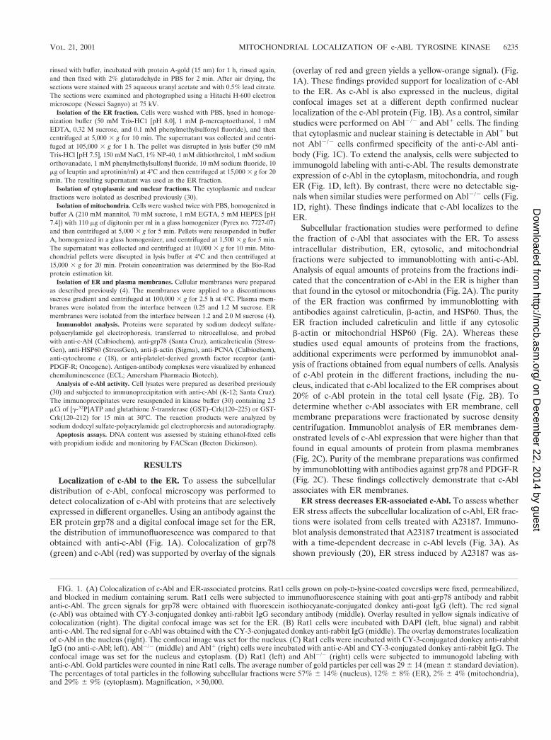
Localization of c-Abl to the ER. To assess the subcellulardistribution of c-Abl, confocal microscopy was performed todetect colocalization of c-Abl with proteins that are selectivelyexpressed in different organelles. Using an antibody against theER protein grp78 and a digital confocal image set for the ER,the distribution of immunofluorescence was compared to thatobtained with anti-c-Abl (Fig. 1A). Colocalization of grp78(green) and c-Abl (red) was supported by overlay of the signals
(overlay of red and green yields a yellow-orange signal). (Fig.1A). These findings provided support for localization of c-Ablto the ER. As c-Abl is also expressed in the nucleus, digitalconfocal images set at a different depth confirmed nuclearlocalization of the c-Abl protein (Fig. 1B). As a control, similarstudies were performed on Abl2/2 and Abl1 cells. The findingthat cytoplasmic and nuclear staining is detectable in Abl1 butnot Abl2/2 cells confirmed specificity of the anti-c-Abl anti-body (Fig. 1C). To extend the analysis, cells were subjected toimmunogold labeling with anti-c-Abl. The results demonstrateexpression of c-Abl in the cytoplasm, mitochondria, and roughER (Fig. 1D, left). By contrast, there were no detectable sig-nals when similar studies were performed on Abl2/2 cells (Fig.1D, right). These findings indicate that c-Abl localizes to theER.
Subcellular fractionation studies were performed to definethe fraction of c-Abl that associates with the ER. To assessintracellular distribution, ER, cytosolic, and mitochondrialfractions were subjected to immunoblotting with anti-c-Abl.Analysis of equal amounts of proteins from the fractions indi-cated that the concentration of c-Abl in the ER is higher thanthat found in the cytosol or mitochondria (Fig. 2A). The purityof the ER fraction was confirmed by immunoblotting withantibodies against calreticulin, b-actin, and HSP60. Thus, theER fraction included calreticulin and little if any cytosolicb-actin or mitochondrial HSP60 (Fig. 2A). Whereas thesestudies used equal amounts of proteins from the fractions,additional experiments were performed by immunoblot anal-ysis of fractions obtained from equal numbers of cells. Analysisof c-Abl protein in the different fractions, including the nu-cleus, indicated that c-Abl localized to the ER comprises about20% of c-Abl protein in the total cell lysate (Fig. 2B). Todetermine whether c-Abl associates with ER membrane, cellmembrane preparations were fractionated by sucrose densitycentrifugation. Immunoblot analysis of ER membranes dem-onstrated levels of c-Abl expression that were higher than thatfound in equal amounts of protein from plasma membranes(Fig. 2C). Purity of the membrane preparations was confirmedby immunoblotting with antibodies against grp78 and PDGF-R(Fig. 2C). These findings collectively demonstrate that c-Ablassociates with ER membranes.
ER stress decreases ER-associated c-Abl. To assess whetherER stress affects the subcellular localization of c-Abl, ER frac-tions were isolated from cells treated with A23187. Immuno-blot analysis demonstrated that A23187 treatment is associatedwith a time-dependent decrease in c-Abl levels (Fig. 3A). Asshown previously (20), ER stress induced by A23187 was as-
FIG. 1. (A) Colocalization of c-Abl and ER-associated proteins. Rat1 cells grown on poly-D-lysine-coated coverslips were fixed, permeabilized,and blocked in medium containing serum. Rat1 cells were subjected to immunofluorescence staining with goat anti-grp78 antibody and rabbitanti-c-Abl. The green signals for grp78 were obtained with fluorescein isothiocyanate-conjugated donkey anti-goat IgG (left). The red signal(c-Abl) was obtained with CY-3-conjugated donkey anti-rabbit IgG secondary antibody (middle). Overlay resulted in yellow signals indicative ofcolocalization (right). The digital confocal image was set for the ER. (B) Rat1 cells were incubated with DAPI (left, blue signal) and rabbitanti-c-Abl. The red signal for c-Abl was obtained with the CY-3-conjugated donkey anti-rabbit IgG (middle). The overlay demonstrates localizationof c-Abl in the nucleus (right). The confocal image was set for the nucleus. (C) Rat1 cells were incubated with CY-3-conjugated donkey anti-rabbitIgG (no anti-c-Abl; left). Abl2/2 (middle) and Abl1 (right) cells were incubated with anti-c-Abl and CY-3-conjugated donkey anti-rabbit IgG. Theconfocal image was set for the nucleus and cytoplasm. (D) Rat1 (left) and Abl2/2 (right) cells were subjected to immunogold labeling withanti-c-Abl. Gold particles were counted in nine Rat1 cells. The average number of gold particles per cell was 29 6 14 (mean 6 standard deviation).The percentages of total particles in the following subcellular fractions were 57% 6 14% (nucleus), 12% 6 8% (ER), 2% 6 4% (mitochondria),and 29% 6 9% (cytoplasm). Magnification, 330,000.
VOL. 21, 2001 MITOCHONDRIAL LOCALIZATION OF c-ABL TYROSINE KINASE 6235
on Decem
ber 22, 2014 by guesthttp://m
cb.asm.org/
Dow
nloaded from

sociated with increases in expression of grp78 (Fig. 3A). Equalloading of the lanes was confirmed by immunoblotting withanticalreticulin (Fig. 3A). ER fractions isolated from cellstreated with brefeldin A to inhibit transport of protein fromthe ER to the Golgi were also subjected to immunoblottingwith anti-c-Abl. The results demonstrate that brefeldin A, likeA23187, decreases levels of c-Abl associated with the ER (Fig.3B). Brefeldin A treatment was also associated with increasesin grp78 and had little if any effect on levels of calreticulin (Fig.3B). These findings demonstrate that ER stress downregulateslocalization of c-Abl to the ER.
ER stress targets c-Abl to mitochondria. The subcellularrelocalization of c-Abl in response to ER stress was investi-gated by measuring intracellular fluorescence. Examination ofthe distribution of fluorescence markers in control Rat1 cellsshowed distinct patterns for anti-c-Abl (red signal) and a mi-
FIG. 2. Subcellular distribution of c-Abl. (A) ER, cytoplasmic(Cyto), and mitochondrial (Mito) fractions were isolated from Rat1cells. Equal amounts of protein (5 mg) from each fraction were sub-jected to immunoblotting (IB) with anti-c-Abl, anticalreticulin, anti-b-actin, or anti-HSP60. (B) Rat1 cells (2 3 107) were divided into fivealiquots for preparation of total cell, nuclear, cytoplasmic, ER, andmitochondrial lysates. The lysates were adjusted to 500 ml with PBS,and aliquots (20 ml) were subjected to immunoblotting with anti-c-Abl.Signal intensities were analyzed by densitometric scanning. The resultsare presented as the percentage of c-Abl in each subcellular fractioncompared to that in the total cell lysate. (C) ER and plasma membranepreparations were isolated from Rat1 cells. Equal amounts of protein(5 mg) were subjected to immunoblot analysis with anti-c-Abl, antical-reticulin, and anti-PDGF-R.
FIG. 3. ER stress decreases ER-associated c-Abl. Rat1 cells weretreated with 10 mM A23187 (A) or 10 mg of brefeldin A per ml (B) andharvested at the indicated times. ER fractions were isolated and sub-jected to immunoblotting with anti-c-Abl (upper panels), anti-grp78(middle panels), or anticalreticulin (lower panels). The signal intensi-ties of c-Abl protein were compared to that of the control.
6236 ITO ET AL. MOL. CELL. BIOL.
on Decem
ber 22, 2014 by guesthttp://m
cb.asm.org/
Dow
nloaded from

tochondrion-selective dye (Mitotracker; green signal) (Fig.4A). By contrast, treatment with A23187 was associated with achange in fluorescence signals (red and green yield yellow-orange) supporting translocation of c-Abl to mitochondria(Fig. 4A). Similar results were obtained with brefeldinA-treated Rat1 cells (Fig. 4A) and with A23187-treated Abl1
cells (Fig. 4B). By contrast, there was little if any change inexpression of c-Abl in the cytoplasm or nucleus (data notshown). These results indicate that ER stress-induced down-regulation of c-Abl in the ER is associated with targeting ofc-Abl to mitochondria.
ER stress activates the c-Abl kinase. To further define thedistribution of c-Abl in response to ER stress, cytoplasmic andnuclear fractions from A23187-treated cells were assessed byimmunoblot analysis with anti-c-Abl. The results demonstratethat A23187 has little if any effect on c-Abl levels in the cyto-plasm or nucleus (Fig. 5A). Purity of the fractions was con-firmed by immunoblotting with anti-b-actin, anti-PCNA, andanticalreticulin (Fig. 5A). In contrast to the cytoplasm andnucleus, immunoblot analysis of the mitochondrial fractionfrom A23187-treated cells demonstrated a time-dependent in-crease in c-Abl protein (Fig. 5B). The mitochondrial fractionwas also subjected to immunoprecipitation with anti-c-Abl.
Analysis of the immunoprecipitates for phosphorylation ofGST-Crk (120–225) demonstrated that A23187 treatment isassociated with increases in mitochondrial c-Abl activity (Fig.5C). As a control, there was no detectable phosphorylation ofGST-Crk (120–212) that lacks the c-Abl Y-221 phosphoryla-tion site (data not shown). Densitometric scanning of the sig-nals obtained for phosphorylation of GST-Crk (120–225) com-pared to those obtained for immunoprecipitated c-Abl proteinindicated that A23187 induces c-Abl activity (Fig. 5C). Theaverage increase in mitochondrial c-Abl activity compared tothat for c-Abl protein for three separate experiments is shown(Fig. 5D). The results support activation of the c-Abl proteinthat localizes to mitochondria.
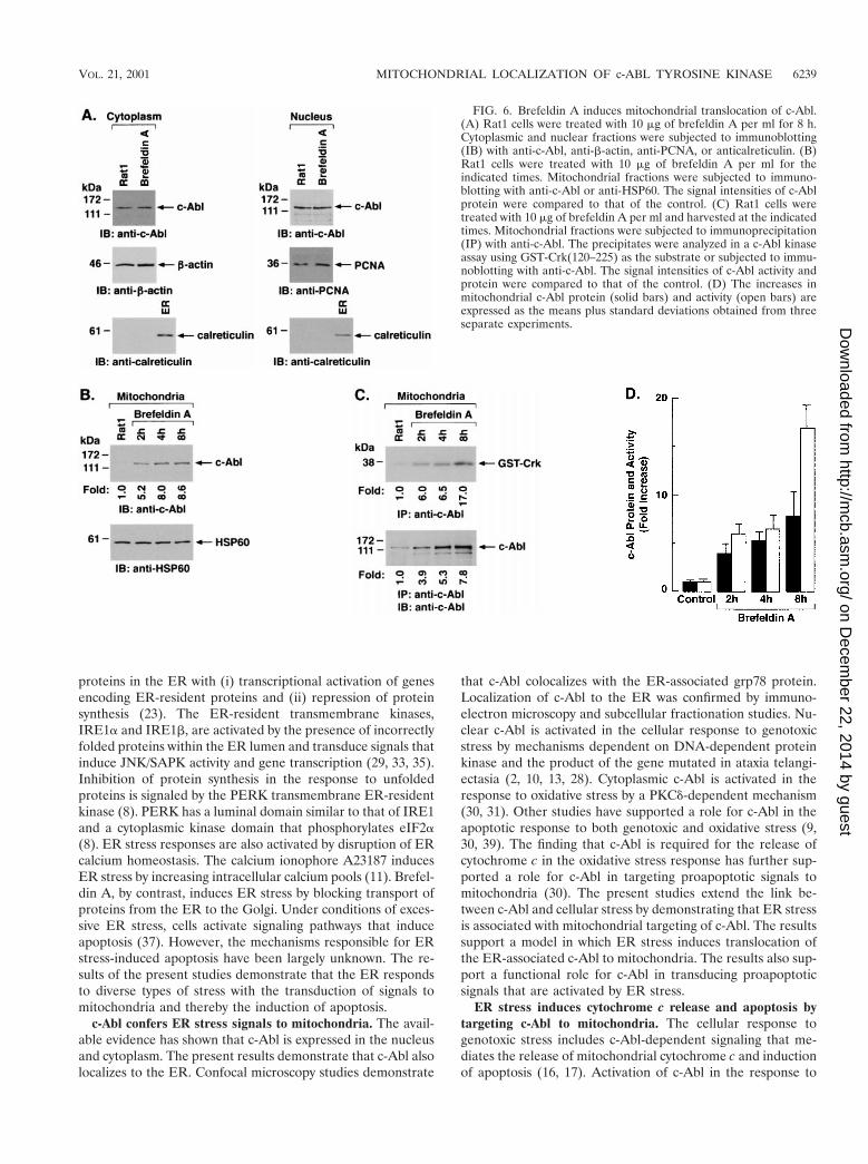
Targeting of c-Abl to mitochondria was similarly assessed incells treated with brefeldin A. Immunoblot analysis of thecytoplasmic and nuclear fractions showed no detectable effectof brefeldin A on c-Abl levels (Fig. 6A). As found with A23187,analysis of the mitochondrial fraction demonstrated brefeldinA-induced increases in c-Abl protein (Fig. 6B). In addition,brefeldin A treatment was associated with increases in mito-chondrial c-Abl activity (Fig. 6C). Comparison of the signalsfound for GST-Crk (120–225) phosphorylation and c-Abl pro-tein indicated that brefeldin A induces activation of the c-Abl
FIG. 4. ER stress targets c-Abl to mitochondria. (A) Rat1 cells (left) were treated with 10 mM A23187 for 6 h (middle) or 10 mg of brefeldinA per ml for 8 h (right). (B) Abl1 cells (left) were treated with 10 mM A23187 for 6 h (right). After being washed, the cells were immobilized onslides, fixed, and incubated with anti-c-Abl antibody followed by Texas red-conjugated goat anti-rabbit IgG. Rat1 cells were also stained with DAPI,while no DAPI was used for staining of the Abl1 cells. Mitochondria were stained with the mitochondrion-selective dye Mitotracker green. Theslides were visualized using a fluorescence microscope coupled to a high-sensitivity charge-coupled device camera and image analyzer. Red signal,c-Abl; green signal, Mitotracker; yellow-orange signals, colocalization of c-Abl and Mitotracker.
VOL. 21, 2001 MITOCHONDRIAL LOCALIZATION OF c-ABL TYROSINE KINASE 6237
on Decem
ber 22, 2014 by guesthttp://m
cb.asm.org/
Dow
nloaded from

kinase (Fig. 6C and D). These findings and those obtained withA23187 demonstrate that ER stress is associated with targetingof c-Abl to mitochondria and stimulation of c-Abl activity.
ER stress induces cytochrome c release and apoptosis by ac-Abl-dependent mechanism. To assess the functional signifi-cance of ER stress-induced targeting of c-Abl to mitochondria,wild-type and Abl2/2 MEFs were treated with A23187. Immu-noblot analysis of the mitochondrial fraction demonstratedA23187-induced increases in mitochondrial c-Abl levels inwild-type but not Abl2/2 cells (Fig. 7A). Cytoplasmic fractionswere also subjected to immunoblot analysis to assess release ofmitochondrial cytochrome c. The results demonstrate thatA23187 induces the release of cytochrome c in wild-type butnot Abl2/2 MEFs (Fig. 7A). Similar results were obtained inwild-type and Abl2/2 cells treated with brefeldin A (Fig. 7B).To confirm dependence on c-Abl, Abl1 cells were treated withinducers of the ER stress. The results demonstrate thatA23187 treatment is associated with targeting of c-Abl to mi-tochondria and cytochrome c release (Fig. 7C). Similar resultswere obtained with brefeldin A and tunicamycin (Fig. 7C). Asadditional controls, wild-type, Abl2/2, and Abl1 cells weretreated with A23187 and analyzed for activation of grp78. Theresults demonstrate that grp78 is activated in the different celltypes (Fig. 7D). Similar findings were obtained with brefeldinA (Fig. 7E). These results demonstrate that c-Abl is not in-
volved in initiating ER stress but is required for transducingER stress signals to mitochondria.
In concert with these findings, A23187 treatment was asso-ciated with the induction of sub-G1 DNA in wild-type cells buthad little effect on the induction of apoptosis in c-Abl2/2 cells(Fig. 8A). The finding that ER stress-induced apoptosis is alsoabolished in Abl2/2 MEFs treated with brefeldin A providedfurther support for involvement of c-Abl in this response (Fig.8B). To extend these studies, Abl1 cells were analyzed for ERstress-induced apoptosis. The results demonstrate that Abl1
cells respond to A23187 with induction of sub-G1 DNA (Fig.8C). Similar results were obtained with brefeldin A and tuni-camycin (Fig. 8C). As additional controls, independently de-rived wild-type, Abl2/2 MEFs (19) were treated with tunica-mycin. Wild-type but not Abl2/2 cells responded totunicamycin with the induction of apoptosis (Fig. 8D). Theseresults demonstrate that ER stress induces cytochrome c re-lease and apoptosis by a c-Abl-dependent mechanism.
DISCUSSION
Stress signaling from the ER to mitochondria. The ERresponds to alterations in homeostasis with the transduction ofsignals to the nucleus and cytoplasm. In this context, eukary-otic cells respond to the accumulation of unfolded or excess
FIG. 5. A23187 induces mitochondrial translocation of c-Abl. (A)Rat1 cells were treated with 10 mM A23187 and harvested at 6 h.Cytoplasmic and nuclear fractions were isolated and subjected to im-munoblotting (IB) with anti-c-Abl, anti-b-actin, anti-PCNA, or anti-calreticulin. (B) Rat1 cells were treated with 10 mM A23187 andharvested at the indicated times. Mitochondrial fractions were isolatedand subjected to immunoblotting with anti-c-Abl or anti-HSP60. Thesignal intensities of c-Abl protein were compared to that of the control.(C) Rat1 cells were treated with 10 mM A23187 and harvested at theindicated times. Mitochondrial fractions were subjected to immuno-precipitation (IP) with anti-c-Abl. The precipitates were analyzed in ac-Abl kinase assay using GST-Crk(120–225) as the substrate or sub-jected to immunoblotting with anti-c-Abl. The signal intensities ofc-Abl activity and protein were compared to that of the controls. (D)The increases in mitochondrial c-Abl protein (solid bars) and activity(open bars) are expressed as the means plus standard deviations ob-tained from three separate experiments.
6238 ITO ET AL. MOL. CELL. BIOL.
on Decem
ber 22, 2014 by guesthttp://m
cb.asm.org/
Dow
nloaded from
proteins in the ER with (i) transcriptional activation of genesencoding ER-resident proteins and (ii) repression of proteinsynthesis (23). The ER-resident transmembrane kinases,IRE1a and IRE1b, are activated by the presence of incorrectlyfolded proteins within the ER lumen and transduce signals thatinduce JNK/SAPK activity and gene transcription (29, 33, 35).Inhibition of protein synthesis in the response to unfoldedproteins is signaled by the PERK transmembrane ER-residentkinase (8). PERK has a luminal domain similar to that of IRE1and a cytoplasmic kinase domain that phosphorylates eIF2a(8). ER stress responses are also activated by disruption of ERcalcium homeostasis. The calcium ionophore A23187 inducesER stress by increasing intracellular calcium pools (11). Brefel-din A, by contrast, induces ER stress by blocking transport ofproteins from the ER to the Golgi. Under conditions of exces-sive ER stress, cells activate signaling pathways that induceapoptosis (37). However, the mechanisms responsible for ERstress-induced apoptosis have been largely unknown. The re-sults of the present studies demonstrate that the ER respondsto diverse types of stress with the transduction of signals tomitochondria and thereby the induction of apoptosis.
c-Abl confers ER stress signals to mitochondria. The avail-able evidence has shown that c-Abl is expressed in the nucleusand cytoplasm. The present results demonstrate that c-Abl alsolocalizes to the ER. Confocal microscopy studies demonstrate
that c-Abl colocalizes with the ER-associated grp78 protein.Localization of c-Abl to the ER was confirmed by immuno-electron microscopy and subcellular fractionation studies. Nu-clear c-Abl is activated in the cellular response to genotoxicstress by mechanisms dependent on DNA-dependent proteinkinase and the product of the gene mutated in ataxia telangi-ectasia (2, 10, 13, 28). Cytoplasmic c-Abl is activated in theresponse to oxidative stress by a PKCd-dependent mechanism(30, 31). Other studies have supported a role for c-Abl in theapoptotic response to both genotoxic and oxidative stress (9,30, 39). The finding that c-Abl is required for the release ofcytochrome c in the oxidative stress response has further sup-ported a role for c-Abl in targeting proapoptotic signals tomitochondria (30). The present studies extend the link be-tween c-Abl and cellular stress by demonstrating that ER stressis associated with mitochondrial targeting of c-Abl. The resultssupport a model in which ER stress induces translocation ofthe ER-associated c-Abl to mitochondria. The results also sup-port a functional role for c-Abl in transducing proapoptoticsignals that are activated by ER stress.
ER stress induces cytochrome c release and apoptosis bytargeting c-Abl to mitochondria. The cellular response togenotoxic stress includes c-Abl-dependent signaling that me-diates the release of mitochondrial cytochrome c and inductionof apoptosis (16, 17). Activation of c-Abl in the response to
FIG. 6. Brefeldin A induces mitochondrial translocation of c-Abl.(A) Rat1 cells were treated with 10 mg of brefeldin A per ml for 8 h.Cytoplasmic and nuclear fractions were subjected to immunoblotting(IB) with anti-c-Abl, anti-b-actin, anti-PCNA, or anticalreticulin. (B)Rat1 cells were treated with 10 mg of brefeldin A per ml for theindicated times. Mitochondrial fractions were subjected to immuno-blotting with anti-c-Abl or anti-HSP60. The signal intensities of c-Ablprotein were compared to that of the control. (C) Rat1 cells weretreated with 10 mg of brefeldin A per ml and harvested at the indicatedtimes. Mitochondrial fractions were subjected to immunoprecipitation(IP) with anti-c-Abl. The precipitates were analyzed in a c-Abl kinaseassay using GST-Crk(120–225) as the substrate or subjected to immu-noblotting with anti-c-Abl. The signal intensities of c-Abl activity andprotein were compared to that of the control. (D) The increases inmitochondrial c-Abl protein (solid bars) and activity (open bars) areexpressed as the means plus standard deviations obtained from threeseparate experiments.
VOL. 21, 2001 MITOCHONDRIAL LOCALIZATION OF c-ABL TYROSINE KINASE 6239
on Decem
ber 22, 2014 by guesthttp://m
cb.asm.org/
Dow
nloaded from

FIG. 7. ER stress induces cytochrome c release and apoptosis by ac-Abl-dependent mechanism. (A) MEF (c-Abl1/1) and c-Abl2/2 cellswere treated with 10 mM A23187 and harvested at the indicated times.Mitochondrial fractions were isolated and subjected to immunoblot-ting (IB) with anti-c-Abl or anti-HSP60. Cytoplasmic fractions weresubjected to immunoblotting with anti-cytochrome c (Cyt c) or anti-b-actin. The signal intensities of c-Abl and cytochrome c were com-pared to that of the control. (B) MEF (c-Abl1/1) and c-Abl2/2 cellswere treated with 10 mg of brefeldin A per ml (Bref A) and harvestedat the indicated times. Mitochondrial fractions were subjected to im-munoblotting with anti-c-Abl or anti-HSP60. Cytoplasmic fractionswere subjected to immunoblotting with anti-cytochrome c or anti-b-actin. (C) MEF (c-Abl1/1), Abl2/2, and Abl1 cells were treated with10 mM A23187 for 6 h, 10 mg of brefeldin A per ml for 8 h, or 10 mgof tunicamycin per ml for 8 h. Mitochondrial fractions were analyzedby immunoblotting with anti-c-Abl and anti-HSP60. Cytoplasmic frac-tions were analyzed by immunoblotting with anti-cytochrome c andanti-b-actin. (D and E) MEF (c-Abl1/1), Abl2/2, and Abl1 cells weretreated with 10 mM A23187 (D) or 10 mM of brefeldin A per ml (E)for the indicated times. ER fractions were analyzed by immunoblottingwith anti-grp78 and anticalreticulin.
6240 ITO ET AL. MOL. CELL. BIOL.
on Decem
ber 22, 2014 by guesthttp://m
cb.asm.org/
Dow
nloaded from

oxidative stress has also been associated with release of cyto-chrome c and the induction of apoptosis by a c-Abl-dependentmechanism (30). In the cytosol, cytochrome c associates with acomplex of Apaf-1 and caspase-9 and thereby induces theactivation of caspase-3 (22, 42). The induction of apoptosis isassociated with caspase-3-mediated cleavage of poly (ADP-ribose) polymerase, PKCd, and other proteins (6, 12, 32).While ER stress can induce apoptosis (37), the involvement ofcytochrome c release in this response has been unknown. Inthe present studies, the finding that ER stress induces therelease of mitochondrial cytochrome c provided further sup-port for signaling from the ER to mitochondria. Importantly,the induction of cytochrome c release by ER stress was atten-uated in Abl2/2 cells. Moreover, Abl2/2 cells were defectivein the apoptotic response to ER stress. These findings in-dicate that ER stress-induced cytochrome c release andapoptosis are mediated by targeting c-Abl from the ER tomitochondria.
ACKNOWLEDGMENTS
This work was supported by grant CA42802 awarded by the NationalCancer Institute, DHHS, and by the office of Health and BiologicalResearch, U.S. Department of Energy, cooperative agreement DE-FC04-96AL76406.
We appreciate the technical assistance of Kamal Chauhan.Y. Ito, P. Pandey, and N. Mishra contributed equally to this work.
REFERENCES
1. Agami, R., G. Blandino, M. Oren, and Y. Shaul. 1999. Interaction of c-Abland p73a and their collaboration to induce apoptosis. Nature 399:809–813.
2. Baskaran, R., L. D. Wood, L. L. Whitaker, Y. Xu, C. Barlow, C. E. Canman,S. E. Morgan, D. Baltimore, A. Wynshaw-Boris, M. B. Kastan, and J. Y. J.Wang. 1997. Ataxia telangiectasia mutant protein activates c-abl tyrosinekinase in response to ionizing radiation. Nature 387:516–519.
3. Dricu, A., M. Carlberg, M. Wang, and O. Larsson. 1997. Inhibition ofN-linked glycosylation using tunicamycin causes cell death in malignant cells:role of down-regulation of the insulin-like growth factor 1 receptor in in-
FIG. 8. ER stress induces apoptosis by a c-Abl-dependent mecha-nism. (A) MEF (c-Abl1/1) and c-Abl2/2 cells were treated with 10 mMA23187 and harvested at the indicated times. After being fixed, cellswere stained with propidium iodide, and sub-G1 DNA content wasmeasured using FACScan. The percentage of apoptotic cells withsub-G1 DNA content is expressed as the means plus standard devia-tions from three independent experiments, each performed in dupli-cate. (B) MEF (c-Abl1/1) and Abl2/2 cells were treated with 10 mg ofbrefeldin A per ml and harvested at the indicated times. (C) MEF(c-Abl1/1), Abl2/2, and Abl1 cells were treated with 10 mM A23187for 6 h, 10 mg of brefeldin A per ml for 8 h, or 10 mg of tunicamycinper ml for 8 h. (D) Independently derived MEFs and Abl2/2 cells weretreated with 10 mg of tunicamycin per ml for 8 h. The percentage ofapoptotic cells with sub-G1 DNA content is expressed as the meansplus standard deviations from three independent experiments, eachperformed in duplicate.
VOL. 21, 2001 MITOCHONDRIAL LOCALIZATION OF c-ABL TYROSINE KINASE 6241
on Decem
ber 22, 2014 by guesthttp://m
cb.asm.org/
Dow
nloaded from

duction of apoptosis. Cancer Res. 57:543–548.4. Frangioni, J. V., P. H. Beahm, V. Shifrin, C. A. Jost, and B. G. Neel. 1992.
The nontransmembrane tyrosine phosphatase PTP-1B localizes to the en-doplasmic reticulum via its 35 amino acid C-terminal sequence. Cell 68:545–560.
5. Gething, M., and J. Sambrook. 1992. Protein folding in the cell. Nature355:33–45.
6. Ghayur, T., M. Hugunin, R. V. Talanian, S. Ratnofsky, C. Quinlan, Y.Emoto, P. Pandey, R. Datta, S. Kharbanda, H. Allen, R. Kamen, W. Wong,and D. Kufe. 1996. Proteolytic activation of protein kinase C d by an ICE/CED 3-like protease induces characteristics of apoptosis. J. Exp. Med. 184:2399–2404.
7. Gong, J., A. Costanzo, H. Yang, G. Melino, W. Kaelin, Jr., M. Levrero, andJ. Y. J. Wang. 1999. The tyrosine kinase c-Abl regulates p73 in apoptoticresponse to cisplatin-induced DNA damage. Nature 399:806–809.
8. Harding, H. P., Y. Zhang, and D. Ron. 1999. Protein translation and foldingare coupled by an endoplasmic-reticulum-resident kinase. Nature 397:271–274.
9. Huang, Y., Z. M. Yuan, T. Ishiko, S. Nakada, T. Utsugisawa, T. Kato, S.Kharbanda, and D. W. Kufe. 1997. Pro-apoptotic effect of the c-Abl tyrosinekinase in the cellular response to 1-b-D-arabinofuranosylcytosine. Oncogene15:1947–1952.
10. Jin, S., S. Kharbanda, B. Mayer, D. Kufe, and D. T. Weaver. 1997. Bindingof Ku and c-Abl at the kinase homology region of DNA-dependent proteinkinase catalytic subunit. J. Biol. Chem. 272:24763–24766.
11. Kaufman, R. J. 1999. Stress signaling from the lumen of the endoplasmicreticulum: coordination of gene transcriptional and translational controls.Genes Dev. 13:1211–1233.
12. Kaufmann, S. H., S. Desnoyers, Y. Ottaviano, N. E. Davidson, and G. G.Poirier. 1993. Specific proteolytic cleavage of poly (ADP-ribose) polymerase:an early marker of chemotherapy-induced apoptosis. Cancer Res. 53:3976–3985.
13. Kharbanda, S., P. Pandey, S. Jin, S. Inoue, A. Bharti, Z.-M. Yuan, R.Weichselbaum, D. Weaver, and D. Kufe. 1997. Functional interaction ofDNA-PK and c-Abl in response to DNA damage. Nature 386:732–735.
14. Kharbanda, S., P. Pandey, R. Ren, S. Feller, B. Mayer, L. Zon, and D. Kufe.1995. c-Abl activation regulates induction of the SEK1/stress activated pro-tein kinase pathway in the cellular response to 1-b-D-arabinofuranosylcy-tosine. J. Biol. Chem. 270:30278–30281.
15. Kharbanda, S., P. Pandey, T. Yamauchi, S. Kumar, M. Kaneki, V. Kumar,A. Bharti, Z. Yuan, L. Ghanem, A. Rana, R. Weichselbaum, G. Johnson, andD. Kufe. 2000. Activation of MEK kinase-1 by the c-Abl protein tyrosinekinase in response to DNA-damage. Mol. Cell. Biol. 20:4979–4989.
16. Kharbanda, S., R. Ren, P. Pandey, T. D. Shafman, S. M. Feller, R. R.Weichselbaum, and D. W. Kufe. 1995. Activation of the c-Abl tyrosine kinasein the stress response to DNA-damaging agents. Nature 376:785–788.
17. Kharbanda, S., S. Saxena, K. Yoshida, P. Pandey, M. Kaneki, Q. Wang, K.Cheng, Y. Chen, A. Campbell, S. Thangrila, Z. Yuan, J. Narula, R. Weich-selbaum, C. Nalin, and D. Kufe. 2000. Translocation of SAPK/JNK to mi-tochondria and interaction with Bcl-xL in response to DNA damage. J. Biol.Chem. 275:322–327.
18. Kirken, R., A. Lincoln, P. Fink, and L. Prochaska. 1995. High yield purifi-cation of a four subunit caa3-type cytochrome oxidase from the thermophilicbacterium Bacillus PS3 using fast protein liquid chromatography. ProteinExpr. Purif. 6:707–715.
19. Koleske, A. J., A. M. Gifford, M. L. Scott, M. Nee, R. T. Bronson, K. A.Miczek, and D. Baltimore. 1998. Essential roles for the Abl and Arg tyrosinekinases in neurulation. Neuron 21:1259–1272.
20. Kozutsumi, Y., M. Segal, K. Normington, M. J. Gething, and J. Sambrook.1988. The presence of malfolded proteins in the endoplasmic reticulumsignals the induction of glucose-regulated proteins. Nature 332:462–464.
21. Larsson, O., M. Carlberg, and A. Zetterberg. 1993. Selective killing inducedby an inhibitor of N-linked glycosylation. J. Cell Sci. 106:299–307.
22. Li, P., D. Nijhawan, I. Budihardjo, S. M. Srinivasula, M. Ahmad, E. S.Alnemri, and X. Wang. 1997. Cytochrome c and dATP-dependent formation
of Apaf-1/caspase-9 complex initiates an apoptotic protease cascade. Cell91:479–489.
23. Mori, K. 2000. Tripartite management of unfolded proteins in the endoplas-mic reticulum. Cell 101:451–454.
24. Nakagawa, T., H. Zhu, N. Morishima, E. Li, J. Xu, B. A. Yankner, and J.Yuan. 2000. Caspase-12 mediates endoplasmic-reticulum-specific apoptosisand cytotoxicity by amyloid-beta. Nature 403:98–103.
25. Pandey, P., J. Raingeaud, M. Kaneki, R. Weichselbaum, R. Davis, D. Kufe,and S. Kharbanda. 1996. Activation of p38 MAP kinase by c-Abl-dependentand -independent mechanisms. J. Biol. Chem. 271:23775–23779.
26. Perez-Sala, D., and F. Mollinedo. 1995. Inhibition of N-linked glycosylationinduces early apoptosis in human promyelocytic HL-60 cells. J. Cell. Physiol.163:523–531.
27. Price, B. D., and S. K. Calderwood. 1992. Gadd45 and Gadd153 messengerRNA levels are increased during hypoxia and after exposure of cells toagents which elevate the levels of the glucose-regulated proteins. CancerRes. 52:3814–3817.
28. Shafman, T., K. K. Khanna, P. Kedar, K. Spring, S. Kozlov, T. Yen, K.Hobson, M. Gatei, N. Zhang, D. Watters, M. Egerton, Y. Shiloh, S. Khar-banda, D. Kufe, and M. F. Lavin. 1997. Interaction between ATM proteinand c-Abl in response to DNA damage. Nature 387:520–523.
29. Shamu, C. E., and P. Walter. 1996. Oligomerization and phosphorylation ofthe Ire1p kinase during intracellular signaling from the endoplasmic reticu-lum to the nucleus. EMBO J. 15:3028–3039.
30. Sun, X., P. Majumder, H. Shioya, F. Wu, S. Kumar, R. Weichselbaum, S.Kharbanda, and D. Kufe. 2000. Activation of the cytoplasmic c-Abl tyrosinekinase by reactive oxygen species. J. Biol. Chem. 275:17237–17240.
31. Sun, X., F. Wu, R. Datta, S. Kharbanda, and D. Kufe. 2000. Interactionbetween protein kinase C d and the c-Abl tyrosine kinase in the cellularresponse to oxidative stress. J. Biol. Chem. 275:7470–7473.
32. Tewari, M., L. T. Quan, K. O’Rourke, S. Desnoyers, Z. Zeng, D. R. Beidler,G. G. Poirier, G. S. Salvesen, and V. M. Dixit. 1995. Yama/CPP32b, amammalian homolog of CED-3, is a CrmA-inhibitable protease that cleavesthe death substrate poly (ADP-ribose) polymerase. Cell 81:801–809.
33. Tirasophon, W., A. A. Welihinda, and R. J. Kaufman. 1998. A stress responsepathway from the endoplasmic reticulum to the nucleus requires a novelbifunctional protein kinase/endoribonuclease (Ire1p) in mammalian cells.Genes Dev. 12:1812–1824.
34. Tybulewicz, V. L. J., C. E. Crawford, P. K. Jackson, R. T. Bronson, and R. C.Mulligan. 1991. Neonatal lethality and lymphopenia in mice with a homozy-gous disruption of the c-abl proto-oncogene. Cell 65:1153–1163.
35. Urano, F., X. Wang, A. Bertolotti, Y. Zhang, P. Chung, H. P. Harding, andD. Ron. 2000. Coupling of stress in the ER to activation of JNK proteinkinases by transmembrane protein kinase. IRE1. Science 287:664–666.
36. Wang, X. Z., H. P. Harding, Y. Zhang, E. M. Jolicoeur, M. Kuroda, and D.Ron. 1998. Cloning of mammalian Ire1 reveals diversity in the ER stressresponses. EMBO J. 17:5708–5717.
37. Welihinda, A. A., W. Tirasophon, and R. J. Kaufman. 1999. The cellularresponse to protein misfolding in the endoplasmic reticulum. Gene Expr.7:293–300.
38. Yuan, Z., Y. Huang, M.-M. Fan, C. Sawers, S. Kharbanda, and D. Kufe.1996. Genotoxic drugs induce interaction of the c-Abl tyrosine kinase andthe tumor suppressor protein p53. J. Biol. Chem. 271:26457–26460.
39. Yuan, Z., Y. Huang, T. Ishiko, S. Kharbanda, R. Weichselbaum, and D.Kufe. 1997. Regulation of DNA damage-induced apoptosis by the c-Abltyrosine kinase. Proc. Natl. Acad. Sci. USA 94:1437–1440.
40. Yuan, Z. M., Y. Huang, Y. Whang, C. Sawyers, R. Weichselbaum, S. Khar-banda, and D. Kufe. 1996. Role for the c-Abl tyrosine kinase in the growtharrest response to DNA damage. Nature 382:272–274.
41. Yuan, Z. M., H. Shioya, T. Ishiko, X. Sun, Y. Huang, H. Lu, S. Kharbanda,R. Weichselbaum, and D. Kufe. 1999. p73 is regulated by the c-Abl tyrosinekinase in the apoptotic response to DNA damage. Nature 399:814–817.
42. Zou, H., W. J. Henzel, X. Liu, A. Lutschg, and X. Wang. 1997. Apaf-1, ahuman protein homologous to C. elegans CED-4, participates in cytochromec-dependent activation of caspase-3. Cell 90:405–413.
6242 ITO ET AL. MOL. CELL. BIOL.
on Decem
ber 22, 2014 by guesthttp://m
cb.asm.org/
Dow
nloaded from