Endoplasmic reticulum stress disrupts placental morphogenesis: implications for human intrauterine...
11
ORIGINAL PAPER Journal of Pathology J Pathol 2012; 228: 554–564 Published online 28 September 2012 in Wiley Online Library (wileyonlinelibrary.com) DOI: 10.1002/path.4068 Endoplasmic reticulum stress disrupts placental morphogenesis: implications for human intrauterine growth restriction Hong wa Yung, 1 Myriam Hemberger, 1,2 Erica D Watson, 1 Claire E Senner, 1,2 Carolyn P Jones, 3 Randal J Kaufman, 4 D Stephen Charnock-Jones 1,5,6 † and Graham J Burton 1,6 * † 1 Centre for Trophoblast Research, University of Cambridge, Cambridge, CB2 3EG, UK 2 Epigenetics Programme, The Babraham Institute, Babraham Research Campus, Cambridge, CB22 3AT, UK 3 Maternal and Fetal Health Research Group, School of Biomedical Sciences, University of Manchester, Manchester, M13 9WL, UK 4 Del E Webb Neuroscience, Aging and Stem Cell Research Center, Sanford/Burnham Medical Research Institute, 10901 North Torrey Pines Road, La Jolla, CA 92037-1062, USA 5 Department of Obstetrics and Gynaecology, University of Cambridge, Cambridge, CB2 0SW, UK 6 National Institute for Health Research, Cambridge Comprehensive Biomedical Research Centre, Cambridge, UK *Correspondence to: Professor Graham J Burton, Centre for Trophoblast Research, Physiological Laboratory, Downing Street, Cambridge, CB2 3EG, UK. e-mail: [email protected] † These authors contributed equally to this work. Abstract We recently reported the first evidence of placental endoplasmic reticulum (ER) stress in the pathophysiology of human intrauterine growth restriction. Here, we used a mouse model to investigate potential underlying mechanisms. Eif2s1 tm1RjK mice, in which Ser51 of eukaryotic initiation factor 2 subunit alpha (eIF2α) is mutated, display a 30% increase in basal translation. In Eif2s1 tm1RjK placentas, we observed increased ER stress and anomalous accumulation of glycoproteins in the endocrine junctional zone (Jz), but not in the labyrinthine zone where physiological exchange occurs. Placental and fetal weights were reduced by 15% (97 mg to 82 mg, p < 0.001) and 20% (1009 mg to 798 mg, p < 0.001), respectively. To investigate whether ER stress affects bioactivity of secreted proteins, mouse embryonic fibroblasts (MEFs) were derived from Eif2s1 tm1RjK mutants. These MEFs exhibited ER stress, grew 50% slower, and showed reduced Akt–mTOR signalling compared to wild-type cells. Conditioned medium (CM) derived from Eif2s1 tm1RjK MEFs failed to maintain trophoblast stem cells in a progenitor state, but the effect could be rescued by exogenous application of FGF4 and heparin. In addition, ER stress promoted accumulation of pro-Igf2 with altered glycosylation in the CM without affecting cellular levels, indicating that the protein failed to be processed after release. Igf2 is the major growth factor for placental development; indeed, activity in the Pdk1–Akt–mTOR pathways was decreased in Eif2s1 tm1RjK placentas, indicating loss of Igf2 signalling. Furthermore, we observed premature differentiation of trophoblast progenitors at E9.5 in mutant placentas, consistent with the in vitro results and with the disproportionate development of the labyrinth and Jz seen in placentas at E18.5. Similar disproportion has been reported in the Igf2-null mouse. These results demonstrate that ER stress adversely affects placental development, and that modulation of post-translational processing, and hence bioactivity, of secreted growth factors contributes to this effect. Placental dysmorphogenesis potentially affects fetal growth through reduced exchange capacity. Copyright 2012 Pathological Society of Great Britain and Ireland. Published by John Wiley & Sons, Ltd. Keywords: endoplasmic reticulum; stress; placental morphogenesis; intrauterine growth restriction; Igf2; Pdk1; Akt; mTOR Received 4 November 2011; Revised 14 June 2012; Accepted 15 June 2012 No conflicts of interest were declared. Introduction Intrauterine growth restriction (IUGR) refers to a fetus that has failed to achieve its genetically determined potential size, and affects approximately 5–8% of human pregnancies worldwide. It is a major cause of neonatal morbidity and mortality, and also increases the risk of developing cardiovascular and metabolic diseases in adulthood. Predisposing factors include maternal smoking, under-nutrition, infection, and con- genital malformations, but in the majority of cases utero-placental insufficiency is thought to be the cause. The placenta serves as the interface between a mother and her fetus, and is critical for fetal nutrition. Reduced placental growth is observed to precede fetal IUGR during human pregnancy [1,2]. Similarly, deletion of the placental-specific Igf2 promoter in the mouse, P0, results in a smaller placenta that in turn restricts fetal growth in late gestation [3]. Consequently, it is widely accepted that placental compromise is one of the major risk factors for the development of IUGR and has been described as ‘placental IUGR’ [4]. Copyright 2012 Pathological Society of Great Britain and Ireland. J Pathol 2012; 228: 554–564 Published by John Wiley & Sons, Ltd. www.pathsoc.org.uk www.thejournalofpathology.com
-
Upload
independent -
Category
Documents
-
view
1 -
download
0
Transcript of Endoplasmic reticulum stress disrupts placental morphogenesis: implications for human intrauterine...
ORIGINAL PAPERJournal of PathologyJ Pathol 2012; 228: 554–564Published online 28 September 2012 in Wiley Online Library(wileyonlinelibrary.com) DOI: 10.1002/path.4068
Endoplasmic reticulum stress disrupts placental morphogenesis:implications for human intrauterine growth restrictionHong wa Yung,1 Myriam Hemberger,1,2 Erica D Watson,1 Claire E Senner,1,2 Carolyn P Jones,3 Randal J Kaufman,4D Stephen Charnock-Jones1,5,6† and Graham J Burton1,6*†
1 Centre for Trophoblast Research, University of Cambridge, Cambridge, CB2 3EG, UK2 Epigenetics Programme, The Babraham Institute, Babraham Research Campus, Cambridge, CB22 3AT, UK3 Maternal and Fetal Health Research Group, School of Biomedical Sciences, University of Manchester, Manchester, M13 9WL, UK4 Del E Webb Neuroscience, Aging and Stem Cell Research Center, Sanford/Burnham Medical Research Institute, 10901 North Torrey PinesRoad, La Jolla, CA 92037-1062, USA5 Department of Obstetrics and Gynaecology, University of Cambridge, Cambridge, CB2 0SW, UK6 National Institute for Health Research, Cambridge Comprehensive Biomedical Research Centre, Cambridge, UK
*Correspondence to: Professor Graham J Burton, Centre for Trophoblast Research, Physiological Laboratory, Downing Street, Cambridge, CB2 3EG,UK. e-mail: [email protected]
†These authors contributed equally to this work.
AbstractWe recently reported the first evidence of placental endoplasmic reticulum (ER) stress in the pathophysiologyof human intrauterine growth restriction. Here, we used a mouse model to investigate potential underlyingmechanisms. Eif2s1tm1RjK mice, in which Ser51 of eukaryotic initiation factor 2 subunit alpha (eIF2α) is mutated,display a 30% increase in basal translation. In Eif2s1tm1RjK placentas, we observed increased ER stress andanomalous accumulation of glycoproteins in the endocrine junctional zone (Jz), but not in the labyrinthinezone where physiological exchange occurs. Placental and fetal weights were reduced by 15% (97 mg to 82 mg,p < 0.001) and 20% (1009 mg to 798 mg, p < 0.001), respectively. To investigate whether ER stress affectsbioactivity of secreted proteins, mouse embryonic fibroblasts (MEFs) were derived from Eif2s1tm1RjK mutants.These MEFs exhibited ER stress, grew 50% slower, and showed reduced Akt–mTOR signalling compared towild-type cells. Conditioned medium (CM) derived from Eif2s1tm1RjK MEFs failed to maintain trophoblast stemcells in a progenitor state, but the effect could be rescued by exogenous application of FGF4 and heparin. Inaddition, ER stress promoted accumulation of pro-Igf2 with altered glycosylation in the CM without affectingcellular levels, indicating that the protein failed to be processed after release. Igf2 is the major growth factorfor placental development; indeed, activity in the Pdk1–Akt–mTOR pathways was decreased in Eif2s1tm1RjK
placentas, indicating loss of Igf2 signalling. Furthermore, we observed premature differentiation of trophoblastprogenitors at E9.5 in mutant placentas, consistent with the in vitro results and with the disproportionatedevelopment of the labyrinth and Jz seen in placentas at E18.5. Similar disproportion has been reported inthe Igf2-null mouse. These results demonstrate that ER stress adversely affects placental development, and thatmodulation of post-translational processing, and hence bioactivity, of secreted growth factors contributes to thiseffect. Placental dysmorphogenesis potentially affects fetal growth through reduced exchange capacity.Copyright 2012 Pathological Society of Great Britain and Ireland. Published by John Wiley & Sons, Ltd.
Keywords: endoplasmic reticulum; stress; placental morphogenesis; intrauterine growth restriction; Igf2; Pdk1; Akt; mTOR
Received 4 November 2011; Revised 14 June 2012; Accepted 15 June 2012
No conflicts of interest were declared.
Introduction
Intrauterine growth restriction (IUGR) refers to a fetusthat has failed to achieve its genetically determinedpotential size, and affects approximately 5–8% ofhuman pregnancies worldwide. It is a major cause ofneonatal morbidity and mortality, and also increasesthe risk of developing cardiovascular and metabolicdiseases in adulthood. Predisposing factors includematernal smoking, under-nutrition, infection, and con-genital malformations, but in the majority of cases
utero-placental insufficiency is thought to be the cause.The placenta serves as the interface between a motherand her fetus, and is critical for fetal nutrition. Reducedplacental growth is observed to precede fetal IUGRduring human pregnancy [1,2]. Similarly, deletion ofthe placental-specific Igf2 promoter in the mouse, P0,results in a smaller placenta that in turn restricts fetalgrowth in late gestation [3]. Consequently, it is widelyaccepted that placental compromise is one of the majorrisk factors for the development of IUGR and has beendescribed as ‘placental IUGR’ [4].
Copyright 2012 Pathological Society of Great Britain and Ireland. J Pathol 2012; 228: 554–564Published by John Wiley & Sons, Ltd. www.pathsoc.org.uk www.thejournalofpathology.com

Endoplasmic reticulum stress and placental function 555
We recently identified endoplasmic reticulum (ER)stress as a feature of the placental pathophysiologyin cases of human IUGR [5]. The ER serves multi-ple functions, including the synthesis, post-translationalmodification, and trafficking of membrane and secretedproteins. Perturbation of ER homeostasis may result inmisfolding or abnormal glycosylation of these proteins,which has the potential to reduce their biological activ-ity. The accumulation of unfolded or misfolded pro-teins within the ER cisternae provokes ER stress andactivation of the unfolded protein response (UPR). TheUPR attempts to restore ER function by attenuation ofprotein translation, increasing the folding capacity andfacilitating degradation of misfolded proteins [6]. Pro-tein synthesis can be regulated through phosphorylationof eIF2α at Ser51 by PKR-like endoplasmic reticulumkinase (PERK) [7], which then acts as a competitiveinhibitor of eIF2B, thereby inhibiting translation initi-ation [8]. If the mechanisms fail to reduce the stress,apoptosis ensues to eliminate the damaged cell.
Organs with major endocrine and/or exocrine activityare more vulnerable to ER stress due to their constitu-tively high level of protein translation. Indeed, boththe pancreas and the placenta suffer low-grade ERstress under normal physiological conditions [9,10].In both Perk knockout and Eif2s1tm1RjK mutant mice(in which eIF2α cannot be inactivated by phosphory-lation at Ser51 and hence protein synthesis is consti-tutively elevated), the β-pancreatic cells exhibit severeER stress and increased apoptosis [11–13]. ER stressis now widely accepted as a major aetiological factorfor human type II diabetes [14].
In this study, we test whether ER stress is a sufficientcause of placental IUGR and further elucidate theunderlying mechanisms using the Eif2s1tm1RjK mutantmouse as an animal model.
Materials and methods
Eif2s1tm1RjK mutant miceEif2s1tm1RjK mutant C57BL/6 mice were generatedas described by Scheuner et al [12]. A total of 26litters of mice containing 57 wild-type, 32 mutant,and 111 heterozygous animals was used. The averagelitter size was ∼8. All experiments were carried outin accordance with the UK Home Office Animals(Scientific Procedures) Act 1986. Placentas and fetuseswere collected at E18.5 and immediately immersedin ice-cold phosphate buffered saline. After weighing,placentas were dissected into junctional zone-rich (Jz)and labyrinthine zone-rich (Lz) pieces, which weresnap-frozen in liquid nitrogen. Tail tips were collectedfor genotyping.
Mouse embryonic fibroblasts (MEFs)MEFs were derived from E13.5 embryos as describedin the Supplementary materials and methods.
Trophoblast stem cellsA bioassay was used to follow trophoblast stem celldifferentiation as described in the Supplementary mate-rials and methods.
EMUltrastructural analysis was performed as described inthe Supplementary materials and methods.
Western blotWestern blotting was performed as previously des-cribed [15], and the antibody details are described inthe Supplementary materials and methods.
Lectin stainingStaining was performed as described by Jones et al[16].
RT-PCR analysis of Xbp-1 mRNA spliced variantsThe RT-PCR procedure was performed as previouslydescribed [15] and in the Supplementary materials andmethods.
TaqMan quantitative real-time RT-PCRThis was conducted as previously described [15].
StereologyThe analysis was conducted as previously described indetail by Coan et al [17] and in the Supplementarymaterials and methods.
Statistical analysisDifferences were tested using either the two-tailed Stu-dent’s t-test or, when appropriate, the non-parametricMann–Whitney U -test, with p < 0.05 being consid-ered significant.
Results
ER stress is increased in the endocrine junctionalzone, but not in the labyrinth, in homozygousEif2s1tm1RjK placentas, and is associated withreductions in both placental and fetal weightThe murine placenta is composed of two major zoneswith distinctive functions: the junctional zone (Jz),which has endocrine functions, and the labyrinthinezone (Lz), which performs nutrient and gas exchange.Ultrastructural analysis revealed that cells in the Jz con-tain more ER cisternae than those in the Lz, consistentwith their high endocrine activity (Figure 1A and Sup-plementary Figure 1). We observed severe dilatationof cisternae in the Jz of Eif2s1tm1RjK (A/A) placentas,indicating loss of homeostasis within the ER lumen(Figure 1A). To confirm that the dilatation resulted
Copyright 2012 Pathological Society of Great Britain and Ireland. J Pathol 2012; 228: 554–564Published by John Wiley & Sons, Ltd. www.pathsoc.org.uk www.thejournalofpathology.com

556 HW Yung et al
Junctional Zone Labyrinth
Wild
-typ
e(S/
S)E
if2s
1tm1R
jK (
A/A
)
Junctional Zone
Labyrinth
Littermate Littermate Littermate Littermate Littermate Littermate
2 µm
S/S A/A S/S A/A S/S A/A S/S A/A S/S A/A S/S A/A
P-eIF2α(Ser51)
A
B
β-actin
P-Perk(Thr980)
Perk
eIF2α
P-eIF2α(Ser51)
β-actin
P-Perk(Thr980)
Perk
eIF2α
400%
300%
200%
100%Rel
ativ
e R
atio
Rel
ativ
e R
atio
0%
250%
200%
150%
100%
50%
0%
S/S A/A
S/S A/A
P-PERKPERKP/T
P-PERKPERKP/T
Figure 1. An increase of ER stress in the junctional zone (Jz) of the Eif2s1tm1RjK (A/A) placenta. (A) Electron micrographs show severedilation of ER cisternae in the spongiotrophoblast cells of the Jz in the Eif2s1tm1RjK placenta compared with only moderate dilation in thewild-type. There are fewer ER cisternae in the syncytiotrophoblast of the Lz, and no dilation was observed in either genotype. (B) Placentaswere dissected into Jz and Lz, and enriched tissue lysates for each zone were studied separately by western blotting analysis using specificantibodies against P-Perk (Thr980), total Perk, P-eIF2α and total eIF2α. β-Actin was used as a loading control. Experiments were carriedout on a paired basis. (C) Densitometry of band intensity is expressed relative to wild-type (100%). Phosphorylation status is presented asthe ratio between phosphorylated and total (P/T) protein. Data are presented as mean ± SEM; n = 6. ∗p ≤ 0.05; ∗∗p ≤ 0.01.
from ER stress, the Jz and Lz were separated bydissection and analysed for ER stress markers (Sup-plementary Figure 2A). There was an approximatelytwo-fold increase in phosphorylation of Perk in the Jzof Eif2s1tm1RjK placentas, but not in the Lz (Figure 1B).However, we observed no change in other ER stressmarkers, such as the ER chaperone proteins, Grp78and 94, and splicing of X-box binding protein-1 (Xbp-1 ) mRNA, in either zone, nor any increase in apop-tosis (Supplementary Figures 2B–2D). These resultsrevealed the existence of low-grade ER stress in theendocrine-active Jz of the Eif2s1tm1RjK placenta.
Despite having an ∼30% higher rate of proteinsynthesis [12], the Eif2s1tm1RjK mutants exhibited an∼15% reduction in placental weight (97 ± 17 mg to82 ± 12 mg, p < 0.001) and ∼20% reduction in fetalweight (1009 ± 203 mg to 798 ± 182 mg, p < 0.001)
at E18.5 (Figure 2). However, placental efficiency,which is calculated as the ratio between fetal andplacental weight, remained unchanged (Figure 2B).
These results are consistent with our data fromhuman IUGR placentas, in which ER stress was onlyobserved in the syncytiotrophoblast, the major sitefor hormone synthesis, and not in the underlyingprogenitor cytotrophoblast layer [5].
Anomalous accumulations of glycoproteins arefound in the junctional zone of the Eif2s1tm1RjK
placentaWith few exceptions, the secreted proteins of eukary-otes are glycosylated within the ER before release.Specific glycan structures determine the in vivo prop-erties of the proteins, including their circulatory life-time, protease resistance, stability, and interaction with
Copyright 2012 Pathological Society of Great Britain and Ireland. J Pathol 2012; 228: 554–564Published by John Wiley & Sons, Ltd. www.pathsoc.org.uk www.thejournalofpathology.com

Endoplasmic reticulum stress and placental function 557
S/SA B
S/S
A/A
A/A
S/A
140%
120%
100%
80%
60%
Rel
ativ
e R
atio
40%
20%
0%Placenta Fetus F/P ratio
wild-type (S/S)
Eif2s1(+/tm1Rjk) (S/A)Eif2s1(tm1Rjk) (A/A)
** **
Figure 2. The Eif2s1tm1RjK mice are associated with reduced placental and fetal growth. (A) Upper panel: the progeny of the mice as shownby PCR. Lower panel: Eif2s1tm1RjK (A/A) homozygous embryo and placenta at E18.5 versus wild-type littermate. (B) Statistical analysis offetal and placental weights from 195 pups from 29 litters. All data are expressed as percentage relative to wild-type (S/S, 100%). Barsindicate mean ± SEM. ∗p ≤ 0.05.
receptors. Perturbation of ER homeostasis disrupts theglycosylation process, and so has a potentially devas-tating impact on the functions of secreted proteins. Forexample, in influenza haemagglutinin, the presence oftunicamycin causes the precursor haemagglutinin to bemisfolded and retained in the ER [18]. A similar phe-nomenon is observed spontaneously in the Eif2s1tm1RjK
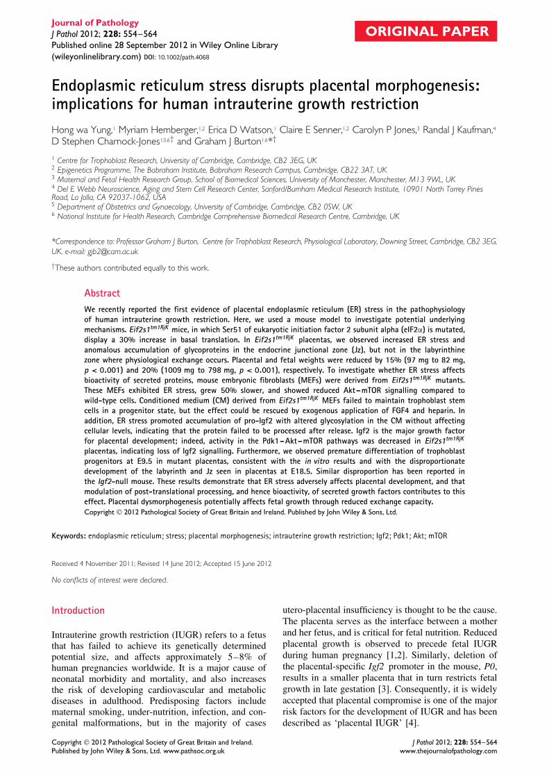
pancreas, where there is an anomalous accumulation ofpro-insulin within β-cells [13]. Therefore, we investi-gated whether there is an equivalent anomalous reten-tion of secreted proteins in the Jz of Eif2s1tm1RjK pla-centas. Placental sections were stained with four differ-ent lectins, [ConA (concanavalin A), e-PHA (Phase-olus vulgaris, erythrohaemagglutinin), DSA (Daturastramonium agglutinin), and STA (Solanum tubero-sum agglutinin)]. Staining with each of the lectinswas rare in the Jz of the wild-type (S/S) placentas.In contrast, strong staining was frequently observed inthe cytoplasm of spongiotrophoblast cells of the Jz inthe Eif2s1tm1RjK (A/A) placentas (Figure 3 and Supple-mentary Figure 3), indicating anomalous intracellularaccumulation of glycoproteins.
Change in glycosylation profile in secreted proteinsreleased from MEFs derived from Eif2s1tm1RjK
embryos
To further elucidate the impact of ER stress onglycosylation of secreted proteins, mouse embry-onic fibroblasts (MEFs) were derived from E13.5Eif2s1tm1RjK and wild-type embryos. Characterizationof Eif2s1tm1RjK MEFs showed an increase of low-gradeER stress, with elevation of phosphorylated and totalPerk, constant Grp78, and no alternative splicing ofXbp-1 mRNA (Figure 4A), equivalent to the patternseen in the Jz. The Eif2s1tm1RjK MEFs grew ∼50%slower, with reduced phosphorylation of Akt at bothThr308 and Ser473, and of eIF4E binding protein 1(4E-BP1) at Ser65, compared with wild-type MEFs,
despite their intrinsically higher translation rate(Figures 4B and 4C).
To examine any change in glycosylation of thesecreted proteins released from Eif2s1tm1RjK MEFs, aconcanavalin A column was used to isolate glyco-proteins from the conditioned media before resolvingby SDS-PAGE. Multiple bands were found to dif-fer between wild-type (S/S) and Eif2s1tm1RjK (A/A)(Figure 5A, arrowed). Similar changes were observedin positive controls, in which wild-type cells weretreated with thapsigargin (Tg) or tunicamycin (Tm),suggesting that these changes likely result from ERstress.
A change in glycosylation pattern may affect thefunction of secreted proteins. Therefore, to investi-gate the bioactivity of secreted proteins released byEif2s1tm1RjK MEFs, a bioassay was established usingtrophoblast stem (TS) cells. TS cells require trophicfactors in MEF conditioned medium for continued pro-liferation and maintenance of their progenitor state,and withdrawal promotes TS cell differentiation intotrophoblast subtypes [19,20]. Therefore, conditionedmedia (CM) from MEFs were used to culture TS cellsfor 3 days. Compared with incubation with wild-typeCM, TS cells cultured in Eif2s1tm1RjK CM showedsignificantly increased levels of transcripts associ-ated with trophoblast differentiation, such as Psx1, anearly differentiation marker; Tpbpa, an ectoplacentalcone/spongiotrophoblast marker; and Pl2 (also knownas Prl3b1 ), a giant cell marker. Correspondingly, therewas a significant decrease in the stem cell marker,Esrrb, and a trend to a reduction in Cdx2 (Figure 5B,left graph). However, supplementation with exogenousFGF4 and heparin in Eif2s1tm1RjK CM prevented thetrophoblast differentiation, as shown by increased Cdx2and reduced Tpbpa mRNA. This finding indicates thatloss of signalling from trophic factors, such as FGF4and TGFβ is the major cause of differentiation in TScells cultured in Eif2s1tm1RjK CM (Figure 5B, rightgraph).
Copyright 2012 Pathological Society of Great Britain and Ireland. J Pathol 2012; 228: 554–564Published by John Wiley & Sons, Ltd. www.pathsoc.org.uk www.thejournalofpathology.com
558 HW Yung et al
Wild-type(S/S) Eif2s1tm1RjK (A/A)
DSA
STA
e-PH
AC
on A
100 µm
Figure 3. Anomalous accumulation of glycoproteins in the endocrine spongiotrophoblast cells of the Jz in the Eif2s1tm1RjK placenta.Biotinylated lectins, ConA (concanavalin A), e-PHA (Phaseolus vulgaris, erythrohaemagglutinin), DSA (Datura stramonium agglutinin), andSTA (Solanum tuberosum agglutinin) were used to stain the placental sections. Note that intensely dark staining accumulations in therounded spongiotrophoblast cells (arrowed) are more common in the mutants. The cells must be distinguished from the irregularly shapedblood channels (asterisks), which also stain positively. All images taken at 20× magnification. Bar = 100 µm.
Igf2 is a major stimulant of placental growth [21] andis released as a heavily glycosylated pro-form. Gen-eration of active Igf2 requires extracellular enzymaticcleavage of the pro-protein. The degree of glycosyla-tion determines its biological activity [22]. The glyco-sylation pattern of pro-Igf2 was changed in the CMfrom Eif2s1tm1RjK (A/A) MEFs compared with wild-type MEFs (Figure 5C). Immunoblotting of the celllysate did not reveal any difference in cellular pro-Igf2levels between the two cell types.
These data indicate that ER stress alters glycosyla-tion of the secreted proteins and can modulate theirbioactivity.
Reduction of Pdk1–Akt–mTOR signalling in theEif2s1tm1RjK placenta
Igf2-deficient mice have smaller placentas and fetuses,and reduced Akt signalling, compared with wild-typelittermates (Supplementary Figure 4) [23]. This sug-gests that the loss of Akt signalling that we observed
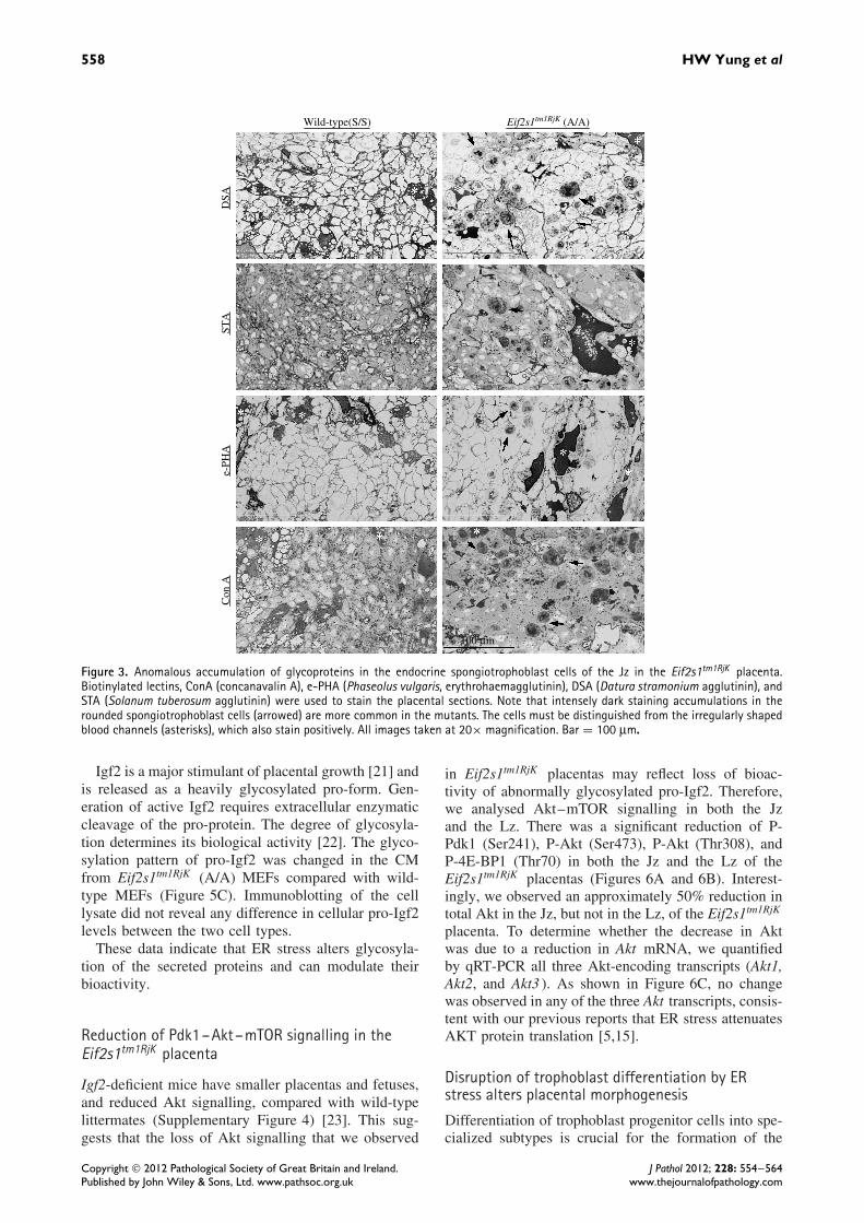
in Eif2s1tm1RjK placentas may reflect loss of bioac-tivity of abnormally glycosylated pro-Igf2. Therefore,we analysed Akt–mTOR signalling in both the Jzand the Lz. There was a significant reduction of P-Pdk1 (Ser241), P-Akt (Ser473), P-Akt (Thr308), andP-4E-BP1 (Thr70) in both the Jz and the Lz of theEif2s1tm1RjK placentas (Figures 6A and 6B). Interest-ingly, we observed an approximately 50% reduction intotal Akt in the Jz, but not in the Lz, of the Eif2s1tm1RjK
placenta. To determine whether the decrease in Aktwas due to a reduction in Akt mRNA, we quantifiedby qRT-PCR all three Akt-encoding transcripts (Akt1,Akt2, and Akt3 ). As shown in Figure 6C, no changewas observed in any of the three Akt transcripts, consis-tent with our previous reports that ER stress attenuatesAKT protein translation [5,15].
Disruption of trophoblast differentiation by ERstress alters placental morphogenesis
Differentiation of trophoblast progenitor cells into spe-cialized subtypes is crucial for the formation of the
Copyright 2012 Pathological Society of Great Britain and Ireland. J Pathol 2012; 228: 554–564Published by John Wiley & Sons, Ltd. www.pathsoc.org.uk www.thejournalofpathology.com

Endoplasmic reticulum stress and placental function 559
S/S
S/S
A/A
A/A
S/S A/A
P-eIF2α(Ser51)
P-Akt(Thr308)
P-Akt(Ser473)
P-4E-BP1(Ser65)
4E-BP1
Ponceau SStaininig
Akt
P-Perk(Thr980)
Perk
Grp78
Ponceau SStaining
Xbp-1mRNA
eIF2α
Tm
US
150%
100%
Rel
ativ
e R
atio
50%
0%
**
Figure 4. Eif2s1tm1RjK MEFs exhibit higher ER stress, slower cell proliferation rate, and lower Akt–mTOR signalling than wild-type controls.(A, C) Both wild-type and Eif2s1tm1RjK (A/A) MEFs were cultured in serum-free medium for 24 h. ER stress markers were investigated usingantibodies against P-Perk, Perk, and Grp78, while Akt–mTOR signalling was examined using specific antibodies against P-Akt(Ser473),P-Akt(Thr308), Akt, P-4E-BP1 (Ser65), and 4E-BP1. Ponceau S staining was used as the loading control. For Xbp-1 mRNA splicing, total RNAwas isolated and RT-PCR was used to detect the spliced variants. Tunicamycin (Tm; 400 ng/ml) treatment for 24 h was used as a positivecontrol. (B) MEFs were seeded at the same density and cultured for 4 days. The number of cells was counted using a haemocytometer.Data are presented as mean ± SEM, n = 4, relative to wild-type (S/S), which is presented as 100%. ∗∗pp ≤ 0.01.
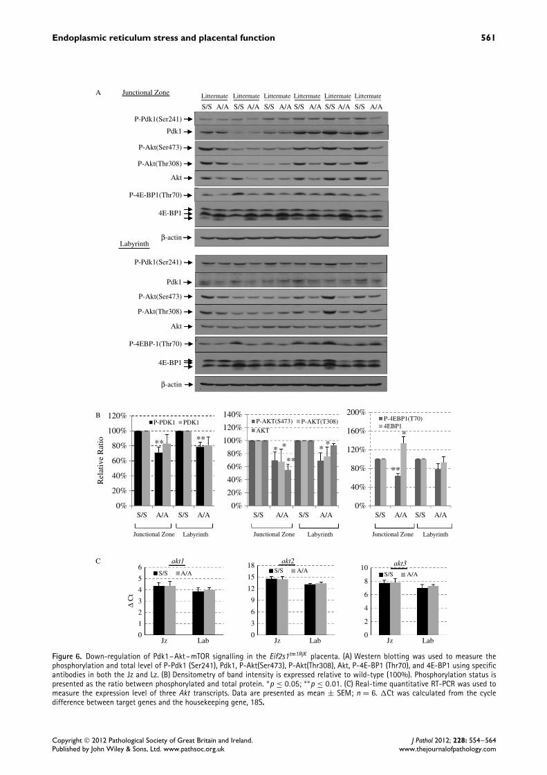
distinct functional zones of the placenta. Trophoblastlineage formation and stem cell maintenance are deter-mined by cell-autonomous nuclear regulators, includ-ing Cdx2 and Esrrb, and paracrine signals such asFgf4 and Activin/Tgfβ/Nodal [19,20,24,25]. Loss ofbioactivity in Eif2s1tm1RjK MEF CM indicated a poten-tial failure in the paracrine signals, thereby alteringtrophoblast differentiation and affecting placental mor-phogenesis. Therefore, we investigated placental struc-ture using stereology to estimate the absolute volumesof the different zones in the Eif2s1tm1RjK placentasat E18.5. As expected, the smaller Eif2s1tm1RjK pla-centas showed an approximately 30% reduction intotal volume (77 mm3 ± 3.9 mm3 versus 112.7 mm3 ±2.8 mm3, p < 0.001, Figure 7A). The compositionof the placenta was also significantly different, witha reduction in both the absolute volume of, andthe proportion occupied by, the Lz (24.77 mm3 ±1.59 mm3 versus 46.49 mm3 ± 2.97 mm3, p < 0.001,or a 21.8% reduction in proportional volume, p <
0.001; Figures 7A and 7B). In contrast, there wasno change in the proportional volume of the Jz inthe Eif2s1tm1RjK placentas (Figure 7B). These resultsindicated the potential disruption of early trophoblastdifferentiation and placental morphogenesis in theEif2s1tm1RjK mice.
During murine development, placental morphogen-esis occurs between E8.0 and E10.5, and formationof the labyrinth and junctional zones is complete byE14.5. Therefore, in order to identify any changein the trophoblast lineage formation, placentas werecollected and analysed by quantitative RT-PCR atE9.5. At E.9.5, all Eif2s1tm1RjK placentas were visuallysmaller than their wild-type counterparts. The early pla-centa is attached to a significant quantity of maternaldecidua. Therefore, in order to examine the propor-tional contribution of different trophoblast cell typesto the entire trophoblast compartment within the pla-centa, a pan-trophoblast lineage marker, Keratin-18,
was used to normalize for trophoblast content. Keratin-18 mRNA level was approximately 50% lower inEif2s1tm1RjK than in wild-type placentas (Figure 7C,left graph). After normalization, the spongiotrophoblastmarker, Tpbpa, was significantly increased, but thegiant cell marker, Pl2, and the syncytiotrophoblastmarker, Gcm1, were both decreased (Figure 7C, rightpanel). As the spongiotrophoblast and syncytiotro-phoblast are the major trophoblast cell lineages withinthe Jz and Lz respectively, these results are consistentwith the stereological data at E18.5 demonstrating asmaller Lz, but no change in the Jz. These findingssuggest that failure of early trophoblast differentia-tion can have long-term consequences for placentaldevelopment.
Discussion
Placental protein synthesis inhibition induced by ERstress was recently identified as a component of thepathophysiology of human IUGR [5]. In this study,we sought an animal model for placental ER stressand used Eif2s1tm1RjK mutant mice because they showER stress due to their elevated basal translation rate[12]. Somatic cells display a range of secretory activ-ity. Those involved in endocrine and exocrine func-tions synthesize and release large quantities of proteins,and hence contain a large amount of rough ER, mak-ing them vulnerable to ER stress. Indeed, ER stressappears to be a normal physiological phenomenon inthe pancreas, one of the most active endocrine andexocrine organs [9]. Excessive protein synthesis hasbeen demonstrated to aggravate ER stress in the pan-creas, promoting accumulation of pro-insulin and apop-tosis in β-cells, reducing circulating levels of insulin,and leading to the development of diabetes [13]. Ame-liorating ER stress is one of the new therapeutic inter-ventions under investigation for the treatment of humandiabetes [26].
Copyright 2012 Pathological Society of Great Britain and Ireland. J Pathol 2012; 228: 554–564Published by John Wiley & Sons, Ltd. www.pathsoc.org.uk www.thejournalofpathology.com

560 HW Yung et al
ConditionedMedia
Con Apurification
A/A A/AS/S S/S- - Tg Tm - - Tg Tm
PM (kDa)
170130
95
55
43
34
17
72
Ponc
eau
Sst
aini
ng
300
A
B
C
250
200
150
Rel
ativ
e L
evel
Rel
ativ
e L
evel
100
50
0 0%
600%S/S CM
A/A CM + FGF & HepS/S CM + FGF & HepA/A CM * *
*
**
**
**
*
**
Cdx2 Esrrb Psx1 Pl1 Pl2Tpbpa Cdx2 Psx1 Pl1 Pl2Tpbpa
500%
400%
300%
200%
100%
A/A
A/A
S/S
S/S
Pro-Igf2 in CM
A/A S/S A/A S/S34
26Anti-Pro-Igf2
Pro-Igf2
β-actin orPonceau S
Ponceau Sstaining
Expt 1 Expt 2 Expt 3
Rel
ativ
e L
evel
CM
Cell L
ysate
200%
150%
100%
50%
0%
Lowest Band Upper Bands
Figure 5. Increased ER stress in Eif2s1tm1RjK (A/A) MEFs modulates the bioactivity of secreted proteins. (A) Alteration of proteinglycosylation profile (arrows). Both Eif2s1tm1RjK and wild-type MEFs, with or without 5 nm thapsigargin (Tg) or 10 ng/ml tunicamycin(Tm), were cultured in serum-free medium for 48 h in the presence of EGF. Conditioned media (CM) were concentrated and glycoproteinsisolated using concanavalin A (ConA) lectin columns, followed by resolving by SDS-PAGE and silver staining. (B) Conditioned medium fromEif2s1tm1RjK MEFs failed to maintain trophoblast stem cells in a multipotent state but this could be reversed by exogenous supplementationwith FGF4 and heparin. Conditioned media prepared from wild-type and Eif2s1tm1RjK MEFs were used to culture TS cells in the absenceor presence of FGF4 and heparin. Total TS cell RNA was isolated, and qRT-PCR was used to assess the relative levels of different nuclearregulators of differentiation including the stem cell markers Cdx2 and Esrrb, the early differentiation marker Psx1, the ectoplacentalcone/spongiotrophoblast marker Tpbpa, and the giant cell markers Pl1 and Pl2. Sdha and Gapdh were used as housekeeping genes. Data arepresented as mean ± SEM from three independent experiments. (C) Altered processing of Igf2 in the Eif2s1tm1RjK MEFs. Immunoblottingof conditioned media or cell lysate prepared from both Eif2s1tm1RjK and wild-type MEFs. Antibodies specific for Pro-Igf2 were used todetect the proteins in both CM and cell lysates. β-Actin and Ponceau S staining were used to show equal loading. Left panel: three blots ofpro-Igf2 in CM and cell lysates are presented to indicate the variation between experiments. The intensities of the lowest band and of theupper few bands were quantified separately to show the mobility shifting of pro-Igf2 due to changes in glycosylation. Right panel: dataare presented as mean ± SEM from four independent experiments. ∗p < 0.05.
The placenta also has a high endocrine activity. Itsecretes numerous hormones and growth factors thatmodulate maternal physiology to ensure continuance ofthe pregnancy and enhance the supply of nutrients, andpromote its own growth through autocrine/paracrinesignalling. The placentally derived insulin-like growthfactors (IGFs) are particularly important for the latter
and, in the mouse, Igf2 is one of the key regulatorsof placental size [21]. Knockout of the P0 placental-specific Igf2 promoter results in a smaller placenta andsubsequent fetal growth restriction [3]. IGF2 also stim-ulates trophoblast proliferation and placental growth inthe human [27], where again reduced placental growthprecedes fetal IUGR [28]. Our findings indicate that
Copyright 2012 Pathological Society of Great Britain and Ireland. J Pathol 2012; 228: 554–564Published by John Wiley & Sons, Ltd. www.pathsoc.org.uk www.thejournalofpathology.com
Endoplasmic reticulum stress and placental function 561
Junctional ZoneA
C
B
Littermate Littermate Littermate Littermate Littermate Littermate
S/S
S/S
S/S S/SA/A A/AS/S A/A
Junctional Zone Labyrinth
A/A S/S A/A S/S
Junctional Zone Labyrinth
A/A S/S A/A S/S
Junctional Zone Labyrinth
A/A S/S A/A
A/A S/S A/A S/S A/A S/S A/A S/S A/A S/S A/A
P-Pdk1(Ser241)
P-Akt(Ser473)
P-Akt(Thr308)
P-4E-BP1(Thr70)
4E-BP1
β-actin
Akt
Pdk1
P-Pdk1(Ser241)
P-Akt(Ser473)
P-Akt(Thr308)
P-4EBP-1(Thr70)
4E-BP1
β-actin
Akt
Pdk1
Labyrinth
120%
100%
80%
60%
40%
Rel
ativ
e R
atio
20%
0%
140%
120%
100%
80%
60%
40%
20%
0%
200%
120%
160%
80%
40%
0%
akt1 akt2 akt3
P-PDK1 P-AKT(S473) P-4EBP1(T70)4EBP1
AKT
P-AKT(T308)PDK1
** **
****
* * * **
6
5
4
3
2
∆ C
t
1
0
18
15
12
9
6
3
0
10
8
6
4
2
0Jz Lab Jz Lab Jz Lab
Figure 6. Down-regulation of Pdk1–Akt–mTOR signalling in the Eif2s1tm1RjK placenta. (A) Western blotting was used to measure thephosphorylation and total level of P-Pdk1 (Ser241), Pdk1, P-Akt(Ser473), P-Akt(Thr308), Akt, P-4E-BP1 (Thr70), and 4E-BP1 using specificantibodies in both the Jz and Lz. (B) Densitometry of band intensity is expressed relative to wild-type (100%). Phosphorylation status ispresented as the ratio between phosphorylated and total protein. ∗p ≤ 0.05; ∗∗p ≤ 0.01. (C) Real-time quantitative RT-PCR was used tomeasure the expression level of three Akt transcripts. Data are presented as mean ± SEM; n = 6. �Ct was calculated from the cycledifference between target genes and the housekeeping gene, 18S.
Copyright 2012 Pathological Society of Great Britain and Ireland. J Pathol 2012; 228: 554–564Published by John Wiley & Sons, Ltd. www.pathsoc.org.uk www.thejournalofpathology.com
562 HW Yung et al
Absolute volumes (mm3) of components in the placenta at E18.5 ofwild-type (S/S) and Eif2s1tm1RjK (A/A)
Placenta Dec Jz Lz Cp
S/S 112.67 ± 2.88 14.67 ± 0.53 43.51 ± 3.21
4.31 ± 0.21
46.49 ± 2.97 8 ± 0.91
24.77 ± 1.5936.78 ± 3.8811.14 ± 1.8277 ± 3.9
<0.0004 <0.0007 <0.0080.1112 0.2297
A/A
t-test (p-value)
S/S A/AD DJz Jz
Lz Lz
Cp Cp
120%
A
B
C
100%
80%
60%
Rel
ativ
e V
olum
eR
elat
ive
Vol
ume
Rat
io a
fter
nor
mal
ized
with
Ker
atin
-18
40%
20%
0%
120%
100%
80%
60%
40%
20%
0%
180%
150%
120%
90%
60%
30%
0%
*
*
*
**
Cp LzJz Dec
S/S A/A
S/S
S/S
A/A
A/A
Pl2 Pl1 Tpbpa Gcm1
**
**
Keratin 18
Figure 7. ER stress disrupts trophoblast stem cell differentiationand placental morphogenesis. (A, B) The absolute volumes andrelative ratios of Lz and chorionic plate, but not of decidua and Jz,were reduced in Eif2s1tm1RjK placentas. Stereological analysis wasused to estimate the absolute volume of different layers. Results arepresented in a table (A) and as a relative ratio in graph (B). A typicalsection of a placenta after H&E staining and used for stereologicalanalysis. D = decidua; Jz = junctional zone; Lz = labyrinth zone;Cp = chorionic plate. Data are expressed as mean ± SEM from fourwild-type and five Eif2s1tm1RjK placentas. (C) Alteration of earlytrophoblast differentiation in Eif2s1tm1RjK placentas. E9.5 placentaswere collected and total RNA was isolated. The levels of the pan-trophoblast marker Keratin-18, the syncytiotrophoblast markerGcm1, Tpbpa, Pl1, and Pl2 were measured. Data are presented asmean ± SEM from five placentas in each group from two litters.All data were normalized with Keratin-18 mRNA before expressedrelative to wild-type (S/S, 100%). ∗p ≤ 0.05.
although excessive protein synthesis did not changethe level of intracellular pro-Igf2, it did alter the gly-cosylation pattern of the secreted protein that maymodulate its bioactivity (Figure 5C). Loss of func-tion of Igf2 released from the Jz may explain thelower Pdk1–Akt–mTOR activity and reduced prolif-eration observed in Eif2s1tm1RjK placentas through areduction in paracrine signalling (Figure 6). Addition-ally, total Akt was lower in the Jz, which sufferedfrom ER stress, but not in the labyrinth, where ERstress was not observed (Figure 6), consistent with ourprevious finding that ER stress attenuates Akt pro-tein translation [15]. The importance of Akt signalling
in placental development is illustrated by the factthat Akt1-knockout mice develop an IUGR phenotype[5,29]. Interestingly, the change in placental structureand Akt signalling observed in the Eif2s1tm1RjK miceis similar to the Igf2 null placenta, with the same dis-proportional reduction of the Jz and Lz, and smallerplacenta size (Supplementary Figure 4) [30].
All secreted and membrane-bound proteins undergomultiple post-translational modifications, such as gly-cosylation and disulphide bond formation, within theER. However, under ER stress these processes are com-promised, resulting in protein misfolding and poten-tially adversely affecting their bioactivity. Indeed, ourresults reveal that the glycosylation pattern of secretedproteins was changed in conditioned medium col-lected from Eif2s1tm1RjK MEFs, and that there wasan anomalous accumulation of glycoproteins in thespongiotrophoblast cells in the placental junctionalzone (Figures 3 and 5A). The change in the glyco-protein profile was associated with altered bioactiv-ity, as the Eif2s1tm1RjK CM failed to maintain TScells in a progenitor cell state. Premature differenti-ation of trophoblast stem/progenitor cells in vivo islikely to limit the expansion of the respective tro-phoblast compartment and to cause a relative over-abundance of post-mitotic trophoblast cell types, lead-ing to smaller placentas, which is what we observed atE9.5. Later, at E18.5, we observed a reduction of theLz, but not the Jz, in the mature Eif2s1tm1RjK mutantplacenta (Figures 7A and 7B). To exclude the possi-bility of a delay in implantation contributing to thesmaller placenta, the number of somites, which rep-resents the developmental stage, was counted in E9.5embryos. No difference was found between wild-typeand Eif2s1tm1RjK mutants (Supplementary Table 1, Sup-plementary Figures 5 and 6, and Supplementary mate-rials and methods).
To conclude, ER stress in placental endocrine cellsaffects the post-translational processing of secretedgrowth factors and other proteins, leading to alteredbioactivity. Compromise of autocrine/paracrine growthfactor signalling provides a mechanistic link betweenER stress, trophoblast differentiation, and altered pla-cental morphogenesis. This may be pertinent to thedevelopment of IUGR in human pregnancies, whereplacental ER stress is a feature of the pathophysiology.This insight may help in the development of novel ther-apeutic interventions targeting placental ER stress forthe treatment of human IUGR.
Acknowledgment
This study was supported by the Wellcome Trust(084804/2/08/Z). Portions of this study were fundedby NIH grants P01HL057346, R37 DK042394, R01DK088227, and R01 HL052173 (RJK). We are gratefulto Melanie Monk for technical assistance.
Copyright 2012 Pathological Society of Great Britain and Ireland. J Pathol 2012; 228: 554–564Published by John Wiley & Sons, Ltd. www.pathsoc.org.uk www.thejournalofpathology.com

Endoplasmic reticulum stress and placental function 563
Author contribution statement
HWY, DSC, and GJB conceived experiments. HWY,MH, EDW, CES, and CPJ carried out experiments.RJK provided Eif2s1tm1RjK mice. HWY, MH, EDW,DSC, and GJB analysed the data. HWY, DSC, and GJBwere involved in writing the manuscript. All authorshad final approval of the submitted and publishedversions.
References
Note: Reference 31 is cited in the Supporting information tothis article.
1. Hafner E, Metzenbauer M, Hofinger D, et al . Placental growthfrom the first to the second trimester of pregnancy in SGA-foetusesand pre-eclamptic pregnancies compared to normal foetuses. Pla-
centa 2003; 24: 336–342.2. Thame M, Osmond C, Bennett F, et al . Fetal growth is directly
related to maternal anthropometry and placental volume. Eur J
Clin Nutr 2004; 58: 894–900.3. Constancia M, Hemberger M, Hughes J, et al . Placental-specific
IGF-II is a major modulator of placental and fetal growth. Nature
2002; 417: 945–948.4. Benton SJ, Hu Y, Xie F, et al . Can placental growth factor in
maternal circulation identify fetuses with placental intrauterinegrowth restriction? Am J Obstet Gynecol ; 2012; 206: 163.e1–7.
5. Yung HW, Calabrese S, Hynx D, et al . Evidence of placentaltranslation inhibition and endoplasmic reticulum stress in theetiology of human intrauterine growth restriction. Am J Pathol
2008; 173: 451–462.6. Schroder M, Kaufman RJ. ER stress and the unfolded protein
response. Mutat Res 2005; 569: 29–63.7. Harding HP, Zhang Y, Bertolotti A, et al . Perk is essential for
translational regulation and cell survival during the unfoldedprotein response. Mol Cell 2000; 5: 897–904.
8. Nika J, Rippel S, Hannig EM. Biochemical analysis of theeIF2beta gamma complex reveals a structural function foreIF2alpha in catalyzed nucleotide exchange. J Biol Chem 2001;276: 1051–1056.
9. Iwawaki T, Akai R, Kohno K, et al . A transgenic mouse modelfor monitoring endoplasmic reticulum stress. Nature Med 2004;10: 98–102.
10. Iwawaki T, Akai R, Yamanaka S, et al . Function of IRE1 alpha inthe placenta is essential for placental development and embryonicviability. Proc Natl Acad Sci U S A 2009; 106: 16657–16662.
11. Harding HP, Zeng H, Zhang Y, et al . Diabetes mellitus andexocrine pancreatic dysfunction in perk−/− mice reveals a rolefor translational control in secretory cell survival. Mol Cell 2001;7: 1153–1163.
12. Scheuner D, Song B, McEwen E, et al . Translational control isrequired for the unfolded protein response and in vivo glucosehomeostasis. Mol Cell 2001; 7: 1165–1176.
13. Scheuner D, Vander Mierde D, Song B, et al . Control of mRNAtranslation preserves endoplasmic reticulum function in betacells and maintains glucose homeostasis. Nature Med 2005; 11:757–764.
14. Oslowski CM, Urano F. The binary switch that controls the lifeand death decisions of ER stressed beta cells. Curr Opin Cell Biol2011; 23: 207–215.
15. Yung HW, Korolchuk S, Tolkovsky AM, et al . Endoplasmic retic-ulum stress exacerbates ischemia–reperfusion-induced apoptosisthrough attenuation of Akt protein synthesis in human choriocar-cinoma cells. FASEB J 2007; 21: 872–884.
16. Jones CJ, Aplin JD, Burton GJ. First trimester histiotrophe showsaltered sialylation compared with secretory phase glycoconjugatesin human endometrium. Placenta 2010; 31: 576–580.
17. Coan PM, Ferguson-Smith AC, Burton GJ. Developmentaldynamics of the definitive mouse placenta assessed by stereology.Biol Reprod 2004; 70: 1806–1813.
18. Singh I, Doms RW, Wagner KR, et al . Intracellular transport ofsoluble and membrane-bound glycoproteins: folding, assembly andsecretion of anchor-free influenza hemagglutinin. EMBO J 1990;9: 631–639.
19. Tanaka S, Kunath T, Hadjantonakis AK, et al . Promotion of tro-phoblast stem cell proliferation by FGF4. Science 1998; 282:2072–2075.
20. Hughes M, Dobric N, Scott IC, et al . The Hand1, Stra13 andGcm1 transcription factors override FGF signaling to promoteterminal differentiation of trophoblast stem cells. Dev Biol 2004;271: 26–37.
21. Roberts CT, Owens JA, Sferruzzi-Perri AN. Distinct actions ofinsulin-like growth factors (IGFs) on placental development andfetal growth: lessons from mice and guinea pigs. Placenta 2008;29 (Suppl A): S42–S47.
22. Valenzano KJ, Heath-Monnig E, Tollefsen SE, et al . Biophysicaland biological properties of naturally occurring high molecularweight insulin-like growth factor II variants. J Biol Chem 1997;272: 4804–4813.
23. DeChiara TM, Efstratiadis A, Robertson EJ. A growth-deficiencyphenotype in heterozygous mice carrying an insulin-like growthfactor II gene disrupted by targeting. Nature 1990; 345: 78–80.
24. Tamai Y, Nakajima R, Ishikawa T, et al . Colonic hamartomadevelopment by anomalous duplication in Cdx2 knockout mice.Cancer Res 1999; 59: 2965–2970.
25. Guzman-Ayala M, Ben-Haim N, Beck S, et al . Nodal proteinprocessing and fibroblast growth factor 4 synergize to maintaina trophoblast stem cell microenvironment. Proc Natl Acad Sci U SA 2004; 101: 15656–15660.
26. Engin F, Hotamisligil GS. Restoring endoplasmic reticulum func-tion by chemical chaperones: an emerging therapeutic approachfor metabolic diseases. Diabetes Obes Metab 2010; 12 (Suppl 2):108–115.
27. Harris LK, Crocker IP, Baker PN, et al . IGF2 actions on tro-phoblast in human placenta are regulated by the insulin-like growthfactor 2 receptor, which can function as both a signaling and clear-ance receptor. Biol Reprod 2010; 84: 440–446.
28. Proctor LK, Toal M, Keating S, et al . Placental size and the pre-diction of severe early-onset intrauterine growth restriction inwomen with low pregnancy-associated plasma protein-A. Ultra-sound Obstet Gynecol 2009; 34: 274–282.
29. Yang ZZ, Tschopp O, Hemmings-Mieszczak M, et al . Proteinkinase B alpha/Akt1 regulates placental development and fetalgrowth. J Biol Chem 2003; 278: 32124–32131.
30. Coan PM, Fowden AL, Constancia M, et al . Disproportionaleffects of Igf2 knockout on placental morphology and diffu-sional exchange characteristics in the mouse. J Physiol 2008; 586:5023–5032.
31. Watson ED, Cross JC. Development of structures and transportfunctions in the mouse placenta. Physiology (Bethesda) 2005; 20:180–193.
Copyright 2012 Pathological Society of Great Britain and Ireland. J Pathol 2012; 228: 554–564Published by John Wiley & Sons, Ltd. www.pathsoc.org.uk www.thejournalofpathology.com
564 HW Yung et al
SUPPORTING INFORMATION ON THE INTERNET
The following supporting information may be found in the online version of this article.
Supplementary materials and methods. Detailed description of materials and methods and data presented in Supplementary Table 1 andSupplementary Figures 5 and 6.
Figure S1. Images of semi-thin toluidine blue-stained sections of the Jz of wild-type (S/S) and Eif2s1tm1RjK (A/A) placentas taken at 20×,40×, and 100× magnification.
Figure S2. No change in other ER stress markers, and no increase in apoptosis in Eif2s1tm1RjK placentas.
Figure S3. Low-power overview of the Jz showing the same pattern of glycoprotein staining as in Figure 3.
Figure S4. Igf2-/- mice display smaller placentas and fetuses, which are associated with reduced Akt signalling.
Figure S5. Litter sizes from Eif2s1+/tm1RjK (S/A) heterozygous inter-crosses at E9.5 and E18.5.
Figure S6. Eif2s1tm1RjK (A/A) mutant embryos within the normal developmental range of E9.5 were growth-restricted compared withwild-type (S/S) and Eif2s1+/tm1RjK (S/A) embryos.
Table S1. Incidence of developmental phenotypes in conceptuses from Eif2s1+/tm1RjK (S/A) heterozygous inter-crosses at E9.5 and E18.5.
Copyright 2012 Pathological Society of Great Britain and Ireland. J Pathol 2012; 228: 554–564Published by John Wiley & Sons, Ltd. www.pathsoc.org.uk www.thejournalofpathology.com