Stress responses at the endometrial-placental interface regulate villous placental differentiation...
17
REPRODUCTION REVIEW Stress responses at the endometrial–placental interface regulate labyrinthine placental differentiation from trophoblast stem cells D A Rappolee 1,2,3,4,5,6,7 , S Zhou 1,4 , E E Puscheck 1,2,3 and Y Xie 1,3 1 CS Mott Center for Human Growth and Development, 2 Department of Obstetrics and Gynecology, 3 Division of Reproductive Endocrinology and Infertility, 4 Program for Reproductive Sciences, Department of Physiology, 5 Institute for Environmental Health and Safety and 6 Karmanos Cancer Institute, Wayne State University School of Medicine, 275 East Hancock, Detroit, Michigan 48201, USA and 7 Department of Biology, University of Windsor, Windsor, Ontario, Canada N9B 3P4 Correspondence should be addressed to D A Rappolee at C S Mott Center for Human Growth and Development, Wayne State University School of Medicine; Email: [email protected] Abstract Development can happen in one of two ways. Cells performing a necessary function can differentiate from stem cells before the need for it arises and stress does not develop. Or need arises before function, stress develops and stress signals are part of the normal stimuli that regulate developmental mechanisms. These mechanisms adjust stem cell differentiation to produce function in a timely and proportional manner. In this review, we will interpret data from studies of null lethal mutants for placental stress genes that suggest the latter possibility. Acknowledged stress pathways participate in stress-induced and -regulated differentiation in two ways. These pathways manage the homeostatic response to maintain stem cells during the stress. Stress pathways also direct stem cell differentiation to increase the first essential lineage and suppress later lineages when stem cell accumulation is diminished. This stress-induced differentiation maintains the conceptus during stress. Pathogenic outcomes arise because population sizes of normal stem cells are first depleted by decreased accumulation. The fraction of stem cells is further decreased by differentiation that is induced to compensate for smaller stem cell populations. Analysis of placental lethal null mutant genes known to mediate stress responses suggests that the labyrinthine placenta develops during, and is regulated by, hypoxic stress. Reproduction (2013) 145 R139–R155 Introduction and summary of goals of the review One goal of this review is to define the trophoblast and embryonic stem cell (TSC and ESC, respectively) lineages in the implanting embryo as the receiving center for normal and pathogenic stress signals for placental development. These signals come from the conceptus and the endometrium. The focus will be on how stress activates adaptive mechanisms that change from low to high exposures (‘exposure’ encompasses duration and magnitude) and pathogenic mechanisms with adverse effects at higher exposures. Adverse effects are mediated by compensatory and prioritized stem cell differentiation (Fig. 1), two phenomena we have discussed elsewhere (Rappolee et al. 2010, 2012, Xie et al. 2011). These stress response mechanisms are important shortly after implan- tation in mice during a period equivalent to the early part of the first-trimester in humans. Another goal is to show that developmental plasticity builds on a foundation of normal mechanisms of development that are overlaid by a rheostat-type control regulated by stress. The normal mechanisms can be modulated by stress developing during normal develop- ment, but elevated stress can lead to adaptation or pathogenesis dependent on the stress exposure. This rheostat regulates the timing and magnitude of the proportional production of differentiated cell lineages. The rheostat regulates stem cells in a sequence of events during normal development and development regulated by elevated stress. What is stress? We have defined this previously (Rappolee 2007, Xie et al. 2011). In developmental terms, stress exposures of sufficient duration and magnitude cause diminished stem cell population growth. This affects the program that balances stem cell potency and differentiation. We use the word stress in general terms, not to uniquely describe oxidative stress, endoplasmic reticu- lum (ER) stress, heat stress, ‘metabolic stress’ of malnutrition, genotoxic stress, or maternal stress hor- mone responses (Houghton et al. 1996, Kwong et al. 2000, Baumann et al. 2007, Burton et al. 2009, q 2013 Society for Reproduction and Fertility DOI: 10.1530/REP-12-0240 ISSN 1470–1626 (paper) 1741–7899 (online) Online version via www.reproduction-online.org
Transcript of Stress responses at the endometrial-placental interface regulate villous placental differentiation...

R
EPRODUCTIONREVIEWStress responses at the endometrial–placental interface regulatelabyrinthine placental differentiation from trophoblast stem cells
D A Rappolee1,2,3,4,5,6,7, S Zhou1,4, E E Puscheck1,2,3 and Y Xie1,3
1CS Mott Center for Human Growth and Development, 2Department of Obstetrics and Gynecology, 3Division ofReproductive Endocrinology and Infertility, 4Program for Reproductive Sciences, Department of Physiology,5Institute for Environmental Health and Safety and 6Karmanos Cancer Institute, Wayne State University School ofMedicine, 275 East Hancock, Detroit, Michigan 48201, USA and 7Department of Biology, University of Windsor,Windsor, Ontario, Canada N9B 3P4
Correspondence should be addressed to D A Rappolee at C S Mott Center for Human Growth and Development, Wayne StateUniversity School of Medicine; Email: [email protected]
Abstract
Development can happen in one of two ways. Cells performing a necessary function can differentiate from stem cells before the need for
it arises and stress does not develop. Or need arises before function, stress develops and stress signals are part of the normal stimuli that
regulate developmental mechanisms. These mechanisms adjust stem cell differentiation to produce function in a timely and proportional
manner. In this review, we will interpret data from studies of null lethal mutants for placental stress genes that suggest the latter
possibility. Acknowledged stress pathways participate in stress-induced and -regulated differentiation in two ways. These pathways
manage the homeostatic response to maintain stem cells during the stress. Stress pathways also direct stem cell differentiation to increase
the first essential lineage and suppress later lineages when stem cell accumulation is diminished. This stress-induced differentiation
maintains the conceptus during stress. Pathogenic outcomes arise because population sizes of normal stem cells are first depleted by
decreased accumulation. The fraction of stem cells is further decreased by differentiation that is induced to compensate for smaller stem
cell populations. Analysis of placental lethal null mutant genes known to mediate stress responses suggests that the labyrinthine placenta
develops during, and is regulated by, hypoxic stress.
Reproduction (2013) 145 R139–R155
Introduction and summary of goals of the review
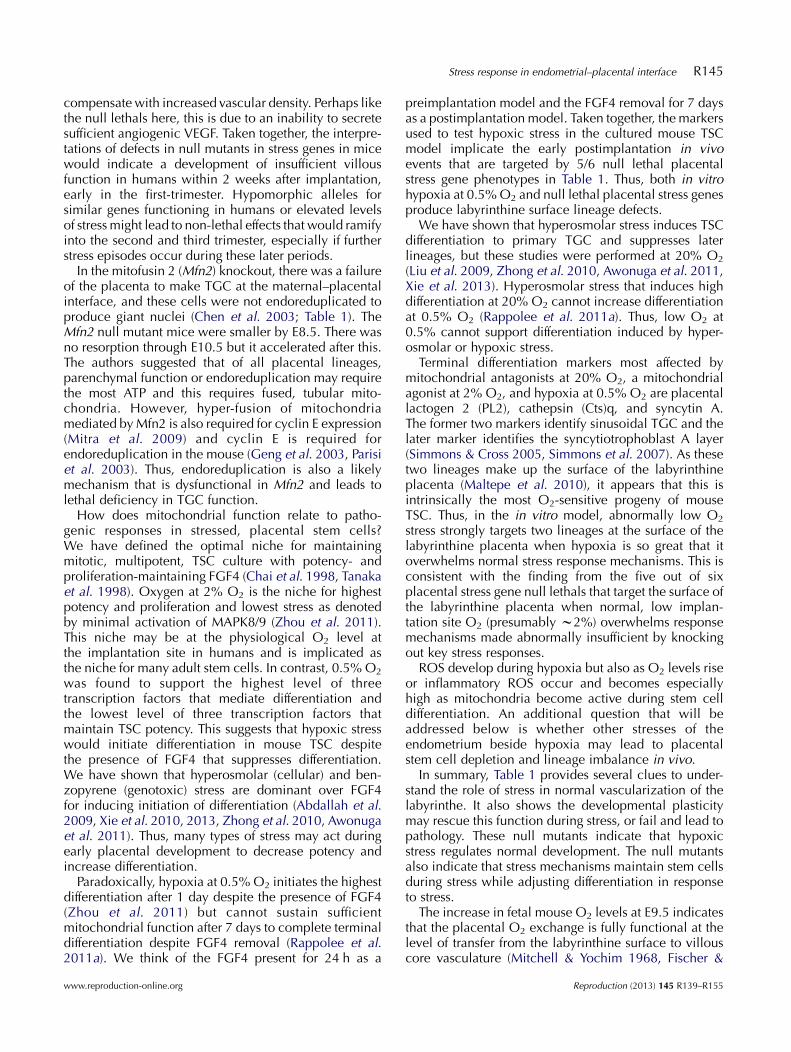
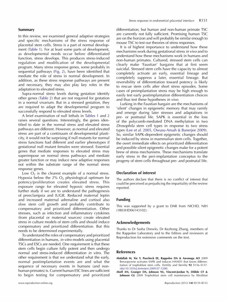
One goal of this review is to define the trophoblast andembryonic stem cell (TSC and ESC, respectively) lineagesin the implanting embryo as the receiving center fornormal and pathogenic stress signals for placentaldevelopment. These signals come from the conceptusand the endometrium. The focus will be on how stressactivates adaptive mechanisms that change from low tohigh exposures (‘exposure’ encompasses duration andmagnitude) and pathogenic mechanisms with adverseeffects at higher exposures. Adverse effects are mediatedby compensatory and prioritized stem cell differentiation(Fig. 1), two phenomena we have discussed elsewhere(Rappolee et al. 2010, 2012, Xie et al. 2011). These stressresponse mechanisms are important shortly after implan-tation in mice during a period equivalent to the early partof the first-trimester in humans.
Another goal is to show that developmental plasticitybuilds on a foundation of normal mechanisms ofdevelopment that are overlaid by a rheostat-type controlregulated by stress. The normal mechanisms can be
q 2013 Society for Reproduction and Fertility
ISSN 1470–1626 (paper) 1741–7899 (online)
modulated by stress developing during normal develop-ment, but elevated stress can lead to adaptation orpathogenesis dependent on the stress exposure. Thisrheostat regulates the timing and magnitude of theproportional production of differentiated cell lineages.The rheostat regulates stem cells in a sequence of eventsduring normal development and development regulatedby elevated stress.
What is stress?
We have defined this previously (Rappolee 2007, Xieet al. 2011). In developmental terms, stress exposuresof sufficient duration and magnitude cause diminishedstem cell population growth. This affects the programthat balances stem cell potency and differentiation.
We use the word stress in general terms, not touniquely describe oxidative stress, endoplasmic reticu-lum (ER) stress, heat stress, ‘metabolic stress’ ofmalnutrition, genotoxic stress, or maternal stress hor-mone responses (Houghton et al. 1996, Kwong et al.2000, Baumann et al. 2007, Burton et al. 2009,
DOI: 10.1530/REP-12-0240
Online version via www.reproduction-online.org

Preimplantation-postimplantation
Increasingstress
Developmentaltime
Unsuccessfulessential lineage 1
Unsuccessfulessential lineage 2
UnsuccessfulEssential lineage 2
Successfulessential lineage 1
Successful Hand1,pTGC, PL1
SuccessfulGcm1. chorionictrophoblast
Essential lineage 1
Essential lineage 2
1
2
4
3
Non-unstress
Minimalessentialdifferentiatedfunction
Differen-tiatedcells
Differentiatedcells
Differentiatedcells
Differentiatedcells
Stem cells
Stem cells
Lineage 1 Lineage 2
Stem cells
Stemcells
Figure 1 Diagram showing stress-inducedcompensatory and prioritized differentiation of TSC.Compensatory differentiation: as stress decreases thegrowth of the stem cell population (examples 2, 3 and4), a higher fraction of differentiated cells compensatesfor fewer cells. For compensatory differentiation, notethat ‘minimal essential differentiated function’ isdenoted by a light green box for lineage 1 that isthe same size as in examples 2, 3 and 4. However, thesize of the total box is smaller from examples 2!3!4resulting in greater differentiated product/cell in 2O3O4. Prioritized differentiation: as stress decreases thegrowth of the stem cell population (examples 3 and 4),early essential lineages are increased and lateressential lineages are suppressed. Example 1, no stemcell growth from pre- to postimplantation results inspontaneous miscarriage before chemical pregnancy.This zero growth level of stress also converts all stemcells into the first lineage. Example 2, stem cellpopulationgrowth only to minimal essential number ofcells sufficient only for first essential lineage results inchemical pregnancy and spontaneous miscarriage.Example 3, sufficient stem cell population growthoccurs for chemical pregnancy and partial essentiallineage 2 then pregnancy loss. Example 4, normalunstressed stem cell population growth accommo-dates first and second essential lineages with stem cellsin reserve. Reversible ‘pseudo-differentiation’ mayrescue a range of decreased stem cell population sizesbetween examples 2, 3 and 4 and rescue pregnancyloss. Between examples 2 and 4, a range of patho-physiological stem cell differentiation strategies, someepigenetic, may result in long-term placental, fetal,and postnatal dysfunctions including preeclampsia,intrauterine growth restriction (IUGR) and develop-mental origins of health and disease (DOHAD) effects.
R140 D A Rappolee and others
Scifres & Nelson 2009, Mu et al. 2011, Xie et al. 2011).But all these are important and mediate shared stressoutcomes such as diminished stem cell accumulation rates.
The early conceptus and its stem cells are highlyanabolic. All cells in the implanting embryo are stemcells, except mural trophectoderm, and all divide. Earlyfunctions require high energy; one example is thepumping of the mural trophectodermal epithelium thatmakes the cyst in the blastocyst before implantation(Brison & Leese 1991, 1994). Before implantation inmouse, energy is required for huge waves of newtranscription at zygotic genome activation at the two-cell stage and around the time of compaction at the eightcell stage (Hamatani et al. 2004, Wang et al. 2004). Atthese two stages, nearly 9000 types of mRNA transcriptsare produced. Immediately after implantation, theproduction of endocrine, antiluteolytic hormones by
Reproduction (2013) 145 R139–R155
the first differentiated placental cells and the epithelialnutrient acquisition of the first extraembryonic endodermare necessary for survival and require high levels of energy(Rappolee 1999). Stressors that trigger reactive oxidativestress (ROS) or ER stress deplete energy that normally goesto stem cell population expansion and early differentiationevents.
We focus on the common outcomes of stressresponses. These are decreased anabolism, decreasedproliferation, and increased fractions of stem cells thatmust differentiate to sustain essential functions afterimplantation. Stressors that decrease stem cell accumu-lation include malnutrition, maternal stress hormones,inflammatory cytokines, shear stress, improper culturemedia, improper O2 levels, environmental toxicants,and many others (Xie et al. 2011). Malnutrition in vivo orserum deprivation in vitro exacerbates other stresses by
www.reproduction-online.org

Stress response in endometrial–placental interface R141
decreasing the carbon supply needed to support thestress response and maintain stress enzymes at a lowlevel (Xie et al. 2011). Maternal leptin provides anendocrine signal proportional to, and produced by,adipocytes which signal that energy reserves aresufficient for reproduction (Krasnow & Steiner 2006).But mouse oviductal and uterine epithelium providesparacrine confirmation of this leptin signal and canincrease stem cell growth rates in the preimplantationembryo (Kawamura et al. 2003). In contrast, maternaladrenaline and cortisol can decrease blastocyst and stemcell growth in vitro and in vivo (Xie et al. 2011). Thesestresses lead to decreased stem cell accumulation and,by the mechanisms of compensatory and prioritizeddifferentiation, stress-induced differentiation.
Are there stress enzymes?
This has been discussed previously (Xie et al. 2011).We will discuss several criteria that enable us to defineas stress enzymes a small subset of approximately 500protein kinases in the kinome (Caenepeel et al. 2004).We will use as an example two subfamilies of the MAPKsuperfamily; these are stress-activated protein kinases(SAPK) and p38MAPKs. Mammalian stress enzymes suchas MAPK8/9/10 (SAPK/JNK1/2/3) and MAPK11–14(p38MAPKs) are activated highly by many stresses(hyperosmolar stress, cytokines, toxicants, DNAdamage, etc.) (Xie et al. 2011) but that mitogenicsignaling MAPK1/3 (aka ERK) is not highly induced bythese stresses. Conversely, mitogenic signals highlyupregulate MAPK1/3 but not MAPK8/9 or MAPK11–14.
Stress enzymes and their immediate substrates arepart of a ‘health insurance policy’ synthesized beforestress, so it is not surprising that they mediate normalfunctions before stress levels increase. This does notdiminish the significance of their role in mediatingsurvival and other responses when stress levels increase.
Cultured cells under certain stressful conditions mayrequire MAPK8/9 to permissively respond to stress sothat cell division can occur. But overexpression ofMAPK8/9 or their upstream activators MEK kinase(MEKK)4/7 has not been shown to instructively activatecell division. In contrast, MEKK1/2 overexpression anddownstream MAPK1/3 activation can lead to transfor-mation of cells in focus-forming assays (Cowley et al.1994). MAPK8/9 function is complex and dependent onstress levels in early embryogenesis. During the stress ofculture, MAPK8/9 is needed to sustain embryos inthe most stressful media but slows development in theleast stressful media (Wang et al. 2005, Xie et al.2006). Thus, other mitogenic MAPKs are instructiveand sufficient but stress enzymes MAPK8/9 may onlybe permissively involved with mitogenesis underspecial conditions. MAPK8/9 can be adaptive atlower stress exposures but can be pathogenic at higher,adverse exposures.
www.reproduction-online.org
Can stress serve as a normal cue for placentaldevelopment?
Emerging evidence suggests that the conceptus buildsdifferentiation programs over time. These programs arepartially guided during synthesis and triggered intoactivity by stress. Mouse null lethal mutants show howessential parenchymal function builds in the extraem-bryonic endoderm soon after implantation at E4.5–6.5(Rappolee 1999), in heart, red blood cells, and a closedvascular system at E8.5, and in placental chorioallantoicfusion, villous vasculature and epithelium, and endo-metrial invasion by E11.5 (Copp 1995).
In this section, we analyze null mutants in knownstress pathways that have a lethal phenotype due toplacental dysfunction. The mutants were found usingEndNote search engine set for ‘all fields’ or ‘abstract’to screen the PubMed Database using key words‘placenta’, ‘knockout’, ‘lethal’, and/or ‘stress’.
It might be expected that the six placental lethalsfound by this search might be distributed throughoutmany placental cell types and throughout the majornecessary period of placental function at E11.5 identifiedby null lethals in the seminal review by Copp (1995).Since this review by Copp, others have cataloged andanalyzed nearly 100 placental null lethals (Rossant &Cross 2001, Watson & Cross 2005). Although ourliterature screen produced only six placental stressgene null lethals, five of these were shown to functionat the surface of the mouse labyrinthine placenta.Furthermore, these lethalities developed over a 2-dayperiod in a normal (unstressed) vivarium, E8.5–10.5,during the initiation of villous function (Table 1). We willinterpret this as support for a model where need developsbefore function during development of the mouselabyrinthine placenta and that this results in stress. Thisstress requires response mechanisms that regulate intra-cellular stem cell adaptation and extracellular mechanismsthat coordinate development of labyrinthine function.
Introduction to Table 1
O2 diffusion limitations trigger responses first observedat E8.5 when the mouse embryo reaches a size whenfunctioning heart, closed vascular system, and red bloodcells are required (Copp 1995). Extraembryonic endo-derm and then placenta are required to provide nutrientsderived from uterine glands and maternal blood.Chorioallantoic fusion occurs at E8.5 when mesenchymedigitates from the allantois into the chorion to form thelabyrinthine cores. As the labyrinthine epithelium,consisting of sinusoidal trophoblast giant cells (sTGC)and syncytiotrophoblasts, differentiates, it develops thecapability to sense low O2 as a stress. This inducesvascular endothelial growth factor (VEGF) that inducesvillous endothelial cells via VEGF receptor (VEGFR)2and tubularization through VEGFR1 (Hanahan 1997).
Reproduction (2013) 145 R139–R155

Table 1 Stress enzyme and other stress mediators are important in placental development, as illustrated by null mutant lethal phenotypesa.
Earliest noted deviationb
Stress gene Onset, site of placental expression Onsetb, cause of lethality Reference
HuR (stress granule) Unique cytoplasmic in labyrinthineepithelium
E10.5 Katsanou et al. (2009)
Required in epithelium, deviatedat E8.5c–9
Failure to initiate labyrinthine invasionand branching
p38MAPKa (stress enzyme) Significant and required in labyrinthineepithelium, deviated at E10.5
E10.5 Mudgett et al. (2000)
Failure of labyrinthine vascularizationb
after invasionHIF1b/ARNT (hypoxic stress) Significant and required in labyrinthine
epithelium, deviated at E8.5cE10.5 Adelman et al. (2000a)
Failure of labyrinthine vascularizationafter invasion
HSP90b (heat stress) Ubiquitous required inmesenchyme, deviated at E10
E10.5 Voss et al. (2000)
Failure of labyrinthine vascularization,yolk sac vascular failure E9.5–10.5
IRE1 (ER stress) In labyrinthine epithelium and core,ubiquitous E13.5 (only test time)
E13.5 Iwawaki et al. (2009)
Failure of labyrinthine vascularizationafter invasion
Mfn2 (highest energy) Ubiquitous smaller embryos at E8.5c E11.5 Chen et al. (2003)Failure to make TGC layer
It is also possible that occult lineage imbalance occurs earlier but is not revealed by deficits in parenchymal function until a later essentialrequirement occurs.aNote that these knockouts were analyzed in mice housed in a normal vivarium and should report endogenous stress-induced cues for normaldevelopment. Other knockouts with placental lethality due to stress responses not discussed in detail include JunB (Schorpp-Kistner et al. 1999),Oct1 (Sebastiano et al. 2010), PPARg (Barak et al. 1999), MEK1 (Giroux et al. 1999), Zfp36L1 tristetraprolin (TTP) family of tandem CCCH fingerproteins (Stumpo et al. 2004), and FoxO1 (Ferdous et al. 2011). bIt should be noted that the timing of deviation from normalcy or lethality is not exactand dissimilar timings may represent differences in assays rather than biological reality. Timing of the beginning of morphological changes wereobserved as early as E8.5, before E9.5 when fetal O2 increases, indicating the start of functioning labyrinth (Fischer & Bavister 1993). cThe earliestvascularization lethality possible is indicated by null lethality of Flk1/VEGFR2 (endothelial cell induction mediator) and Flt1/VEGR1 (vascular tubulemediator), which occur at E8.5 (Hanahan 1997).
R142 D A Rappolee and others
A series of null mutant genes are in stress pathways thatfeed into VEGF induction in the labyrinthine epithelium.We hypothesize that the VEGF pathway may be activatedwithout stress, but it also has the capacity to be adjustedby hypoxic stress during development of the labyrinthineplacenta.
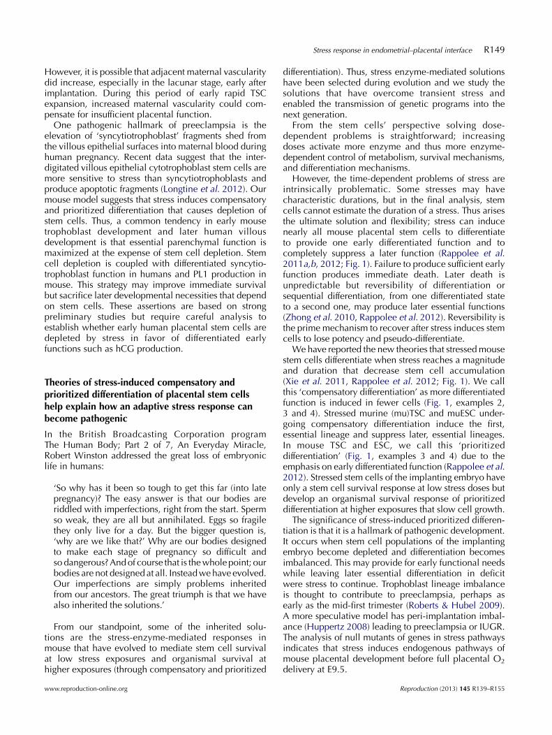
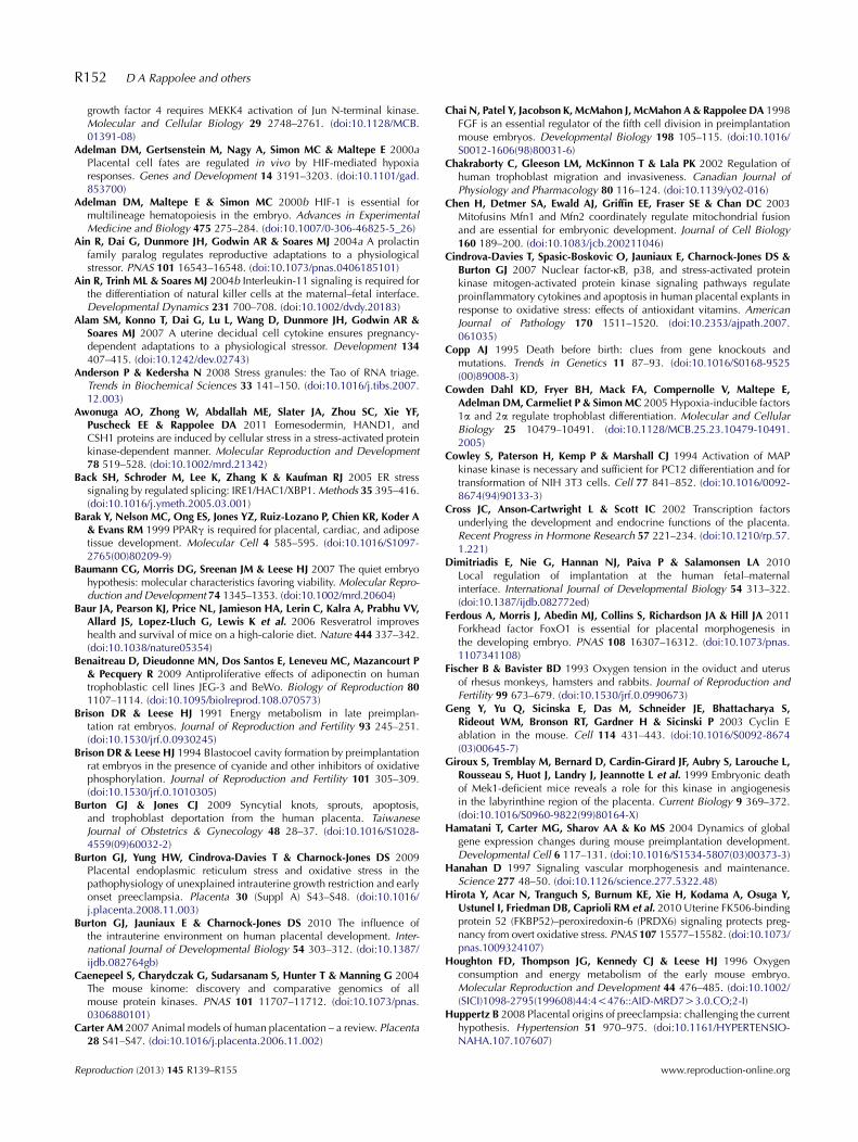
Table 1 shows a number of mouse null mutants forstress enzymes and stress response genes that are neededfor survival of the conceptus when gestational femalesare housed in a normal vivarium. These are multi-function genes, so it is not clear which functions in deficitlead to death. But these genes are in acknowledged stresspathways and similar stress response functions of the genesargue for their activation by an endogenous stress (Fig. 2).The stress is hypoxia and it induces autonomous cellsurvival and non-cell-autonomous differentiated neovas-cular and angiogenic induction by VEGF. We hypothesizethat without the functions of these genes, VEGF inductionis insufficient and/or the survival response of cells isinsufficient.
Inositol-requiring enzyme 1 (IRE1) is the ER stresssensor (Table 1, Fig. 2) that activates protein kinase RNA(PKR), PKR-like ER kinase 1 (PERK1), and general controlnon-derepressible 2 (GCN2) that are able to phosphor-ylate eukaryotic translation initiation factor 2A (eIF2a)(Back et al. 2005, Oikawa & Kimata 2011). This leads to
Reproduction (2013) 145 R139–R155
cessation of protein synthesis and is a canonical stressresponse to resist difficulties and stress and reestablishnormal levels of ATP by slowing anabolism andincreasing catabolism. eIF2a phosphorylation leads totransfer of mRNA, translating in ribosomes into storage inhuman antigen R (HuR)-positive stress granules, not intoP-bodies for destruction (Anderson & Kedersha 2008).In humans, PERK1 is elevated in preeclamptic placenta,suggesting that these are under stress high enough toactivate the unfolded protein response (Burton et al. 2010).In humans, this stress may be due to insufficient responseto the oxidative stress of hypoxia–reoxygenation at thestart of the second trimester. Second-trimester arterialhypoxia arises from insufficient trophoblast invasion inthe first trimester. However, in cultured mouse trophoblaststem cell (TSC) models, implantation site hypoxia at 0.5%O2 or lower initiates the greatest induction of differen-tiation factor and greatest loss of potency factors (Zhouet al. 2011). This would impair initiation of normal mouselabyrinth differentiation that would emulate decreasedhuman villous function in the first trimester.
The mouse null mutant shows that IRE1 is activatedand functions through VEGFA to vascularize thelabyrinthine placental core to create function thatbecomes essential by E12.5 (Iwawaki et al. 2009).However, this phenotype develops in a normal vivarium
www.reproduction-online.org

Hypoxic stressHyperosmolar(cellular) stressInflammation(cytokine) stressInfection (TLR4)Toxic (BaP) stress
ATP consumptionanabolism
ATP synthesiscatabolism
Metabolic stress(undernutrition,
serum deprivation)
Stemness
Glycolysis
Glycogen
P-body
Ribosome
?
?
ID2 protein
NucleusVEGF
SAPK
Endoplasmicreticulum
Stressedstemcell
Genotoxicstress
αβ
γ
AMPK
MAPK14
HIF1α/2α
HIF1α
JunB
HSP90β
PhosphoelF2a
Stressgranule
HuR
SAPK
Growth,apoptosis,Gcm1
Plasmamembrane
ER stress
(Id 2)mRNA
Hand1mRNAprotein
PERK
IRE1
Mitochondrion
Proteosome
Figure 2 Multiple intracellular and extracellularstresses convert anabolism to catabolism, suppressapoptosis at low doses, and at high doses suppressgrowth and induce loss of potency factors andprioritized gain of early essential differentiationfactors. BaP, benzopyrene; TLR4, toll-like receptor 4,lipopolysaccharide receptor; Id2, inhibitor of differ-entiation 2; HuR, human antigen R; Hand1, heartand neural crest derivative inducer; Gcm1, glial cellsmissing 1; eIF2a, eukaryotic translation initiationfactor 2A; SAPK, stress-activated protein kinase;AMPK, AMP-activated protein kinase; aka Jun kinase,MAPK8/9; PERK, PKR-like ER kinase; IRE1, inositol-requiring enzyme 1. Red ellipses show placentalstress null lethals discussed in Table 1 and in the text(JunB). The rough endoplasmic reticulum andnuclear figures were used with kind permission ofMolecular Expressions (http://micro.magnet.fsu.edu)and the proteasome figure was used with kindpermission of wikipedia (http://en.wikipedia.org/wiki/Proteasome)
Stress response in endometrial–placental interface R143
and occurs during an earlier formative period of themouse labyrinth structure compared with the inductionof elevated PERK1 in later preeclamptic human placenta.It indicates that IRE1 is activated by hypoxic stress inmouse and acts to regulate vascularization of thelabyrinth during placentation to provide sufficient gas/nutrient exchange. Taken together with the interpretationof human data, it suggests that the IRE1 sensor receivesnormal hypoxic signals to feed forward into trophoblastdevelopment, but elevated stress signals lead to sloweranabolism and labyrinthine growth. In other words, IRE1and ER stress mechanisms are part of the developmentalplasticity shared by normal and pathogenic responsesearly and late in placental development.
The similar requirements during placental deve-lopment for p38MAPKa (aka MAPK14), HIF1a/2a,and its requisite heterodimer partner HIF1b (akahypoxia-inducible factor 1a/2a and HIF1b/ARNTrespectively) has been reviewed elsewhere (Rappolee2007). Interestingly, MAPK14a is much more highlyexpressed than other p38MAPK isoforms (p38MAPKd/MAPK13 and p38MAPKg/MAPK12) in preimplantationmouse embryos (Zhong et al. 2004) (Table 1, Fig. 2).
Although it is not known whether MAPK14a is thedominant isoform in first-trimester human placenta, forthe SAPK/JNK subfamily of the MAPK superfamily,
www.reproduction-online.org
SAPK/JNK2 (MAPK9) is the dominant isoform (comparedwith JNK1/MAPK8 and JNK3/MAPK10) in mousepreimplantation embryos and in weeks 5–7 humanvillous explants. In early postimplantation mouse,MAPK9 is the dominantly expressed form of theSAPK/JNK subfamily of MAPK and contributes tohyperglycemia-induced diabetic embryopathy in amouse model for diabetes (Yang et al. 2007, 2008a).Thus, for MAPK9, dominant expression before and afterimplantation is correlated with dominant mediation ofdiabetic stress effects early after implantation. Byexpression and function, it is likely that for thep38MAPK subfamily, MAPK14 is dominant in humansin the early first-trimester placenta.
MAPK14 and HIF1b null mouse mutants developlethal placental phenotypes that are phenocopies.Similar to the IRE1 null mutant phenotype, the nullmutants for MAPK14 and HIF1b or HIF1a/2a have adeficiency in labyrinthine villous core vascularization.MAPK14 nulls fail to develop sufficient labyrinthinevasculature and this results in apoptosis and labyrinthinefailure by E9.5, earlier than the IRE1 null mutant failure(Mudgett et al. 2000). Mouse ARNT null embryos haveinsufficient vascularization of the yolk sac and labyr-inthine villous mesenchyme and die by E10.5, andARNT null ESC chimeras are normal while ARNT null
Reproduction (2013) 145 R139–R155
R144 D A Rappolee and others
lethality is prevented by tetraploid fusion rescue(Maltepe et al. 1997, Adelman et al. 2000a). BothAMP-activated protein kinase (AMPK) and MAPK14(which is often downstream of AMPK) are knownto activate HIF in other cell models (Xie et al. 2011).It is likely that HIF is required in TSC-derived placentalectoderm of the villi to induce vascularization ofthe ESC-derived mesenchymal core, which does notrequire HIF.
The common phenotype for MAPK14 and HIF1 ishypoxia-induced placental ectoderm-to-mesenchymesignaling to induce vascularization (Adelman et al.2000a, 2000b, Mudgett et al. 2000, Cowden Dahl et al.2005, Maltepe et al. 2005). HIF is required for normalspongiotrophoblast lineage and subsequent productionof invasive TGC. But the initial requirement is for normalmesenchymal invasion of the chorion and its vascular-ization. Labyrinthine villous vascular failure is dominantalthough spongiotrophoblast and invasive TGC are alsopartially reduced in the HIF1b/ARNT nulls.
HIF functions have been most-studied duringresponses to hypoxic stimulation. However, it shouldbe noted that HIF1a/1b gene products are induced andhave essential functions when fibroblast growth factor 4(FGF4) is removed and cultured mouse TSC differentiatenormally at ambient O2 levels (Maltepe et al. 2005). Thelineage-governing effects of HIFs during normal differ-entiation or hypoxic differentiation are different inquantity and quality (Maltepe & Simon 1998, CowdenDahl et al. 2005, Maltepe et al. 2005), suggesting thatHIFs are part of a developmentally plastic program tocreate placental and labyrinthine function. HIF isimportant in normal mouse TSC differentiation inmediating lineage choice. But HIF acts to maintainpotency and block differentiation when hypoxic stresselevates as O2 levels drop below the optimum for TSCproliferation and potency at 2% O2.
HuR is another multifunctional protein that is inducedby many stresses with pleiotropic effects, the mostimportant involves the eIF2a phosphorylation event(Kedersha & Anderson 2002; Table 1, Fig. 2). Theactivation of the HuR response can occur via oxidativeor ER stress through IRE1 and PERK. This leads to thetransition of mRNA from active ribosomes to inactiveHuR-positive stress granules where the mRNA isprotected (Anderson & Kedersha 2008). This saveslarge amounts of energy after stress subsides, andexisting mRNA returns to the ribosomes from stressgranules. An alternate strategy would be to destroymRNA during stress and re-synthesize it after stress; butdestruction and resynthesis of mRNA require largeamounts of energy. For an implanting conceptus withlarge energy requirements for proliferation and differen-tiation, protecting mRNA during stress is a key function.The mouse HuR knockout is a placental lethal withfailure to initiate the mesenchymal interdigitation intothe chorion at chorioallantoic fusion (Katsanou et al.
Reproduction (2013) 145 R139–R155
2009). It results in an early initiation of dysfunction atE8.5–9.0. Use of conditional Cre-lox-driven recombina-tion showed that only trophoblasts required HuR, notvillous mesenchyme and endothelium. HuR is mostlycytoplasmic in villous placental epithelium where it mightbe involved with stress granules. The HuR null mutantconceptuses developed normal spongiotrophoblasts andtrophoblast giant cells (TGCs) and only the chorionicinteraction with the allantois was faulty. An importantdefect was the failure of the allantoic mesoderm tovascularize. Thus, like IRE1, MAPK14, and HIF1b, HuR isa stress response gene that is necessarily activated in thechorionic epithelium to develop and vascularize mouselabyrinthine mesenchymal cores.
Interestingly, we have shown that stress induces areversible decrease in mouse TSC accumulation ratesand an w80% loss of ID2 protein (Zhong et al. 2010).ID2 protein loss is proteasome-dependent and notdependent on mRNA loss. However, Id2 mRNA isretained at 100% of unstressed levels (Liu et al. 2009)during the w80% loss of ID2 protein. Thus, it is possiblethat the essential role of HuR (Mazroui et al. 2007) is inthe reversible adaptation to stress by preserving mRNAlike Id2 in stress granules for use in new translation oncethe stress subsides. Hypoxic stress at !2% O2 is likely tosubside at E9.5 in mice when fetal oxygen rises sharplyand indicates that both maternal and fetal placentalvascularization is in full operation.
Heat-shock protein 90b (HSP90b) is a master HSPepistatically near to end function and the mouse HSP90bnull mutant generates placental lethality due to labyr-inthine vascularization failure at E10.5 (Voss et al. 2000;Table 1, Fig. 2). Although HSP90b is expressedubiquitously, chimeric analysis of the null mutantshowed that the primary failure occurred when themutant was in the allantois and mesenchymal core, notthe epithelium. Lack of HSP90b in the labyrinthinemesenchyme indirectly led to incomplete differentiationof the placental epithelium and decreased expression ofepithelial VEGF. Thus, failure of the mesenchymal triggerresulted in a decreased epithelium VEGF similar to theother null mutants we have discussed whose directeffects were in the chorionic epithelium.
The human embryo implants at the end of the thirdweek of pregnancy, the chorion develops by the fourthweek, is invaginated by allantoic mesoderm by thefifth week, and the mesodermal core vascularizes by thesixth week with villous branching continuing for sometime (Schoenwolf & Larsen 2009). These events occurduring days E8.0–10.5 in mouse with labyrinthinebranching continuing for some time thereafter (Watson& Cross 2005). In mice with proliferation defects like theretinoblastoma (Rb) null mutant, a relative increase inlabyrinthine villous vascular density can compensate forthe excessive trophoblast proliferation and reducedlabyrinthine branching (Watson & Cross 2005). However,most mouse null mutants with a small labyrinth cannot
www.reproduction-online.org
Stress response in endometrial–placental interface R145
compensate with increased vascular density. Perhaps likethe null lethals here, this is due to an inability to secretesufficient angiogenic VEGF. Taken together, the interpre-tations of defects in null mutants in stress genes in micewould indicate a development of insufficient villousfunction in humans within 2 weeks after implantation,early in the first-trimester. Hypomorphic alleles forsimilar genes functioning in humans or elevated levelsof stress might lead to non-lethal effects that would ramifyinto the second and third trimester, especially if furtherstress episodes occur during these later periods.
In the mitofusin 2 (Mfn2) knockout, there was a failureof the placenta to make TGC at the maternal–placentalinterface, and these cells were not endoreduplicated toproduce giant nuclei (Chen et al. 2003; Table 1). TheMfn2 null mutant mice were smaller by E8.5. There wasno resorption through E10.5 but it accelerated after this.The authors suggested that of all placental lineages,parenchymal function or endoreduplication may requirethe most ATP and this requires fused, tubular mito-chondria. However, hyper-fusion of mitochondriamediated by Mfn2 is also required for cyclin E expression(Mitra et al. 2009) and cyclin E is required forendoreduplication in the mouse (Geng et al. 2003, Parisiet al. 2003). Thus, endoreduplication is also a likelymechanism that is dysfunctional in Mfn2 and leads tolethal deficiency in TGC function.
How does mitochondrial function relate to patho-genic responses in stressed, placental stem cells?We have defined the optimal niche for maintainingmitotic, multipotent, TSC culture with potency- andproliferation-maintaining FGF4 (Chai et al. 1998, Tanakaet al. 1998). Oxygen at 2% O2 is the niche for highestpotency and proliferation and lowest stress as denotedby minimal activation of MAPK8/9 (Zhou et al. 2011).This niche may be at the physiological O2 level atthe implantation site in humans and is implicated asthe niche for many adult stem cells. In contrast, 0.5% O2
was found to support the highest level of threetranscription factors that mediate differentiation andthe lowest level of three transcription factors thatmaintain TSC potency. This suggests that hypoxic stresswould initiate differentiation in mouse TSC despitethe presence of FGF4 that suppresses differentiation.We have shown that hyperosmolar (cellular) and ben-zopyrene (genotoxic) stress are dominant over FGF4for inducing initiation of differentiation (Abdallah et al.2009, Xie et al. 2010, 2013, Zhong et al. 2010, Awonugaet al. 2011). Thus, many types of stress may act duringearly placental development to decrease potency andincrease differentiation.
Paradoxically, hypoxia at 0.5% O2 initiates the highestdifferentiation after 1 day despite the presence of FGF4(Zhou et al. 2011) but cannot sustain sufficientmitochondrial function after 7 days to complete terminaldifferentiation despite FGF4 removal (Rappolee et al.2011a). We think of the FGF4 present for 24 h as a
www.reproduction-online.org
preimplantation model and the FGF4 removal for 7 daysas a postimplantation model. Taken together, the markersused to test hypoxic stress in the cultured mouse TSCmodel implicate the early postimplantation in vivoevents that are targeted by 5/6 null lethal placentalstress gene phenotypes in Table 1. Thus, both in vitrohypoxia at 0.5% O2 and null lethal placental stress genesproduce labyrinthine surface lineage defects.
We have shown that hyperosmolar stress induces TSCdifferentiation to primary TGC and suppresses laterlineages, but these studies were performed at 20% O2
(Liu et al. 2009, Zhong et al. 2010, Awonuga et al. 2011,Xie et al. 2013). Hyperosmolar stress that induces highdifferentiation at 20% O2 cannot increase differentiationat 0.5% O2 (Rappolee et al. 2011a). Thus, low O2 at0.5% cannot support differentiation induced by hyper-osmolar or hypoxic stress.
Terminal differentiation markers most affected bymitochondrial antagonists at 20% O2, a mitochondrialagonist at 2% O2, and hypoxia at 0.5% O2 are placentallactogen 2 (PL2), cathepsin (Cts)q, and syncytin A.The former two markers identify sinusoidal TGC and thelater marker identifies the syncytiotrophoblast A layer(Simmons & Cross 2005, Simmons et al. 2007). As thesetwo lineages make up the surface of the labyrinthineplacenta (Maltepe et al. 2010), it appears that this isintrinsically the most O2-sensitive progeny of mouseTSC. Thus, in the in vitro model, abnormally low O2
stress strongly targets two lineages at the surface of thelabyrinthine placenta when hypoxia is so great that itoverwhelms normal stress response mechanisms. This isconsistent with the finding from the five out of sixplacental stress gene null lethals that target the surface ofthe labyrinthine placenta when normal, low implan-tation site O2 (presumably w2%) overwhelms responsemechanisms made abnormally insufficient by knockingout key stress responses.
ROS develop during hypoxia but also as O2 levels riseor inflammatory ROS occur and becomes especiallyhigh as mitochondria become active during stem celldifferentiation. An additional question that will beaddressed below is whether other stresses of theendometrium beside hypoxia may lead to placentalstem cell depletion and lineage imbalance in vivo.
In summary, Table 1 provides several clues to under-stand the role of stress in normal vascularization of thelabyrinthe. It also shows the developmental plasticitymay rescue this function during stress, or fail and lead topathology. These null mutants indicate that hypoxicstress regulates normal development. The null mutantsalso indicate that stress mechanisms maintain stem cellsduring stress while adjusting differentiation in responseto stress.
The increase in fetal mouse O2 levels at E9.5 indicatesthat the placental O2 exchange is fully functional at thelevel of transfer from the labyrinthine surface to villouscore vasculature (Mitchell & Yochim 1968, Fischer &
Reproduction (2013) 145 R139–R155

R146 D A Rappolee and others
Bavister 1993). This is probably similar to the end of thefirst-trimester in humans when the trophoblast plugblocking the spiral artery is broken and the villoussurface is first bathed in maternal blood (Burton et al.2010). It is significant to note that the three lethal mousenull mutant phenotypes begin before E8.5, in a periodsimilar to the early first-trimester in humans before theplug breaks and maternal blood bathes the labyrinthinesurface. Considering the early mouse null mutantphenotypes together with the later-developing humanplacental oxygenation, this suggests that diseases ofplacental insufficiency (e.g. intrauterine growth restric-tion (IUGR), preeclampsia) that present clinically in thesecond and third trimester may have their origins in earlyfirst-trimester.
It is also significant that three of five of the lethal mutantphenotypes of the labyrinthine vascularization have adefect in VEGF production by the labyrinthine epithelialcells. Expression analysis and elegant experiments usingESC chimeras, tetraploid fusion rescue, and induciblepromoters for labyrinthine mesenchymal core or epitheliumshows that four lethality phenotypes are due to function inthe epithelium, but even the mesenchymal deficiency leadsto reduced VEGF production by the epithelium.
Although five null labyrinthine placental lethal mutantsis a small fraction of all placental null lethal mutants, it is ahigh fraction of placental stress pathway null lethals (fiveout of six). Bolstering the five null mutants are additionalmutants like JunB (discussed later), which were notclassified as a ‘stress’ pathway genes by their authors butclassified as such by others in the field. In addition,gestational hypoxia induces increased trophoblast inva-sion but also diminishes the labyrinthine layer of therodent placenta (Rosario et al. 2008). Moreover, data fromour laboratory that models hypoxic stress effects incultured mouse TSC pinpoint severest hypoxic suppres-sion of the syncytiotrophoblast A layer and sinusoidal TGC(Zhou et al. 2011, Rappolee et al. 2011a), both celllineages at the surface of the labyrinthine placenta. Alsonearly all the hypoxia-induced, VEGF-regulating genes
Table 2 Stress enzyme and other stress mediators are important in placentalethality during stressed pregnanciesa.
Earliest note
Stress geneOnset, site of placental,decidual expression
Onsetduring
DPRP Decidua expression initiates by E5.5–6.5Hypoxia induced deviation by E9.5
Insuffimes
PLPA Labyrinthine trophoblasts peak expressionE10.5 normally
IrreveInsuffi
of tlaby
PRDX6 (FKBP52K/K) Decidua expression peaking atE8.0 normally
Insuffiinje
Failur
DPRP, decidual prolactin-related protein; FKBP52, FK506-binding protein 5aPLPA suppresses maternal NK feedback on villous epithelial development
Reproduction (2013) 145 R139–R155
from Table 1 are necessary in placental developmental,suggesting that stress is a normal part of labyrinthinedevelopment. Thus, several lines of evidence suggest thathypoxia arises normally and regulates many genes that arenecessary in regulating the cell lineages at the surface ofthe mouse labyrinthine placenta.
Together, data from the null mutants, gestationalhypoxic stress studies, and analysis of the effects ofhypoxic stress during differentiation of cultured TSCsupport the hypothesis that stress causes deficits oflabyrinthine function. This is a primary type of placentalinsufficiency and originates well before function isactivated at E9.5 and suggests that function does notarise before stress but because of stress. Pathology wouldoccur when elevated stress hyperactivates the normalresponse but depletes stem cells due to stress-induceddifferentiation or apoptosis in human placental path-ology (Xie et al. 2011, Longtine et al. 2012, Rappoleeet al. 2012). The mouse placental null mutant genes instress pathways that are lethal in normal vivariumindicate that stress arises during normal development.
Null mutants that are not lethal until the gestationalfemales are stressed
A separate category is composed of null mutants withnormal or near normal fertility until the pregnancy isstressed (Table 2). Trophoblast prolactin-like protein A(PLPA) and decidual prolactin-related protein arenecessary in the response to gestational hypoxia inmouse (Ain et al. 2004a, Alam et al. 2007) but have noor only minor phenotypes without gestational stress.These prolactin-related proteins are required from E9.5to 11.5 and both act on mesometrial and decidualresponding cells that are needed for chorioallantoicplacental development.
The peroxiredoxin 6 (PRXD6) null is not an embryoniclethal, but the immunophilin FK506-binding protein 52(FKBP52) null is an implantation lethal due to insufficientprogesterone signaling and insufficient antioxidant
l development, as illustrated by null mutants that show or increase
d deviation
, cause of lethalityelevated stress response Reference
cient response to hypobaric O2, failure inometrial decidua, chorioallantoic placenta
Alam et al. (2007)
rsible collapse by E11.5cient response to hypobaric O2 and failurerophoblast to NK maternal response andrinthine response E9.5
Ain et al. (2004a)
cient response to oxidative stress to E4.0ction of paraquat
Hirota et al. (2010)
e of blastocysts to implant by E5.0
2; PRDX6, peroxiredoxin 6; PLPA, prolactin-like protein A.and mesometrial invasion.
www.reproduction-online.org

Stress response in endometrial–placental interface R147
effects of FKBP52-dependent PRXD6 (Hirota et al.2010; Table 2). When paraquat is used to induceoxidative stress, it is likely that the insufficient antiox-idant capacity of PRXD6 in the endometrial implantationsite in FKBP52 nulls is the cause of severe loss of theimplanting embryo.
These null mutants show that stress response pathwaysexist that are not essential until stress becomes elevatedabove normal levels. It is likely that many other stressresponse genes under study, such as MAPK8/9, haveessential function during placental development undernormal conditions but become essential under hypoxicand other types of stress.
Stress can serve as an endogenous inducer of normalplacental development, but at higher exposuresbecomes pathogenic
The null mutant lethals discussed here are catastrophicoutcomes that illustrate the essential role of a gene in anormal process that includes stress. The catastrophes aredue to missing copies of both alleles due to experimentalmanipulation in the mouse. If both alleles weredysfunctional in humans, the catastrophic end of acontinuum of development plasticity would be spon-taneous miscarriage. More numerous, lesser pathologiesconstitute the lesser range of the continuum. Thesewould result due to a combination of genetic hetero-zygous loss or less functional alleles and/or greater orrepeated hypoxic stress (or other stresses that diminishstem cell population expansion). Hypofunctional allelesand repeated hypoxic stress that overwhelms theresponse mechanisms should cause runting, incompleteparenchymal function, lineage imbalance, stem celldepletion, and epigenetic changes. The lesser range ofthe continuum might include non-lethal pathologiessuch as preeclampsia or IUGR in humans.
The null lethal mutants suggest an interpretationthat is significant to developmental plasticity. Theysupport the second-alternate hypothesis from the Intro-duction section: ‘need arises before function and stresssignals are part of the normal stimuli that regulatedevelopmental mechanisms’. Endogenous stresses pro-portionally modify essential developmental programsthat make the labyrinthine placenta. In this case, hypoxiaarises before placental function and this is the apparentstress for the five placenta null lethals analyzed here.Thus, the mechanisms shown by the null lethals mustbe in place to respond to normal stress. These mecha-nisms can be overwhelmed by high stress exposures,but interesting sublethal responses are likely to requirecentral regulation by stress enzymes that control manyof their short- and long-term outcomes.
Shallow invasion is a hallmark of placental patho-genesis and is also regulated by oxygen levels in mouseand human (Jauniaux et al. 2006, Rosario et al. 2008,
www.reproduction-online.org
Pringle et al. 2010), so it is not clear why this literaturescreen did not also reveal a cluster of null mutants thataffect invasion. It could be that mouse invasion isshallow compared with human (Carter 2007) and nonull mutation is lethal because it limits an alreadyshallow invasion.
A defect in the methodology we used is that the nullmutant gene may not be identified as a ‘stress’ gene bythe EndNote search engine. For example, the activatorprotein 1 (AP1) heterodimeric signaling complex isversatile in inducing or suppressing growth (Shaulian &Karin 2002). In the AP1 complex, JunC mediatesmitogenic signaling and JunB mediates SAPK- andp38MAPK-induced suppression of growth duringinflammatory and genotoxic stress. The mouse JunBnull described previously creates lethality betweenE8.5 and 10.0 due to placental failure to inducevascularization of the decidua and the labyrinthinecore (Schorpp-Kistner et al. 1999). But the authors didnot classify JunB as a stress gene. The significance ofthis is that JunB is a stress response factor that enableslabyrinthine epithelium early on to induce maternaldecidual vascularization as well as mesenchymal core.In addition, hypoxia induces VEGF through collabora-tive binding of AP-1/JunB and HIF1a, so JunBcontributes to intercellular vascular induction (Schmidtet al. 2007). Thus, JunB should be considered as a sixthstress gene, of seven considered here, that mediatesessential differentiated function of the labyrinthineepithelium in response to hypoxic stress.
Although several lines of evidence from the rodentmodels suggest that stress guides normal labyrinthineplacental development and pathogenically imbalancethis development at high exposures, some cautionarynotes need to be mentioned. One is the large expansion ofprolactin-like genes in large-litter-size rodents (Wiemerset al. 2003, Soares et al. 2007) that make rodents uniquecompared with humans. The other important reservationfor interpreting data is that rodents (especially mice)have high surface-to-volume ratios and low fat reservesand are therefore much more susceptible to nutritionimbalances and stress than mammalian models suchas humans and large farm mammals (Krasnow & Steiner2006). But this suggests that aspects of the stress responsewill have larger emphasis in mouse, not that lessonslearned in mouse are incorrect.
Some data have been gathered in humans that supportthe stress model of placentation. For example, two ofthree maternal stress hormones increased humanchorionic gonadotrophin (hCG) secretion/stressedhuman trophoblast cells during culture (Tal et al.1991). But diminished cellular growth was not testedin these studies. Tong et al. (2006) found that very earlypostimplantation hCG levels can predict miscarriagemuch later; ‘The mechanisms underlying late first- andsecond-trimester miscarriages may have begun as earlyas the first week of implantation’. In humans a diagnostic
Reproduction (2013) 145 R139–R155

R148 D A Rappolee and others
of successful pregnancies in the normal population orfollowing IVF, is the continuing, sequential increase inhCG early after implantation (Seeber 2012). Althoughthere are many differences between hCG in humans andPL1 in mice, both increase rapidly after implantation, areupregulated by similar transcription factor subsets (Crosset al. 2002, Roberts et al. 2004), bind receptors in thecorpus luteum that lead to progesterone secretion, andcontribute strongly to maintenance of early implantedconceptuses. Thus, it can be anticipated that thecompensatory and prioritized differentiation that is aresponse of stressed, cultured mouse TSC and featuresPL1 induction will occur in vivo during gestational stressin rodents and humans.
Well-planned studies are needed to test whether stressinduces early placental hormones in vivo in mice andwhether they are preferentially induced by stress inhumans. Stress decreases placental stem cell populationexpansion and this would reduce total hormoneproduction, although it increases the ratio of hormonesto cells during compensatory differentiation (Zhong et al.2010, Awonuga et al. 2011). Studies of placentalhormone production in vivo do not normalize hormoneproduction to the size of the conceptus or placenta.Cultured human villous explants or first-trimester celllines would enable the highest accuracy of tests ofcompensatory and prioritized differentiation. Unfortu-nately, the earliest villous explants may be too late toassay early postimplantation stress effects and humantrophoblast cell lines are limited in their potencycompared with mouse TSC.
What are the endometrial stress signals that caninitiate early stress responses that include placentalstem cell lineage imbalance and stem cell depletion?
There are stress inputs from the endometrium toplacental surface
Placental dysfunction and death may result from a directattack on placental surface by intolerant maternalimmune and non-immune cells (Myatt & Miodovnik1999, Schiessl 2007). Indirect negative, stressful stimuliby second-hand cytokine signaling by maternal cells,hypoxia, infection, and other inflammatory signalsare received by and harm placental cells (Red-Horseet al. 2004a, 2004b). Aside from leukemia inhibitoryfactor, colony stimulating factor 1 (CSF1), and granulo-cyte-monocyte–CSF, which we reviewed previously(Rappolee 2007, Xie et al. 2011), other maternal andfetal ligands mediate implantation. Ligands that signalthrough specific receptors coupled with gp130 areimportant in signaling trophectoderm before and afterimplantation. These ligands include interleukin 11(IL11), C-X3-C motif ligand 1 (CX3CL), and C-C motifligand 14 (CCL14) and are important in the human mid-secretory phase, and IL11 is essential in mouse
Reproduction (2013) 145 R139–R155
implantation (Robb et al. 1998, Ain et al. 2004b,Salamonsen et al. 2007, Dimitriadis et al. 2010). Muchof the function of these ligands is to directly supportdecidual and natural killer cell development andregulate trophoblast development and migration.However, these ligands also regulate trophoblastresponses such as adhesion and migration but are notstressful in terms of decreasing proliferation. Interest-ingly, in humans, progesterone signaling can be blockedby MAPK8/9 activity and MAPK8/9 signaling mustremain attenuated for decidualization to continuesuccessfully (Leitao et al. 2010). We think that placentaldysfunction, also mediated in part by MAPK8/9 signal-ing, due to indirect effects of maternal cytokines, canlead to insufficient placental development and ensuingpreeclampsia and IUGR.
In the two-stage theory of preeclampsia, initialdysfunction is in the trophoblast lineage. But thisdysfunction can be triggered by endometrial dysfunc-tion. The major cause of preeclampsia is the shallowplacental invasion of the endometrium that leads tolower blood and O2 flow to the placental exchangesurface (Chakraborty et al. 2002, Myatt 2002, Knofler2010). This occurs when insufficient placental invasionand trophoblast endo-vascularization of maternal spiralarteries leads to abnormally high resistance to maternalblood flow and resulting insufficient blood flow to theplacental surface. A second cause is the failure of properplacental exchange functions at the surface of thelabyrinthine placenta and insufficient vascularization ofthe villous core in humans (Burton & Jones 2009).
Thus, placental dysfunction results from a failure tobring maternal blood to the placental surface or a failureat the surface to mediate sufficient nutrient/gasexchange. If the development of the villous exchangesurface is sufficient and normal, then insufficientdevelopment of maternal blood delivery is dominantand severe forms of preeclampsia and IUGR can develop(Burton et al. 2010, Maltepe et al. 2010). However, ifmaternal blood delivery is normal and sufficient,malformation of the epithelial surface or mesenchymalcore of the labyrinthine placental can still lead to lesssevere forms of preeclampsia and IUGR as well as moresevere forms (Myatt & Webster 2009).
The data in Table 1 indicate that normal levels of stressare sensed by the developing labyrinthine placenta andmediate aspects of vascularization. Higher levels ofstress would truncate this program by decreasing stemcell growth and increasing the essential program ofvascularization and hormone production by the humanvillous epithelial cells. A less extensive villous layer ofthe labyrinth occurs in a normal vivarium with themouse Rb null, which appears to increase vasculariza-tion in compensation for decreased size, but most nullswith reduced labyrinths do not compensate by increas-ing vascularity (Watson & Cross 2005). The amount ofvascularity is proportional to the size of the placenta.
www.reproduction-online.org
Stress response in endometrial–placental interface R149
However, it is possible that adjacent maternal vascularitydid increase, especially in the lacunar stage, early afterimplantation. During this period of early rapid TSCexpansion, increased maternal vascularity could com-pensate for insufficient placental function.
One pathogenic hallmark of preeclampsia is theelevation of ‘syncytiotrophoblast’ fragments shed fromthe villous epithelial surfaces into maternal blood duringhuman pregnancy. Recent data suggest that the inter-digitated villous epithelial cytotrophoblast stem cells aremore sensitive to stress than syncytiotrophoblasts andproduce apoptotic fragments (Longtine et al. 2012). Ourmouse model suggests that stress induces compensatoryand prioritized differentiation that causes depletion ofstem cells. Thus, a common tendency in early mousetrophoblast development and later human villousdevelopment is that essential parenchymal function ismaximized at the expense of stem cell depletion. Stemcell depletion is coupled with differentiated syncytio-trophoblast function in humans and PL1 production inmouse. This strategy may improve immediate survivalbut sacrifice later developmental necessities that dependon stem cells. These assertions are based on strongpreliminary studies but require careful analysis toestablish whether early human placental stem cells aredepleted by stress in favor of differentiated earlyfunctions such as hCG production.
Theories of stress-induced compensatory andprioritized differentiation of placental stem cellshelp explain how an adaptive stress response canbecome pathogenic
In the British Broadcasting Corporation programThe Human Body; Part 2 of 7, An Everyday Miracle,Robert Winston addressed the great loss of embryoniclife in humans:
‘So why has it been so tough to get this far (into latepregnancy)? The easy answer is that our bodies areriddled with imperfections, right from the start. Spermso weak, they are all but annihilated. Eggs so fragilethey only live for a day. But the bigger question is,‘why are we like that?’ Why are our bodies designedto make each stage of pregnancy so difficult andso dangerous? And of course that is the whole point; ourbodies are not designed at all. Instead we have evolved.Our imperfections are simply problems inheritedfrom our ancestors. The great triumph is that we havealso inherited the solutions.’
From our standpoint, some of the inherited solu-tions are the stress-enzyme-mediated responses inmouse that have evolved to mediate stem cell survivalat low stress exposures and organismal survival athigher exposures (through compensatory and prioritized
www.reproduction-online.org
differentiation). Thus, stress enzyme-mediated solutionshave been selected during evolution and we study thesolutions that have overcome transient stress andenabled the transmission of genetic programs into thenext generation.
From the stem cells’ perspective solving dose-dependent problems is straightforward; increasingdoses activate more enzyme and thus more enzyme-dependent control of metabolism, survival mechanisms,and differentiation mechanisms.
However, the time-dependent problems of stress areintrinsically problematic. Some stresses may havecharacteristic durations, but in the final analysis, stemcells cannot estimate the duration of a stress. Thus arisesthe ultimate solution and flexibility; stress can inducenearly all mouse placental stem cells to differentiateto provide one early differentiated function and tocompletely suppress a later function (Rappolee et al.2011a,b, 2012; Fig. 1). Failure to produce sufficient earlyfunction produces immediate death. Later death isunpredictable but reversibility of differentiation orsequential differentiation, from one differentiated stateto a second one, may produce later essential functions(Zhong et al. 2010, Rappolee et al. 2012). Reversibility isthe prime mechanism to recover after stress induces stemcells to lose potency and pseudo-differentiate.
We have reported the new theories that stressed mousestem cells differentiate when stress reaches a magnitudeand duration that decrease stem cell accumulation(Xie et al. 2011, Rappolee et al. 2012; Fig. 1). We callthis ‘compensatory differentiation’ as more differentiatedfunction is induced in fewer cells (Fig. 1, examples 2,3 and 4). Stressed murine (mu)TSC and muESC under-going compensatory differentiation induce the first,essential lineage and suppress later, essential lineages.In mouse TSC and ESC, we call this ‘prioritizeddifferentiation’ (Fig. 1, examples 3 and 4) due to theemphasis on early differentiated function (Rappolee et al.2012). Stressed stem cells of the implanting embryo haveonly a stem cell survival response at low stress doses butdevelop an organismal survival response of prioritizeddifferentiation at higher exposures that slow cell growth.
The significance of stress-induced prioritized differen-tiation is that it is a hallmark of pathogenic development.It occurs when stem cell populations of the implantingembryo become depleted and differentiation becomesimbalanced. This may provide for early functional needswhile leaving later essential differentiation in deficitwere stress to continue. Trophoblast lineage imbalanceis thought to contribute to preeclampsia, perhaps asearly as the mid-first trimester (Roberts & Hubel 2009).A more speculative model has peri-implantation imbal-ance (Huppertz 2008) leading to preeclampsia or IUGR.The analysis of null mutants of genes in stress pathwaysindicates that stress induces endogenous pathways ofmouse placental development before full placental O2
delivery at E9.5.
Reproduction (2013) 145 R139–R155

R150 D A Rappolee and others
When we use a stressed mouse TSC model are wemodeling later mouse developmental responses andhuman second-trimester defects, first-trimester defects,or both? Primary TGC are derived from the implantingmouse blastocysts and is the first lineage arising fromcultured TSC when FGF4 is removed or stress added.Thus, this culture system is a model of normal andstressed differentiation soon after implantation.
During normal human placental development, stemcell proliferation occurs at high velocity during the firsttrimester when the stem cells are behind the endovas-cular plug where O2 levels are low (Burton et al. 2010).Some cytotrophoblasts invade toward the higher O2
levels of the mesometrial endometrium and thesedifferentiate to increase invasiveness. Differentiatedlineages also produce factors that promote maternalvascular angiogenesis and permeability with the aim ofincreasing the vascular supply to the conceptus.
One type of abnormal human placentation occursbecause of signals in the endometrial milieu, whichcreates placental stress, both for stem cells anddifferentiating placental cells (Burton et al. 2010).Inflammation, infection, hypoxia, and/or under-nutritionlead to a decrease in macromolecular synthesis such astranslation. This suppresses growth pathways such asAkt/mTOR and suppresses proliferation of endometrialcells and placental cells in vivo. Although causation ofconditions such as preeclampsia and IUGR are complex,shared mechanisms and effects between in vivo and invitro models are significant (Burton et al. 2010).
Cellular stress signaling pathways
In cultured human cytotrophoblasts, and mouse TSC,placental cytokines produced by inflammation (TNFa),toll-like receptor 4 stimulation, serum starvation,hypoxia, toxicants such as benzopyrene and dioxin,and hyperosmolar stress lead to activation of stressenzymes such as SAPK, decrease in macromolecularsynthesis, and suppression of proliferation (Fig. 2; Zhonget al. 2007, 2010, Xie et al. 2008, 2010, Liu et al. 2009,Awonuga et al. 2011, Zhou et al. 2011). The stressenzymes our laboratory has studied most are stress-activated protein kinase protein (aka MAPK8/9, SAPK/JNK1/2) and AMPK (aka AMPKa1/2, PRKAA1/2). Theseenzymes have complementary and integrated roles inthe stem cell survival and organismal survival (prioritizeddifferentiation) stress responses in cultured mouse TSC.
MAPK8/9 mediates rapid suppression of apoptosis inhuman first-trimester placental HTR but mediatesincreased apoptosis if stress persists at high enoughlevels (Zhong et al. 2007). In mouse, stress-induced,activated MAPK8/9 also transiently induces eomesoder-min but chronically induces heart and neural crestderivative inducer 1 (aka Hand1) and Hand1-dependentPL1 (aka PL1, chorionic somatomammotropin/Prl3b1/
Reproduction (2013) 145 R139–R155
CSH1) (Awonuga et al. 2011). Thus, MAPK8/9 mediatesstem cell survival and differentiation responses.
MAPK8/9 is activated in mouse and human placentalcells in vivo and in vitro and plays a role in adhesion,invasion, survival, and apoptosis (Zhong et al. 2004,2007, Cindrova-Davies et al. 2007, Kang et al. 2007,Lucchi & Moore 2007, Xie et al. 2007, Yang et al. 2008b,Abell et al. 2009, Jessmon et al. 2010, Zhu et al. 2010).Unlike AMPK, SAPK has no role in mediating loss ofpotency factors in cultured mouse TSC (Abell et al. 2009)or ESC (Xu & Davis 2010). However, when activated bythe stress of cell culture, SAPK mediates choice ofdifferentiated lineages in TSC and ESC (Abell et al. 2009,Xu & Davis 2010, Rappolee et al. 2011a, 2011b).
AMPK is activated in placental cells by nutrientrestriction in vivo (Ma et al. 2011) and benzopyrene,hyperosmolar stress, and adiponectin in vitro (Benaitreauet al. 2009, Xie et al. 2010, 2013, Zhong et al. 2010).In contrast, AMPK is suppressed in placenta duringobesogenic diets (Zhu et al. 2009). Nutrient restrictionand diminished ATP synthesis through serum deprivationin vitro hypersensitizes cells to hypoxic stress byactivating as much AMPK in 20 min as is activatedovernight when full serum is present (Liu et al. 2006,reviewed in Xie et al. (2011)). When AMPK suppression isreversed by an AMPK activator during an obesogenicdiet, this reverses the generation of obesity (Baur et al.2006), suggesting a causative role of AMPK in preventinganabolic activity. Thus, AMPK is activated by ATP-depleting stress stimuli in vitro and in vivo and causes aswitch from anabolic to catabolic metabolism in attemptto replenish ATP.
In mouse, AMPK mediates rapid low-dose suppressionof anabolic enzymes such as acetyl-CoA reductase(aka ACACA), which starts cell-autonomous anabolicto catabolic conversion (Zhong et al. 2010). But only athigher doses does AMPK cause loss of the TSC potencyfactor inhibitor of differentiation 2 (Id2). ID2 proteinloss is necessary for normal TSC differentiation inmouse and cytotrophoblast differentiation in humans(Xie et al. 2011). Thus, at low stress exposures, AMPKmediates metabolic adaptation to stress, but at higher,proliferation-decreasing exposures, AMPK mediatesdifferentiation by potency loss.
In mouse, stress induces rapid, transient AMPK activitythat mediates loss of potency with reversibility as a keyfeature (Zhong et al. 2010). SAPK arises and attenuatesmuch more slowly. During its longer activity, SAPK doesnot regulate potency but increases differentiation to anearly, necessary lineage while suppressing later lineages(Zhong et al. 2007, Awonuga et al. 2011, Rappolee et al.2011a). Other enzymes may play a role in TSC and otherstem cells may use other enzymes to perform functionssimilar to those of AMPK and SAPK. But the kinetics of fast,reversible potency loss and slower prioritized differen-tiation gain are likely to be hallmarks of stress enzymesin the stress response of many types of stem cells.
www.reproduction-online.org
Stress response in endometrial–placental interface R151
Summary
In this review, we examined general adaptive strategiesand specific mechanisms of the stress response ofplacental stem cells. Stress is a part of normal develop-ment (Table 1). For at least some parts of development,as developmental needs occur before differentiatedfunction, stress develops. This produces stress-inducedregulation and modification of the developmentalprogram. Many stress response genes, some probably insequential pathways (Fig. 2), have been identified thatmediate the role of stress in normal development. Inaddition, as these stress response pathways are presentand necessary, they may also play key roles in theadaptation to elevated stress.
Supra-normal stress levels during gestation identifyother genes (Table 2) that are not required for gestationin a normal vivarium. But in a stressed gestation, theyare required to adapt the developmental program tosuccessfully respond to elevated stress levels.
A brief examination of null lethals in Tables 1 and 2raises several questions. Interestingly, the genes iden-tified to date in the normal stress and elevated stresspathways are different. However, as normal and elevatedstress are part of a continuum of developmental plasti-city, it would not be surprising if null mutants for normalstress functions had different and earlier phenotypes ifgestational null mutant females were stressed. Essentialgenes that mediate responses to elevated stress maysuperimpose on normal stress pathways and mediategreater function or may induce new adaptive responsesnot within the substrate range of the normal stressresponse genes.
Low O2 is the clearest example of a normal stress.Hypoxia below the 2% O2 physiological optimum forpotency/proliferation creates elevated stress. Thisexposure range for elevated hypoxic stress requiresfurther study if we are to understand the pathogenesisof preeclampsia and IUGR. Reduced maternal leptinand increased maternal adrenaline and cortisol alsoslow stem cell growth and probably contribute tocompensatory and prioritized differentiation. Otherstresses, such as infection and inflammatory cytokines(from placental or maternal sources) create elevatedstress in culture models of stem cells and should inducecompensatory and prioritized differentiation. But thisneeds to be determined experimentally.
To understand the roles of compensatory and prioritizeddifferentiation in humans, in vitro models using placentalTSCs and ESCs are needed. One requirement is that thesestem cells begin culture fully potent and then undergonormal and stress-induced differentiation in vitro. Theother requirement is that we understand what the early,normal postimplantation events are and what thesequence of necessary events in humans (and non-human primates) is. Current human ESC lines are sufficientto begin testing for compensatory and prioritized
www.reproduction-online.org
differentiation, but human and non-human primate TSCare currently not fully sufficient. Promising human TSCare on the horizon and will probably be similar enough tomouse TSC to test our theories of stress responses.
It is of highest importance to understand how thesemechanisms work during gestational stress in vivo and tounderstand how these mechanisms work in humans andnon-human primates. Cultured, stressed stem cells canclearly make ‘Faustian’ bargains that at first seemsuicidal. Stressed stem cells have the capacity to almostcompletely activate an early, essential lineage andcompletely suppress a later, essential lineage. Butreversibility of differentiation toward potency is likelyto rescue stem cells after short stress episodes. Somecases of preimplantation stress may be high enough toeasily test early postimplantation differentiation choicesand thus test these hypotheses in vivo.
Lurking in the Faustian bargain are the mechanisms of‘silent’ changes in epigenetic memory that may ramifyand emerge during later stresses and adaptations inpre- or postnatal life. SAPK is essential in the lossof the polycomb-mediated DNA methylation in twoDrosophila stem cell types in response to two stresstypes (Lee et al. 2005, Owusu-Ansah & Banerjee 2009).So, similar SAPK-dependent epigenetic changes shouldbe induced by stress in mammalian stem cells. Together,the overt immediate effects on prioritized differentiationand possible silent epigenetic changes make for a potentbrew of stress mechanisms. These mechanisms translateearly stress in the peri-implantation conceptus to theprogeny of stem cells throughout pre- and postnatal life.
Declaration of interest
The authors declare that there is no conflict of interest thatcould be perceived as prejudicing the impartiality of the reviewreported.
Funding
This was supported by a grant to DAR from NICHD, NIH(1R03HD06143102).
Acknowledgements
Thanks to Dr Sasha Drewlo, Dr Kezhong Zhang, members ofthe Rappolee Laboratory and to the Editors and reviewers atReproduction for extensive comments on the text.
References
Abdallah M, Xie Y, Puscheck EE, Rappolee DA & Awonuga AO 2009Benzopyrene activates SAPK and induces HAND1 that favors differen-tiation of trophoblast stem cells. Fertility and Sterility 92 S136–S137.(doi:10.1016/j.fertnstert.2009.07.1208)
Abell AN, Granger DA, Johnson NL, Vincent-Jordan N, Dibble CF &Johnson GL 2009 Trophoblast stem cell maintenance by fibroblast
Reproduction (2013) 145 R139–R155
R152 D A Rappolee and others
growth factor 4 requires MEKK4 activation of Jun N-terminal kinase.Molecular and Cellular Biology 29 2748–2761. (doi:10.1128/MCB.01391-08)
Adelman DM, Gertsenstein M, Nagy A, Simon MC & Maltepe E 2000aPlacental cell fates are regulated in vivo by HIF-mediated hypoxiaresponses. Genes and Development 14 3191–3203. (doi:10.1101/gad.853700)
Adelman DM, Maltepe E & Simon MC 2000b HIF-1 is essential formultilineage hematopoiesis in the embryo. Advances in ExperimentalMedicine and Biology 475 275–284. (doi:10.1007/0-306-46825-5_26)
Ain R, Dai G, Dunmore JH, Godwin AR & Soares MJ 2004a A prolactinfamily paralog regulates reproductive adaptations to a physiologicalstressor. PNAS 101 16543–16548. (doi:10.1073/pnas.0406185101)
Ain R, Trinh ML & Soares MJ 2004b Interleukin-11 signaling is required forthe differentiation of natural killer cells at the maternal–fetal interface.Developmental Dynamics 231 700–708. (doi:10.1002/dvdy.20183)
Alam SM, Konno T, Dai G, Lu L, Wang D, Dunmore JH, Godwin AR &Soares MJ 2007 A uterine decidual cell cytokine ensures pregnancy-dependent adaptations to a physiological stressor. Development 134407–415. (doi:10.1242/dev.02743)
Anderson P & Kedersha N 2008 Stress granules: the Tao of RNA triage.Trends in Biochemical Sciences 33 141–150. (doi:10.1016/j.tibs.2007.12.003)
Awonuga AO, Zhong W, Abdallah ME, Slater JA, Zhou SC, Xie YF,Puscheck EE & Rappolee DA 2011 Eomesodermin, HAND1, andCSH1 proteins are induced by cellular stress in a stress-activated proteinkinase-dependent manner. Molecular Reproduction and Development78 519–528. (doi:10.1002/mrd.21342)
Back SH, Schroder M, Lee K, Zhang K & Kaufman RJ 2005 ER stresssignaling by regulated splicing: IRE1/HAC1/XBP1. Methods 35 395–416.(doi:10.1016/j.ymeth.2005.03.001)
Barak Y, Nelson MC, Ong ES, Jones YZ, Ruiz-Lozano P, Chien KR, Koder A& Evans RM 1999 PPARg is required for placental, cardiac, and adiposetissue development. Molecular Cell 4 585–595. (doi:10.1016/S1097-2765(00)80209-9)
Baumann CG, Morris DG, Sreenan JM & Leese HJ 2007 The quiet embryohypothesis: molecular characteristics favoring viability. Molecular Repro-duction and Development 74 1345–1353. (doi:10.1002/mrd.20604)
Baur JA, Pearson KJ, Price NL, Jamieson HA, Lerin C, Kalra A, Prabhu VV,Allard JS, Lopez-Lluch G, Lewis K et al. 2006 Resveratrol improveshealth and survival of mice on a high-calorie diet. Nature 444 337–342.(doi:10.1038/nature05354)
Benaitreau D, Dieudonne MN, Dos Santos E, Leneveu MC, Mazancourt P& Pecquery R 2009 Antiproliferative effects of adiponectin on humantrophoblastic cell lines JEG-3 and BeWo. Biology of Reproduction 801107–1114. (doi:10.1095/biolreprod.108.070573)
Brison DR & Leese HJ 1991 Energy metabolism in late preimplan-tation rat embryos. Journal of Reproduction and Fertility 93 245–251.(doi:10.1530/jrf.0.0930245)
Brison DR & Leese HJ 1994 Blastocoel cavity formation by preimplantationrat embryos in the presence of cyanide and other inhibitors of oxidativephosphorylation. Journal of Reproduction and Fertility 101 305–309.(doi:10.1530/jrf.0.1010305)
Burton GJ & Jones CJ 2009 Syncytial knots, sprouts, apoptosis,and trophoblast deportation from the human placenta. TaiwaneseJournal of Obstetrics & Gynecology 48 28–37. (doi:10.1016/S1028-4559(09)60032-2)
Burton GJ, Yung HW, Cindrova-Davies T & Charnock-Jones DS 2009Placental endoplasmic reticulum stress and oxidative stress in thepathophysiology of unexplained intrauterine growth restriction and earlyonset preeclampsia. Placenta 30 (Suppl A) S43–S48. (doi:10.1016/j.placenta.2008.11.003)
Burton GJ, Jauniaux E & Charnock-Jones DS 2010 The influence ofthe intrauterine environment on human placental development. Inter-national Journal of Developmental Biology 54 303–312. (doi:10.1387/ijdb.082764gb)
Caenepeel S, Charydczak G, Sudarsanam S, Hunter T & Manning G 2004The mouse kinome: discovery and comparative genomics of allmouse protein kinases. PNAS 101 11707–11712. (doi:10.1073/pnas.0306880101)
Carter AM 2007 Animal models of human placentation – a review. Placenta28 S41–S47. (doi:10.1016/j.placenta.2006.11.002)
Reproduction (2013) 145 R139–R155
Chai N, Patel Y, Jacobson K, McMahon J, McMahon A & Rappolee DA 1998FGF is an essential regulator of the fifth cell division in preimplantationmouse embryos. Developmental Biology 198 105–115. (doi:10.1016/S0012-1606(98)80031-6)
Chakraborty C, Gleeson LM, McKinnon T & Lala PK 2002 Regulation ofhuman trophoblast migration and invasiveness. Canadian Journal ofPhysiology and Pharmacology 80 116–124. (doi:10.1139/y02-016)
Chen H, Detmer SA, Ewald AJ, Griffin EE, Fraser SE & Chan DC 2003Mitofusins Mfn1 and Mfn2 coordinately regulate mitochondrial fusionand are essential for embryonic development. Journal of Cell Biology160 189–200. (doi:10.1083/jcb.200211046)
Cindrova-Davies T, Spasic-Boskovic O, Jauniaux E, Charnock-Jones DS &Burton GJ 2007 Nuclear factor-kB, p38, and stress-activated proteinkinase mitogen-activated protein kinase signaling pathways regulateproinflammatory cytokines and apoptosis in human placental explants inresponse to oxidative stress: effects of antioxidant vitamins. AmericanJournal of Pathology 170 1511–1520. (doi:10.2353/ajpath.2007.061035)
Copp AJ 1995 Death before birth: clues from gene knockouts andmutations. Trends in Genetics 11 87–93. (doi:10.1016/S0168-9525(00)89008-3)
Cowden Dahl KD, Fryer BH, Mack FA, Compernolle V, Maltepe E,Adelman DM, Carmeliet P & Simon MC 2005 Hypoxia-inducible factors1a and 2a regulate trophoblast differentiation. Molecular and CellularBiology 25 10479–10491. (doi:10.1128/MCB.25.23.10479-10491.2005)
Cowley S, Paterson H, Kemp P & Marshall CJ 1994 Activation of MAPkinase kinase is necessary and sufficient for PC12 differentiation and fortransformation of NIH 3T3 cells. Cell 77 841–852. (doi:10.1016/0092-8674(94)90133-3)
Cross JC, Anson-Cartwright L & Scott IC 2002 Transcription factorsunderlying the development and endocrine functions of the placenta.Recent Progress in Hormone Research 57 221–234. (doi:10.1210/rp.57.1.221)
Dimitriadis E, Nie G, Hannan NJ, Paiva P & Salamonsen LA 2010Local regulation of implantation at the human fetal–maternalinterface. International Journal of Developmental Biology 54 313–322.(doi:10.1387/ijdb.082772ed)
Ferdous A, Morris J, Abedin MJ, Collins S, Richardson JA & Hill JA 2011Forkhead factor FoxO1 is essential for placental morphogenesis inthe developing embryo. PNAS 108 16307–16312. (doi:10.1073/pnas.1107341108)
Fischer B & Bavister BD 1993 Oxygen tension in the oviduct and uterusof rhesus monkeys, hamsters and rabbits. Journal of Reproduction andFertility 99 673–679. (doi:10.1530/jrf.0.0990673)
Geng Y, Yu Q, Sicinska E, Das M, Schneider JE, Bhattacharya S,Rideout WM, Bronson RT, Gardner H & Sicinski P 2003 Cyclin Eablation in the mouse. Cell 114 431–443. (doi:10.1016/S0092-8674(03)00645-7)
Giroux S, Tremblay M, Bernard D, Cardin-Girard JF, Aubry S, Larouche L,Rousseau S, Huot J, Landry J, Jeannotte L et al. 1999 Embryonic deathof Mek1-deficient mice reveals a role for this kinase in angiogenesisin the labyrinthine region of the placenta. Current Biology 9 369–372.(doi:10.1016/S0960-9822(99)80164-X)
Hamatani T, Carter MG, Sharov AA & Ko MS 2004 Dynamics of globalgene expression changes during mouse preimplantation development.Developmental Cell 6 117–131. (doi:10.1016/S1534-5807(03)00373-3)
Hanahan D 1997 Signaling vascular morphogenesis and maintenance.Science 277 48–50. (doi:10.1126/science.277.5322.48)
Hirota Y, Acar N, Tranguch S, Burnum KE, Xie H, Kodama A, Osuga Y,Ustunel I, Friedman DB, Caprioli RM et al. 2010 Uterine FK506-bindingprotein 52 (FKBP52)–peroxiredoxin-6 (PRDX6) signaling protects preg-nancy from overt oxidative stress. PNAS 107 15577–15582. (doi:10.1073/pnas.1009324107)
Houghton FD, Thompson JG, Kennedy CJ & Leese HJ 1996 Oxygenconsumption and energy metabolism of the early mouse embryo.Molecular Reproduction and Development 44 476–485. (doi:10.1002/(SICI)1098-2795(199608)44:4!476::AID-MRD7O3.0.CO;2-I)
Huppertz B 2008 Placental origins of preeclampsia: challenging the currenthypothesis. Hypertension 51 970–975. (doi:10.1161/HYPERTENSIO-NAHA.107.107607)
www.reproduction-online.org
Stress response in endometrial–placental interface R153
Iwawaki T, Akai R, Yamanaka S & Kohno K 2009 Function of IRE1a inthe placenta is essential for placental development and embryonicviability. PNAS 106 16657–16662. (doi:10.1073/pnas.0903775106)
Jauniaux E, Poston L & Burton GJ 2006 Placental-related diseases ofpregnancy: involvement of oxidative stress and implications in humanevolution. Human Reproduction Update 12 747–755. (doi:10.1093/humupd/dml016)
Jessmon P, Kilburn BA, Romero R, Leach RE & Armant DR 2010 Function-specific intracellular signaling pathways downstream of heparin-bindingEGF-like growth factor utilized by human trophoblasts. Biology ofReproduction 82 921–929. (doi:10.1095/biolreprod.109.082305)
Kang SS, Woo SS, Im J, Yang JS, Yun CH, Ju HR, Son CG, Moon EY &Han SH 2007 Human placenta promotes IL-8 expression throughactivation of JNK/SAPK and transcription factors NF-kB and AP-1 inPMA-differentiated THP-1 cells. International Immunopharmacology 71488–1495. (doi:10.1016/j.intimp.2007.07.011)
Katsanou V, Milatos S, Yiakouvaki A, Sgantzis N, Kotsoni A, Alexiou M,Harokopos V, Aidinis V, Hemberger M & Kontoyiannis DL 2009The RNA-binding protein Elavl1/HuR is essential for placental branchingmorphogenesis and embryonic development. Molecular and CellularBiology 29 2762–2776. (doi:10.1128/MCB.01393-08)
Kawamura K, Sato N, Fukuda J, Kodama H, Kumagai J, Tanikawa H,Murata M & Tanaka T 2003 The role of leptin during the development ofmouse preimplantation embryos. Molecular and Cellular Endocrinology202 185–189. (doi:10.1016/S0303-7207(03)00081-9)
Kedersha N & Anderson P 2002 Stress granules: sites of mRNA triagethat regulate mRNA stability and translatability. Biochemical SocietyTransactions 30 963–969. (doi:10.1042/BST0300963)
Knofler M 2010 Critical growth factors and signalling pathways controllinghuman trophoblast invasion. International Journal of DevelopmentalBiology 54 269–280. (doi:10.1387/ijdb.082769mk)
Krasnow SM & Steiner RA 2006 Physiological mechanisms integratingmetabolism and reproduction. In Knobil and Neill’s Physiology ofReproduction, third edition, ch 47, pp 2553–2626. Eds E Knobil E & JDNeill. Amsterdam; Boston: Elsevier.
Kwong WY, Wild AE, Roberts P, Willis AC & Fleming TP 2000 Maternalundernutrition during the preimplantation period of rat developmentcauses blastocyst abnormalities and programming of postnatal hyper-tension. Development 127 4195–4202.
Lee N, Maurange C, Ringrose L & Paro R 2005 Suppression ofPolycomb group proteins by JNK signalling induces transdeterminationin Drosophila imaginal discs. Nature 438 234–237. (doi:10.1038/nature04120)
Leitao B, Jones MC, Fusi L, Higham J, Lee Y, Takano M, Goto T, Christian M,Lam EW & Brosens JJ 2010 Silencing of the JNK pathway maintainsprogesterone receptor activity in decidualizing human endometrialstromal cells exposed to oxidative stress signals. FASEB Journal 241541–1551. (doi:10.1096/fj.09-149153)
Liu L, Cash TP, Jones RG, Keith B, Thompson CB & Simon MC 2006 Hypoxia-induced energy stress regulates mRNA translation and cell growth.Molecular Cell 21 521–531. (doi:10.1016/j.molcel.2006.01.010)
Liu J, Xu W, Sun T, Wang F, Puscheck E, Brigstock D, Wang QT, Davis R &Rappolee DA 2009 Hyperosmolar stress induces global mRNA responsesin placental trophoblast stem cells that emulate early post-implantationdifferentiation.Placenta 30 66–73. (doi:10.1016/j.placenta.2008.10.009)
Longtine MS, Chen B, Odibo AO, Zhong Y & Nelson DM 2012 Caspase-mediated apoptosis of trophoblasts in term human placental villi isrestricted to cytotrophoblasts and absent from the multinucleated syn-cytiotrophoblast. Reproduction 143 107–121. (doi:10.1530/REP-11-0340)
Lucchi NW & Moore JM 2007 LPS induces secretion of chemokines byhuman syncytiotrophoblast cells in a MAPK-dependent manner.Journal of Reproductive Immunology 73 20–27. (doi:10.1016/j.jri.2006.05.005)
Ma Y, Zhu MJ, Uthlaut AB, Nijland MJ, Nathanielsz PW, Hess BW &Ford SP 2011 Upregulation of growth signaling and nutrient transportersin cotyledons of early to mid-gestational nutrient restricted ewes.Placenta 32 255–263. (doi:10.1016/j.placenta.2011.01.007)
Maltepe E & Simon MC 1998 Oxygen, genes, and development: ananalysis of the role of hypoxic gene regulation during murine vasculardevelopment. Journal of Molecular Medicine 76 391–401. (doi:10.1007/s001090050231)
www.reproduction-online.org
Maltepe E, Schmidt JV, Baunoch D, Bradfield CA & Simon MC 1997Abnormal angiogenesis and responses to glucose and oxygen depri-vation in mice lacking the protein ARNT. Nature 386 403–407. (doi:10.1038/386403a0)
Maltepe E, Krampitz GW, Okazaki KM, Red-Horse K, Mak W, Simon MC &Fisher SJ 2005 Hypoxia-inducible factor-dependent histone deacetylaseactivity determines stem cell fate in the placenta. Development 1323393–3403. (doi:10.1242/dev.01923)
Maltepe E, Bakardjiev AI & Fisher SJ 2010 The placenta: transcriptional,epigenetic, and physiological integration during development. Journal ofClinical Investigation 120 1016–1025. (doi:10.1172/JCI41211)
Mazroui R, Di Marco S, Kaufman RJ & Gallouzi IE 2007 Inhibition of theubiquitin–proteasome system induces stress granule formation. Molecu-lar Biology of the Cell 18 2603–2618. (doi:10.1091/mbc.E06-12-1079)
Mitchell JA & Yochim JM 1968 Intrauterine oxygen tension during theestrous cycle in the rat: its relation to uterine respiration and vascularactivity. Endocrinology 83 701–705. (doi:10.1210/endo-83-4-701)
Mitra K, Wunder C, Roysam B, Lin G & Lippincott-Schwartz J 2009 Ahyperfused mitochondrial state achieved at G1–S regulates cyclin Ebuildup and entry into S phase. PNAS 106 11960–11965. (doi:10.1073/pnas.0904875106)
Mu XF, Jin XL, Farnham MM, Li Y & O’Neill C 2011 DNA damage-sensingkinases mediate the mouse 2-cell embryo’s response to genotoxic stress.Biology of Reproduction 85 524–535. (doi:10.1095/biolreprod.110.089334)
Mudgett JS, Ding J, Guh-Siesel L, Chartrain NA, Yang L, Gopal S &Shen MM 2000 Essential role for p38a mitogen-activated protein kinasein placental angiogenesis. PNAS 97 10454–10459. (doi:10.1073/pnas.180316397)
Myatt L 2002 Role of placenta in preeclampsia. Endocrine 19 103–111.(doi:10.1385/ENDO:19:1:103)
Myatt L & Miodovnik M 1999 Prediction of preeclampsia. Seminars inPerinatology 23 45–57. (doi:10.1016/S0146-0005(99)80059-7)
Myatt L & Webster RP 2009 Vascular biology of preeclampsia. Journal ofThrombosis and Haemostasis 7 375–384. (doi:10.1111/j.1538-7836.2008.03259.x)
Oikawa D & Kimata Y 2011 Experimental approaches for elucidation ofstress-sensing mechanisms of the IRE1 family proteins. Methods inEnzymology 490 195–216. (doi:10.1016/B978-0-12-385114-7.00012-X)
Owusu-Ansah E & Banerjee U 2009 Reactive oxygen species primeDrosophila haematopoietic progenitors for differentiation. Nature 461537–541. (doi:10.1038/nature08313)
Parisi T, Beck AR, Rougier N, McNeil T, Lucian L, Werb Z & Amati B 2003Cyclins E1 and E2 are required for endoreplication in placentaltrophoblast giant cells. EMBO Journal 22 4794–4803. (doi:10.1093/emboj/cdg482)
Pringle KG, Kind KL, Sferruzzi-Perri AN, Thompson JG & Roberts CT 2010Beyond oxygen: complex regulation and activity of hypoxia induciblefactors in pregnancy. Human Reproduction Update 16 415–431. (doi:10.1093/humupd/dmp046)
Rappolee DA 1999 It’s not just baby’s babble/Babel: recent progressin understanding the language of early mammalian development: aminireview. Molecular Reproduction and Development 52 234–240.(doi:10.1002/(SICI)1098-2795(199902)52:2!234::AID-MRD15O3.0.CO;2-H)
Rappolee DA 2007 Impact of transient stress and stress enzymes ondevelopment. Developmental Biology 304 1–8. (doi:10.1016/j.ydbio.2006.12.032)
Rappolee DA, Awonuga AO, Puscheck EE, Zhou S & Xie Y 2010Benzopyrene and experimental stressors cause compensatory differen-tiation in placental trophoblast stem cells. Systems Biology in Reproduc-tive Medicine 56 168–183. (doi:10.3109/19396360903431638)
Rappolee DA, Xie YF, Zhou SC, Huttemann M, Lee IS & Puscheck EE 2011aHypoxia induces maximal preparation for, but incomplete differentiationof stem cells that is associated with decreased mitochondrial functionand increased expression of two potency factors. Fertility and Sterility 96S169. (doi:10.1016/j.fertnstert.2011.07.661)
Rappolee DA, Xie YF, Zhou SC, Slater J & Puscheck EE 2011b Cellular andtoxicological stress cause adaptive “prioritized” and “compensatory”differentiation of embryonic and placental stem cells of the implantingblastocyst. Fertility and Sterility 96 S171. (doi:10.1016/j.fertnstert.2011.07.666)
Reproduction (2013) 145 R139–R155

R154 D A Rappolee and others
Rappolee DA, Xie Y, Slater JA, Zhou S & Puscheck EE 2012 Toxic stressprioritizes and imbalances stem cell differentiation: implications for newbiomarkers and in vitro toxicology tests. Systems Biology in ReproductiveMedicine 58 33–40. (doi:10.3109/19396368.2011.647381)
Red-Horse K, Drake PM & Fisher SJ 2004a Human pregnancy: the roleof chemokine networks at the fetal–maternal interface. Expert Reviewsin Molecular Medicine 2004 1–14. (doi:http://dx.doi.org/10.1017/S1462399404007720)
Red-Horse K, Zhou Y, Genbacev O, Prakobphol A, Foulk R, McMaster M &Fisher SJ 2004b Trophoblast differentiation during embryo implantationand formation of the maternal–fetal interface. Journal of ClinicalInvestigation 114 744–754. (doi:10.1172/JCI22991)
Robb L, Li R, Hartley L, Nandurkar HH, Koentgen F & Begley CG 1998Infertility in female mice lacking the receptor for interleukin 11 isdue to a defective uterine response to implantation. Nature Medicine4 303–308. (doi:10.1038/nm0398-303)
Roberts JM & Hubel CA 2009 The two stage model of preeclampsia:variations on the theme. Placenta 30 S32–S37. (doi:10.1016/j.placenta.2008.11.009)
Roberts RM, Ezashi T & Das P 2004 Trophoblast gene expression:transcription factors in the specification of early trophoblast. Reproduc-tive Biology and Endocrinology 2 47. (doi:10.1186/1477-7827-2-47)
Rosario GX, Konno T & Soares MJ 2008 Maternal hypoxia activatesendovascular trophoblast cell invasion. Developmental Biology 314362–375. (doi:10.1016/j.ydbio.2007.12.007)
Rossant J & Cross JC 2001 Placental development: lessons from mousemutants. Nature Reviews. Genetics 2 538–548. (doi:10.1038/35080570)
Salamonsen LA, Hannan NJ & Dimitriadis E 2007 Cytokines andchemokines during human embryo implantation: roles in implantationand early placentation. Seminars in Reproductive Medicine 25 437–444.(doi:10.1055/s-2007-991041)
Schiessl B 2007 Inflammatory response in preeclampsia. Molecular Aspectsof Medicine 28 210–219. (doi:10.1016/j.mam.2007.04.004)
Schmidt D, Textor B, Pein OT, Licht AH, Andrecht S, Sator-Schmitt M,Fusenig NE, Angel P & Schorpp-Kistner M 2007 Critical role for NF-kB-induced JunB in VEGF regulation and tumor angiogenesis. EMBO Journal26 710–719. (doi:10.1038/sj.emboj.7601539)
Schoenwolf GC & Larsen WJ 2009 In Larsen’s Human Embryology. 4thedition. Philadelphia, PA, USA: Churchill Livingstone/Elsevier.
Schorpp-Kistner M, Wang ZQ, Angel P & Wagner EF 1999 JunB is essentialfor mammalian placentation. EMBO Journal 18 934–948. (doi:10.1093/emboj/18.4.934)
Scifres CM & Nelson DM 2009 Intrauterine growth restriction, humanplacental development and trophoblast cell death. Journal of Physiology587 3453–3458. (doi:10.1113/jphysiol.2009.173252)
Sebastiano V, Dalvai M, Gentile L, Schubart K, Sutter J, Wu GM, Tapia N,Esch D, Ju JY, Hubner K et al. 2010 Oct1 regulates trophoblastdevelopment during early mouse embryogenesis. Development 1373551–3560. (doi:10.1242/dev.047027)
Seeber BE 2012 What serial hCG can tell you, and cannot tell you, aboutan early pregnancy. Fertility and Sterility 98 1074–1077. (doi:10.1016/j.fertnstert.2012.09.014)
Shaulian E & Karin M 2002 AP-1 as a regulator of cell life and death. NatureCell Biology 4 E131–E136. (doi:10.1038/ncb0502-e131)
Simmons DG & Cross JC 2005 Determinants of trophoblast lineage and cellsubtype specification in the mouse placenta. Developmental Biology284 12–24. (doi:10.1016/j.ydbio.2005.05.010)
Simmons DG, Fortier AL & Cross JC 2007 Diverse subtypes anddevelopmental origins of trophoblast giant cells in the mouse placenta.Developmental Biology 304 567–578. (doi:10.1016/j.ydbio.2007.01.009)
Soares MJ, Konno T & Alam SM 2007 The prolactin family: effectorsof pregnancy-dependent adaptations. Trends in Endocrinology andMetabolism 18 114–121. (doi:10.1016/j.tem.2007.02.005)
Stumpo DJ, Byrd NA, Phillips RS, Ghosh S, Maronpot RR, Castranio T,Meyers EN, Mishina Y & Blackshear PJ 2004 Chorioallantoic fusiondefects and embryonic lethality resulting from disruption of Zfp36L1, agene encoding a CCCH tandem zinc finger protein of the Tristetraprolinfamily. Molecular and Cellular Biology 24 6445–6455. (doi:10.1128/MCB.24.14.6445-6455.2004)
Reproduction (2013) 145 R139–R155
Tal J, Kaplan M, Sharf M & Barnea ER 1991 Stress-related hormones affecthuman chorionic gonadotrophin secretion from the early humanplacenta in vitro. Human Reproduction 6 766–769.
Tanaka S, Kunath T, Hadjantonakis AK, Nagy A & Rossant J 1998 Promotionof trophoblast stem cell proliferation by FGF4. Science 282 2072–2075.(doi:10.1126/science.282.5396.2072)
Tong S, Wallace EM & Rombauts L 2006 Association between low day 16hCG and miscarriage after proven cardiac activity. Obstetrics and Gyne-cology 107 300–304. (doi:10.1097/01.AOG.0000196505.09016.f3)
Voss AK, Thomas T & Gruss P 2000 Mice lacking HSP90b fail to developa placental labyrinth. Development 127 1–11.
Wang QT, Piotrowska K, Ciemerych MA, Milenkovic L, Scott MP, Davis RW& Zernicka-Goetz M 2004 A genome-wide study of gene activityreveals developmental signaling pathways in the preimplantation mouseembryo. Developmental Cell 6 133–144. (doi:10.1016/S1534-5807(03)00404-0)
Wang Y, Puscheck EE, Lewis JJ, Trostinskaia AB, Wang F & Rappolee DA2005 Increases in phosphorylation of SAPK/JNK and p38MAPK correlatenegatively with mouse embryo development after culture in differentmedia. Fertility and Sterility 83 (Suppl 1) 1144–1154. (doi:10.1016/j.fertnstert.2004.08.038)
Watson ED & Cross JC 2005 Development of structures and transportfunctions in the mouse placenta. Physiology 20 180–193. (doi:10.1152/physiol.00001.2005)
Wiemers DO, Shao LJ, Ain R, Dai G & Soares MJ 2003 The mouse prolactingene family locus. Endocrinology 144 313–325. (doi:10.1210/en.2002-220724)
Xie Y, Puscheck EE & Rappolee DA 2006 Effects of SAPK/JNK inhibitorson preimplantation mouse embryo development are influenced greatlyby the amount of stress induced by the media. Molecular HumanReproduction 12 217–224. (doi:10.1093/molehr/gal021)
Xie Y, Zhong W, Wang Y, Trostinskaia A, Wang F, Puscheck EE &Rappolee DA 2007 Using hyperosmolar stress to measure biologic andstress-activated protein kinase responses in preimplantation embryos.Molecular Human Reproduction 13 473–481. (doi:10.1093/molehr/gam027)
Xie Y, Liu J, Proteasa S, Proteasa G, Zhong W, Wang Y, Wang F, Puscheck EE& Rappolee DA 2008 Transient stress and stress enzyme responses havepractical impacts on parameters of embryo development, from IVF todirected differentiation of stem cells. Molecular Reproduction andDevelopment 75 689–697. (doi:10.1002/mrd.20787)
Xie Y, Abdallah ME, Awonuga AO, Slater JA, Puscheck EE & Rappolee DA2010 Benzo(a)pyrene causes PRKAA1/2-dependent ID2 loss in tropho-blast stem cells. Molecular Reproduction and Development 77 533–539.(doi:10.1002/mrd.21178)
Xie Y, Awonuga AO, Zhou S, Puscheck EE & Rappolee DA 2011 Interpretingthe stress response of early mammalian embryos and their stem cells.International Review of Cell and Molecular Biology 287 43–95.(doi:10.1016/B978-0-12-386043-9.00002-5)
Xie Y, Awonuga A, Liu J, Rings E, Puscheck E & Rappolee D 2013Stress induces AMPK-dependent loss of potency factors Cdx2 and Id2in early embryos and stem cells. Stem Cells and Development .(doi:10.1089/scd.2012.0352)
Xu P & Davis RJ 2010 c-Jun NH2-terminal kinase is required for lineage-specific differentiation but not stem cell self-renewal. Molecular andCellular Biology 30 1329–1340. (doi:10.1128/MCB.00795-09)
Yang P, Zhao Z & Reece EA 2007 Involvement of c-Jun N-terminalkinases activation in diabetic embryopathy. Biochemical and BiophysicalResearch Communications 357 749–754. (doi:10.1016/j.bbrc.2007.04.023)
Yang P, Zhao Z & Reece EA 2008a Activation of oxidative stress signalingthat is implicated in apoptosis with a mouse model of diabeticembryopathy. American Journal of Obstetrics and Gynecology 198130.e1–130.e7. (doi:10.1016/j.ajog.2007.06.070)
Yang P, Zhao Z & Reece EA 2008b Blockade of c-Jun N-terminalkinase activation abrogates hyperglycemia-induced yolk sac vasculo-pathy in vitro. American Journal of Obstetrics and Gynecology 198321.e1–321.e7. (doi:10.1016/j.ajog.2007.09.010)
Zhong W, Sun T, Wang QT, Wang Y, Xie Y, Johnson A, Leach R, Puscheck EE &Rappolee DA 2004 SAPKg/JNK1 and SAPKa/JNK2 mRNA transcripts
www.reproduction-online.org

Stress response in endometrial–placental interface R155
are expressed in early gestation human placenta and mouse eggs,preimplantation embryos, and trophoblast stem cells. Fertility and Sterility82 (Suppl 3) 1140–1148. (doi:10.1016/j.fertnstert.2004.06.014)
Zhong W, Xie Y, Wang Y, Lewis J, Trostinskaia A, Wang F, Puscheck EE &Rappolee DA 2007 Use of hyperosmolar stress to measure stress-activated protein kinase activation and function in human HTR cellsand mouse trophoblast stem cells. Reproductive Sciences 14 534–547.(doi:10.1177/1933719107307182)
Zhong W, Xie Y, Abdallah M, Awonuga AO, Slater JA, Sipahi L, Puscheck EE& Rappolee DA 2010 Cellular stress causes reversible, PRKAA1/2-,and proteasome-dependent ID2 protein loss in trophoblast stem cells.Reproduction 140 921–930. (doi:10.1530/REP-10-0268)
Zhou S, Xie Y, Puscheck EE & Rappolee DA 2011 Oxygen levels thatoptimize TSC culture are identified by maximizing growth rates andminimizing stress. Placenta 32 475–481. (doi:10.1016/j.placenta.2011.03.013)
www.reproduction-online.org
Zhu MJ, Du M, Nijland MJ, Nathanielsz PW, Hess BW, Moss GE & Ford SP2009 Down-regulation of growth signaling pathways linked to areduced cotyledonary vascularity in placentomes of over-nourished,obese pregnant ewes. Placenta 30 405–410. (doi:10.1016/j.placenta.2009.02.001)
Zhu MJ, Du M, Nathanielsz PW & Ford SP 2010 Maternal obesityup-regulates inflammatory signaling pathways and enhances cytokineexpression in the mid-gestation sheep placenta. Placenta 31 387–391.(doi:10.1016/j.placenta.2010.02.002)
Received 5 July 2012
First decision 10 August 2012
Revised manuscript received 17 February 2013
Accepted 5 March 2013
Reproduction (2013) 145 R139–R155