Evaluation of endometrial biomarkers for semi-invasive diagnosis of endometriosis
9
Evaluation of endometrial biomarkers for semi-invasive diagnosis of endometriosis Cleophas M. Kyama, Ph.D., a,b,c Attila Mihalyi, Ph.D., a,b Olivier Gevaert, Ph.D., d Etienne Waelkens, M.D., Ph.D., e Peter Simsa, M.Sc., a,b Raf Van de Plas, Ph.D., d Christel Meuleman, M.D., a Bart De Moor, Ph.D., d and Thomas M. D’Hooghe, M.D., Ph.D. a,c a Leuven University Fertility Center, and b Experimental Gynecology Laboratory, Department of Obstetrics and Gynecology, University Hospital Gasthuisberg, Leuven, Belgium; c Division of Reproductive Health and Biology, Institute of Primate Research, Nairobi, Kenya; d Department of Electrical Engineering, Heverlee, Belgium; and e Biochemistry Section, Department of Molecular Cell Biology, Campus Gasthuisberg, Leuven, Belgium Objective: To test the hypothesis that specific proteins and peptides are expressed differentially in eutopic endo- metrium of women with and without endometriosis and at specific stages of the disease (minimal, mild, moderate, or severe) during the secretory phase. Design: Patients with endometriosis were compared with controls. Setting: University hospital. Patient(s): A total of 29 patients during the secretory phase were selected for this study on the basis of cycle phase and presence or absence of endometriosis. Intervention(s): Endometriosis was confirmed laparoscopically and histologically in 19 patients with endometri- osis of revised American Society for Reproductive Medicine stages (9 minimal-mild and 10 moderate-severe), and the presence of a normal pelvis was documented by laparoscopy in 10 controls. Main Outcome Measure(s): Protein expression of endometrium was evaluated with use of surface-enhanced laser desorption/ionization time-of-flight mass spectrometry. The differential expression of protein mass peaks was analyzed with use of support vector machine algorithms and logistic regression models. Result(s): Data preprocessing resulted in differential expression of 73, 30, and 131 mass peaks between controls and patients with endometriosis (all stages), with minimal-mild endometriosis, and with moderate-severe endometriosis, respectively. Endometriosis was diagnosed with high sensitivity (89.5%) and specificity (90%) with use of five down-regulated mass peaks (1.949 kDa, 5.183 kDa, 8.650 kDa, 8.659 kDa, and 13.910 kDa) obtained after support vector machine ranking and logistic regression classification. With use of a similar analysis, minimal-mild endometriosis was diagnosed with four mass peaks (two up-regulated: 35.956 kDa and 90.675 kDa and two down-regulated: 1.924 kDa and 2.504 kDa) with maximal sensitivity (100%) and specificity (100%). The 90.675-kDa and 35.956-kDa mass peaks were identified as T-plastin and annexin V, respectively. Conclusion(s): Surface-enhanced laser desorption/ionization time-of-flight mass spectrometry analysis of secretory phase endometrium combined with bioinformatics puts forward a prospective panel of potential biomarkers with sensitivity of 100% and specificity of 100% for the diagnosis of minimal to mild endometriosis. (Fertil Steril Ò 2011;95:1338–43. Ó2011 by American Society for Reproductive Medicine.) Key Words: Endometrium, proteomics, SELDI-TOF-MS, secretory phase, endometriosis, support vector machine Endometriosis, defined as the ectopic presence of endometrial-like tissue, is an enigmatic, benign, estrogen-dependent disease, associ- ated with infertility and pain. It is progressive in 40% to 50% of women (1), and recurrence of endometriosis is often observed after surgery and after cessation of medical therapy, especially in women with moderate to severe endometriosis (2). Endometriosis is associ- ated with a high cost (2–4), estimated to be higher than the cost for Crohn’s disease (4). Early detection of endometriosis is crucial for its timely diagnosis and treatment. Studies report an average delay of 11.7 years in the United States and 8.0 years in the United Kingdom (5) before women get a correct diagnosis after the initial onset of symptoms for endometriosis. However, endometriosis can be diagnosed only via laparoscopy, ideally combined with histologic confirmation. A noninvasive or semi-invasive diagnostic test in easily accessible fluid or tissue (i.e., plasma, serum, urine, saliva, endometrium) would be beneficial to both physicians and patients but does not exist (6–9). Protein analysis using two-dimensional gel electrophoresis or more advanced technology may represent a promising method for developing noninvasive diagnosis of endometriosis, on the basis of Received August 18, 2009; revised June 24, 2010; accepted June 25, 2010; published online August 30, 2010. T.M.D. has been an adviser, consultant, or research collaborator for the following companies: Merck-Serono, Ferring, Centocor, Pfizer, Ipsen, Schering-Plough, Bayer Schering Pharma. C.M.K. has nothing to disclose. A.M. has nothing to disclose. O.G. has nothing to disclose. E.W. has nothing to disclose. P.S. has nothing to disclose. R.V.d.P. has nothing to disclose. C.M. has nothing to disclose. B.D.M. has nothing to disclose. Supported by grants from the Leuven University Council (Dienst Onder- zoekscoordinatie, K. U. Leuven, Leuven, Belgium); the Flemish Fund for Scientific Research (FWO), Leuven, Belgium; and K. U. Leuven Inter- faculty Council for Development Cooperation, Leuven, Belgium. Reprint requests: Thomas M. D’Hooghe, M.D., Ph.D., Director, Leuven University Fertility Center, Department of Obstetrics and Gynecology, University Hospital Gasthuisberg, Herestraat 49, B-3000 Leuven, Belgium (E-mail: [email protected]). Fertility and Sterility â Vol. 95, No. 4, March 15, 2011 0015-0282/$36.00 Copyright ª2011 American Society for Reproductive Medicine, Published by Elsevier Inc. doi:10.1016/j.fertnstert.2010.06.084 1338
Transcript of Evaluation of endometrial biomarkers for semi-invasive diagnosis of endometriosis

Received
2010; pu
T.M.D. has
following
Schering
disclose
E.W. ha
has not
nothing
Supported
zoeksco
for Scie
faculty
Reprint re
Universit
Universit
(E-mail:
FC
1338
Evaluation of endometrial biomarkers forsemi-invasive diagnosis of endometriosis
Cleophas M. Kyama, Ph.D.,a,b,c Attila Mihalyi, Ph.D.,a,b Olivier Gevaert, Ph.D.,d Etienne Waelkens, M.D., Ph.D.,e
Peter Simsa, M.Sc.,a,b Raf Van de Plas, Ph.D.,d Christel Meuleman, M.D.,a Bart De Moor, Ph.D.,d
and Thomas M. D’Hooghe, M.D., Ph.D.a,c
a Leuven University Fertility Center, and b Experimental Gynecology Laboratory, Department of Obstetrics and Gynecology,
University Hospital Gasthuisberg, Leuven, Belgium; c Division of Reproductive Health and Biology, Institute of Primate
Research, Nairobi, Kenya; d Department of Electrical Engineering, Heverlee, Belgium; and e Biochemistry Section,
Department of Molecular Cell Biology, Campus Gasthuisberg, Leuven, Belgium
Objective: To test the hypothesis that specific proteins and peptides are expressed differentially in eutopic endo-metrium of women with and without endometriosis and at specific stages of the disease (minimal, mild, moderate,or severe) during the secretory phase.Design: Patients with endometriosis were compared with controls.Setting: University hospital.Patient(s): A total of 29 patients during the secretory phase were selected for this study on the basis of cycle phaseand presence or absence of endometriosis.Intervention(s): Endometriosis was confirmed laparoscopically and histologically in 19 patients with endometri-osis of revised American Society for Reproductive Medicine stages (9 minimal-mild and 10 moderate-severe), andthe presence of a normal pelvis was documented by laparoscopy in 10 controls.Main Outcome Measure(s): Protein expression of endometrium was evaluated with use of surface-enhanced laserdesorption/ionization time-of-flight mass spectrometry. The differential expression of protein mass peaks wasanalyzed with use of support vector machine algorithms and logistic regression models.Result(s): Data preprocessing resulted in differential expression of 73, 30, and 131 mass peaks between controlsand patients with endometriosis (all stages), with minimal-mild endometriosis, and with moderate-severeendometriosis, respectively. Endometriosis was diagnosed with high sensitivity (89.5%) and specificity (90%)with use of five down-regulated mass peaks (1.949 kDa, 5.183 kDa, 8.650 kDa, 8.659 kDa, and 13.910 kDa)obtained after support vector machine ranking and logistic regression classification. With use of a similaranalysis, minimal-mild endometriosis was diagnosed with four mass peaks (two up-regulated: 35.956 kDaand 90.675 kDa and two down-regulated: 1.924 kDa and 2.504 kDa) with maximal sensitivity (100%) andspecificity (100%). The 90.675-kDa and 35.956-kDa mass peaks were identified as T-plastin and annexinV, respectively.Conclusion(s): Surface-enhanced laser desorption/ionization time-of-flight mass spectrometry analysis ofsecretory phase endometrium combined with bioinformatics puts forward a prospective panel of potentialbiomarkers with sensitivity of 100% and specificity of 100% for the diagnosis of minimal to mildendometriosis. (Fertil Steril� 2011;95:1338–43. �2011 by American Society for Reproductive Medicine.)
Key Words: Endometrium, proteomics, SELDI-TOF-MS, secretory phase, endometriosis, support vector machine
Endometriosis, defined as the ectopic presence of endometrial-liketissue, is an enigmatic, benign, estrogen-dependent disease, associ-
August 18, 2009; revised June 24, 2010; accepted June 25,
blished online August 30, 2010.
been an adviser, consultant, or research collaborator for the
companies: Merck-Serono, Ferring, Centocor, Pfizer, Ipsen,
-Plough, Bayer Schering Pharma. C.M.K. has nothing to
. A.M. has nothing to disclose. O.G. has nothing to disclose.
s nothing to disclose. P.S. has nothing to disclose. R.V.d.P.
hing to disclose. C.M. has nothing to disclose. B.D.M. has
to disclose.
by grants from the Leuven University Council (Dienst Onder-
ordinatie, K. U. Leuven, Leuven, Belgium); the Flemish Fund
ntific Research (FWO), Leuven, Belgium; and K. U. Leuven Inter-
Council for Development Cooperation, Leuven, Belgium.
quests: Thomas M. D’Hooghe, M.D., Ph.D., Director, Leuven
y Fertility Center, Department of Obstetrics and Gynecology,
y Hospital Gasthuisberg, Herestraat 49, B-3000 Leuven, Belgium
ertility and Sterility� Vol. 95, No. 4, March 15, 2011opyright ª2011 American Society for Reproductive Medicine, P
ated with infertility and pain. It is progressive in 40% to 50% ofwomen (1), and recurrence of endometriosis is often observed aftersurgery and after cessation of medical therapy, especially in womenwith moderate to severe endometriosis (2). Endometriosis is associ-ated with a high cost (2–4), estimated to be higher than the cost forCrohn’s disease (4).
Early detection of endometriosis is crucial for its timely diagnosisand treatment. Studies report an average delay of 11.7 years in theUnited States and 8.0 years in the United Kingdom (5) before womenget a correct diagnosis after the initial onset of symptoms forendometriosis. However, endometriosis can be diagnosed only vialaparoscopy, ideally combined with histologic confirmation.A noninvasive or semi-invasive diagnostic test in easily accessiblefluid or tissue (i.e., plasma, serum, urine, saliva, endometrium) wouldbe beneficial to both physicians and patients but does not exist (6–9).
Protein analysis using two-dimensional gel electrophoresis ormore advanced technology may represent a promising method fordeveloping noninvasive diagnosis of endometriosis, on the basis of
0015-0282/$36.00ublished by Elsevier Inc. doi:10.1016/j.fertnstert.2010.06.084

previous reports showing differential protein expression in womenwith endometriosis when compared with controls in peritoneal fluid(10, 11) or in endometrium (12–15). Although two-dimensionalelectrophoresis has high-resolution capacity, it is labor intensiveand requires large quantities of intact proteins. Protein profilingusing surface-enhanced laser desorption ionization time-of-flightmass spectrometry (SELDI-TOF-MS) allows study of the expres-sion of peptides and proteins that are poorly detected by other ana-lytic methods, but precautions should be taken when designingSELDI-TOF-MS experiments to avoid bias in data interpretation(16). Surface-enhanced laser desorption/ionization time-of-flightmass spectrometry was used initially (17) to identify biomarkersfor ovarian cancer. Although this first study had some serious flaws(18), the SELDI-TOF-MS technique has been improved over theyears and has been used to identify biomarkers for women withmutations of breast cancer 1, breast cancer (19), and early ovariancancer (20). In our preliminary study, SELDI-TOF-MS protein pro-filing of eutopic endometrium showed that several proteins and pep-tides were expressed differentially in women with endometriosiswhen compared with controls (21), suggesting that proteomic anal-ysis of endometrium may be a promising method for the diagnosis ofendometriosis. The aim of this study was to test the hypothesis thatspecific proteins and peptides are differentially expressed by eutopicendometrium of women with and without endometriosis and atspecific stages of the disease (minimal, mild, moderate, or severe)during the secretory phase.
MATERIALS AND METHODSPatients and Tissue SelectionThe biobank of the Leuven University Fertility Center was searched to iden-
tify 10 endometrial samples obtained during the secretory phase (day 16–26
of a 28-day menstrual cycle (22) from each of the following three groups:
women with a normal pelvis (controls, n ¼ 10), women with minimal to
mild endometriosis (stages I–II, n ¼ 9), and women with moderate to severe
endometriosis (stages III–IV, n¼ 10). Endometriosis was staged according to
the classification system of the American Society for Reproductive Medicine
(23) during laparoscopy and confirmed by histopathology. Endometrial sam-
ples from patients with and without endometriosis had been collected retro-
spectively between April 2003 and July 2005 by endometrial biopsy during
hysteroscopy or laparoscopy procedures for subfertility with or without pain
(Table 1) and had been frozen at �80�C until use. All patients were white,
with similar age among women with endometriosis (mean 31.7 � 4.2 years,
median 30 years, range 27–40 years) and controls (mean 31.5 � 6.0 years,
median 31.5 years, range 23–41 years). All patients had signed a written
informed consent before surgery and had agreed on the collection of tissues
for research. The study protocol had been approved by the institutional
ethical and review board of the University Hospital Gasthuisberg for the
protection of human subjects.
Patients with pelvic inflammatory disease, myomas, or urinary tract infec-
tion; patients using the oral contraceptive pill; patients taking long-term
medication; and patients operated on within 6 months before the time of
TABLE 1Demographic data of women with and without endometriosis wit
SubjectsTotal no. of
subjects
Infe
Yes, n (%
Controls 10 9 (90)Endometriosis stage I–II 9 7 (77.8
Endometriosis stage III–IV 10 10 (100)
Kyama. Biomarkers for early detection of endometriosis. Fertil Steril 2011.
Fertility and Sterility�
sample collection were excluded from this study. Only endometrial samples
obtained during the secretory phase were selected for this study to rule out
cycle-dependent changes in endometrial protein or peptide expression. One
control patient was noted to have subacute focal endometritis. A blind screen-
ing approach was applied on these samples with use of SELDI-TOF-MS to
search for potential biomarkers.
Preparation of Protein Lysate From Endometrial SamplesHomogenization of tissue Frozen endometrial tissue biopsy samples
were weighed (100 mg/mL lysis buffer) and immediately thawed in
phosphate-buffered saline solution while on ice. Tissues were washed five
times in phosphate-buffered saline solution to rinse off any adhering hemo-
globin. The tissue homogenization was realized as previously described (21),
followed by hemoglobin depletion (Supplemental Materials and Methods).
Statistical analysis: data preprocessing The SELDI-TOF mass spec-
tra were baseline corrected and normalized on the basis of total ion current
with use of the Biomarker Wizard Program (Ciphergen, Fremont, CA).
The same application was used for peak detection and the determination of
P values. For P value calculations, peaks exceeding a peak threshold of
20% of the total ion current and exhibiting a signal-to-noise ratio of at least
3 were selected and analyzed with the nonparametric Mann-Whitney U test.
All univariate analyses were carried out with use of ProteinChip Software
(v3.1.1; Ciphergen) and the Prism 3 software (GraphPad, San Diego, CA).
Results are expressed as mean � SD, median, range. A differentially
expressed mass peak with P value< .05 was considered to be statistically sig-
nificant. Multivariate analysis was applied to evaluate and identify potential
biomarkers with diagnostic value. Feature selection was accomplished
through a support vector machine (SVM)–based feature ranking algorithm
(24), stepwise logistic regression, and logistic ridge regression (25) to rank
the selected mass peaks according to their classification power. The perfor-
mance of these models was evaluated with use of leave-one-out cross valida-
tion (LOO-CV) to avoid overfitting. Subsequently, the highly ranked mass
peaks overlapping between these three models (selected via simple stepwise
logistic regression, with an odds ratio of at least 2 in logistic ridge regression,
and highly ranked by the SVM were selected and their performance was
checked by a logistic ridge regression model with 10-fold CV.
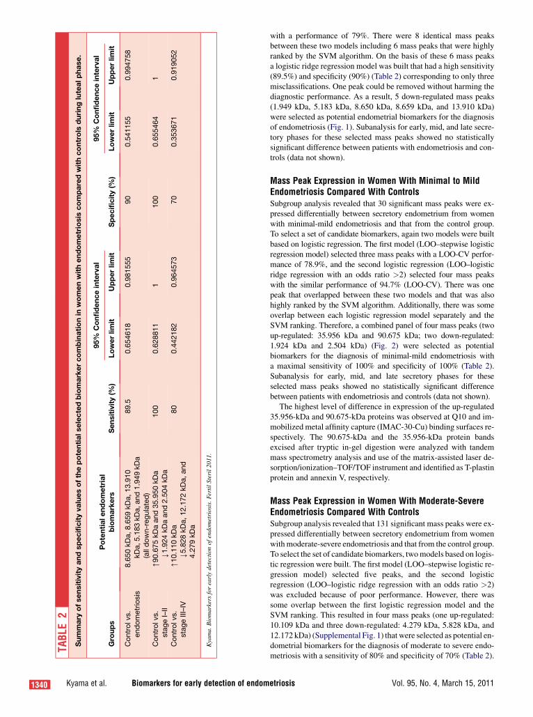
RESULTSMass Peak Expression in Women With EndometriosisCompared With ControlsAfter preprocessing the mass spectra with use of the CiphergenProteinChip Software, 73 mass peaks were expressed differentiallyin the secretory endometrium of all women with endometriosis(stages I–IV) compared with all controls. The mass of the differen-tially expressed peaks ranged between 1.923 kDa and 133.810kDa. Peaks with a mass-over-charge <1.6 kDa were excluded toavoid interference from matrix ions. To select a set of candidate bio-markers, two models based on logistic regression were built. The firstmodel (LOO–stepwise logistic regression model) selected 14 peakswith a performance of 76% (LOO-CV), and the second model(LOO–logistic ridge regression odds ratio >2) selected 16 peaks
h indications for laparoscopic surgery.
rtility symptoms Pain symptoms
) No, n (%) Yes, n (%) No, n (%)
1 (10) 3 (30) 7 (70)) 2 (22.2) 7 (77.8) 2 (22.2)
0 7 (70) 3 (30)
1339
TAB
LE2
Su
mm
ary
of
se
nsit
ivit
ya
nd
sp
ec
ific
ity
va
lue
so
fth
ep
ote
nti
alse
lec
ted
bio
ma
rke
rc
om
bin
ati
on
inw
om
en
wit
he
nd
om
etr
iosis
co
mp
are
dw
ith
co
ntr
ols
du
rin
glu
tea
lp
ha
se
.
Gro
up
sP
ote
nti
ale
nd
om
etr
ial
bio
ma
rke
rsS
en
sit
ivit
y(%
)
95%
Co
nfi
de
nc
ein
terv
al
Sp
ec
ific
ity
(%)
95%
Co
nfi
de
nc
ein
terv
al
Lo
we
rlim
itU
pp
er
lim
itL
ow
er
lim
itU
pp
er
lim
it
Co
ntr
olv
s.
end
om
etr
iosis
8.6
50
kD
a,8.6
59
kD
a,13.9
10
kD
a,5.1
83
kD
a,and
1.9
49
kD
a
(all
do
wn-r
eg
ula
ted
)
89.5
0.6
54618
0.9
81555
90
0.5
41155
0.9
94758
Co
ntr
olv
s.
sta
ge
I–II
[90.6
75
kD
aand
35.9
50
kD
a
Y1.9
24
kD
aand
2.5
04
kD
a
100
0.6
28811
1100
0.6
55464
1
Co
ntr
olv
s.
sta
ge
III–
IV[
10.1
10
kD
aY
5.8
28
kD
a,12.1
72
kD
a,and
4.2
79
kD
a
80
0.4
42182
0.9
64573
70
0.3
53671
0.9
19052
Kya
ma.
Bio
mar
kers
for
earl
ydet
ecti
onof
endo
met
riosi
s.F
erti
lSte
ril
201
1.
1340 Kyama et al. Biomarkers for early detection of endom
with a performance of 79%. There were 8 identical mass peaksbetween these two models including 6 mass peaks that were highlyranked by the SVM algorithm. On the basis of these 6 mass peaksa logistic ridge regression model was built that had a high sensitivity(89.5%) and specificity (90%) (Table 2) corresponding to only threemisclassifications. One peak could be removed without harming thediagnostic performance. As a result, 5 down-regulated mass peaks(1.949 kDa, 5.183 kDa, 8.650 kDa, 8.659 kDa, and 13.910 kDa)were selected as potential endometrial biomarkers for the diagnosisof endometriosis (Fig. 1). Subanalysis for early, mid, and late secre-tory phases for these selected mass peaks showed no statisticallysignificant difference between patients with endometriosis and con-trols (data not shown).
Mass Peak Expression in Women With Minimal to MildEndometriosis Compared With ControlsSubgroup analysis revealed that 30 significant mass peaks were ex-pressed differentially between secretory endometrium from womenwith minimal-mild endometriosis and that from the control group.To select a set of candidate biomarkers, again two models were builtbased on logistic regression. The first model (LOO–stepwise logisticregression model) selected three mass peaks with a LOO-CV perfor-mance of 78.9%, and the second logistic regression (LOO–logisticridge regression with an odds ratio >2) selected four mass peakswith the similar performance of 94.7% (LOO-CV). There was onepeak that overlapped between these two models and that was alsohighly ranked by the SVM algorithm. Additionally, there was someoverlap between each logistic regression model separately and theSVM ranking. Therefore, a combined panel of four mass peaks (twoup-regulated: 35.956 kDa and 90.675 kDa; two down-regulated:1.924 kDa and 2.504 kDa) (Fig. 2) were selected as potentialbiomarkers for the diagnosis of minimal-mild endometriosis witha maximal sensitivity of 100% and specificity of 100% (Table 2).Subanalysis for early, mid, and late secretory phases for theseselected mass peaks showed no statistically significant differencebetween patients with endometriosis and controls (data not shown).
The highest level of difference in expression of the up-regulated35.956-kDa and 90.675-kDa proteins was observed at Q10 and im-mobilized metal affinity capture (IMAC-30-Cu) binding surfaces re-spectively. The 90.675-kDa and the 35.956-kDa protein bandsexcised after tryptic in-gel digestion were analyzed with tandemmass spectrometry analysis and use of the matrix-assisted laser de-sorption/ionization–TOF/TOF instrument and identified as T-plastinprotein and annexin V, respectively.
Mass Peak Expression in Women With Moderate-SevereEndometriosis Compared With ControlsSubgroup analysis revealed that 131 significant mass peaks were ex-pressed differentially between secretory endometrium from womenwith moderate-severe endometriosis and that from the control group.To select the set of candidate biomarkers, two models based on logis-tic regression were built. The first model (LOO–stepwise logistic re-gression model) selected five peaks, and the second logisticregression (LOO–logistic ridge regression with an odds ratio >2)was excluded because of poor performance. However, there wassome overlap between the first logistic regression model and theSVM ranking. This resulted in four mass peaks (one up-regulated:10.109 kDa and three down-regulated: 4.279 kDa, 5.828 kDa, and12.172 kDa) (Supplemental Fig. 1) that were selected as potential en-dometrial biomarkers for the diagnosis of moderate to severe endo-metriosis with a sensitivity of 80% and specificity of 70% (Table 2).
etriosis Vol. 95, No. 4, March 15, 2011

FIGURE 1
Women with endometriosis versus controls during luteal phase. Endometrial protein-peptide expression of 8.650 kDa (fold difference [n-fold]
¼ 2.5), 8.659 kDa (n-fold ¼ 1.64), 13.91 kDa (n-fold ¼ 1.32), 5.183 kDa (n-fold ¼ 1.52), and 1.949 kDa (n-fold ¼ 1.8) was significantly lower inwomen with endometriosis when compared with the controls. *P< .05; **P< .01; ***P< .005.
Kyama. Biomarkers for early detection of endometriosis. Fertil Steril 2011.
DISCUSSIONOur data strongly support the concept that endometrial profiling us-ing SELDI-TOF-MS and selecting mass peaks with stepwise logis-tic regression combined with SVM provides a clinically relevantpanel of biomarkers that demonstrate high sensitivity (100%) andspecificity (100%) for the diagnosis of minimal to mild endometri-osis. Leave-one-out cross validation was applied to rank and selectthe peaks according to their discriminatory power from womenwithout endometriosis and those with early (minimal-mild) and ad-vanced (moderate-severe) stages of the disease. When comparedwith our results, other investigators (26) reported a different set of20 mass peaks with lower sensitivity (87%) and specificity (80%)for the diagnosis of endometriosis, using another learning algorithmwith a combination of three selected mass peaks (mass-to-charge ra-tio values of 3.957 kDa, 11.711 kDa, and 6.987 kDa). However, theirstudy was fundamentally different in design and methodology: theystudied plasma samples (26) instead of endometrium, their studypopulation was not defined clearly with respect to menstrual cycle
Fertility and Sterility�
phase and stages of endometriosis (26), and they used only onetype of surface chemistry (26) whereas we used four different typesof surface chemistries. The set of mass peaks selected in our studymay be a fingerprint associated with endometrial tissue and maybe absent in plasma or only present in low concentrations hamperingtheir direct detection.
A possible limitation of our study is that our controls were infer-tile women including some with pelvic pain (30%), instead ofwomen of proven fertility without pain at the moment of laparo-scopic sterilization. However, such a well-defined control groupwas unavailable in our hospital in sufficient number because ofthe decline in the number of laparoscopic sterilizations and thefact that a significant proportion of women undergoing laparoscopicsterilization also may have some degree of pelvic pain.
In our study, T-plastin, a cytoplasmic protein regulating actin as-sembly and cellular motility, was up-regulated in the secretory phaseendometrium from women with minimal to mild endometriosis com-pared with controls. T-plastin protein functions in the formation of
1341

FIGURE 2
Women with minimal-mild endometriosis versus controls during luteal phase. Endometrial protein expression of 90.675 kDa (fold difference
[n-fold] ¼ 2.0) and 35.95 kDa (n-fold ¼ 1.7) was significantly higher in women with minimal-mild endometriosis compared with controls.Decreased endometrial peptide levels for 1.924 kDa (n-fold ¼ 1.8) and 2.504 kDa (n-fold ¼ 3.0) in women with minimal-mild endometriosis
were observed compared with controls. *P< .05; **P< .01. Stg ¼ stage.
Kyama. Biomarkers for early detection of endometriosis. Fertil Steril 2011.
actin bundles that are required for cell locomotion and maintenanceof the cellular architecture (27), and studies have shown that culturedfibroblasts overexpressing T-plastin demonstrated increased mobil-ity and altered cellular architecture (28). It has been reported thatthe surface mesothelium of pelvic peritoneum adjacent to activeendometriosis is marked by a sequence of morphologic changesfrom flat cell to cuboidal or columnar epithelium (29, 30). Wehypothesize that T-plastin may play a role in the development ofthese structural abnormalities. It is not clear whether T-plastin ispresent in peripheral blood, because we could not find any data inthe literature showing the concentration of T-plastin in serum orplasma.
Additionally, annexin V, a calcium phospholipid-binding proteinbelonging to the annexin family, was also up-regulated in secretoryphase endometrium from women with minimal to mild endometri-osis when compared with controls. The role of annexin V in thepathogenesis of endometriosis is unclear, but, in cancer studies, an-nexin V is expressed exclusively in the periphery of invasivelygrowing tumor areas, suggesting that it may play a role in prolifer-ation and/or cell mobility and may have metastatic potential (31).Endometriosis can be considered as benign metastatic diseasewith characteristics similar to malignancy, including aggressivegrowth and localized invasion. We hypothesize that annexin Vplays a role in the early invasion of endometrial cells into the me-sothelium after initial attachment to the peritoneal wall. Annexin Vis detectable in plasma, with higher levels in patients with acutemyocardial infarction (11.0 � 4.9 ng/mL) compared with controls(1.9 � 0.7 ng/mL) (32).
1342 Kyama et al. Biomarkers for early detection of endom
Because of the pure exploratory nature of the present study, theidentification of proteins was limited to the two proteins mentionedearlier, because they have a high molecular weight and were up-regulated in the primary group of interest to us, women with mini-mal to mild endometriosis, when compared with controls. Wewere unable to identify other potentially relevant mass peaks be-cause of the low amounts of protein lysates that were available inthe samples.
Although our preliminary data are interesting, it can be ques-tioned how statistical significance can be translated into clinical use-fulness. Therefore, our next step is to plan a validation study ina larger number of patients with and without endometriosis to con-firm our findings and directly identify the up- and down-regulatedproteins and peptides. Obviously, an endometrial biopsy is stilla semi-invasive procedure, and we also plan to use proteomics tech-nology to develop a less-invasive test based on blood, urine, or salivathat would be better applicable for adolescents with pelvic pain or inwomen at risk for endometriosis.
In conclusion, SELDI-TOF proteomics analysis revealed a pro-spective panel of potential endometrial biomarkers includingT-plastin and annexin V with a sensitivity of 100% and specificityof 100% for the diagnosis of minimal to mild endometriosis. Confir-mation of these data in a larger and independent patient population,together with identification of all up- and down-regulated proteinsand peptides, is needed and planned as the next phase of our research.
Acknowledgments: The authors acknowledge the important technical assis-
tance of Ms. Karin Schildermans.
etriosis Vol. 95, No. 4, March 15, 2011

REFERENCES
1. D’Hooghe TM, Hill JA. Endometriosis. In: Berek JS,
ed. Novak’s gynecology. Philadelphia: Williams &
Wilkins, 2002:931–72.
2. Gao X, Outley J, Botteman M, Spalding J, Simon JA,
Pashos CL. Economic burden of endometriosis. Fertil
Steril 2006;86:1561–72.
3. Zhao SZ, Wong JM, Davis MB, Gersh GE,
Johnson KE. The cost of in patient endometriosis
treatment: an analysis based on the Healthcare Cost
and Utilization Project Nationwide Inpatient
Sample. Am J Manag Care 1998;4:1127–34.
4. Simoens S, Hummelshoj L, D’Hooghe T.
Endometriosis: cost estimates and methodological
perspective. Hum Reprod Update 2007;13:395–404.
5. Hadfield R, Mardon H, Barlow D, Kennedy S. Delay in
the diagnosis of endometriosis: a survey of women
from USA and the UK. Hum Reprod 1996;11:878–80.
6. Martinez S, Garrido N, Coperias JL, Pardo F, Desco J,
Garcia-Velasco JA, et al. Serum interleukin-6 levels
are elevated in women with minimal-mild endometri-
osis. Hum Reprod 2007;22:836–42.
7. Rosa E, Silva AC, Rosa E, Silva JC, Ferriani RA.
Serum CA-125 in the diagnosis of endometriosis.
Int J Gynaecol Obstet 2007;96:206–7.
8. Agic A, Xu H, Rehbein M, Wolfler MM, Ebert AD,
Hornung D. Cognate chemokine receptor 1
messenger ribonucleic acid expression in peripheral
blood as a diagnostic test for endometriosis. Fertil
Steril 2007;87:982–4.
9. Othmam EE, Hornung D, Al-Hendy A. Biomarkers of
endometriosis. Expert Opin Med Diagn 2008;2:741–52.
10. Tabibzadeh S, Becker JL, Parsons AK. Endometriosis
is associated with alterations in the relative
abundance of proteins and IL-10 in the peritoneal
fluid. Front Biosci 2003;1:a70–8.
11. Ferrero S, Gillott DJ, Remorgida V, Anserini P,
Leung KY, Ragni N, et al. Proteomic analysis of
peritoneal fluid in women with endometriosis. J
Proteome Res 2007;6:3402–11.
12. Fowler PA, Tattum J, Bhattacharya S, Klonisch T,
Hombach-Klonisch S, Gazvani R, et al. An
investigation of the effects of endometriosis on the
proteome of human eutopic endometrium:
a heterogeneous tissue with a complex disease.
Proteomics 2007;7:30–42.
Fertility and Sterility�
13. Have ST, Fraser I, Markham R, Lam A, Matsumoto I.
Proteomic analysis of protein expression in the
eutopic endometrium of women with endometriosis.
Proteomics Clin Appl 2007;1:1243–51.
14. Ametzazurra A, Matorras R, Garc�ıa-Velasco JA,
Prieto B, Sim�on L, Mart�ınez A, et al. Endometrial
fluid is a specific and non-invasive biological sample
for protein biomarker identification in endometriosis.
Hum Reprod 2009;24:954–65.
15. Casado-Vela J, Rodriguez-Suarez E, Iloro I,
Ametzazurra A, Alkorta N, Garc�ıa-Velasco JA,
et al. Comprehensive proteomic analysis of human
endometrial fluid aspirate. J Proteome Res 2009;8:
4622–32.
16. Hu J, Coombes KR, Morris JS, Baggerly KA. The
importance of experimental design in proteomic
mass spectrometry experiments: some cautionary
tales. Brief Funct Genomic Proteomic 2005;3:
322–31.
17. Petricoin EF, Ardekani AM, Hitt BA, Levine PJ,
Fusaro VA, Steinberg SM, et al. Use of proteomic
patterns in serum to identify ovarian cancer. Lancet
2002;359:572–7.
18. Baggerly KA, Morris JS, Coombes KR.
Reproducibility of SELDI-TOF protein patterns in se-
rum: comparing datasets from different experiments.
Bioinformatics 2004;20:777–85.
19. Becker S, Cazares LH, Watson P, Lynch H,
Semmes OJ, Drake RR, et al. Surfaced-enhanced
laser desorption/ionization time-of-flight (SELDI-
TOF) differentiation of serum protein profiles of
BRCA-1 and sporadic breast cancer. Ann Surg Oncol
2004;11:907–14.
20. Zhang Z, Bast RC Jr, Yu Y, Li J, Sokoll LJ,
Rai AJ, et al. Three biomarkers identified from
serum proteomic analysis for the detection of
early stage ovarian cancer. Cancer Res 2004;64:
5882–90.
21. Kyama CM, T’Jampens D, Mihalyi A, Simsa P,
Debrock S, Waelkens E, et al. ProteinChip
technology is a useful method in the pathogenesis
and diagnosis of endometriosis: a preliminary study.
Fertil Steril 2006;86:203–9.
22. Noyes RW, Hertig AT, Rock J. Dating the endometrial
biopsy. Fertil Steril 1950;1:3–25.
23. The American Society for Reproductive Medicine.
Revised American Society for Reproductive
Medicine classification of endometriosis: 1996.
Fertil Steril 1997;67:817–21.
24. Guyon I, Weston J, Barnhill S, Vapnik V. Gene
selection for cancer classification using support
vector machines. Machine Learning 2002;46:
389–422.
25. le Cessie S, van Houwelingen JC. Ridge estimators in
logistic regression. Applied Statistics 1992;41:
191–201.
26. Liu H, Lang J, Zhou Q, Shan D, Li Q. Detection of
endometriosis with the use of plasma protein
profiling by surface-enhanced laser desorption/ioni-
zation time-of-flight mass spectrometry. Fertil Steril
2007;87:988–90.
27. Delanote V, Vandekerckhove J, Gettemans J. Plastins:
versatile modulators of actin organization in
(patho)physiological cellular processes. Acta
Pharmacol Sin 2005;26:769–79.
28. Arpin M, Friederich E, Algrain M, Vernel F,
Louvard D. Functional differences between L- and
T-plastin isoforms. J Cell Biol 1994;127:1995–2008.
29. Ishimaru T, Khan KN, Fujishita A, Kitajima M,
Masuzaki H. Hepatocyte growth factor may be
involved in cellular changes to the peritoneal
mesothelium adjacent to pelvic endometriosis. Fertil
Steril 2004;81:810–8.
30. Sugawara J, Fukaya T, Murakami T, Yoshida H,
Yajima A. Hepatocyte growth factor stimulated
proliferation, migration, and lumen formation of
human endometrial epithelial cells in vitro. Biol
Reprod 1997;57:936–42.
31. Melle C, Ernst G, Schimmel B, Bleul A, Koscielny S,
Wiesner A, et al. Biomarker discovery and
identification in laser microdissected head and neck
squamous cell carcinoma with ProteinChip
technology, two-dimensional gel electrophoresis, tan-
dem mass spectrometry, and immunohistochemistry.
Mol Cell Proteomics 2003;2:443–52.
32. Matsuda R, Kaneko N, Kikuchi M, Chiwaki F,
Toda M, Ieiri T, et al. Clinical significance of
measurement of plasma annexin V concentration of
patients in the emergency room. Resuscitation
2003;57:171–7.
1343
SUPPLEMENTAL MATERIALS AND METHODS
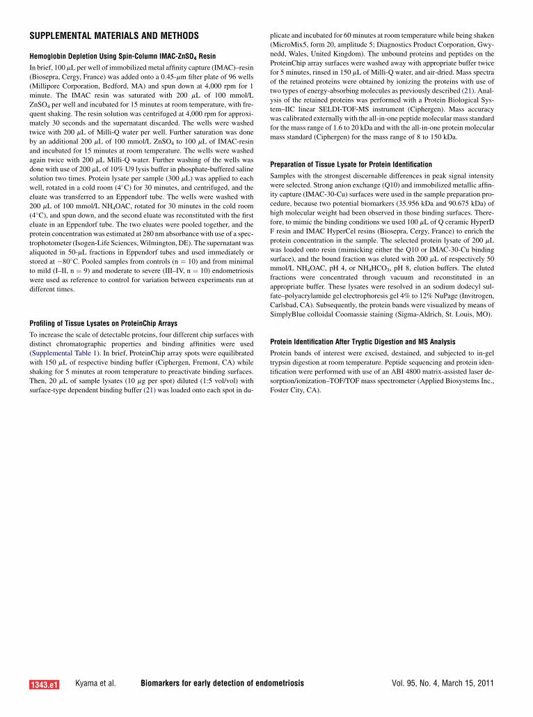
Hemoglobin Depletion Using Spin-Column IMAC-ZnSO4 Resin
In brief, 100 mL per well of immobilized metal affinity capture (IMAC)–resin
(Biosepra, Cergy, France) was added onto a 0.45-mm filter plate of 96 wells
(Millipore Corporation, Bedford, MA) and spun down at 4,000 rpm for 1
minute. The IMAC resin was saturated with 200 mL of 100 mmol/L
ZnSO4 per well and incubated for 15 minutes at room temperature, with fre-
quent shaking. The resin solution was centrifuged at 4,000 rpm for approxi-
mately 30 seconds and the supernatant discarded. The wells were washed
twice with 200 mL of Milli-Q water per well. Further saturation was done
by an additional 200 mL of 100 mmol/L ZnSO4 to 100 mL of IMAC-resin
and incubated for 15 minutes at room temperature. The wells were washed
again twice with 200 mL Milli-Q water. Further washing of the wells was
done with use of 200 mL of 10% U9 lysis buffer in phosphate-buffered saline
solution two times. Protein lysate per sample (300 mL) was applied to each
well, rotated in a cold room (4�C) for 30 minutes, and centrifuged, and the
eluate was transferred to an Eppendorf tube. The wells were washed with
200 mL of 100 mmol/L NH4OAC, rotated for 30 minutes in the cold room
(4�C), and spun down, and the second eluate was reconstituted with the first
eluate in an Eppendorf tube. The two eluates were pooled together, and the
protein concentration was estimated at 280 nm absorbance with use of a spec-
trophotometer (Isogen-Life Sciences, Wilmington, DE). The supernatant was
aliquoted in 50-mL fractions in Eppendorf tubes and used immediately or
stored at �80�C. Pooled samples from controls (n ¼ 10) and from minimal
to mild (I–II, n ¼ 9) and moderate to severe (III–IV, n ¼ 10) endometriosis
were used as reference to control for variation between experiments run at
different times.
Profiling of Tissue Lysates on ProteinChip Arrays
To increase the scale of detectable proteins, four different chip surfaces with
distinct chromatographic properties and binding affinities were used
(Supplemental Table 1). In brief, ProteinChip array spots were equilibrated
with 150 mL of respective binding buffer (Ciphergen, Fremont, CA) while
shaking for 5 minutes at room temperature to preactivate binding surfaces.
Then, 20 mL of sample lysates (10 mg per spot) diluted (1:5 vol/vol) with
surface-type dependent binding buffer (21) was loaded onto each spot in du-
1343.e1 Kyama et al. Biomarkers for early detection of end
plicate and incubated for 60 minutes at room temperature while being shaken
(MicroMix5, form 20, amplitude 5; Diagnostics Product Corporation, Gwy-
nedd, Wales, United Kingdom). The unbound proteins and peptides on the
ProteinChip array surfaces were washed away with appropriate buffer twice
for 5 minutes, rinsed in 150 mL of Milli-Q water, and air-dried. Mass spectra
of the retained proteins were obtained by ionizing the proteins with use of
two types of energy-absorbing molecules as previously described (21). Anal-
ysis of the retained proteins was performed with a Protein Biological Sys-
tem–IIC linear SELDI-TOF-MS instrument (Ciphergen). Mass accuracy
was calibrated externally with the all-in-one peptide molecular mass standard
for the mass range of 1.6 to 20 kDa and with the all-in-one protein molecular
mass standard (Ciphergen) for the mass range of 8 to 150 kDa.
Preparation of Tissue Lysate for Protein Identification
Samples with the strongest discernable differences in peak signal intensity
were selected. Strong anion exchange (Q10) and immobilized metallic affin-
ity capture (IMAC-30-Cu) surfaces were used in the sample preparation pro-
cedure, because two potential biomarkers (35.956 kDa and 90.675 kDa) of
high molecular weight had been observed in those binding surfaces. There-
fore, to mimic the binding conditions we used 100 mL of Q ceramic HyperD
F resin and IMAC HyperCel resins (Biosepra, Cergy, France) to enrich the
protein concentration in the sample. The selected protein lysate of 200 mL
was loaded onto resin (mimicking either the Q10 or IMAC-30-Cu binding
surface), and the bound fraction was eluted with 200 mL of respectively 50
mmol/L NH4OAC, pH 4, or NH4HCO3, pH 8, elution buffers. The eluted
fractions were concentrated through vacuum and reconstituted in an
appropriate buffer. These lysates were resolved in an sodium dodecyl sul-
fate–polyacrylamide gel electrophoresis gel 4% to 12% NuPage (Invitrogen,
Carlsbad, CA). Subsequently, the protein bands were visualized by means of
SimplyBlue colloidal Coomassie staining (Sigma-Aldrich, St. Louis, MO).
Protein Identification After Tryptic Digestion and MS Analysis
Protein bands of interest were excised, destained, and subjected to in-gel
trypsin digestion at room temperature. Peptide sequencing and protein iden-
tification were performed with use of an ABI 4800 matrix-assisted laser de-
sorption/ionization–TOF/TOF mass spectrometer (Applied Biosystems Inc.,
Foster City, CA).
ometriosis Vol. 95, No. 4, March 15, 2011

SUPPLEMENTARY FIGURE 1
Women with moderate-severe endometriosis versus controls during luteal phase. Increased endometrial protein levels for 10.1094 kDa (fold
difference [n-fold] ¼ 1.53) in women with moderate-severe endometriosis when compared with controls. Protein and peptide levels of 5.828kDa (n-fold¼ 1.9), 12.172 kDa (n-fold¼ 2.33), and 4.279 kDa (n-fold¼ 2.9) were significantly lower in endometrium of women with moderate-
severe endometriosis when compared with controls. *P< .05; **P< .01; ***P< .005.
Kyama. Biomarkers for early detection of endometriosis. Fertil Steril 2011.
Fertility and Sterility� 1343.e2

SUPPLEMENTARY TABLE 1Different ProteinChip surfaces with their respective binding buffer that were used in the study.
ProteinChip surfaces Binding buffers
Weak cation exchange surface (CM10) Low stringency binding buffer (50 mmol/L NaOAC, pH 4.0)
Immobilized metallic affinity capture surface
(IMAC-30-Cu) loaded with CuSO4
0.1 mol/L phosphate, 0.5 mol/L NaCl, pH 7.0
Hydrophobic surface (H50) 10% acetonitrile, 0.1% trifluoroacetic acidStrong anion exchange surface (Q10) 50 mmol/L Tris-HCl, pH 8.0.
Note: Tris ¼ tris(hydroxymethyl)aminomethane.
Kyama. Biomarkers for early detection of endometriosis. Fertil Steril 2011.
1343.e3 Kyama et al. Biomarkers for early detection of endometriosis Vol. 95, No. 4, March 15, 2011