Mitigation of endometriosis using regenerative cerium oxide nanoparticles
10
Research Article Mitigation of endometriosis using regenerative cerium oxide nanoparticles Koel Chaudhury, PhD a, ⁎ ,1 , Narendra Babu K, PhD a,1 , Abhay K. Singh, MSc a , Soumen Das, PhD b , Amit Kumar, MTech b , Sudipta Seal, PhD b, ⁎ a School of Medical Science and Technology, Indian Institute of Technology, Kharagpur, West Bengal, India b Department of Mechanical, Materials and Aerospace Engineering, Advanced Materials Processing Analysis Center, Nanoscience Technology Center, University of Central Florida Orlando, Florida, USA Received 13 November 2011; accepted 2 August 2012 Abstract Cerium oxide nanoparticles (nanoceria) have recently received attention from the scientific community due to their unique free radicals (specially superoxide radical and hydrogen peroxide) scavenging property in biological system, both in vitro and in vivo. It is suggested that free radicals play an important role in the pathogenesis of endometriosis. In this study we have shown that nanoceria mitigate the endometrial lesions induced in mice model by decreasing oxidative stress and inhibiting angiogenesis. Moreover, nanoceria were also observed to protect endometriosis-related adverse effects on the oocytes, which is critical for successful pregnancy. Summarizing, nanoceria have shown promising efficacy against endometriosis related pathogenesis. From the Clinical Editor: Free radicals have been implemented in the pathogenesis of endometriosis. In a murine model the authors demonstrated successful treatment of endometriosis with nanoceria, and protection of endometriosis-related adverse effects on the oocytes, paving the way to potential clinical translational applications in the future. © 2013 Elsevier Inc. All rights reserved. Key words: Cerium oxide nanoparticles; Antioxidant; Endometriosis; Anti-angiogenesis Worldwide approximately 176 million women and girls suffer from endometriosis (Endometriosis Facts & Statistics, Endometriosis Foundation of America) and is the third leading cause of gynecologic hospitalization in the United States. 1 It is defined by the presence of endometrial-like tissues external to the uterus, which induce a chronic, inflammatory reaction. 2,3 While symptoms of endometriosis usually include pelvic pain, dysmenorrhea and infertility, some women affected by this disease often remain asymptomatic. Though hormonal suppres- sion and surgical intervention are the conventional treatment modalities for endometriosis, results are not always satisfactory thereby necessitating the need to develop new approaches for the management of the disease. It is evidenced that a positive association exists between oxidative stress (OS) and endometriosis. Several studies have documented a higher concentration of reactive oxygen species (ROS), lipid peroxidation markers and reduced activity of antioxidants such as superoxide dismutase (SOD), glutathione peroxidise (GPx) and vitamin E in serum and peritoneal fluid of women with endometriosis. 4–6 Following vitamin C and E supplementation, a decrease in the concentration of OS markers in women with endometriosis is reported. 7 Recently, Melo et al (2010) have shown that OS and inflammation are associated with the progression and develop- ment of endometriosis. 8 Excessive endometrial angiogenesis is thought to be a critical mechanism in the pathogenesis of this disease. 9 Long-term survival and proliferation of endometrial lesions are significantly dependent on neovascularization, which ensures supply of oxygen and nutrients. 10 It is hypothesized that high concentrations of VEGF may help the endometrial implant to revascularize and proliferate, which possibly is the explanation for the increased VEGF levels in the peritoneal POTENTIAL CLINICAL RELEVANCE Nanomedicine: Nanotechnology, Biology, and Medicine 9 (2013) 439 – 448 nanomedjournal.com Sources of Support: This research was supported by Indian Council of Medical Research, and Department of Science and Technology, Government of India. Dr. Sudipta Seal acknowledges the grant from National Science Foundation (NIRT: 0708172) for funding this research. ⁎ Corresponding authors: Koel Chaudhury, School of Medical Science and Technology, Indian Institute of Technology, Kharagpur, West Bengal, India. Sudipta Seal, Department of Mechanical, Materials and Aerospace Engineering, Advanced Materials Processing Analysis Center, Nanoscience Technology Center, 4000 Central Florida Blvd, Engineering I, Room Number 381, Orlando, Florida 32816, USA. E-mail addresses: [email protected] (K. Chaudhury), [email protected] (S. Seal). 1 Authors contributed equally. 1549-9634/$ – see front matter © 2013 Elsevier Inc. All rights reserved. http://dx.doi.org/10.1016/j.nano.2012.08.001 Please cite this article as: Chaudhury K, Babu K N, Singh AK, Das S, Kumar A, Seal S, Mitigation of endometriosis using regenerative cerium oxide nanoparticles. Nanomedicine: NBM 2013;9:439-448, http://dx.doi.org/10.1016/j.nano.2012.08.001
-
Upload
independent -
Category
Documents
-
view
7 -
download
0
Transcript of Mitigation of endometriosis using regenerative cerium oxide nanoparticles

POTENTIAL CLINICAL RELEVANCE
Nanomedicine: Nanotechnology, Biology, and Medicine9 (2013) 439–448
Research Article
Mitigation of endometriosis using regenerative cerium oxide nanoparticlesKoel Chaudhury, PhDa,⁎, 1, Narendra Babu K, PhDa,1, Abhay K. Singh, MSca,
Soumen Das, PhDb, Amit Kumar, MTechb, Sudipta Seal, PhDb,⁎aSchool of Medical Science and Technology, Indian Institute of Technology, Kharagpur, West Bengal, India
bDepartment of Mechanical, Materials and Aerospace Engineering, Advanced Materials Processing Analysis Center, Nanoscience Technology Center,University of Central Florida Orlando, Florida, USA
Received 13 November 2011; accepted 2 August 2012
nanomedjournal.com
Abstract
Cerium oxide nanoparticles (nanoceria) have recently received attention from the scientific community due to their unique free radicals(specially superoxide radical and hydrogen peroxide) scavenging property in biological system, both in vitro and in vivo. It is suggested thatfree radicals play an important role in the pathogenesis of endometriosis. In this study we have shown that nanoceria mitigate the endometriallesions induced in mice model by decreasing oxidative stress and inhibiting angiogenesis. Moreover, nanoceria were also observed to protectendometriosis-related adverse effects on the oocytes, which is critical for successful pregnancy. Summarizing, nanoceria have shownpromising efficacy against endometriosis related pathogenesis.
From the Clinical Editor: Free radicals have been implemented in the pathogenesis of endometriosis. In a murine model the authorsdemonstrated successful treatment of endometriosis with nanoceria, and protection of endometriosis-related adverse effects on the oocytes,paving the way to potential clinical translational applications in the future.© 2013 Elsevier Inc. All rights reserved.
Key words: Cerium oxide nanoparticles; Antioxidant; Endometriosis; Anti-angiogenesis
Worldwide approximately 176 million women and girlssuffer from endometriosis (Endometriosis Facts & Statistics,Endometriosis Foundation of America) and is the third leadingcause of gynecologic hospitalization in the United States.1 It isdefined by the presence of endometrial-like tissues external tothe uterus, which induce a chronic, inflammatory reaction.2,3
While symptoms of endometriosis usually include pelvic pain,dysmenorrhea and infertility, some women affected by thisdisease often remain asymptomatic. Though hormonal suppres-
Sources of Support: This research was supported by Indian Council ofMedical Research, and Department of Science and Technology, Governmentof India. Dr. Sudipta Seal acknowledges the grant from National ScienceFoundation (NIRT: 0708172) for funding this research.
⁎Corresponding authors: Koel Chaudhury, School of Medical Scienceand Technology, Indian Institute of Technology, Kharagpur, West Bengal,India. Sudipta Seal, Department of Mechanical, Materials and AerospaceEngineering, Advanced Materials Processing Analysis Center, NanoscienceTechnology Center, 4000 Central Florida Blvd, Engineering I, RoomNumber 381, Orlando, Florida 32816, USA.
E-mail addresses: [email protected] (K. Chaudhury),[email protected] (S. Seal).
1 Authors contributed equally.
1549-9634/$ – see front matter © 2013 Elsevier Inc. All rights reserved.http://dx.doi.org/10.1016/j.nano.2012.08.001
Please cite this article as: Chaudhury K, Babu K N, Singh AK, Das S, Kumananoparticles. Nanomedicine: NBM 2013;9:439-448, http://dx.doi.org/10.1016
sion and surgical intervention are the conventional treatmentmodalities for endometriosis, results are not always satisfactorythereby necessitating the need to develop new approaches for themanagement of the disease.
It is evidenced that a positive association exists betweenoxidative stress (OS) and endometriosis. Several studies havedocumented a higher concentration of reactive oxygen species(ROS), lipid peroxidation markers and reduced activity ofantioxidants such as superoxide dismutase (SOD), glutathioneperoxidise (GPx) and vitamin E in serum and peritoneal fluid ofwomen with endometriosis.4–6 Following vitamin C and Esupplementation, a decrease in the concentration of OS markersin women with endometriosis is reported.7
Recently, Melo et al (2010) have shown that OS andinflammation are associated with the progression and develop-ment of endometriosis.8 Excessive endometrial angiogenesis isthought to be a critical mechanism in the pathogenesis of thisdisease.9 Long-term survival and proliferation of endometriallesions are significantly dependent on neovascularization, whichensures supply of oxygen and nutrients.10 It is hypothesizedthat high concentrations of VEGF may help the endometrialimplant to revascularize and proliferate, which possibly is theexplanation for the increased VEGF levels in the peritoneal
r A, Seal S, Mitigation of endometriosis using regenerative cerium oxide/j.nano.2012.08.001

440 K. Chaudhury et al / Nanomedicine: Nanotechnology, Biology, and Medicine 9 (2013) 439–448
fluid.11 Consequently, several research groups are investigatingtowards the development of anti-angiogenic therapeutic strate-gies for endometriosis management.12,13
In the recent years, nanoceria or cerium oxide nanoparticleshave gained increasing importance in the treatment of variousdiseases caused by ROS or their intermediates14–18 (ROI). Theability of nanoceria to switch between oxidation states iscomparable to that of biological antioxidants,16 which impartsit the radical scavenging property. SOD and catalase mimickingactivity19 and anti-inflammatory activity15 of nanoceria arewell established. Potential of nanoceria in preventing inheritedretinal degeneration and ocular diseases17 and ROI-inducedblindness in in vivo mice model is reported.20 It is alsosuggested that nanoceria have tremendous therapeutic potentialin treating neurodegenerative diseases,21 protecting againstcardiac dysfunction22 and providing radioprotection againstgastrointestinal epithelium.19
Presently, there is no report on the role of nanoceria in femalereproductive health. It may be worthwhile to investigate theeffect of nanoceria in endometriosis, a gynecological disorderassociated with OS and neovascularization. The objective of thepresent study is to explore the antioxidant and anti-angiogenicproperties of nanoceria in mice induced with endometriosis.
Methods
Nanoceria synthesis and characterization
Nanoceria of size 3–5 nm were synthesized by themicroemulsion method.23 The micro-emulsion system con-sisted of AOT (surfactant), water (polar solvent) and toluene(non-polar solvent). AOT was dissolved in 50 mL of toluene,0.1 mol/L aqueous cerium nitrate solution was added, and themixture was stirred. Next, 5 mL of 30% hydrogen peroxidesolution was added dropwise and the reaction was carried outfor 2h. The reaction mixture was then allowed to separate intotwo layers. The upper toluene layer was separated and 30%ammonium hydroxide was added to it. The resultant mixturewas stirred and allowed to settle down. The particles were thenwashed with acetone and water to remove the surfactant andother impurities. High resolution transmission electron micros-copy (HRTEM), with FEI Tecnai F30 having an energydispersive X-ray (EDX) analyzer, was carried out to study thesize and morphology of nanoparticles. Hydrodynamic radiusand surface charge were estimated using Zetasizer (Nano-ZSfrom Malvern Instruments).
Cell proliferation/cytotoxicity assay
Cell proliferation/cytotoxicity was assayed by colorimetricassay using MTT (3-(4,5 dimethylthiazol-2-yl)-2,5-diphenyltetrazolium bromide) dye in human skin keratinocytes(HACAT) cell line. Cells were cultured in 96-well plate at adensity of 3×10³ cells/well, treated with different concentrationof nanoceria and incubated for 48h. MTT was added to the cell ata final concentration of 1.2mM and incubated for 4h. Cells werelysed and insoluble formazan product dissolved using buffer(10% SDS, 0.1M HCl). Absorbance of each well was measured
using SpectraMax 190 spectrophotometer (Molecular Devices,Sunnyvale, California). Cell proliferation was calculated byabsorbing the nanoceria-treated samples/untreated controlsin percentage.24
Animal study design
CD-1 strain Swiss Albino female mice weighing 31±3 g and5–6 weeks of age were used in this study. These mice weremaintained at 14-h light and 10-h dark photoperiod underconstant temperature (25°±2°C) and provided with standardfood and water, ad libitum according to the guidelines ofCommittee for the Purpose of Supervision and Control ofExperiments on Animals (CPCSEA), Chennai, India. Approvalwas obtained from the Animal Ethics Committee of the Instituteto carry out this study.
A total of 22 mice were selected for the first part of the study.12 mice were induced with endometriosis, out of which two weresacrificed to confirm the disease. 10 mice remained withoutinduction of endometriosis and were considered as controls forcomparison purposes. OS markers, including ROS, LPO andTAC, and angiogenic markers including ADM and VEGF wereassessed in both controls (n=10) and endometriosis inducedmice (n=10).
In the second part of the study, the anti-angiogenic effect ofnanoceria and a known antioxidant (N-acetyl cysteine; NAC) onendometriosis was investigated. 40 additional mice wereincluded in this part of the study, out of which 30 were inducedwith endometriosis (Group A) and the remaining 10 normalhealthy mice (not induced with endometriosis) were consideredas controls (Group B). The endometriosis induced mice (GroupA; n=30) were divided into three groups (10 in each group):mice injected with 100 μL of 0.9% NaCl (Group A1; n=10),mice injected with NAC at a dose of 250 mg/kg body weightthrice a week for 15 days (Group A2; n=10) and mice injectedwith Nanoceria at a single dose of 0.5mg/kg body weight (GroupA3; n=10).
Nanoceria, NAC and NaCl were injected into the peritonealcavity of the abdominal wall with an 18-gauge needle on themidline just below the mice umbilicus.25 The rationale forchoosing nanoceria concentration (0.5mg/kg body weight) isbased on a previous study where it has been shown that micetwice administered with 0.5 mgkg−1 dose of nanoceria on day 1and 15 did not experience any tissue damage up to at least 30days.15 The dosage of NAC was adopted in this study from aprevious work where they have shown that NAC reduced OS andstress-responsive signaling kinases in mice model.26
Blood samples were collected from the saphenous vein of allthe groups for estimation of OS and angiogenic markers. 5 mice,from each Group, i.e. A1, A2, A3 and B were superovulated,oocytes collected and their characteristics studied. Remaining 5mice of the respective groups were sacrificed for viewing theperitoneal cavity and collecting endometrial tissue sections forhistology and immunohistochemistry.
Endometriosis induction and confirmation
Induction of endometriosis was performed according toa method reported by Nowak et al (2008) with slight
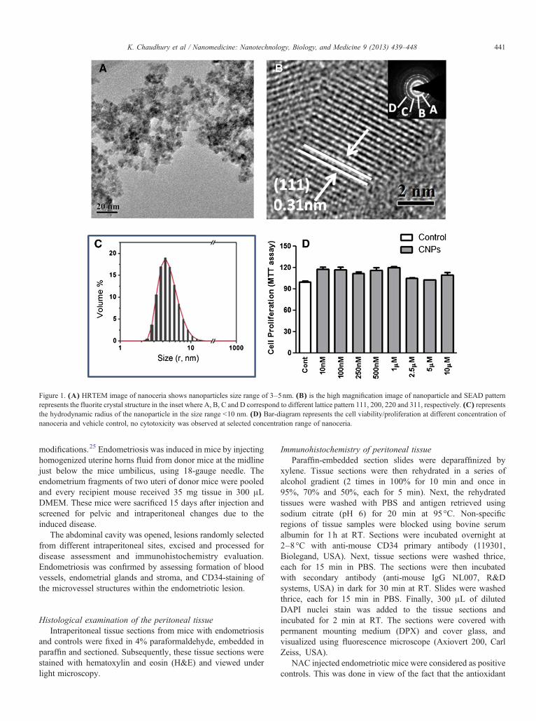
Figure 1. (A) HRTEM image of nanoceria shows nanoparticles size range of 3–5nm. (B) is the high magnification image of nanoparticle and SEAD patternrepresents the fluorite crystal structure in the inset where A, B, C and D correspond to different lattice pattern 111, 200, 220 and 311, respectively. (C) representsthe hydrodynamic radius of the nanoparticle in the size range b10 nm. (D) Bar-diagram represents the cell viability/proliferation at different concentration ofnanoceria and vehicle control, no cytotoxicity was observed at selected concentration range of nanoceria.
441K. Chaudhury et al / Nanomedicine: Nanotechnology, Biology, and Medicine 9 (2013) 439–448
modifications.25 Endometriosis was induced in mice by injectinghomogenized uterine horns fluid from donor mice at the midlinejust below the mice umbilicus, using 18-gauge needle. Theendometrium fragments of two uteri of donor mice were pooledand every recipient mouse received 35 mg tissue in 300 μLDMEM. These mice were sacrificed 15 days after injection andscreened for pelvic and intraperitoneal changes due to theinduced disease.
The abdominal cavity was opened, lesions randomly selectedfrom different intraperitoneal sites, excised and processed fordisease assessment and immunohistochemistry evaluation.Endometriosis was confirmed by assessing formation of bloodvessels, endometrial glands and stroma, and CD34-staining ofthe microvessel structures within the endometriotic lesion.
Histological examination of the peritoneal tissueIntraperitoneal tissue sections from mice with endometriosis
and controls were fixed in 4% paraformaldehyde, embedded inparaffin and sectioned. Subsequently, these tissue sections werestained with hematoxylin and eosin (H&E) and viewed underlight microscopy.
Immunohistochemistry of peritoneal tissueParaffin-embedded section slides were deparaffinized by
xylene. Tissue sections were then rehydrated in a series ofalcohol gradient (2 times in 100% for 10 min and once in95%, 70% and 50%, each for 5 min). Next, the rehydratedtissues were washed with PBS and antigen retrieved usingsodium citrate (pH 6) for 20 min at 95°C. Non-specificregions of tissue samples were blocked using bovine serumalbumin for 1h at RT. Sections were incubated overnight at2–8°C with anti-mouse CD34 primary antibody (119301,Biolegand, USA). Next, tissue sections were washed thrice,each for 15 min in PBS. The sections were then incubatedwith secondary antibody (anti-mouse IgG NL007, R&Dsystems, USA) in dark for 30 min at RT. Slides were washedthrice, each for 15 min in PBS. Finally, 300 μL of dilutedDAPI nuclei stain was added to the tissue sections andincubated for 2 min at RT. The sections were covered withpermanent mounting medium (DPX) and cover glass, andvisualized using fluorescence microscope (Axiovert 200, CarlZeiss, USA).
NAC injected endometriotic mice were considered as positivecontrols. This was done in view of the fact that the antioxidant
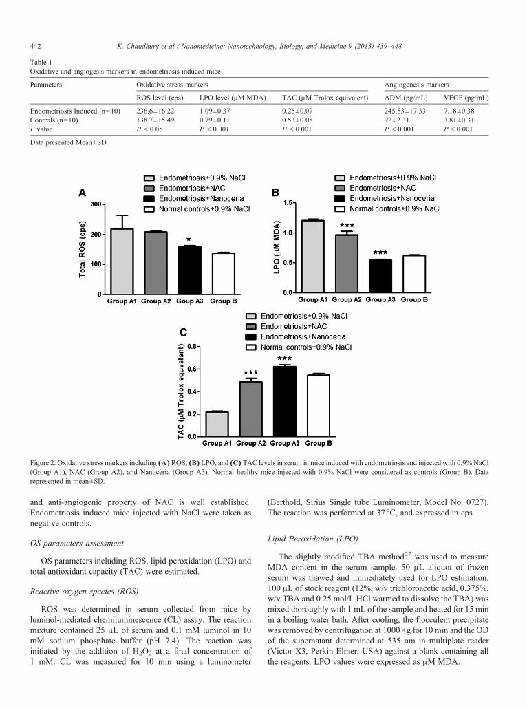
Table 1Oxidative and angiogesis markers in endometriosis induced mice
Parameters Oxidative stress markers Angiogenesis markers
ROS level (cps) LPO level (μM MDA) TAC (μM Trolox equivalent) ADM (pg/mL) VEGF (pg/mL)
Endometriosis Induced (n=10) 236.6±16.22 1.09±0.37 0.25±0.07 245.83±17.33 7.18±0.38Controls (n=10) 138.7±15.49 0.79±0.11 0.53±0.08 92±2.31 3.81±0.31P value P b 0.05 P b 0.001 P b 0.001 P b 0.001 P b 0.001
Data presented Mean±SD.
Figure 2. Oxidative stress markers including (A) ROS, (B) LPO, and (C) TAC levels in serum in mice induced with endometriosis and injected with 0.9%NaCl(Group A1), NAC (Group A2), and Nanoceria (Group A3). Normal healthy mice injected with 0.9% NaCl were considered as controls (Group B). Datarepresented in mean±SD.
442 K. Chaudhury et al / Nanomedicine: Nanotechnology, Biology, and Medicine 9 (2013) 439–448
and anti-angiogenic property of NAC is well established.Endometriosis induced mice injected with NaCl were taken asnegative controls.
OS parameters assessment
OS parameters including ROS, lipid peroxidation (LPO) andtotal antioxidant capacity (TAC) were estimated.
Reactive oxygen species (ROS)
ROS was determined in serum collected from mice byluminol-mediated chemiluminescence (CL) assay. The reactionmixture contained 25 μL of serum and 0.1 mM luminol in 10mM sodium phosphate buffer (pH 7.4). The reaction wasinitiated by the addition of H2O2 at a final concentration of1 mM. CL was measured for 10 min using a luminometer
(Berthold, Sirius Single tube Luminometer, Model No. 0727).The reaction was performed at 37°C, and expressed in cps.
Lipid Peroxidation (LPO)
The slightly modified TBA method27 was used to measureMDA content in the serum sample. 50 μL aliquot of frozenserum was thawed and immediately used for LPO estimation.100 μL of stock reagent (12%, w/v trichloroacetic acid, 0.375%,w/v TBA and 0.25 mol/L HCl warmed to dissolve the TBA) wasmixed thoroughly with 1 mL of the sample and heated for 15 minin a boiling water bath. After cooling, the flocculent precipitatewas removed by centrifugation at 1000×g for 10 min and the ODof the supernatant determined at 535 nm in multiplate reader(Victor X3, Perkin Elmer, USA) against a blank containing allthe reagents. LPO values were expressed as μM MDA.
Figure 3. Angiogenic markers including (A) ADM and (B) VEGF levels ofserum in mice induced with endometriosis and injected with 0.9% NaCl(Group A1), NAC (Group A2), and Nanoceria (Group A3). Normal healthymice injected with 0.9% NaCl were considered as controls (Group B). Datarepresented as mean±SD.
443K. Chaudhury et al / Nanomedicine: Nanotechnology, Biology, and Medicine 9 (2013) 439–448
Total Antioxidant Capacity (TAC)
TAC was measured in serum using a modified enhanced CLassay.28 Serum was thawed at room temperature (RT) and theirantioxidant capacity assessed. Serum was diluted 1:10 withdeionized water. Signal reagent was prepared using a CL kit (GEHealthcare, United Kingdom). A constant peak of ROS wasproduced using HRP conjugated IgG (Santa Cruz Biotechnology,Santa Cruz, California). HRP was diluted with deionized water togive a desired CL output. Luminometer was set in the kinetic modeand 900 μL reaction mixture added to the cuvette (100 μL signalreagent+700 μL deionized water +100 μL 1:350 diluted HRP) tohave the desired CL output. After 100s of HRP addition, 50 μL ofthe diluted serum sample was added to the mixture and 10%recovery of CL recorded. Trolox, a water soluble tocopherolanalogue, was used as a standard. The antioxidant capacity of themice serum samples was expressed in μM Trolox equivalents.
Angiogenic factors assessment
Angiogenic markers including vascular endothelial growthfactor (VEGF) and adrenomedullin (ADM) were assessed.
Vascular endothelial growth factor (VEGF)
VEGF level was assessed by ELISA using a mousemonoclonal anti-VEGF antibody (ab1316, Abcam, USA).Serum collected from mice was diluted in 50 mM carbonatebuffer at pH 9.0, added to ELISA plate and left overnight at 4 °C.The antigen solution was removed and 200 μL/well of blockingbuffer (PBS containing 1% BSA) added to block non-specificprotein binding. After incubation for 1–2h at RT, the wells werewashed with TBS (PBS containing 0.1% Tween 20), and anti-VEGF (1:1000 dilution of 1 mg/mL stock) added followed byincubation for 2h at RT. The wells were washed again, and 50μL/well of goat anti-mouse alkaline phosphatase conjugatedsecondary antibody (sc2008, Santacruz, California) diluted to1:1000, was added. Following incubation for one hour at RT,wells were washed three times with TBS. 50 μL/well of para-nitrophenylphosphate (Santacruz, California) dissolved indiethanolamine buffer to a final concentration of 1 mg/mL wasadded and allowed to develop for 10–20 min or until positivecontrol reached an OD of about 1.0 at 405 nm. Plate readings onmicrotiter plate reader were taken at OD 405 nm. A standardcurve was obtained for VEGF using recombinant mouse VEGFprotein (0.1–10,000 pg/mL) (580906, Recombinant MouseVEGF, Biolegand, San Diego, USA). Data were normalized tothe standard curve to yield values in pg/mL for VEGF.
Adrenomedullin (ADM)
Similar to the VEGF protocol, ADM levels were assessedby ELISA using a rabbit polyclonal anti-ADM antibody(sc33187, Santacruz, California). Goat anti-rabbit alkalinephosphatase conjugated antibody (ab6722) was used assecondary antibody. A standard curve was obtained for ADMusing recombinant mouse ADM protein (0.1–10,000 pg/mL)(ADM partial recombinant Protein, H00000133-Q01, NovusBiologicals, California). Data were normalized to the standardcurve to yield values in pg/mL for ADM.
Ovarian stimulation and spindle imaging
Superovulation was achieved using 5 IU rFSH injection(intraperitoneal; I.P.) followed by 5 IU hCG injection (I.P.) after48h. Oocyte cumulus complex (OCC) was flushed fromoviducts of mice 16h after hCG and transferred into Ham F-10medium for further investigation.29 The OCC was examinedunder a stereo zoom microscope and morphologically graded asmature/metaphase II (MII) oocytes when radiating corona cellsand expanded cumulus were present. Next, OCCs were culturedfor 2–3h in vitro in a humidified atmosphere of 5% CO2 at 37°Cfollowed by complete denudation of the cumulus by briefexposure to 300 IU/mL hyaluronidase (Sigma, St. Louis,Missouri) and subsequently passed through a micropipette.Live denuded MII oocytes were examined for the presence ofmeiotic spindle (MS) using a Polscope at 37°C (Oosight; CRi,Cambridge, Massachusetts). Briefly, oocytes were placed on aglass bottomed Polscope dish (Willco-dish; Willco Wells,Amsterdam, The Netherlands) and examined under the micro-scope attached to a 37°C temperature-controlled stage warmer.MS and its characteristics were examined under ×200 magni-fication and analyzed using Oosight META image systemsoftware version 1.6.
Statistical analysis
One way analysis of variance (ANOVA), t-test and chi-squaretest were used to analyze the data, as appropriate. Ky Plot version
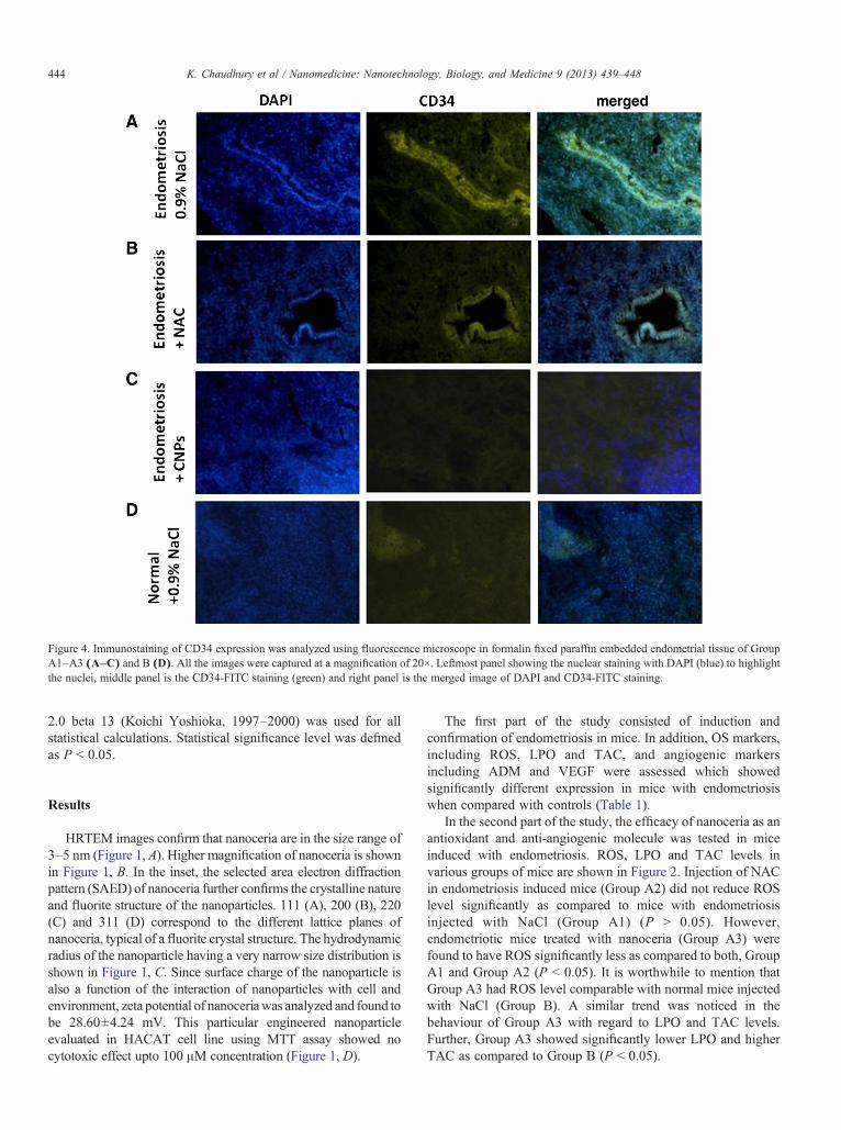
Figure 4. Immunostaining of CD34 expression was analyzed using fluorescence microscope in formalin fixed paraffin embedded endometrial tissue of GroupA1–A3 (A–C) and B (D). All the images were captured at a magnification of 20×. Leftmost panel showing the nuclear staining with DAPI (blue) to highlightthe nuclei, middle panel is the CD34-FITC staining (green) and right panel is the merged image of DAPI and CD34-FITC staining.
444 K. Chaudhury et al / Nanomedicine: Nanotechnology, Biology, and Medicine 9 (2013) 439–448
2.0 beta 13 (Koichi Yoshioka, 1997–2000) was used for allstatistical calculations. Statistical significance level was definedas P b 0.05.
Results
HRTEM images confirm that nanoceria are in the size range of3–5 nm (Figure 1, A). Higher magnification of nanoceria is shownin Figure 1, B. In the inset, the selected area electron diffractionpattern (SAED) of nanoceria further confirms the crystalline natureand fluorite structure of the nanoparticles. 111 (A), 200 (B), 220(C) and 311 (D) correspond to the different lattice planes ofnanoceria, typical of a fluorite crystal structure. The hydrodynamicradius of the nanoparticle having a very narrow size distribution isshown in Figure 1, C. Since surface charge of the nanoparticle isalso a function of the interaction of nanoparticles with cell andenvironment, zeta potential of nanoceriawas analyzed and found tobe 28.60±4.24 mV. This particular engineered nanoparticleevaluated in HACAT cell line using MTT assay showed nocytotoxic effect upto 100 μM concentration (Figure 1, D).
The first part of the study consisted of induction andconfirmation of endometriosis in mice. In addition, OS markers,including ROS, LPO and TAC, and angiogenic markersincluding ADM and VEGF were assessed which showedsignificantly different expression in mice with endometriosiswhen compared with controls (Table 1).
In the second part of the study, the efficacy of nanoceria as anantioxidant and anti-angiogenic molecule was tested in miceinduced with endometriosis. ROS, LPO and TAC levels invarious groups of mice are shown in Figure 2. Injection of NACin endometriosis induced mice (Group A2) did not reduce ROSlevel significantly as compared to mice with endometriosisinjected with NaCl (Group A1) (P N 0.05). However,endometriotic mice treated with nanoceria (Group A3) werefound to have ROS significantly less as compared to both, GroupA1 and Group A2 (P b 0.05). It is worthwhile to mention thatGroup A3 had ROS level comparable with normal mice injectedwith NaCl (Group B). A similar trend was noticed in thebehaviour of Group A3 with regard to LPO and TAC levels.Further, Group A3 showed significantly lower LPO and higherTAC as compared to Group B (P b 0.05).
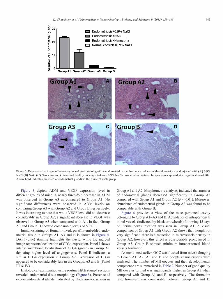
Figure 5. Representative image of hematoxylin and eosin staining of the endometrial tissue from mice induced with endometriosis and injected with (A) 0.9%NaCl (B) NAC (C) Nanoceria and (D) normal healthy mice injected with 0.9% NaCl considered as controls. Images were captured at a magnification of 20×.Arrow head indicates presence of endometrial glands in the tissue of each group.
445K. Chaudhury et al / Nanomedicine: Nanotechnology, Biology, and Medicine 9 (2013) 439–448
Figure 3 depicts ADM and VEGF expression level indifferent groups of mice. A nearly three-fold decrease in ADMwas observed in Group A3 as compared to Group A1. Nosignificant differences were observed in ADM levels oncomparing Group A3 with Group A2 and Group B, respectively.It was interesting to note that while VEGF level did not decreaseconsiderably in Group A2, a significant decrease in VEGF wasobserved in Group A3 when compared with A1. In fact, GroupA3 and Group B showed comparable levels of VEGF.
Immunostaining of formalin-fixed, paraffin-embedded endo-metrial tissue in Groups A1–A3 and B is shown in Figure 4.DAPI (blue) staining highlights the nuclei while the mergedimage represents localization of CD34 expression. Panel I showsintense membrane localization of CD34 (green) in Group A1depicting higher level of angiogenesis. Panel II indicates asimilar CD34 expression in Group A2. Expression of CD34appeared to be considerably less in the Groups, A3 and B (PanelIII & IV).
Histological examination using routine H&E stained sectionsrevealed endometrial tissue morphology (Figure 5). Presence ofexcess endometrial glands, indicated by black arrows, is seen in
Group A1 and A2. Morphometric analyses indicated that numberof endometrial glands decreased significantly in Group A3compared with Group A1 and Group A2 (P b 0.01). Moreover,abundance of endometrial glands in Group A3 was found to becomparable with Group B.
Figure 6 provides a view of the mice peritoneal cavitybelonging to Group A1–A3 and B. Abundance of intraperitonealblood vessels (indicated by black arrowheads) following 15daysof uterine horns injection was seen in Group A1. A visualcomparison of Group A1 with Group A2 shows that though notvery significant, there is a reduction in microvessels density inGroup A2; however, this effect is considerably pronounced inGroup A3. Group B showed minimum intraperitoneal bloodvessels formation.
As mentioned earlier, OCC was flushed from mice belongingto Group A1, A2, A3 and B and oocyte characteristics wereanalyzed. The number of MII oocytes and their developmentalcompetence are summarized in Table 2. Number of good qualityMII oocytes formed was significantly higher in Group A3 whencompared with Group A1 and B, respectively. The formationrate, however, was comparable between Group A3 and B.
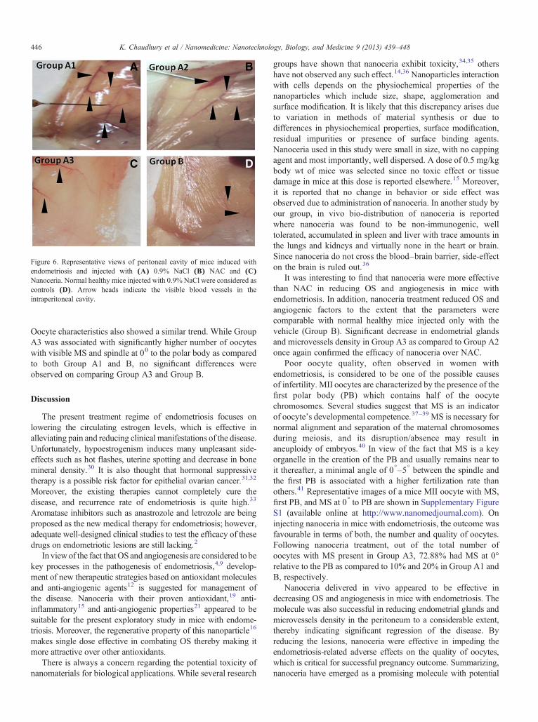
Figure 6. Representative views of peritoneal cavity of mice induced withendometriosis and injected with (A) 0.9% NaCl (B) NAC and (C)Nanoceria. Normal healthy mice injected with 0.9% NaCl were considered ascontrols (D). Arrow heads indicate the visible blood vessels in theintraperitoneal cavity.
446 K. Chaudhury et al / Nanomedicine: Nanotechnology, Biology, and Medicine 9 (2013) 439–448
Oocyte characteristics also showed a similar trend. While GroupA3 was associated with significantly higher number of oocyteswith visible MS and spindle at 00 to the polar body as comparedto both Group A1 and B, no significant differences wereobserved on comparing Group A3 and Group B.
Discussion
The present treatment regime of endometriosis focuses onlowering the circulating estrogen levels, which is effective inalleviating pain and reducing clinical manifestations of the disease.Unfortunately, hypoestrogenism induces many unpleasant side-effects such as hot flashes, uterine spotting and decrease in bonemineral density.30 It is also thought that hormonal suppressivetherapy is a possible risk factor for epithelial ovarian cancer.31,32
Moreover, the existing therapies cannot completely cure thedisease, and recurrence rate of endometriosis is quite high.33
Aromatase inhibitors such as anastrozole and letrozole are beingproposed as the new medical therapy for endometriosis; however,adequate well-designed clinical studies to test the efficacy of thesedrugs on endometriotic lesions are still lacking.2
In viewof the fact thatOS and angiogenesis are considered to bekey processes in the pathogenesis of endometriosis,4,9 develop-ment of new therapeutic strategies based on antioxidant moleculesand anti-angiogenic agents12 is suggested for management ofthe disease. Nanoceria with their proven antioxidant,19 anti-inflammatory15 and anti-angiogenic properties21 appeared to besuitable for the present exploratory study in mice with endome-triosis. Moreover, the regenerative property of this nanoparticle16
makes single dose effective in combating OS thereby making itmore attractive over other antioxidants.
There is always a concern regarding the potential toxicity ofnanomaterials for biological applications. While several research
groups have shown that nanoceria exhibit toxicity,34,35 othershave not observed any such effect.14,36 Nanoparticles interactionwith cells depends on the physiochemical properties of thenanoparticles which include size, shape, agglomeration andsurface modification. It is likely that this discrepancy arises dueto variation in methods of material synthesis or due todifferences in physiochemical properties, surface modification,residual impurities or presence of surface binding agents.Nanoceria used in this study were small in size, with no cappingagent and most importantly, well dispersed. A dose of 0.5 mg/kgbody wt of mice was selected since no toxic effect or tissuedamage in mice at this dose is reported elsewhere.15 Moreover,it is reported that no change in behavior or side effect wasobserved due to administration of nanoceria. In another study byour group, in vivo bio-distribution of nanoceria is reportedwhere nanoceria was found to be non-immunogenic, welltolerated, accumulated in spleen and liver with trace amounts inthe lungs and kidneys and virtually none in the heart or brain.Since nanoceria do not cross the blood–brain barrier, side-effecton the brain is ruled out.36
It was interesting to find that nanoceria were more effectivethan NAC in reducing OS and angiogenesis in mice withendometriosis. In addition, nanoceria treatment reduced OS andangiogenic factors to the extent that the parameters werecomparable with normal healthy mice injected only with thevehicle (Group B). Significant decrease in endometrial glandsand microvessels density in Group A3 as compared to Group A2once again confirmed the efficacy of nanoceria over NAC.
Poor oocyte quality, often observed in women withendometriosis, is considered to be one of the possible causesof infertility. MII oocytes are characterized by the presence of thefirst polar body (PB) which contains half of the oocytechromosomes. Several studies suggest that MS is an indicatorof oocyte's developmental competence.37–39 MS is necessary fornormal alignment and separation of the maternal chromosomesduring meiosis, and its disruption/absence may result inaneuploidy of embryos.40 In view of the fact that MS is a keyorganelle in the creation of the PB and usually remains near toit thereafter, a minimal angle of 0°–5° between the spindle andthe first PB is associated with a higher fertilization rate thanothers.41 Representative images of a mice MII oocyte with MS,first PB, and MS at 0° to PB are shown in Supplementary FigureS1 (available online at http://www.nanomedjournal.com). Oninjecting nanoceria in mice with endometriosis, the outcome wasfavourable in terms of both, the number and quality of oocytes.Following nanoceria treatment, out of the total number ofoocytes with MS present in Group A3, 72.88% had MS at 0°relative to the PB as compared to 10% and 20% in Group A1 andB, respectively.
Nanoceria delivered in vivo appeared to be effective indecreasing OS and angiogenesis in mice with endometriosis. Themolecule was also successful in reducing endometrial glands andmicrovessels density in the peritoneum to a considerable extent,thereby indicating significant regression of the disease. Byreducing the lesions, nanoceria were effective in impeding theendometriosis-related adverse effects on the quality of oocytes,which is critical for successful pregnancy outcome. Summarizing,nanoceria have emerged as a promising molecule with potential

Table 2Oocyte characterization in control, NAC and nanoceria treated groups
Oocyte parameters Group A1 Group A2 Group A3 Group B P value
No. of MII oocytes 75.80% (30/62) 79.45% (58/73) 92.50% (74/80) 66.66% (48/72) A3 A1≤0.05A3B≤0.05A3 A2 NSNo. of oocytes with spindle present 33.33% (10/30) 72.41% (42/58) 79.72% (59/74) 41.66% (20/48) A3 A1≤0.05A3B≤0.05A3 A2 NSNo. of spindles at 0° to polar body 10% (1/10) 69.04% (29/42) 72.88% (43/59) 20% (4/20) A3 A1≤0.05A3B≤0.05A3 A2 NS
447K. Chaudhury et al / Nanomedicine: Nanotechnology, Biology, and Medicine 9 (2013) 439–448
application in clinical therapy for endometriosis. This is a proof ofconcept study. Before considering pre-clinical trials, toxicitystudies in greater detail using these nanoparticles are necessary.We suggest pre-clinical non-human primate studies42 for testingthe safety and efficacy of these nanoparticles at larger doses inendometriosis. A pilot study exploring nanoceria as an adjuvant toletrozole in mice with endometriosis is presently underway.
Appendix A. Supplementary data
Supplementary data to this article can be found online athttp://dx.doi.org/10.1016/j.nano.2012.08.001.
References
1. McLeod BS, Retzloff MG. Epidemiology of endometriosis: anassessment of risk factors. Clin Obstet Gynecol 2010;53:389-96.
2. Colette SB, Donnez J. Are aromatase inhibitors effective in endometri-osis treatment? Expert Opin Investig Drugs 2011;20:917-31.
3. Martin DC. Hysterectomy for treatment of pain associated withendometriosis. J Minim Invasive Gynecol 2006;13:566-72.
4. Augoulea A, Mastorakos G, Lambrinoudaki I, Christodoulakos G,Creatsas G. The role of the oxidative-stress in the endometriosis-relatedinfertility. Gynecol Endocrinol 2009;25:75-81.
5. Szczepańska M, Koźlik J, Skrzypczak J, Mikołajczyk M. Oxidativestress may be a piece in the endometriosis puzzle. Fertil Steril 2003;79:1288-93.
6. Jackson LW, Schisterman EF, Dey-Rao R, Browne R, Armstrong D.Oxidative stress and endometriosis. Hum Reprod 2005;20:2014-20.
7. Mier-Cabrera J, Aburto-Soto T, Burrola-Méndez S, Jiménez-Zamudio L,Tolentino M, Casanueva E, et al. Women with endometriosis improvedtheir peripheral antioxidant markers after the application of a highantioxidant diet. Reprod Biol Endocrinol 2009;28:54.
8. Melo AS, Rosa-e-Silva JCs, Rosa-e-Silva ACJdS, Poli-Neto OB,Ferriani RA, Vieira CS. Unfavorable lipid profile in women withendometriosis. Fertil Steril 2010;93:2433-6.
9. Healy DL, Rogers PAW, Hii L, Wingfield M. Angiogenesis: a newtheory for endometriosis. Hum Reprod Update 1998;4:736-40.
10. Taylor RN, Jie Y, Torres PB, Schickedanz AC, Park JK, Mueller MD,et al. Mechanistic and therapeutic implications of angiogenesis inendometriosis. Reprod Sci 2009;16:140-6.
11. McLaren J, Prentice A, Charnock-Jones DS, Smith SK. Vascularendothelial growth factor (VEGF) concentrations are elevated inperitoneal fluid of women with endometriosis. Hum Reprod 1996;11:220-3.
12. Ferrero S, Ragni N, Remorgida V. Antiangiogenic therapies inendometriosis. Br J Pharmacol 2006;149:133-5.
13. Taylor HS, Osteen KG, Bruner-Tran KL, Krikun G, Sokalska A,Duleba AJ. Novel therapies targeting endometriosis. Reprod Sci 2011;18:814-23.
14. Celardo I, Traversa E, Ghibelli L. Cerium oxide nanoparticles: a promisefor applications in therapy. J Exp Ther Oncol 2011;9:47-51.
15. Hirst SM, Karakoti AS, Tyler RD, Sriranganathan N, Seal S, Reilly CM.Anti-inflammatory properties of cerium oxide nanoparticles. Small2009;5:2848-56.
16. Karakoti A, Monteiro-Riviere N, Aggarwal R, Davis J, Narayan R, SelfW, et al. Nanoceria as antioxidant: synthesis and biomedical applica-tions. JOM J Minerals, Metals Mater Soc 2008;60:33-7.
17. Kong L, Cai X, Zhou X, Wong LL, Karakoti AS, Seal S, et al. Nanoceriaextend photoreceptor cell lifespan in tubby mice by modulation ofapoptosis/survival signaling pathways. Neurobiol Dis 2011;42:514-23.
18. Estevez AY, Pritchard S, Harper K, Aston JW, Lynch A, Lucky JJ, et al.Neuroprotective mechanisms of cerium oxide nanoparticles in a mousehippocampal brain slice model of ischemia. Free Radic Biol Med 2011;51:1155-63.
19. Colon J, Hsieh N, Ferguson A, Kupelian P, Seal S, Jenkins DW, et al.Cerium oxide nanoparticles protect gastrointestinal epithelium fromradiation-induced damage by reduction of reactive oxygen species andupregulation of superoxide dismutase 2. Nanomedicine: Nanotechnol,Biol Med 2010;6:698-705.
20. Anderson RE, LaVail MM, Hollyfield JG, Chen J, Patil S, Seal S, et al.Nanoceria particles prevent ROI-induced blindness. Recent advances inretinal degeneration. New York: Springer; 2008. p. 53-9.
21. Zhou X, Wong LL, Karakoti AS, Seal S, McGinnis JF. Nanoceria inhibitthe development and promote the regression of pathologic retinalneovascularization in the knockout mouse. PLoS One 2011;6:e16733.
22. Niu J, Azfer A, Rogers LM, Wang X, Kolattukudy PE. Cardioprotectiveeffects of cerium oxide nanoparticles in a transgenic murine model ofcardiomyopathy. Cardiovasc Res 2007;73:549-59.
23. Das M, Patil S, Bhargava N, Kang J-F, Riedel LM, Seal S, et al. Auto-catalytic ceria nanoparticles offer neuroprotection to adult rat spinal cordneurons. Biomaterials 2007;28:1918-25.
24. Karakoti AS, Singh S, Kumar A, Malinska M, Kuchibhatla SVNT,Wozniak K, et al. PEGylated nanoceria as radical scavenger with tunableredox chemistry. J Am Chem Soc 2009;131:14144-5.
25. Nowak NM, Fischer OM, Gust TC, Fuhrmann U, Habenicht UF,Schmidt A. Intraperitoneal inflammation decreases endometriosis in amouse model. Hum Reprod 2008;23:2466-74.
26. Marian AJ, Senthil V, Chen SN, Lombardi R. Antifibrotic effects ofantioxidant N-acetylcysteine in a mouse model of human hypertrophiccardiomyopathy mutation. J Am Coll Cardiol 2006;47(4):827-34.
27. Buege J, Aust S. Microsomal lipid peroxidation. Methods Enzymol1978;52:02-10.
28. Chapple I, Mason G, Garner I, Matthews J, Thorpe G, SM SR, et al.Enhanced chemiluminescent assay for measuring the total antioxidantcapacity of serum, saliva and crevicular fluid. Ann Clin Biochem 1997;34:412-21.
29. Ganesh A, Chattopadhyay R, Narendra Babu K, Chakravarty B,Chaudhury K. Analysis of spindle characteristics and embryo qualityin mice stimulated with letrozole using Polscope imaging. Fertil Steril2010;93:1477-81.
30. Helms RA, Quan DJ, editors. Textbook of therapeutics: drug anddisease management Philadelphia. Lippincott Williams & Wilkins;2006.
31. Blumenfeld Z. Hormonal suppressive therapy for endometriosis may notimprove patient health. Fertil Steril 2004;81:487-92.
32. Greer JB, Modugno F, Allen GO, Ness RB. Androgenic progestins inoral contraceptives and the risk of epithelial ovarian cancer. ObstetGynecol 2005;105:731-40.
448 K. Chaudhury et al / Nanomedicine: Nanotechnology, Biology, and Medicine 9 (2013) 439–448
33. Guo S-W. Recurrence of endometriosis and its control. Hum ReprodUpdate 2009;15:441-61.
34. Srinivas A, Rao PJ, Selvam G, Murthy PB, Reddy PN. Acute inhalationtoxicity of cerium oxide nanoparticles in rats. Toxicol Lett 2011;205:105-15.
35. Wingard C, Walters D, Cathey B, Hilderbrand S, Katwa P, Lin S, et al.Mast cells contribute to altered vascular reactivity and ischemia–reperfusion injury following cerium oxide nanoparticle instillation. Na-notoxicology 2010;5:531-45.
36. Hirst SM, Karakoti A, Singh S, Self W, Tyler R, Seal S, et al. Bio-distribution and in vivo antioxidant effects of cerium oxide nanoparticlesin mice. Environ Toxicol 2011.
37. Wang WH, Meng L, Hackett RJ, Keefe DL. Developmental ability ofhuman oocytes with or without birefringent spindles imaged by Polscopebefore insemination. Hum Reprod 2001;16:1464-8.
38. Moon JH, Hyun CS, Lee SW, Son WY, Yoon SH, Lim JH.Visualization of the metaphase II meiotic spindle in living human
oocytes using the Polscope enables the prediction of embryonicdevelopmental competence after ICSI. Hum Reprod 2003;18:817-20.
39. Rienzi L, Martinez F, Ubaldi F, Minasi MG, Iacobelli M, Tesarik J, et al.Polscope analysis of meiotic spindle changes in living metaphase IIhuman oocytes during the freezing and thawing procedures. HumReprod 2004;19:655-9.
40. Wang W-H, Meng L, Hackett RJ, Odenbourg R, Keefe DL. Thespindle observation and its relationship with fertilization afterintracytoplasmic sperm injection in living human oocytes. FertilSteril 2001;75:348-53.
41. Fang C, Tang M, Li T, Peng W-L, Zhou C-Q, Zhuang G-L, et al.Visualization of meiotic spindle and subsequent embryonic developmentin in vitro and in vivo matured human oocytes. J Assist Reprod Genet2007;24:547-51.
42. D'Hooghe TM, Kyama CM, Chai D, Fassbender A, Vodolazkaia A,Bokor A, et al. Nonhuman primate models for translational research inendometriosis. Reprod Sci 2009;16:152-61.