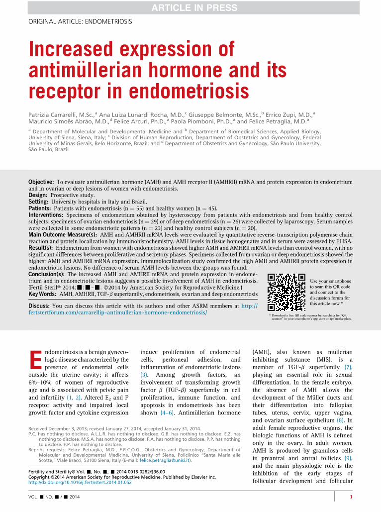
Increased expression of antimüllerian hormone and its receptor in endometriosis
6
Increased expression of antim € ullerian hormone and its receptor in endometriosis Patrizia Carrarelli, M.Sc., a Ana Luiza Lunardi Rocha, M.D., c Giuseppe Belmonte, M.Sc., b Errico Zupi, M.D., a Mauricio Simoẽs Abr~ ao, M.D., d Felice Arcuri, Ph.D., a Paola Piomboni, Ph.D., a and Felice Petraglia, M.D. a a Department of Molecular and Developmental Medicine and b Department of Biomedical Sciences, Applied Biology, University of Siena, Siena, Italy; c Division of Human Reproduction, Department of Obstetrics and Gynecology, Federal University of Minas Gerais, Belo Horizonte, Brazil; and d Department of Obstetrics and Gynecology, S~ ao Paulo University, S~ ao Paulo, Brazil Objective: To evaluate antim€ ullerian hormone (AMH) and AMH receptor II (AMHRII) mRNA and protein expression in endometrium and in ovarian or deep lesions of women with endometriosis. Design: Prospective study. Setting: University hospitals in Italy and Brazil. Patients: Patients with endometriosis (n ¼ 55) and healthy women (n ¼ 45). Interventions: Specimens of endometrium obtained by hysteroscopy from patients with endometriosis and from healthy control subjects; specimens of ovarian endometriosis (n ¼ 29) or of deep endometriosis (n ¼ 26) were collected by laparoscopy. Serum samples were collected in some endometriotic patients (n ¼ 23) and healthy control subjects (n ¼ 20). Main Outcome Measure(s): AMH and AMHRII mRNA levels were evaluated by quantitative reverse-transcription polymerase chain reaction and protein localization by immunohistochemistry. AMH levels in tissue homogenates and in serum were assessed by ELISA. Result(s): Endometrium from women with endometriosis showed higher AMH and AMHRII mRNA levels than control women, with no significant differences between proliferative and secretory phases. Specimens collected from ovarian or deep endometriosis showed the highest AMH and AMHRII mRNA expression. Immunolocalization study confirmed the high AMH and AMHRII protein expression in endometriotic lesions. No difference of serum AMH levels between the groups was found. Conclusion(s): The increased AMH and AMHRII mRNA and protein expression in endome- trium and in endometriotic lesions suggests a possible involvement of AMH in endometriosis. (Fertil Steril Ò 2014;-:-–-. Ó2014 by American Society for Reproductive Medicine.) Key Words: AMH, AMHRII, TGF-b superfamily, endometriosis, ovarian and deep endometriosis Discuss: You can discuss this article with its authors and other ASRM members at http:// fertstertforum.com/carrarellip-antimullerian-hormone-endometriosis/ Use your smartphone to scan this QR code and connect to the discussion forum for this article now.* * Download a free QR code scanner by searching for “QR scanner” in your smartphone’s app store or app marketplace. E ndometriosis is a benign gyneco- logic disease characterized by the presence of endometrial cells outside the uterine cavity; it affects 6%–10% of women of reproductive age and is associated with pelvic pain and infertility (1, 2). Altered E 2 and P receptor activity and impaired local growth factor and cytokine expression induce proliferation of endometrial cells, peritoneal adhesion, and inflammation of endometriotic lesions (3). Among growth factors, an involvement of transforming growth factor b (TGF-b) superfamily in cell proliferation, immune function, and apoptosis in endometriosis has been shown (4–6). Antim€ ullerian hormone (AMH), also known as m€ ullerian inhibiting substance (MIS), is a member of TGF-b superfamily (7), playing an essential role in sexual differentiation. In the female embryo, the absence of AMH allows the development of the M€ uller ducts and their differentiation into fallopian tubes, uterus, cervix, upper vagina, and ovarian surface epithelium (8). In adult female reproductive organs, the biologic functions of AMH is defined only in the ovary. In adult women, AMH is produced by granulosa cells in preantral and antral follicles (9), and the main physiologic role is the inhibition of the early stages of follicular development and follicular Received December 3, 2013; revised January 27, 2014; accepted January 31, 2014. P.C. has nothing to disclose. A.L.L.R. has nothing to disclose. G.B. has nothing to disclose. E.Z. has nothing to disclose. M.S.A. has nothing to disclose. F.A. has nothing to disclose. P.P. has nothing to disclose. F.P. has nothing to disclose. Reprint requests: Felice Petraglia, M.D., F.R.C.O.G., Obstetrics and Gynecology, Department of Molecular and Developmental Medicine, University of Siena, Policlinico ‘‘Santa Maria alle Scotte,’’ Viale Bracci, 53100 Siena, Italy (E-mail: [email protected]). Fertility and Sterility® Vol. -, No. -, - 2014 0015-0282/$36.00 Copyright ©2014 American Society for Reproductive Medicine, Published by Elsevier Inc. http://dx.doi.org/10.1016/j.fertnstert.2014.01.052 VOL. - NO. - / - 2014 1 ORIGINAL ARTICLE: ENDOMETRIOSIS
-
Upload
independent -
Category
Documents
-
view
1 -
download
0
Transcript of Increased expression of antimüllerian hormone and its receptor in endometriosis
ORIGINAL ARTICLE: ENDOMETRIOSIS
Increased expression ofantim€ullerian hormone and itsreceptor in endometriosis
Patrizia Carrarelli, M.Sc.,a Ana Luiza Lunardi Rocha, M.D.,c Giuseppe Belmonte, M.Sc.,b Errico Zupi, M.D.,aMauricio Simoẽs Abr~ao, M.D.,d Felice Arcuri, Ph.D.,a Paola Piomboni, Ph.D.,a and Felice Petraglia, M.D.a
a Department of Molecular and Developmental Medicine and b Department of Biomedical Sciences, Applied Biology,University of Siena, Siena, Italy; c Division of Human Reproduction, Department of Obstetrics and Gynecology, FederalUniversity of Minas Gerais, Belo Horizonte, Brazil; and d Department of Obstetrics and Gynecology, S~ao Paulo University,S~ao Paulo, Brazil
Objective: To evaluate antim€ullerian hormone (AMH) and AMH receptor II (AMHRII) mRNA and protein expression in endometriumand in ovarian or deep lesions of women with endometriosis.Design: Prospective study.Setting: University hospitals in Italy and Brazil.Patients: Patients with endometriosis (n ¼ 55) and healthy women (n ¼ 45).Interventions: Specimens of endometrium obtained by hysteroscopy from patients with endometriosis and from healthy controlsubjects; specimens of ovarian endometriosis (n¼ 29) or of deep endometriosis (n¼ 26) were collected by laparoscopy. Serum sampleswere collected in some endometriotic patients (n ¼ 23) and healthy control subjects (n ¼ 20).Main Outcome Measure(s): AMH and AMHRII mRNA levels were evaluated by quantitative reverse-transcription polymerase chainreaction and protein localization by immunohistochemistry. AMH levels in tissue homogenates and in serum were assessed by ELISA.Result(s): Endometrium fromwomen with endometriosis showed higher AMH and AMHRII mRNA levels than control women, with nosignificant differences between proliferative and secretory phases. Specimens collected from ovarian or deep endometriosis showed thehighest AMH and AMHRII mRNA expression. Immunolocalization study confirmed the high AMH and AMHRII protein expression in
Use your smartphone
endometriotic lesions. No difference of serum AMH levels between the groups was found.Conclusion(s): The increased AMH and AMHRII mRNA and protein expression in endome-trium and in endometriotic lesions suggests a possible involvement of AMH in endometriosis.(Fertil Steril� 2014;-:-–-. �2014 by American Society for Reproductive Medicine.)KeyWords: AMH, AMHRII, TGF-b superfamily, endometriosis, ovarian and deep endometriosis
Discuss: You can discuss this article with its authors and other ASRM members at http://fertstertforum.com/carrarellip-antimullerian-hormone-endometriosis/
to scan this QR codeand connect to thediscussion forum forthis article now.*
* Download a free QR code scanner by searching for “QRscanner” in your smartphone’s app store or app marketplace.
E ndometriosis is a benign gyneco-logic disease characterized by thepresence of endometrial cells
outside the uterine cavity; it affects6%–10% of women of reproductiveage and is associated with pelvic painand infertility (1, 2). Altered E2 and Preceptor activity and impaired localgrowth factor and cytokine expression
Received December 3, 2013; revised January 27, 201P.C. has nothing to disclose. A.L.L.R. has nothing to
nothing to disclose. M.S.A. has nothing to discloto disclose. F.P. has nothing to disclose.
Reprint requests: Felice Petraglia, M.D., F.R.C.O.GMolecular and Developmental Medicine, UniScotte,’’ Viale Bracci, 53100 Siena, Italy (E-mail:
Fertility and Sterility® Vol. -, No. -, - 2014 0015-Copyright ©2014 American Society for Reproductivehttp://dx.doi.org/10.1016/j.fertnstert.2014.01.052
VOL. - NO. - / - 2014
induce proliferation of endometrialcells, peritoneal adhesion, andinflammation of endometriotic lesions(3). Among growth factors, aninvolvement of transforming growthfactor b (TGF-b) superfamily in cellproliferation, immune function, andapoptosis in endometriosis has beenshown (4–6). Antim€ullerian hormone
4; accepted January 31, 2014.disclose. G.B. has nothing to disclose. E.Z. hasse. F.A. has nothing to disclose. P.P. has nothing
., Obstetrics and Gynecology, Department ofversity of Siena, Policlinico ‘‘Santa Maria [email protected]).
0282/$36.00Medicine, Published by Elsevier Inc.
(AMH), also known as m€ullerianinhibiting substance (MIS), is amember of TGF-b superfamily (7),playing an essential role in sexualdifferentiation. In the female embryo,the absence of AMH allows thedevelopment of the M€uller ducts andtheir differentiation into fallopiantubes, uterus, cervix, upper vagina,and ovarian surface epithelium (8). Inadult female reproductive organs, thebiologic functions of AMH is definedonly in the ovary. In adult women,AMH is produced by granulosa cellsin preantral and antral follicles (9),and the main physiologic role is theinhibition of the early stages offollicular development and follicular
1

ORIGINAL ARTICLE: ENDOMETRIOSIS
recruitment (10). Recently, serum AMH has been recognizedas a useful diagnostic and prognostic tool as a reliablemarker of the ovarian reserve and predictor of the ovarianresponse to controlled ovarian hyperstimulation (11), aswell as an early indicator of relapse of ovarian granulosacell tumors (12).
As a member of the TGF-b superfamily, AMH exertsits effect by binding to a heterodimeric transmembraneserine/threonine kinase cell surface receptor complex,consisting of its specific type II receptor (AMHRII), whichthen binds to a type I receptor of the activin-like kinase family(ALK2, ALK3, and ALK6) (13). The heterodimeric type I–type IIreceptor complex triggers a downstream signaling cascade ofphosphorylation, activating Smads 1, 5, and 8 (14). The Smadsignaling complex then enters the nucleus and interacts withtranscription factors to induce gene expression for apoptosis(15), specifically for regression of m€ullerian ducts in the maleembryo (13).
Glandular and stromal endometrial cells express mRNAand protein for AMH and AMHRII, resulting in measurableAMH in culture media (16); in agreement with the proapopto-tic effect (13, 15), when AMH is added to the cultured humanendometrial stromal cells, an increase of caspase 3/7 occurs,causing a decrease of cell viability (16). An effect of AMHon cell proliferation and cell death has been found also inendometrial/cervical cancer cells (17, 18), and even morepronounced in cultures of endometrial cells fromendometriotic patients, where AMH was found to inducesignificant increase of annexin V (17).
Because of the involvement of the AMH/AMHRII pathwayin modulating endometrial proliferative and invasivebehavior, and because this is a typical characteristic of bothendometrium and ectopic lesions (19), we aimed to evaluatethe expression of AMH/AMHRII in eutopic and ectopic endo-metrium of endometriosis. In particular, we tested the hypoth-esis that: (1) endometrial AMH andAMHRII mRNA and proteinexpression in endometriotic patients is different from that inhealthy women; (2) ovarian (endometrioma) and extraovarian(deep) endometriotic lesions express a specific AMH/AMHRIIpattern (the two sites were also selected to exclude thatthe expression in endometrioma may depend by a possiblecontamination of ovarian tissue); and (3) the different expres-sion of AMH in endometrium and lesions does not affectcirculating AMH secretion of endometriotic patients.
MATERIALS AND METHODSPatients and Tissue Collection
The study population consisted of women with regular men-strual cycles (28–32 days) undergoing gynecologic surgery(age range 21–39 years) and was divided into:
Endometriosis group: ovarian endometrioma (n ¼ 29)and deep endometriosis (n ¼ 26) undergoing laparoscopictreatment (for pain and/or infertility).
Nonendometriosis group: women without endometriosis(n ¼ 45; control subjects) undergoing laparoscopy for tubalsterilization.
For each subject, a complete medical history was obtainedand physical examination performed. Patients with hormonal
2
treatment in the past 3 months were excluded from the study.Each of the patients gave written informed consent. The studywas approved by the local Human Investigation Committee.
In all of the women, a specimen of endometrium wascollected by hysteroscopy and separated into proliferative(days 5–14) and secretory phases (day 15 onward), accordingto the last day of menstruation, transvaginal ultrasound scan,and histologic criteria. In the endometriosis group, all patientsshowed stage III or IV endometriosis according to the Ameri-can Society for Reproductive Medicine classification (20).Specimens of endometriotic lesions were collected duringlaparoscopic surgery. Ovarian endometriotic tissues (cystdiameter measured by ultrasound ranged from 38 to72 mm) were carefully stripped from the inner cyst wallavoiding contamination with ovarian tissues. Deep endome-triosis specimens were collected from cases with bowel orrectovaginal (n ¼ 23) or bladder (n ¼ 3) involvement. Serumsamples were also collected in endometriotic (n¼ 23; 11 withendometriosis and 12 with deep endometriosis) and healthy(n ¼ 20) women 1 week before surgery.
All specimens were immediately frozen and stored inliquid nitrogen for RNA and protein extraction or paraffinembedded for immunohistochemistry; they were all analyzedat the same time.
RNA Extraction and cDNA Preparation
Total RNA was extracted with the use of the SV Total RNAIsolation System (Promega), according to manufacturer's in-structions. RNA was quantified by ultraviolet absorptionand RNA integrity checked before downstream analysiswith the use of the Flashgel System (Lonza Group). ForcDNA synthesis, one microgram of total RNA was reversetranscribed with the use of Improm-II Reverse Transcriptase(Promega).
Reverse Transcription Polymerase Chain Reaction
Endometrial AMH and AMHRII expression was analyzed byquantitative reverse-transcription (RT) polymerase chain re-action (PCR). AMH and AMHRII mRNA levels, normalizedfor the transferrin receptor (TFRC) as housekeeping genewere measured in triplicate with the use of the 2� Sybr SelectMaster Mix for CFX (Applied Biosystems) according to themanufacturer's protocol, on a CFX Connect 96 (Bio-RadLaboratories) real-time PCR system.
Gene-specific primers used for PCR were chosen accord-ing to the published sequences of human AMH (Genbankaccession no. NM_000479.3), AMHRII (Genbank accessionno. NM_002192.2), and TFRC (Genbank accession no.NM_003234.2). The AMH forward primer was 50–TCC GAGAAG ACT TGG ACT GG–30, the reverse primer was 50- TCCTCC AGG TGT AGG ACC AC–30, and the expected size ofthe amplified fragment was 296 bp; the AMHRII forwardprimer was 50–GAG ATC ATC ACG TTT GCC GAG–30, thereverse primer was 50- GAA GAG CCA GAC TTC TGC ACG–30, and the expected size of the amplified fragment was258 bp; the TFRC forward primer was 50–ACC GGC ACCATC AAG CT–30, the reverse primer was 50- TGA TCA CGC
VOL. - NO. - / - 2014
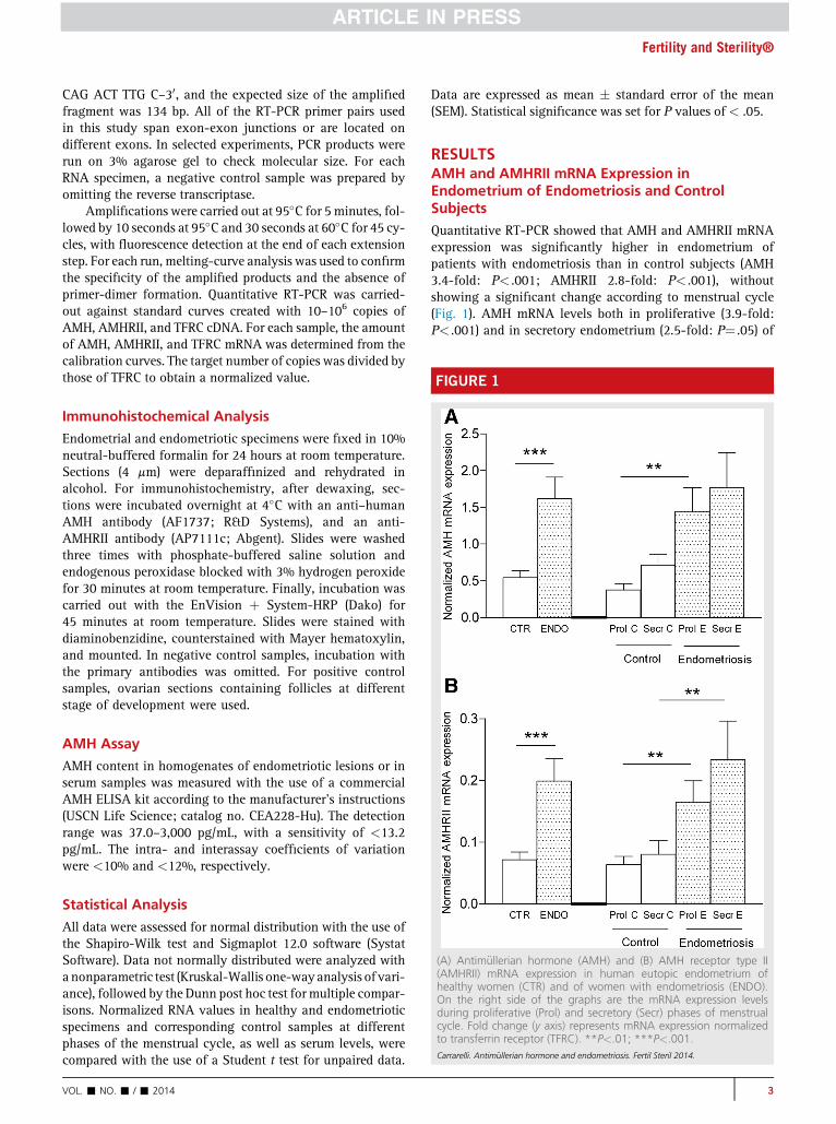
FIGURE 1
Fertility and Sterility®
CAG ACT TTG C–30, and the expected size of the amplifiedfragment was 134 bp. All of the RT-PCR primer pairs usedin this study span exon-exon junctions or are located ondifferent exons. In selected experiments, PCR products wererun on 3% agarose gel to check molecular size. For eachRNA specimen, a negative control sample was prepared byomitting the reverse transcriptase.
Amplifications were carried out at 95�C for 5 minutes, fol-lowed by 10 seconds at 95�C and 30 seconds at 60�C for 45 cy-cles, with fluorescence detection at the end of each extensionstep. For each run, melting-curve analysis was used to confirmthe specificity of the amplified products and the absence ofprimer-dimer formation. Quantitative RT-PCR was carried-out against standard curves created with 10–106 copies ofAMH, AMHRII, and TFRC cDNA. For each sample, the amountof AMH, AMHRII, and TFRC mRNA was determined from thecalibration curves. The target number of copies was divided bythose of TFRC to obtain a normalized value.
Immunohistochemical Analysis
Endometrial and endometriotic specimens were fixed in 10%neutral-buffered formalin for 24 hours at room temperature.Sections (4 mm) were deparaffinized and rehydrated inalcohol. For immunohistochemistry, after dewaxing, sec-tions were incubated overnight at 4�C with an anti–humanAMH antibody (AF1737; R&D Systems), and an anti-AMHRII antibody (AP7111c; Abgent). Slides were washedthree times with phosphate-buffered saline solution andendogenous peroxidase blocked with 3% hydrogen peroxidefor 30 minutes at room temperature. Finally, incubation wascarried out with the EnVision þ System-HRP (Dako) for45 minutes at room temperature. Slides were stained withdiaminobenzidine, counterstained with Mayer hematoxylin,and mounted. In negative control samples, incubation withthe primary antibodies was omitted. For positive controlsamples, ovarian sections containing follicles at differentstage of development were used.
AMH Assay
AMH content in homogenates of endometriotic lesions or inserum samples was measured with the use of a commercialAMH ELISA kit according to the manufacturer's instructions(USCN Life Science; catalog no. CEA228-Hu). The detectionrange was 37.0–3,000 pg/mL, with a sensitivity of <13.2pg/mL. The intra- and interassay coefficients of variationwere <10% and <12%, respectively.
(A) Antim€ullerian hormone (AMH) and (B) AMH receptor type II(AMHRII) mRNA expression in human eutopic endometrium ofhealthy women (CTR) and of women with endometriosis (ENDO).On the right side of the graphs are the mRNA expression levelsduring proliferative (Prol) and secretory (Secr) phases of menstrualcycle. Fold change (y axis) represents mRNA expression normalizedto transferrin receptor (TFRC). **P<.01; ***P<.001.Carrarelli. Antim€ullerian hormone and endometriosis. Fertil Steril 2014.
Statistical Analysis
All data were assessed for normal distribution with the use ofthe Shapiro-Wilk test and Sigmaplot 12.0 software (SystatSoftware). Data not normally distributed were analyzed withanonparametric test (Kruskal-Wallis one-wayanalysis of vari-ance), followed by the Dunn post hoc test formultiple compar-isons. Normalized RNA values in healthy and endometrioticspecimens and corresponding control samples at differentphases of the menstrual cycle, as well as serum levels, werecompared with the use of a Student t test for unpaired data.
VOL. - NO. - / - 2014
Data are expressed as mean � standard error of the mean(SEM). Statistical significance was set for P values of < .05.
RESULTSAMH and AMHRII mRNA Expression inEndometrium of Endometriosis and ControlSubjects
Quantitative RT-PCR showed that AMH and AMHRII mRNAexpression was significantly higher in endometrium ofpatients with endometriosis than in control subjects (AMH3.4-fold: P< .001; AMHRII 2.8-fold: P< .001), withoutshowing a significant change according to menstrual cycle(Fig. 1). AMH mRNA levels both in proliferative (3.9-fold:P< .001) and in secretory endometrium (2.5-fold: P¼ .05) of
3

ORIGINAL ARTICLE: ENDOMETRIOSIS
endometriosis were significantly higher than in controlsamples (Fig. 1A); similarly, endometrial AMHRII mRNAlevels in endometriosis were significantly in both proliferative(2.61-fold: P< .01) and secretory (2.9-fold: P< .01; Fig. 1B)phases.
AMH and AMHRII mRNA and Protein Expression inEndometriotic Lesions
AMH mRNA expression analysis showed significantly higherlevels in endometrioma and deep endometriosis comparedwith eutopic endometrium (endometrioma 34.2-fold:P< .001; deep endometriosis 18.8-fold: P< .001; Fig. 2A).
FIGURE 2
(A) AMH and (B) AMHRII mRNA expression in eutopic endometriumof women with endometriosis (ENDO), in ectopic endometrium ofwomen with ovarian endometriosis, and in ectopic endometrium ofwomen with deep endometriosis. Fold change (y axis) representsmRNA expression normalized to TFRC. ***P<.001. Abbreviationsas in Figure 1.Carrarelli. Antim€ullerian hormone and endometriosis. Fertil Steril 2014.
4
AMHRII mRNA expression pattern was similar, with signifi-cantly higher levels in both endometrioma (704-fold:P< .001) and deep endometriosis (57-fold: P< .001) than inendometrium (Fig. 2B).
Immunohistochemical localization of AMH and AMHRIIshowed the presence of both proteins in endometrial glan-dular epithelium and stromal cells (Fig. 3A and B), with anintense staining in endometrioma (Fig. 3C and D) and deependometriosis (Fig. 3E and F). Immunohistochemical stainingin positive control ovarian sections confirmed AMH andAMHRII antibody specificity, and negative control samplesdid not stain at all (data not shown).
When AMH concentration was measured by ELISA, highprotein levels were detected in both endometrioma (44 � 7.2pg/mg of protein; n ¼ 7) and deep endometriosis (85.6 � 10.3pg/mg of protein; n ¼ 7). Blood concentrations of AMH werein the normal range without any significant differencebetween healthy control subjects (mean 2.13 � 0.38 ng/mL)and endometriotic patients (mean 1.75 � 0.43 ng/mL).
DISCUSSIONThe present study showed that: 1) endometrial AMH andAMHRII mRNA and protein expression are elevatedin endometrium of women with endometriosis; 2) endometri-otic lesions express even higher levels of AMH and AMHRIImRNA and protein (and the elevated AMH expressionin deep endometriosis lesions clearly rules out a possiblecontamination from ovarian tissues); and 3) the unchangedlevels of serum AMH in women with endometriosis excludea secretion from endometriotic lesions into bloodstream.
The present endometrial findings support the concept thatendometrium is a source and apossible target ofAMHandTGF-b–related factors. Cell growth and differentiation (21, 22),injury repair (23), immune response, and extracellular matrixremodeling (21, 22) are the main mechanisms regulated byTGF-b–related factors in endometrium. In particular, activinA (24, 25), bone morphogenetic proteins (BMPs), and growthdifferentiation factors (26) modulate cell differentiation andproliferation, apoptosis, and tissue remodeling, and AMHin endometrial cell cultures increases apoptosis (16).The effect of AMH on proliferation and cell death was evenmore pronounced in cultured endometrial/cervical cancercells (18, 27), as well as in endometrium of patients withendometriosis (16, 17). The lack of significant changes inAMH/AMHRII mRNA levels throughout the menstrual cyclereported in the present study, suggests a sex hormone–independent expression like that of BMP-4, another TGF-bfamily member whose expression is dysregulated in endome-trium of womenwith endometriosis (28). The increased expres-sion of AMH in the endometrium of endometriotic patients isconsistent with increased expression of TGF-b1, TGF-b2,TGF-b3 (5), and activin A (6) in endometrium of endometriosis,supporting the hypothesis that TGF-b family members maycontribute to impaired endometrial functions and infertilityin endometrium, owing to their effects on stromal-epithelial in-teractions and local inflammatory events.
The present study is the first to show increased AMH/AMHRII expression in lesions from endometrioma or deep
VOL. - NO. - / - 2014

FIGURE 3
Immunohistochemical analysis of AMH and AMHRII expression in endometriosis. Immunohistochemistry was performed with the use of an indirectperoxidase technique (see Methods). AMH expression in (A) control endometrium, (C) ovarian endometriosis, (E) and deep endometriosis;expression of AMHRII in (B) control endometrium, (D) ovarian endometriosis, and (F) deep endometriosis. Original magnification 100�.Positivity is shown by brown staining. Arrows indicate epithelial cells; asterisks indicate stroma cells. Abbreviations as in Figure 1.Carrarelli. Antim€ullerian hormone and endometriosis. Fertil Steril 2014.
Fertility and Sterility®
endometriosis in terms of gene and proteins and opens up thequestion of the biologic significance. In endometrioma anddeep endometriosis lesions, chronic inflammation andfibrosis represents a common finding and TGF-b signalingis critically involved in the fibrotic reaction (29, 30),enhancing the expression of a fibrosis marker (31). TGF-bfamily members and AMH act through serine/threoninekinase receptors and Smad effectors, and the regulate Smadexpression and phosphorylation in endometrial epithelialand stromal cells (13, 14, 32, 33). In particular, AMH gainsaccess to the Smad system through its specific type IIreceptor, sharing some biologic action with other TFG-bfamily members (34). Earlier studies showed that TGF-b/Smad signaling is activated in endometrioma lesions (35),as well as via the activin/crypto pathway (6), and isimpaired in peritoneal endometriosis (36). Therefore, theincreased AMH/AMHRII pathway in endometriotic lesionsmay have an impact on the development of the disease,probably affecting inflammation and apoptosis.
The evidence that serum AMH levels are unaffectedby increased AMH mRNA and protein expression in endo-metriotic lesions excludes possible secretion in general circu-lation and its possible use as a marker of endometriosis andunderscores a local autocrine/paracrine action of endome-trial AMH. Moreover, our findings support earlier studies
VOL. - NO. - / - 2014
on infertility in women with endometriosis (37, 38),excluding a possible direct effect of ectopically producedAMH on ovarian reserve of women with endometriosis.
In conclusion, the present study showed an increasedexpression of AMH and AMHRII in endometrium and endo-metriotic lesions, suggesting a possible involvement ofAMH in the pathogenic development of endometriosis.
Acknowledgments: The authors thank Dr. Paolo Toti fortechnical support in protein immunolocalization and imageselection and Dr. Lucia Funghi for molecular experimentsand data analysis.
REFERENCES1. de Ziegler D, Borghese B, Chapron C. Endometriosis and infertility: patho-
physiology and management. Lancet 2010;376:730–8.2. Giudice LC. Clinical practice. Endometriosis. N Engl J Med 2010;362:
2389–98.3. Lebovic DI, Mueller MD, Taylor RN. Immunobiology of endometriosis. Fertil
Steril 2001;75:1–10.4. Reis FM, Petraglia F, Taylor RN. Endometriosis: hormone regulation and clin-
ical consequences of chemotaxis and apoptosis. Hum Reprod Update 2013;19:406–18.
5. Omwandho CO, Konrad L, Halis G, Oehmke F, Tinneberg HR. Role of TGF-betas in normal human endometrium and endometriosis. Hum Reprod2010;25:101–9.
5

ORIGINAL ARTICLE: ENDOMETRIOSIS
6. Rocha AL, Carrarelli P, Novembri R, Sabbioni L, Luisi S, Reis FM, et al. Alteredexpression of activin, cripto, and follistatin in the endometrium of womenwith endometrioma. Fertil Steril 2011;95:2241–6.
7. Pepinsky RB, Sinclair LK, Chow EP, Mattaliano RJ, Manganaro TF,Donahoe PK, et al. Proteolytic processing of m€ullerian inhibiting substanceproduces a transforming growth factor-beta–like fragment. J Biol Chem1988;263:18961–4.
8. Behringer RR, Finegold MJ, Cate RL. M€ullerian-inhibiting substance functionduring mammalian sexual development. Cell 1994;79:415–25.
9. Weenen C, Laven JS, von Bergh AR, Cranfield M, Groome NP, Visser JA,et al. Anti-m€ullerian hormone expression pattern in the human ovary: poten-tial implications for initial and cyclic follicle recruitment. Mol Hum Reprod2004;10:77–83.
10. Visser JA, Themmen AP. Anti-m€ullerian hormone and folliculogenesis. MolCell Endocrinol 2005;234:81–6.
11. Broer SL, van Disseldorp J, Broeze KA, Dolleman M, Opmeer BC, Bossuyt P,et al. Added value of ovarian reserve testing on patient characteristics in theprediction of ovarian response and ongoing pregnancy: an individual patientdata approach. Hum Reprod Update 2013;19:26–36.
12. la Marca A, Volpe A. The anti-m€ullerian hormone and ovarian cancer. HumReprod Update 2007;13:265–73.
13. Visser JA. AMH signaling: from receptor to target gene. Mol Cell Endocrinol2003;211:65–73.
14. Tran TT, Segev DL, Gupta V, Kawakubo H, Yeo G, Donahoe PK, et al. M€ulle-rian inhibiting substance regulates androgen-induced gene expression andgrowth in prostate cancer cells through a nuclear factor-kappaB–dependentSmad-independent mechanism. Mol Endocrinol 2006;20:2382–91.
15. Catlin EA, Tonnu VC, Ebb RG, Pacheco BA, Manganaro TF, Ezzell RM, et al.M€ullerian inhibiting substance inhibits branching morphogenesis and in-duces apoptosis in fetal rat lung. Endocrinology 1997;138:790–6.
16. Wang J, Dicken C, Lustbader JW, Tortoriello DV. Evidence for a m€ullerian-inhibiting substance autocrine/paracrine system in adult human endome-trium. Fertil Steril 2009;91:1195–203.
17. Namkung J, Song JY, Jo HH, Kim MR, Lew YO, Donahoe PK, et al. M€ullerianinhibiting substance induces apoptosis of human endometrial stromal cellsin endometriosis. J Clin Endocrinol Metab 2012;97:3224–30.
18. Renaud EJ, MacLaughlin DT, Oliva E, Rueda BR, Donahoe PK. Endometrialcancer is a receptor-mediated target for m€ullerian inhibiting substance.Proc Natl Acad Sci U S A 2005;102:111–6.
19. Borghese B, Mondon F, Noel JC, Fayt I, Mignot TM, Vaiman D, et al. Geneexpression profile for ectopic versus eutopic endometrium provides new in-sights into endometriosis oncogenic potential. Mol Endocrinol 2008;22:2557–62.
20. American Fertility Society. Revised American Fertility Society classification ofendometriosis: 1985. Fertil Steril 1985;43:351–2.
21. Jones RL, Stoikos C, Findlay JK, Salamonsen LA. TGF-beta superfamilyexpression and actions in the endometrium and placenta. Reproduction2006;132:217–32.
22. Chang H, Brown CW, Matzuk MM. Genetic analysis of the mammaliantransforming growth factor-beta superfamily. Endocr Rev 2002;23:787–823.
6
23. O'Kane S, Ferguson MW. Transforming growth factor beta s and woundhealing. Int J Biochem Cell Biol 1997;29:63–78.
24. Jones RL, Salamonsen LA, Findlay JK. Activin A promotes human endome-trial stromal cell decidualization in vitro. J Clin Endocrinol Metab 2002;87:4001–4.
25. Tierney EP, Giudice LC. Role of activin A as a mediator of in vitro endometrialstromal cell decidualization via the cyclic adenosine monophosphatepathway. Fertil Steril 2004;81(Suppl 1):899–903.
26. Stoikos CJ, Harrison CA, Salamonsen LA, Dimitriadis E. A distinct cohort ofthe TGFbeta superfamily members expressed in human endometrium regu-late decidualization. Hum Reprod 2008;23:1447–56.
27. Barbie TU, Barbie DA, MacLaughlin DT, Maheswaran S, Donahoe PK.M€ullerian inhibiting substance inhibits cervical cancer cell growth via apathway involving p130 and p107. Proc Natl Acad Sci U S A 2003;100:15601–6.
28. Crispi S, Piccolo MT, d'Avino A, Donizetti A, Viceconte R, Spyrou M, et al.Transcriptional profiling of endometriosis tissues identifies genes relatedto organogenesis defects. J Cell Physiol 2013;228:1927–34.
29. van Kaam KJ, Schouten JP, Nap AW, Dunselman GA, Groothuis PG. Fibro-muscular differentiation in deeply infiltrating endometriosis is a reaction ofresident fibroblasts to the presence of ectopic endometrium. Hum Reprod2008;23:2692–700.
30. Derynck R, Akhurst RJ, Balmain A. TGF-beta signaling in tumor suppressionand cancer progression. Nat Genet 2001;29:117–29.
31. Xie R, Schlumbrecht MP, Shipley GL, Xie S, Bassett RL Jr, Broaddus RR.S100A4 mediates endometrial cancer invasion and is a target of TGF-beta1 signaling. Lab Invest 2009;89:937–47.
32. Belville C, Jamin SP, Picard JY, Josso N, di Clemente N. Role of type I recep-tors for anti-m€ullerian hormone in the SMAT-1 Sertoli cell line. Oncogene2005;24:4984–92.
33. Kamato D, Burch ML, Piva TJ, Rezaei HB, RostamMA, Xu S, et al. Transform-ing growth factor-beta signalling: role and consequences of Smad linker re-gion phosphorylation. Cell Signal 2013;25:2017–24.
34. Gouedard L, Chen YG, Thevenet L, Racine C, Borie S, Lamarre I, et al.Engagement of bone morphogenetic protein type IB receptor and Smad1signaling by anti-m€ullerian hormone and its type II receptor. J Biol Chem2000;275:27973–8.
35. Mabuchi Y, Yamoto M, Minami S, Umesaki N. Immunohistochemical local-ization of inhibin and activin subunits, activin receptors and Smads in ovarianendometriosis. Int J Mol Med 2010;25:17–23.
36. Li CL, Leng JH, Li MH, Shi JH, Jia SZ, Lang JH. [Expressions and roles of TGFbe-ta/Smad signal pathway in peritoneum of endometriosis]. Zhonghua FuChan Ke Za Zhi 2011;46:826–30 [Chinese].
37. Streuli I, de Ziegler D, Gayet V, Santulli P, Bijaoui G, de Mouzon J, et al. Inwomen with endometriosis anti-m€ullerian hormone levels are decreasedonly in those with previous endometrioma surgery. Hum Reprod 2012;27:3294–303.
38. Prieto L, Quesada JF, Cambero O, Pacheco A, Pellicer A, Codoceo R, et al.Analysis of follicular fluid and serum markers of oxidative stress in womenwith infertility related to endometriosis. Fertil Steril 2012;98:126–30.
VOL. - NO. - / - 2014