Ligand selectivity by seeking hydrophobicity in thyroid hormone receptor
Upload
independentCategory
view
2download
0
THYROIDVolume 15, Number 8, 2005© Mary Ann Liebert, Inc.
Thyroid Hormone Deiodination in Fish
Aurea Orozco and Carlos Valverde-R
We review the experimental evidence accumulated within the past decade regarding the physiologic, bio-chemical, and molecular characterization of iodothyronine deiodinases (IDs) in piscine species. Agnathans,chondrichthyes, and teleosts express the three isotypes of IDs: ID1, ID2, and ID3, which are responsible for theperipheral fine-tuning of thyroid hormone (TH) bioactivity. At the molecular and operational level, fish IDsshare properties with their corresponding vertebrate counterparts. However, fish IDs also exhibit discrete fea-tures that seem to be distinctive for piscine species. Indeed, teleostean ID1 is conspicuously resistant to propyl-thiouracil (PTU) inhibition, and its response to thyroidal status differs from that exhibited by other ID1s. More-over, both the high level of ID2 activity and its expression in the liver of teleosts are unique among vertebrates.The physiologic role of iodothyronine deiodination in functions regulated by TH in fish is not entirely clear.Nevertheless, current experimental evidence suggests that IDs may coordinate and facilitate, in a tissue-spe-cific fashion, the action of iodothyronines and other hormones involved in such processes.
799
Introduction
FISH ACCOUNT FOR approximately 60% of all vertebrates,and they have the greatest variety of species including
many of great economic importance. For instance, there aremore than 21,000 species of osteichthyes (bony fishes), by farthe greatest number of living species of any class of crani-ates (1,2). Furthermore, fish have adapted to habitats vary-ing greatly in salinity, pH, pressure, and other conditionsthat distinguish them from the habitats of most terrestrialspecies. Accordingly, the systematic study of peripheraliodothyronine deiodination activity in representative indi-viduals of this vast array of organisms offers unique insightsinto the regulation of a critical metabolic pathway.
The goal of this review is to critically discuss currentknowledge regarding peripheral ID activity in fish. Takingadvantage of several authoritative reviews that have coveredthe field from its beginning to the middle of the 1990s (3–10),here we will mostly deal with the literature from the pastdecade. The present survey mainly focuses on those studiesdealing with a more canonical biochemical and physiologiccharacterization of the set of enzymes catalyzing iodothyro-nine outer- and inner-ring deiodination (ORD and IRD, re-spectively) in fish. This information supports the notion that,similar to vertebrates, with the conspicuous exception of am-phibia, fish express the three isotypes of IDs responsible forthe peripheral fine-tuning of thyroid hormone (TH) bioac-tivity. Furthermore, teleostean iodothyronine deiodinasestypes 1, 2, and 3 (ID1, ID2, and ID3) are all selenoproteinsand share with other vertebrate IDs their dependence on
thiol donors and their in vitro substrate preference. This in-formation also reveals a number of biochemical and physi-ologic differences between fish IDs and those in other ver-tebrates. For instance, fish IDs differ markedly in theirsusceptibility to inhibition by various iodothyronines andthiourea derivatives. Aside from their distinctive organ-otypic distribution, piscine IDs exhibit noticeable differencesin their response to substrate supply and other regulatorysignals. Accordingly, the present discussion will be centeredon these divergent traits that characterize fish IDs, summa-rizing those aspects that appear to be generally accepted,while highlighting controversial or uncertain issues. In areasof agreement, references are restricted to key contributionsor review articles, and in areas of controversy, greater detailis provided.
Iodothyronine Deiodinases in Fish: Overview
ID is the essential first step in the mechanism of thyroidhormone action. The stereospecific and sequential enzyme-catalyzed removal of iodine atoms from the prohormone thy-roxine (T4) generates active and inactive isomers of both tri-iodothyronine and diiodothyronine (T3 and T2, respectively).This biotransformation of TH occurs in practically every tis-sue of the organism; thus, it is referred to as peripheraliodothyronine deiodination and is catalyzed by a family ofreductive dehalogenases generically called iodothyroninedeiodinases or IDs. These enzymes have differing catalyticproperties and are expressed in a tissue- and developmen-tal-specific fashion. Two deiodinases, ID1 and ID2, serve the
Laboratorio de Fisiología Evolutiva, Instituto de Neurobiología, Campus UNAM-UAQ, Juriquilla, Querétaro, México.
activating or outer ring-deiodinating pathway by convertingT4 to T3. The inactivating or inner ring-deiodinating path-way is catalyzed primarily by ID3, which converts T4 and T3
to inactive metabolites (reverse triiodothyronine [rT3] and 3,3�-diiodothyronine [T2], respectively). Thus, peripheral IDtightly regulates, in an organ-specific manner, both circulat-ing levels and the local intracellular concentrations of activeand inactive THs (11–14). All IDs cloned to date contain ahighly conserved (approximately 70% identity) molecularsignature of 48 amino acids, including the modified aminoacid selenocysteine (SeCys) in the active site. This SeCys isencoded in all selenoproteins by an in-frame UGA triplet thatfunctions as a codon for the incorporation of the rare aminoacid (11–16). All IDs also contain in their 3� untranslated re-gions a selenocysteine insertion sequence (SECIS), which isa cis-acting signal required for the incorporation of seleno-cysteine into the protein during translation. SECIS elementshave been found to adopt two alternative hairpin loops des-ignated form 1 or form 2 that differ in the location of the es-sential AA(A) nucleotides. In addition, deiodinase tran-scription requires a complex trans-acting machinery, whichincludes a SeCys synthase (selA), a selenocystyl-tRNA–spe-cific elongation factor (Efsec), a SECIS-binding factor that in-teracts with (Efsec tRNA [Ser]Sec SBP2), and a selenophos-phate synthetase (SPS2) (for a comprehensive review seeBianco et al. [13] and Krol [17]).
Hepatic ORD activity has been by far the most studied IDpathway in all vertebrate classes. In endotherms, the liver isthe organ that expresses the highest levels of ORD activity,which is catalyzed exclusively by ID1, at least in adults. IDexpression in ectotherms, differs both quantitatively andqualitatively from that in endotherms. For example, hepaticID1 activity in reptiles is among the highest so far reportedin vertebrates, the isoenzyme is strikingly thermostable, andunexpectedly, its mRNA has been the most elusive to iden-tify (18–20). By contrast, in amphibians, the liver does notexpress ID1 at any ontogenetic stage (13,21).
In the case of fish, although early in vivo experiments sug-gested that peripheral ID of T4 occurred in salmonids (22,23),the in vitro conversion of hepatic and renal T4 to T3 was notdemonstrated until 1981 (24). However, initial attempts tocharacterize T4 deiodination in fish were inconclusive withregard to the biochemical and physiologic properties of theirdeiodinating systems. For example, the coexistence of twotypes of T4-ORD deiodinase activity in the rainbow troutliver was suggested, but their strict operational characteri-zation and correspondence to the ID1 and ID2 isotypes wereambiguous. These two T4-ORD processes were reported tohave Km values of 0.098 nM and 10 nM, and although thelatter could correspond to ID2, the former, with such a lowKm, had no precedent in other species (25). Furthermore, thelow-Km T4-ORD was sensitive to inhibition by 6-N-propyl-2-thiouracil (PTU), a selective inhibitor of ID1, whereas thehigher Km process was relatively resistant to this agent, ex-actly the opposite of the observations in the mammalian low-and high-Km ORD pathways. These findings and the failureto consistently demonstrate the presence of rT3 in plasma orbile of fish led to questions regarding the corresponding gen-erative enzymatic pathway in these species and contributedto the methodological parameters initially used to charac-terize the complete set of IDs in fish (for review see Eales [4]and McNabb [6]). Thus, only T4 had been used as substrate
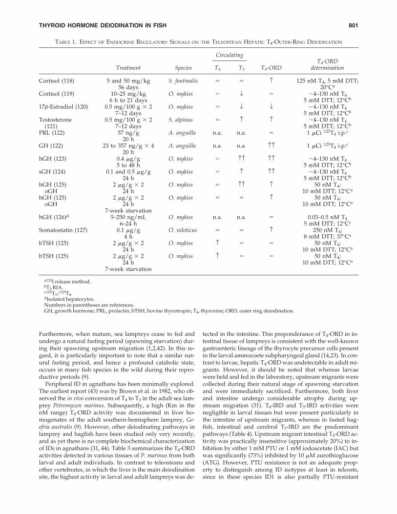
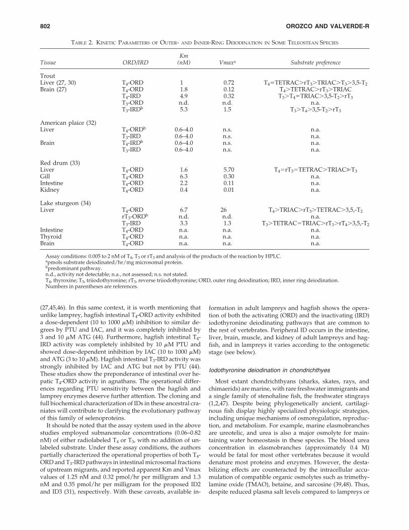
and only within a concentration range that could not distin-guish separate enzymatic pathways such as the mammalianID1 and ID2 types. Even when tested with a broader rangeof concentrations, T4 is not the best substrate to demonstratemammalian ID1 in assays that measure iodine release (26).Nevertheless, a number of studies analyzing the regulatoryfactors involved in the T4-ORD pathway of fish were con-ducted in the 1980s and the early 1990s (Table 1). Becausethe biochemical identity of the analyzed IDs is not clear, theresults of these studies are difficult to interpret. Currently,the identity of teleostean deiodinases as counterparts of themammalian ID1, ID2, and ID3 is still an unresolved issue forsome research groups. They consider that while fish deiod-inases show partial molecular similarity to mammalian deio-dinases in having selenocysteines in their active site, theirfunctional equivalence is debatable (27,28). By using T4, T3,and rT3 at substrate concentrations that range, depending onthe study, between 0.005 and 2 nM and analyzing the prod-ucts of the reaction by high-performance liquid chromatog-raphy (HPLC), the T4-ORD, T4-IRD, T3-ORD, T3-IRD, andrT3-ORD pathways have been partially characterized in dif-ferent tissues of sea lamprey, rainbow trout, American plaice,red drum, and lake sturgeon (27,29–34). The kinetic datafrom these studies is summarized in Tables 2 and 3.
Iodothyronine Deiodination in Agnatha
Extant vertebrates comprise two major superclasses; theagnatha or jawless fish, and the gnathostomata or jawed ver-tebrates. The phylogenetic relationship between these twomain members of the taxon Craniata is still debated. How-ever, recent molecular data support the notion that hagfishes(Myxiniformes) and lampreys (Petromyzontiforms) aremonophyletic and form a basal clade that may correspondto a sister group to all other craniata (35,36). Indeed, hag-fishes are regarded as the most primitive living craniates,whose evolutionary history dates back well into the Or-dovician (approximately 470 Ma). To date, close to 60 ac-cepted hagfish species are recognized, and a recent compre-hensive study of mtDNA sequence data divides them intotwo major subfamilies (37,38). The systematic study of thelamprey genome has not been undertaken, and little isknown about the phylogeny of the 40 or so lamprey speciesrecognized so far. However, recent phylogenetic analysis ofdifferent lamprey gene families supports the concept thatthese agnathans are a sister group of gnathostomes (36). Allhagfish species are stenohaline and live in the ocean, mostlyin temperate regions down to 2000 m. Hagfishes are the onlyvertebrates in which the body fluids are isosmotic (or verynearly so) to seawater (SW); thus, they are generally con-sidered to be osmoconformers (1,2,39). In contrast, all lam-prey species are osmoregulators, often euryhaline, and themajority live exclusively in rivers exhibiting an antitropicalor bipolar distribution. Whereas hagfish development is di-rect and there is no larval stage, lampreys, independent oftheir parasitic or nonparasitic lifestyle, are invariably fresh-water in their ammocoete or larval stage. Their prolongedlarval stage is associated with a steady increase of circulat-ing TH levels, which are among the highest recorded in anyvertebrate (40–42). The extended growth stage is followedby a nontrophic metamorphic phase of 3 to 4 months, dur-ing which the larval ammocoete transforms into a juvenile.
OROZCO AND VALVERDE-R800
Furthermore, when mature, sea lampreys cease to fed andundergo a natural fasting period (spawning starvation) dur-ing their spawning upstream migration (1,2,42). In this re-gard, it is particularly important to note that a similar nat-ural fasting period, and hence a profound catabolic state,occurs in many fish species in the wild during their repro-ductive periods (9).
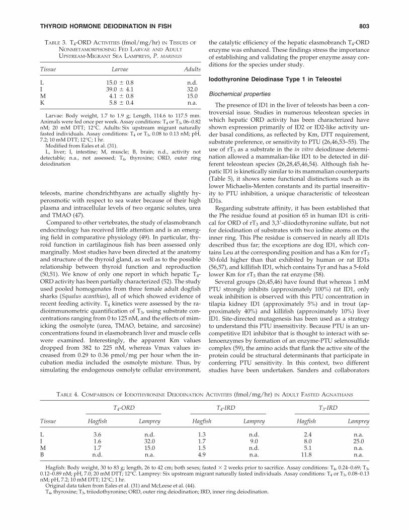
Peripheral ID in agnathans has been minimally explored.The earliest report (43) was by Brown et al. in 1982, who ob-served the in vivo conversion of T4 to T3 in the adult sea lam-prey Petromyzon marinus. Subsequently, a high (Km in thenM range) T4-ORD activity was documented in liver ho-mogenates of the adult southern-hemisphere lamprey, Ge-ofria australis (9). However, other deiodinating pathways inlamprey and hagfish have been studied only very recently,and as yet there is no complete biochemical characterizationof IDs in agnathans (31, 44). Table 3 summarizes the T4-ORDactivities detected in various tissues of P. marinus from bothlarval and adult individuals. In contrast to teleosteans andother vertebrates, in which the liver is the main deiodinationsite, the highest activity in larval and adult lampreys was de-
tected in the intestine. This preponderance of T4-ORD in in-testinal tissue of lampreys is consistent with the well-knowngastroenteric lineage of the thyrocyte precursor cells presentin the larval ammocoete subpharyngeal gland (14,23). In con-trast to larvae, hepatic T4-ORD was undetectable in adult mi-grants. However, it should be noted that whereas larvaewere held and fed in the laboratory, upstream migrants werecollected during their natural stage of spawning starvationand were immediately sacrificed. Furthermore, both liverand intestine undergo considerable atrophy during up-stream migration (31). T4-IRD and T3-IRD activities werenegligible in larval tissues but were present particularly inthe intestine of upstream migrants, whereas in fasted hag-fish, intestinal and cerebral T3-IRD are the predominantpathways (Table 4). Upstream migrant intestinal T4-ORD ac-tivity was practically insensitive (approximately 20%) to in-hibition by either 1 mM PTU or 1 mM iodoacetate (IAC) butwas significantly (73%) inhibited by 10 �M aurothioglucose(ATG). However, PTU resistance is not an adequate prop-erty to distinguish among ID isotypes at least in teleosts,since in these species ID1 is also partially PTU-resistant
THYROID HORMONE DEIODINATION IN FISH 801
TABLE 1. EFFECT OF ENDOCRINE REGULATORY SIGNALS ON THE TELEOSTEAN HEPATIC T4-OUTER-RING DEIODINATION
T4-ORDTreatment Species T4 T3 T4-ORD determination
Cortisol (118) 5 and 50 mg/kg S. fontinalis � � ↑ 125 nM T4, 5 mM DTT;56 days 20°Ca
Cortisol (119) 10–25 mg/kg O. mykiss � ↓ � �4–130 nM T46 h to 21 days 5 mM DTT; 12°Cb
17�-Estradiol (120) 0.5 mg/100 g � 2 O. mykiss � ↓ ↓ �4–130 nM T47–12 days 5 mM DTT; 12°Cb
Testosterone 0.5 mg/100 g � 2 S. alpinus � ↑ ↑ �4–130 nM T4(121) 7–12 days 5 mM DTT; 12°Cb
PRL (122) 57 ng/g A. anguilla n.a. n.a. � 1 �Ci 125T4 i.p.c20 h
GH (122) 23 to 357 ng/g � 4 A. anguilla n.a. n.a. ↑↑ 1 �Ci 125T4 i.p.c20 h
hGH (123) 0.4 �g/g O. mykiss � ↑↑ ↑↑ �4–130 nM T45 to 48 h 5 mM DTT; 12°Cb
sGH (124) 0.1 and 0.5 �g/g O. mykiss � ↑ ↑↑ �4–130 nM T424 h 5 mM DTT; 12°Cb
hGH (125) 2 �g/g � 2 O. mykiss � ↑↑ ↑ 50 nM T4;oGH 24 h 10 mM DTT; 12°Ca
hGH (125) 2 �g/g � 2 O. mykiss � � ↑ 50 nM T4;oGH 24 h 10 mM DTT; 12°Ca
7-week starvationhGH (126)d 5–250 ng/mL O. mykiss n.a. n.a. � 0.03–0.5 nM T4
6–24 h 5 mM DTT; 12°Cc
Somatostatin (127) 0.1 �g/g O. niloticus � � ↑ 250 nM T4;4 h 8 mM DTT; 37°Ca
bTSH (125) 2 �g/g � 2 O. mykiss ↑ � � 50 nM T4;24 h 10 mM DTT; 12°Ca
bTSH (125) 2 �g/g � 2 O. mykiss ↑ � � 50 nM T4;24 h 10 mM DTT; 12°Ca
7-week starvation
a125I release method.bT3 RIA.c125T3/125T4.dIsolated hepatocytes.Numbers in parentheses are references.GH, growth hormone; PRL, prolactin; bTSH, bovine thyrotropin; T4, thyroxine; ORD, outer ring deiodination.
Circulating
(27,45,46). In this same context, it is worth mentioning thatunlike lamprey, hagfish intestinal T4-ORD activity exhibiteda dose-dependent (10 to 1000 �M) inhibition to similar de-grees by PTU and IAC, and it was completely inhibited by3 and 10 �M ATG (44). Furthermore, hagfish intestinal T4-IRD activity was completely inhibited by 10 �M PTU andshowed dose-dependent inhibition by IAC (10 to 1000 �M)and ATG (3 to 10 �M). Hagfish intestinal T3-IRD activity wasstrongly inhibited by IAC and ATG but not by PTU (44).These studies show the preponderance of intestinal over he-patic T4-ORD activity in agnathans. The operational differ-ences regarding PTU sensitivity between the hagfish andlamprey enzymes deserve further attention. The cloning andfull biochemical characterization of IDs in these ancestral cra-niates will contribute to clarifying the evolutionary pathwayof this family of selenoproteins.
It should be noted that the assay system used in the abovestudies employed subnanomolar concentrations (0.06–0.82nM) of either radiolabeled T4 or T3, with no addition of un-labeled substrate. Under these assay conditions, the authorspartially characterized the operational properties of both T4-ORD and T3-IRD pathways in intestinal microsomal fractionsof upstream migrants, and reported apparent Km and Vmaxvalues of 1.25 nM and 0.32 pmol/hr per milligram and 1.3nM and 0.35 pmol/hr per milligram for the proposed ID2and ID3 (31), respectively. With these caveats, available in-
formation in adult lampreys and hagfish shows the opera-tion of both the activating (ORD) and the inactivating (IRD)iodothyronine deiodinating pathways that are common tothe rest of vertebrates. Peripheral ID occurs in the intestine,liver, brain, muscle, and kidney of adult lampreys and hag-fish, and in lampreys it varies according to the ontogeneticstage (see below).
Iodothyronine deiodination in chondrichthyes
Most extant chondrichthyans (sharks, skates, rays, andchimaerids) are marine, with rare freshwater immigrants anda single family of stenohaline fish, the freshwater stingrays(1,2,47). Despite being phylogenetically ancient, cartilagi-nous fish display highly specialized physiologic strategies,including unique mechanisms of osmoregulation, reproduc-tion, and metabolism. For example, marine elasmobranchesare ureotelic, and urea is also a major osmolyte for main-taining water homeostasis in these species. The blood ureaconcentration in elasmobranches (approximately 0.4 M)would be fatal for most other vertebrates because it woulddenature most proteins and enzymes. However, the desta-bilizing effects are counteracted by the intracellular accu-mulation of compatible organic osmolytes such as trimethy-lamine oxide (TMAO), betaine, and sarcosine (39,48). Thus,despite reduced plasma salt levels compared to lampreys or
OROZCO AND VALVERDE-R802
TABLE 2. KINETIC PARAMETERS OF OUTER- AND INNER-RING DEIODINATION IN SOME TELEOSTEAN SPECIES
KmTissue ORD/IRD (nM) Vmaxa Substrate preference
TroutLiver (27, 30) T4-ORD 1.0 0.72 T4�TETRAC�rT3�TRIAC�T3�3,5-T2Brain (27) T4-ORD 1.8 0.12 T4�TETRAC�rT3�TRIAC
T4-IRD 4.9 0.32 T3�T4�TRIAC�3,5-T2�rT3T3-ORD n.d. n.d. n.a.T3-IRDb 5.3 1.50 T3�T4�3,5-T2�rT3
American plaice (32)Liver T4-ORDb 0.6–4.0 n.s. n.a.
T3-IRD 0.6–4.0 n.s. n.a.Brain T4-IRDb 0.6–4.0 n.s. n.a.
T3-IRD 0.6–4.0 n.s. n.a.
Red drum (33)Liver T4-ORD 1.6 5.70 T4�rT3�TETRAC�TRIAC�T3Gill T4-ORD 6.3 0.30 n.a.Intestine T4-ORD 2.2 0.11 n.a.Kidney T4-ORD 0.4 0.01 n.a.
Lake sturgeon (34)Liver T4-ORD 6.7 26.00 T4�TRIAC�rT3�TETRAC�3,5,-T2
rT3-ORDb n.d. n.d. n.a.T3-IRD 3.3 1.30 T3�TETRAC�TRIAC�rT3�rT4�3,5,-T2
Intestine T4-ORD n.a. n.a. n.a.Thyroid T4-ORD n.a. n.a. n.a.Brain T4-ORD n.a. n.a. n.a.
Assay conditions: 0.005 to 2 nM of T4, T3 or rT3 and analysis of the products of the reaction by HPLC.apmols substrate deiodinated/hr/mg microsomal protein.bpredominant pathway.n.d., activity not detectable; n.a., not assessed; n.s. not stated.T4, thyroxine; T3, triiodothyronine; rT3, reverse triiodothyronine; ORD, outer ring deiodination; IRD, inner ring deiodination.Numbers in parentheses are references.
teleosts, marine chondrichthyans are actually slightly hy-perosmotic with respect to sea water because of their highplasma and intracellular levels of two organic solutes, ureaand TMAO (47).
Compared to other vertebrates, the study of elasmobranchendocrinology has received little attention and is an emerg-ing field in comparative physiology (49). In particular, thy-roid function in cartilaginous fish has been assessed onlymarginally. Most studies have been directed at the anatomyand structure of the thyroid gland, as well as to the possiblerelationship between thyroid function and reproduction(50,51). We know of only one report in which hepatic T4-ORD activity has been partially characterized (52). The studyused pooled homogenates from three female adult dogfishsharks (Squalus acanthias), all of which showed evidence ofrecent feeding activity. T4 kinetics were assessed by the ra-dioimmunometric quantification of T3, using substrate con-centrations ranging from 0 to 125 nM, and the effects of mim-icking the osmolyte (urea, TMAO, betaine, and sarcosine)concentrations found in elasmobranch liver and muscle cellswere examined. Interestingly, the apparent Km valuesdropped from 382 to 225 nM, whereas Vmax values in-creased from 0.29 to 0.36 pmol/mg per hour when the in-cubation media included the osmolyte mixture. Thus, bysimulating the endogenous osmolyte cellular environment,
the catalytic efficiency of the hepatic elasmobranch T4-ORDenzyme was enhanced. These findings stress the importanceof establishing and validating the proper enzyme assay con-ditions for the species under study.
Iodothyronine Deiodinase Type 1 in Teleostei
Biochemical properties
The presence of ID1 in the liver of teleosts has been a con-troversial issue. Studies in numerous teleostean species inwhich hepatic ORD activity has been characterized haveshown expression primarily of ID2 or ID2-like activity un-der basal conditions, as reflected by Km, DTT requirement,substrate preference, or sensitivity to PTU (26,46,53–55). Theuse of rT3 as a substrate in the in vitro deiodinase determi-nation allowed a mammalian-like ID1 to be detected in dif-ferent teleostean species (26,28,45,46,54). Although fish he-patic ID1 is kinetically similar to its mammalian counterparts(Table 5), it shows some functional distinctions such as itslower Michaelis-Menten constants and its partial insensitiv-ity to PTU inhibition, a unique characteristic of teleosteanID1s.
Regarding substrate affinity, it has been established thatthe Phe residue found at position 65 in human ID1 is criti-cal for ORD of rT3 and 3,3�-diiodothyronine sulfate, but notfor deiodination of substrates with two iodine atoms on theinner ring. This Phe residue is conserved in nearly all ID1sdescribed thus far; the exceptions are dog ID1, which con-tains Leu at the corresponding position and has a Km for rT3
30-fold higher than that exhibited by human or rat ID1s(56,57), and killifish ID1, which contains Tyr and has a 5-foldlower Km for rT3 than the rat enzyme (58).
Several groups (26,45,46) have found that whereas 1 mMPTU strongly inhibits (approximately 100%) rat ID1, onlyweak inhibition is observed with this PTU concentration intilapia kidney ID1 (approximately 5%) and in trout (ap-proximately 40%) and killifish (approximately 10%) liverID1. Site-directed mutagenesis has been used as a strategyto understand this PTU insensitivity. Because PTU is an un-competitive ID1 inhibitor that is thought to interact with se-lenoenzymes by formation of an enzyme-PTU selenosulfidecomplex (59), the amino acids that flank the active site of theprotein could be structural determinants that participate inconferring PTU sensitivity. In this context, two differentstudies have been undertaken. Sanders and collaborators
THYROID HORMONE DEIODINATION IN FISH 803
TABLE 3. T4-ORD ACTIVITIES (fmol/mg/hr) IN TISSUES OF
NONMETAMORPHOSING FED LARVAE AND ADULT
UPSTREAM-MIGRANT SEA LAMPREYS, P. MARINUS
Tissue Larvae Adults
L 15.0 � 0.8 n.d.I 39.0 � 4.1 32.0M 4.1 � 0.8 15.0K 5.8 � 0.4 n.a.
Larvae: Body weight, 1.7 to 1.9 g; Length, 114.6 to 117.5 mm.Animals were fed once per week. Assay conditions: T4 or T3, 06–0.82nM; 20 mM DTT; 12°C. Adults: Six upstream migrant naturallyfasted individuals. Assay conditions: T4 or T3, 0.08 to 0.13 nM; pH,7.2; 10 mM DTT; 12°C; 1 hr.
Modified from Eales et al. (31).L, liver; I, intestine; M, muscle; B, brain; n.d., activity not
detectable; n.a., not assessed; T4, thyroxine; ORD, outer ring deiodination
TABLE 4. COMPARISON OF IODOTHYRONINE DEIODINATION ACTIVITIES (fmol/mg/hr) IN ADULT FASTED AGNATHANS
T4-ORD T4-IRD T3-IRD
Tissue Hagfish Lamprey Hagfish Lamprey Hagfish Lamprey
L 3.6 n.d. 1.3 n.d. 2.4 n.a.I 1.6 32.0 1.7 9.0 8.0 25.0M 1.7 15.0 1.5 n.d. 5.1 n.a.B n.d. n.a. 4.9 n.a. 11.8 n.a.
Hagfish: Body weight, 30 to 83 g; length, 26 to 42 cm; both sexes; fasted � 2 weeks prior to sacrifice. Assay conditions: T4, 0.24–0.69; T3,0.12–0.89 nM; pH, 7.0; 20 mM DTT; 12°C. Lamprey: Six upstream migrant naturally fasted individuals. Assay conditions: T4 or T3, 0.08–0.13nM; pH, 7.2; 10 mM DTT; 12°C; 1 hr.
Original data taken from Eales et al. (31) and McLeese et al. (44).T4, thyroxine; T3, triiodothyronine; ORD, outer ring deiodination; IRD, inner ring deiodination.
(60) constructed a recombinant form of a tilapia ID1 in whichproline 128 was replaced by a serine. Amino acid 128 is twopositions downstream from the SeCys residue, and the Pro-128 residue is present in all PTU-insensitive deiodinases(ID2s, ID3s, and fish ID1s) while Ser is the amino acid pres-ent in all the PTU-sensitive ID1s (i.e., human, rat, mouse, anddog; (15,56,61,62). PTU sensitivity was not modified in theP128S mutant, suggesting that the Ser residue does not con-fer PTU sensitivity by itself. In addition to the amino acidsthat flank the active site, other residues that seem to be keyfor ID1 activity are the histidines in positions 158 and 174(63). These histidines are conserved in all ID1s, are locatedclose to the active site, and are essential for substrate bind-ing. Experiments using site-directed mutagenesis showedthat the substitution of His-158 by Ala in rat D1 resulted ina totally inactive enzyme, suggesting that this residue is keyfor catalysis and/or the conformation of the enzyme (63).Fish ID1s contain a serine in position 159, whereas all otherID1s have an alanine, and all PTU-insensitive deiodinaseshave Pro-159. Thus, the substitution of Ser-159 by Pro was
expected to decrease sensitivity of the enzyme to PTU. Un-expectedly, this mutated protein was more sensitive to PTUinhibition (58% inhibition for Ser-159 versus 80% inhibitionfor Pro-159) (58).
Regulation by iodothyronines
In mammals, THs are among the most potent physiologicregulators of deiodinase activity. Hyperthyroidism increasesID1 activity and transcription, whereas hypothyroidism ex-erts the opposite effects. In humans, the observed responsesto substrate have been explained by the presence of two thy-roid responsive elements (TREs) in the 5� flanking region ofthe Dio1 gene (for review Bianco et al. [13]). In contrast, sev-eral studies have shown that fish ID1 exhibits a distinct thy-roid hormone response. Thus, neither T4, T3, nor 3,5-T2-treat-ment upregulates hepatic ID1 activity in the rainbow troutor the killifish (64–66). Furthermore, hyperthyroidism doesnot alter renal ID1 activity in tilapia (67). However, despitethe lack of effects upon hepatic ID1 activity, TH treatment
OROZCO AND VALVERDE-R804
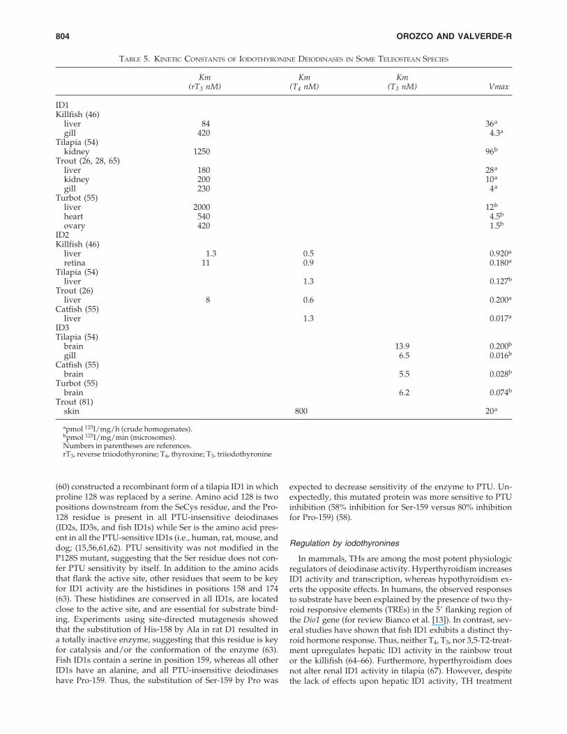
TABLE 5. KINETIC CONSTANTS OF IODOTHYRONINE DEIODINASES IN SOME TELEOSTEAN SPECIES
Km Km Km(rT3 nM) (T4 nM) (T3 nM) Vmax
ID1Killfish (46)
liver 84 36.agill 420 4.3a
Tilapia (54)kidney 1250 96.b
Trout (26, 28, 65)liver 180 28.akidney 200 10.agill 230 4.a
Turbot (55)liver 2000 12.bheart 540 4.5b
ovary 420 1.5b
ID2Killfish (46)
liver 1.3 0.5 0.920a
retina 11 0.9 0.180a
Tilapia (54)liver 1.3 0.127b
Trout (26)liver 8 0.6 0.200a
Catfish (55)liver 1.3 0.017a
ID3Tilapia (54)
brain 13.9 0.200b
gill 6.5 0.016b
Catfish (55)brain 5.5 0.028b
Turbot (55)brain 6.2 0.074b
Trout (81)skin 800 20.a
apmol 125I/mg/h (crude homogenates).bpmol 125I/mg/min (microsomes).Numbers in parentheses are references.rT3, reverse triiodothyronine; T4, thyroxine; T3, triiodothyronine
for 24 hours decreases ID1 mRNA levels in the killifish liver(66). One feasible interpretation could be that ID1 in theteleosts does not deiodinate the inner ring of the iodothyro-nine, and thus the excess substrate would downregulate ID1transcription. This interpretation is supported by the obser-vation that ID1 of tilapia renal microsomes catalyzes negli-gible IRD deiodination of T3 and T3S (60). The fact that de-creased ID1 transcription is not reflected in the activity ofthe enzyme is intriguing. The longer half-life of ID1 (68) mayaccount in part for this discrepancy. Nevertheless, fishtreated for up to 15 days with T4 or T3 showed no effect onhepatic (28,34) or renal (67) ID1 activity.
The opposite response has been observed in hypothy-roidal conditions. Although almost no ID1 activity or mRNAexpression can be detected in the liver of euthyroid tilapia(54,55), both hepatic ID1 activity and mRNA are upregulatedin long-term (90 days) hypothyroid tilapia (69). Furthermore,tilapia kidney ID1 is not affected by short-term (11 day) hy-pothyroidism (67), but kidney ID1 activity decreases duringlong-term hypothyroidism in the Nile tilapia (69).
mRNA characteristics
Only two teleostean ID1 mRNAs have been cloned andcharacterized (58,60). Killifish ID1 mRNA is the smallest (1.3kb), whereas tilapia is 2.4 Kb. As previously described, all ID1mRNAs contain an in-frame UGA that codes for the insertionof SeCys at a position that is conserved in IDs. The predictedsecondary structure of the SECIS element in tilapia resemblesthat of form 1, while the killifish SECIS element resemblesform 2. ID1 mRNA expression has been detected in killifishliver (58). In tilapia, the highest mRNA levels were observedin kidney, followed by gut, testis, gill, and brain (60).
Iodothyronine Deiodinase Type 2 in Teleostei
Biochemical properties
ID2 controls the intracellular concentration of T3. The cellsthat express ID2 locally produce T3 that can more rapidly ac-cess the thyroid receptors in the nucleus than T3 from plasma(13). Studies in different teleostean species have shown theexpression primarily of a low Km (1 nM) hepatic T4-ORD ac-tivity (Table 2). However, as discussed above, the identifi-cation of this T4-ORD activity as a counterpart of the mam-malian ID2 is not fully established. There are only a fewstudies in which the kinetic and operational characteristicsof fish ID2s have been fully evaluated. These studies showthat teleostean ID2 has substrate preferences, iodothyronineinhibition profiles, and apparent Km values similar to thosefor mammalian ID2 (26,45,46,55, and Table 5). Furthermore,in teleosts, the highest ID2 activity is detected in the liver,where its expression is up to 20 times higher (46) than in hy-pothyroid rat brain (12).
Regulation by iodothyronines
As in the case of ID1, thyroidal status is the main factorin higher vertebrates that controls ID2 activity both at thepretranscriptional and posttranscriptional levels. Hyperthy-roidism suppresses ID2 activity and expression of its mRNA,whereas hypothyroidism increases them. The posttranscrip-tional mechanisms through which THs regulate ID2 activityhave been elucidated in part. The very short half-life (less
than 1 hour) of ID2 is further shortened in cells exposed toits substrates, T4, rT3, and even high concentrations of T3. Ex-perimental data suggest that the enzyme–substrate interac-tion induces proteolysis of the complex and that this down-regulatory process is mediated by ubiquitin. The molecularmechanisms that could explain the pretranscriptional ID2regulation by substrate are not yet understood in any verte-brate species (for review Bianco et al. [13]).
In teleosts, ID2 also seems to be tightly regulated by sub-strate. Short-term (24 hour) treatment with T4 and T3 down-regulates killifish hepatic ID2 activity. The effects upon tran-scription were evident earlier, with both iodothyroninesinducing a decrease in ID2 mRNA levels after 12 hours (66).Longer treatments (7 to 11 days) exert the same downregu-latory effect. A reduction in hepatic T4-ORD has been re-ported in rainbow trout after administration of T3-supple-mented food as well as after fasting (64), while T3-inducedhyperthyroidism significantly lowers hepatic ID2 in tilapialiver (67). In contrast, methimazole-induced hypothyroidismsignificantly increases hepatic ID2 activity (67,69) as well ashepatic ID2 mRNA levels (69). The mechanism throughwhich thyroidal status controls teleostean ID2 activity is notknown. Nevertheless, there is experimental evidence sug-gesting that the regulation occurs at a pretranslational level.The decrement in hepatic ID2 activity from T4- and T3-treatedfish is preceded (12 hours) by a decrease in ID2 mRNA; also,the apparent Vmax values declined in the TH-treated groups,whereas the substrate affinity was not modified (66). Thesefindings support the notion of a decrease in transcriptionand/or an increase in protein degradation in response toacute administration of iodothyronines. In this same context,hypothyroidism in tilapia liver is accompanied by an in-crease in ID2 activity and mRNA expression (69). The mo-lecular mechanisms that could explain the pretranslationalID2 regulation by substrate have not been elucidated inmammalian (13,70) or teleostean species (66). Available in-formation suggests that while the final observed effects ofiodothyronines on ID2 are similar in mammals and fish, thespecific mechanisms of this regulation could differ.
It has recently been reported that in killifish liver, 3,5-T2
exerts downregulatory effects on ID2 activity and mRNAlevels (66). Little is known about the kinetics of 3,5-T2 in ver-tebrates; nevertheless, the fact that this iodothyronine exertsa clear inhibitory effect on ID2 suggests that it could play aphysiologic role in teleostean thyroidal systems. Althoughthere has been no demonstration of conversion of T3 to 3,5-T2 in teleosts in vitro (A. Orozco, unpublished observations;29) or in vivo, recent experimental evidence suggests that thisconversion can occur in mammalian species (71). The ob-served effects of 3,5-T2 on hepatic ID2 could be the conse-quence of an interaction of 3,5-T2 with TH nuclear receptors.A very low binding of 3,5-T2 to salmon hepatic nuclei hasbeen reported (72,73). Further work is required to under-stand the mechanism of action of 3,5-T2.
mRNA characteristics
Sequenced cDNAs that encode teleostean ID2s have beencloned from trout (74), killifish (75,76), and lungfish (77). Ofthese cDNAs, only the killifish homologue has been ex-pressed and characterized (75). With the exception of the pu-tative ID2 from lungfish, the structure of the teleostean ID2
THYROID HORMONE DEIODINATION IN FISH 805
mRNAs comprises an ORF of approximately 1 kb and an ex-tended 3� untranslated region (3�-UTR). The killifish ID2mRNA is the largest yet described (4.7 Kb) as compared totrout (2.7 Kb) and lungfish (1.3 Kb). As in their mammaliancounterparts, the 3�-UTR of trout and killifish ID2 mRNAscontains a SECIS region positioned approximately 250 bp be-fore the poly(A) tail. Lungfish ID2 appears to contain twoputative SECIS form 1 regions. In all other ID2 mRNAs, thepredicted secondary structure closely resembles that of SE-CIS form 2. ID2 mRNA expression has been detected in kil-lifish liver (75), trout liver (69,74) and gonads (74), and lung-fish liver, gills, heart, and kidney (77).
Genes
The structure of a dio2 gene has been partially character-ized in rainbow trout (74) and more extensively studied inkillifish (75). As in its mammalian counterparts (78,79), thetrout and killifish dio2 gene comprises two exons separatedby a single intron. The second exon contains the SeCys TGAcodon, the SECIS region required for the incorporation of Se-Cys during translation, and the polyadenylation signal. Thekillifish Dio2 intron (4.7 kb) is smaller than those identifiedin mammals, but it is located in the same position as the 8.5and 7.4 kb introns in the mouse and human genes, respec-tively (75,78,79). There are nevertheless some differences inthe 5�-FR of the Dio2 genes that could be of phylogenetic rel-evance. In contrast to the mammalian Dio2 genes, no TATAor CCAAT boxes were found in the killifish homologue. Fur-thermore, the human, rat, and mouse Dio2 genes all containa single, completely conserved canonical cyclic adenosinemonophosphate (cAMP) response element (CRE) approxi-mately 70 bp upstream of the TATA box. No clear CRE se-quence is found within 1.3 Kb upstream of the transcriptionstart site of the killifish dio2 gene. In addition, only the hu-man dio2 gene is stimulated by thyroid transcription factor1 (TTF-1) (80). These observations may explain the differen-tial expression of dio2 in the different vertebrate species.
Iodothyronine Deiodinase Type 3 in Teleostei
Biochemical properties
ID3 possesses IRD activity almost exclusively and cat-alyzes the conversion of T4 to rT3 and of T3 to 3,3�-T2, bothof which are biologically inactive. Thus, the main physio-logic role of ID3 is its contribution to thyroid hormone ho-meostasis by protecting tissues from an excess of active THs.In fish, ID3 is highly expressed in brain, gill (55), and skin(81). The kinetic constants for these tissues are summarizedin Table 5. Furthermore, T4-IRD activity has been docu-mented in the brain of trout (27), American plaice (32), andlake sturgeon (34), whereas T3-IRD has been found in troutbrain (27), American plaice brain and liver (32), and lake stur-geon liver (34). The kinetic information of these studies iscompiled in Table 2.
Regulation
As previously mentioned, changes in the activity of ID3modulate both circulating and tissue thyroidal status by in-creasing or reducing T3 inactivation to maintain homeosta-sis. Thyroidal status parallels ID3 activity in several species,increasing during hyperthyroidism and decreasing duringhypothyroidism in the CNS (for review see Bianco et al. [13]).
In tilapia, T3-induced hyperthyroidism strongly stimulateshepatic but not brain or gill ID3 (67). Similar changes havebeen observed in trout, in which T3 challenge increases T3-IRD in liver but not in gill (82–84). These data suggest thatthere is a tissue-specific regulatory response of ID3 toiodothyronines. On the other hand, methimazole-inducedhypothyroidism significantly decreases ID3 in tilapia liverand brain after 11 (67) or 90 days of treatment (69), suggest-ing a protective mechanism against very low T3 concentra-tions, as in other vertebrate species.
mRNA characteristics
Teleostean ID3 cDNAs have been cloned from tilapia (85)and Australian lungfish (86). Of these, only the tilapia clonehas been expressed and characterized. Both ID3 cDNAs in-clude a SeCys-encoding TGA codon as well as a SECIS ele-ment in the 3�-UTR. The identity of the two teleostean ID3sis approximately 67% at the ORF nucleotide level. TilapiaID3 mRNA is predominantly expressed in brain and gill,whereas the putative lungfish ID3 mRNA was amplifiedfrom liver, lung, kidney, brain, heart, and gill.
Iodothyronine Deiodination and Fish Ontogeny
Thyroidal systems are unique to vertebrates and comprisethe so-called hypothalamic-pituitary-thyroid axis (HPT) andits complementary network of peripheral IDs. Thyroidal sys-tems have evolved to provide the systemic prodevelopmen-tal, iodine-containing signal thyroxine or T4 with the preciseontogenetic timing required by each species. Furthermore,peripheral IDs, by determining the local intracellular avail-ability of active or inactive TH, play a crucial role in regu-lating the specific developmental actions of these iodine-con-taining messengers (13,14,87).
Embryo and larval stages
Regardless of their reproductive strategy, after hatchingand yolk resorption, the life history of all fish continues witha free-larval phase of variable length, which is followed bymetamorphosis into the juvenile immature adult stage. Thepresence of significant amounts of maternal TH in the yolkof different teleostean marine and fresh water species is am-ply documented. However, contrary to other nonneotenicvertebrates, an absolute requirement for TH during early fishembryogenesis and larval development has not been dem-onstrated unequivocally (for review see Leatherland [9] andPower et al. [88]). Nevertheless, a distinct tissue- and devel-opmental-specific expression pattern of dio1 and dio2 genesduring zebrafish embryogenesis has been reported recently(89). Interestingly, whereas dio1 expression occurs until 72hours postfertilization and its mRNA accumulates in theliver, kidney, and gut, dio2 was expressed at earlier devel-opmental stages and was confined to the retina, intestinalbulb, and a subpopulation of adenohypophyseal cells. Thesedata are in close agreement with studies in rat embryos (90)and strongly suggest that IDs are able to generate, at the pre-cise time and site, the required amount of TH for early fishembryogenesis.
Metamorphosis
As a developmental strategy that ultimately prepares theindividual for life as a sexually mature adult, metamorpho-
OROZCO AND VALVERDE-R806

sis is found only within a small number of extant fish, all ofwhich follow indirect development (42,91). With the notableexception of lampreys, available information supports theidea that THs are essential for fish larval growth and meta-morphosis (for review see Leatherland [9] and Power et al.[88]). In the case of lampreys, although the mechanisms arefar from clear, the prevailing idea is that THs play an in-hibitory role in the metamorphosis of the larval ammocoeteinto the juvenile organism. In fact, a distinctive feature ofmetamorphosing ammocoetes is the rapid fall in circulatingTH levels, which then remain relatively low throughout theprocess. Furthermore, because precocious metamorphosis inlamprey is induced by administering a goitrogen, KClO4,and because T4 or T3 treatment blocks KClO4-induced meta-morphosis, it has been suggested that THs, particularly T3,may be acting as a antimetamorphic or juvenile hormone (forreview see Youson [42] and Power et al. [88]).
Lamprey larvae grow gradually and accumulate lipids un-til they attain a critical metamorphic somatic-weight ratioknown as the condition factor [(weight (g)/length (mm)3) �106]. At this premetamorphic stage, approximately 13% ofthe ammocoete body weight corresponds to lipid reserves.Furthermore, it is well known that lipogenesis or hyper-lipexia is an essential and critical feature of premetamor-phosis, whereas lipolysis is decisive for the survival of themetamorphosing animals, because during this ontogeneticstage lampreys do not feed but instead rely on their lipid de-pots (42). To date, we are aware of only one correlative studyregarding peripheral ID activity during spontaneous meta-morphosis in P. marinus larvae. The study reports a recipro-cal relationship between intestinal T4-ORD and T4-IRD ac-tivities from nonmetamorphosing larvae to the adultparasitic stage (92). Accordingly, T4-ORD activity is rela-tively low in nonmetamorphosing individuals (7 fmol/mgper hour), reaches a peak during premetamorphosis andearly metamorphosis (approximately 40 fmol/mg per hourat stages 1 and 2), and then decreases dramatically duringmetamorphosis to eventually rise again in parasitic-stagelampreys. In contrast, intestinal T4-IRD activity is negligibleuntil midmetamorphosis (stage 3). It then peaks (approxi-mately 70 fmol/mg per hour) at stage 7 and falls to modestlevels in the adult stage (approximately 15 fmol/mg/hour).Interestingly, liver and intestinal T4-ORD activities were sim-ilar in nonmetamorphosing larvae, but as larvae enter meta-morphosis (stage 2), liver activity significantly declines andintestine becomes the predominant ID site. This course ofevents in lamprey intestinal ORD and IRD activities is con-sistent with the well-known changes exhibited by circulat-ing TH levels during spontaneous lamprey metamorphosis.Although the evidence is circumstantial, these authors con-clude that during the initiation of metamorphosis there is amajor increase in intestinal T3 production, followed by ashutdown of T4 to T3 conversion and a major induction ofIRD of both T4 and T3 during midmetamorphosis. Thus, thepreponderance of T4-ORD activity at early metamorphosis,followed by its abrupt reduction and the simultaneous in-crease in T4-IRD activity, may be responsible for both thehigh plasma concentrations of TH prior to metamorphosisand the ensuing decline in these levels during metamor-phosis.
A similar correlative study assessing circulating levels ofT4, T3, and rT3 and the activity of the three ID isotypes dur-ing rainbow trout transition from fry to fingerling and adult
individuals has been recently conducted (93). As fry devel-oped into fingerlings, blood levels of T3 increased, whilethose of its inactive isomer rT3 decreased. These changes incirculating concentrations of T3 and rT3 were associated withchanges in the corresponding deiodinative enzymes (i.e., theactivity of liver ID2 increased) while that of skin ID3 de-creased. The time course of these changes was directly (T3)or inversely (rT3 and D3) correlated to an increase in age,length, and body weight, suggesting a temporal associationwith growth and differentiation processes. Of note is the factthat hepatic D1 activity remained low during the entirestudy.
Overall, these two studies rely on indirect evidence to as-sociate circulating TH levels and ID activities during thetransition of larvae to juvenile and/or adult stages in fish.Consequently, there is a need to extend and complementthese types of studies using a direct temporal and tissue ap-proach to enhance the understanding of the role played bythe ID system during fish ontogenesis.
Smoltification
Smoltification or parr-smolt transformation (PST) is an an-ticipatory ontogenetic stage of nontrophic metamorphosisthough which juvenile anadromous salmonids undergophysiological, morphological, and behavioral changes inpreparation for downstream migration and marine life. It iswell established that plasma T4 rises to a distinct peak dur-ing smoltification and that the survival of some migratorysalmonids is directly related to the extent and completion ofthis hormonal surge prior to sea water entry (94–97). Nev-ertheless, the function of this T4 surge is still of uncertainphysiologic significance in the regulation of PST (9,98).
Several studies have been undertaken to study deiodina-tion during PST. As previously mentioned, because of theassay conditions used, it is difficult to determine the enzy-matic type assessed in these experiments. Regardless of themethodological considerations, several authors have ob-served an increase in hepatic T4-ORD activity in Atlanticsalmon induced by photoperiod to undergo smoltificationcombined with an increased temperature and augmentedfood ratio (99,100). In contrast, advanced PST under naturalconditions is accompanied by a depressed hepatic T4-ORDand T4-ORD/T3IRD activity ratio. These evidently contra-dictory observations have been explained as a consequenceof the increase in food ratio and not as a normal PST feature(101). Other studies by the same research group agree withthis notion. A low hepatic T4-ORD activity accompanied bysignificant hepatic T4-IRD and T3-IRD activities was ob-served in smolting coho salmon (102). In studies of TH deio-dination during PST (99,101,102), the authors have inter-preted the observed T4-ORD decrement as indicating thatthere is no enhanced potential for hepatic production of T3
at this physiologic stage. Because they infer that this sub-nanomolar Km deiodinating enzyme is the probable primaryregulator of systemic T3 levels, they explain the low T4-ORDactivity as a means to inhibit growth and growth-related en-ergy expenditure during the period of migration by curtail-ing the availability of circulating T3. However, as discussedabove, this low-Km hepatic deiodinating enzyme has beencharacterized as an ID2 isotype (26,45,46). As for other ver-tebrate ID2 enzymes, T3 and T4 challenges depress ID2 ac-tivity in other teleostean species (66,67); thus, it is not sur-
THYROID HORMONE DEIODINATION IN FISH 807
prising that the T4 peak that accompanies PST would reducethis enzymatic activity. As far as we know, there is no evi-dence that the teleostean hepatic ID2 serves as a major sourceof circulating T3. Therefore, a depressed hepatic ID2 activitywould not necessarily represent a way to downregulate sys-temic T3 at a life-cycle stage when a number of thyroid hor-mone-dependent physiologic changes are occurring. Itwould be of interest to investigate how the ID1 of differenttissues, including the liver, is regulated during PST. It seemsreasonable to suggest that during PST, T4 to T3 conversioncould be enhanced in other tissues expressing ID1, and thatthe resultant T3 would be rapidly captured by other tissues,thereby ensuring an increased metabolic clearance rate of thehormone. This proposal is consistent with the increase in he-patic IRD-activity reported in smolting coho salmon (102),and it would explain the lack of changes in plasma T3 con-centrations during PST.
Although not conclusive, the pattern of deiodinating ac-tivity in the brain of smolting Atlantic salmon is similarwhether PST occurred under natural photoperiod (101) orwas induced by a combination of photoperiod, temperature,and feeding conditions (99,100). In both situations, a poten-tial to form T3 from T4 as well as to degrade T3 to T2 and T4
to rT3 was observed. In contrast, T4 challenge in smolting At-lantic salmon did not alter the T4-ORD and T3-IRD pathways(103). A role for TH in regulating neural function during ad-vanced PST has been suggested. The authors propose thatthe surge in plasma T4 accompanying the advanced stagesof PST may act to decrease certain aspects of brain olfactoryfunction and terminate a period of enhanced olfactory learn-ing. This may serve to preserve intact the memory of thehome stream odor as the fish moves downstream to differ-ent chemical environments (101,103). In support of this in-terpretation, T4 treatment in advanced smolting salmon depressed brain T4-ORD activity and olfactory bulb elec-troencephalographic responses to L-alanine (103), accompa-nied by enhanced dopamine metabolism of the olfactorybulb (104).
Iodothyronine Deiodination and Energy Balance
In all vertebrates studied thus far, energy imbalances (i.e.,alterations in the caloric intake/caloric expenditure) areknown to significantly modify TH circulating levels and pe-ripheral ID activity. Thus, complete or partial food depriva-tion decreases plasma T3, while the effect upon the levels ofT4 varies depending on the species and the duration of foodrestriction. On the contrary, overfeeding produces changesopposite to those of fasting, and refeeding reverses the fast-ing-induced changes in circulating TH levels and ID-activ-ity. These changes result primarily from alterations in pe-ripheral conversion of T4 to T3 as well as from changes inthe rate of T3 inactivation. The mechanisms subserving theseadaptive thyroid responses to altered nutritional intake arestill not clearly understood. However, they are related to thecomposition of the diet, particularly to its carbohydrate andprotein content (6,9,10,105). Although there are species dif-ferences in the ID pathways involved in these homeorheticchanges, in all cases, the liver is a key effector in the install-ment of the response. In fish, hepatic ID2 and gill ID3 arethe major regulatory enzymes responsible for these adjust-ments, and they may be the primary sources for plasma T3.
Unlike the rat, in tilapia there is a significant decrease in he-patic ID2 but not ID1 activity during fasting (106,107). Fur-thermore, whereas hepatic ID3 increased in fasted rats andchickens, in starved tilapia, ID3 decreased in the gill, brain,and liver (107). Interestingly, refeeding in tilapia rapidly (4hours) restored plasma T4 but not T3 levels, which remainedlow after 2 days of refeeding. This slow recuperation of cir-culating T3 was matched by hepatic ID2 and gill ID3 activi-ties, which also returned to normal (increased and decreased,respectively) 2 days after food was available again (107).These data add to those provided by studies in hyperthy-roid and/or hypothyroid fish indicating distinct differencesin fish thyroid physiology compared to other vertebrates.Thus, considering the importance of the endocrine system inregulating intermediary metabolism in fish, more systematicstudies to analyze the participation of IDs will be of value.
Iodothyronine Deiodination and Osmotic Balance
Cell volume regulation is among the most ancient home-ostatic mechanisms and is essential to life. When confrontingosmoregulatory demands, all living systems install immedi-ate, common responses characterized by the adaptive intra-cellular accumulation of compatible organic osmolytes (48).This accumulation is transcriptionally regulated and is me-diated by a highly conserved regulatory element called theosmotic response element, also known as tonicity responsiveenhancer (ORE or TonE, respectively). The concentration ofthe cognate transcription factor, known as the osmotic re-sponse element-binding protein/tonicity-responsive en-hancer-binding protein or OREBP/TonEBP, varies directlywith extracellular NaCl concentration (108,109). Osmolari-ties of aqueous environments range from a few milliosmolesper liter (mosm/L�1) in freshwater lakes to about 1000mosm/L�1 in ordinary seawater, or even more in landlockedsalt seas. Consequently, aquatic animals display quite dis-tinct strategies to cope with this wide range of osmotic de-mands. With the notable exception of hagfishes and elas-mobranchs, most vertebrates are strict osmoregulators (i.e.,they maintain an internal osmolarity different from themedium in which they are immersed; (1,2,39,47). In fish, theability to precisely regulate their internal milieu in the faceof either rapid (tidal cycles) or slow (seasonal, ontogenetic)changes in the salinity of their external environments de-pends primarily on the coordinated interplay of a major por-tion of the neuroendocrine apparatus. Among the major endocrine messengers involved in the regulation and main-tenance of fish hydroosmotic homeostasis are prolactin(PRL), growth hormone/insulin-like growth factor-1(GH/IGF-1), cortisol, and thyroid hormones (for review seeMcCormick [110] and Sakamoto et al. [111]). Although scarseand confusing, the available data regarding the response ofthe thyroid gland to alterations in ambient salinity suggesta close interaction between iodothyronine bioactivity and os-motic balance in teleosts (for review see Grau [96], Mc-Cormick [110], and Sakamoto [111]). Indeed, the interactionbetween thyroid function and hydroosmotic balance in non-mammalian vertebrates has historically been a confusing is-sue (112). Nevertheless, in fish species such as migratorysalmon, it is clear that THs are pivotal for the PST, wherefunctional changes (i.e., increase in Na/K-adenosine triphos-phatase [ATPase] activity and the number of mitochondria-
OROZCO AND VALVERDE-R808
rich cells in the gill) allow the adaptation to the new envi-ronmental salinity (94–97,110). As previously discussed,most of the information regarding the effects of osmoregu-latory demands on fish IDs stems from studies in migratorysalmon during the smoltification process. In both migratoryand nonmigratory euryhaline species, it has consistentlybeen shown that, whereas FW exposure does not elicitchanges in circulating levels of TH, SW exposure is accom-panied by significant increases in circulating T4
(65,94,112–116). These T4 elevations have been explained ei-ther as a rapid adaptive response of the HPT axis to the novelenvironment or as an element of the known role of TH inpromoting long-term responses, such as enhancing the sen-sitivity to hormones normally considered to play a signifi-cant osmoregulatory role in fish (e.g., PRL, GH, and cortisol;96,97,110,111). In close agreement with these data, deiodi-nase activity also responds to changes in environmentalsalinity. Both hypertonic and hypotonic demands elicit a dif-ferential ID regulation in an organ- and enzyme-specificmanner. Thus, despite the increase in circulating T4 levels(65), hepatic ID1 activity is not modified by a hypoosmotic(killifish; 117) or hyperosmotic (rainbow trout; 65) challenge.Furthermore, renal ID1 is depressed during a mild hyper-tonic demand, while branchial ID1 is not modified through-out the challenge (65). Beyond suggesting that hepatic ID1activity is not osmo-responsive, these findings add furthersupport to the notion that T4 does not regulate hepatic ID1in these teleosts (see above). In contrast, hepatic ID2 is theenzyme most acutely modified by osmotic stress. A hyper-osmotic challenge abruptly decreases hepatic ID2. Becausethis downregulatory response was preceded by a significantelevation in circulating T4, hepatic ID2 could be respondingto the thyroidal status more that to the osmoregulatory de-mand. However, a hypoosmotic demand increases hepaticID2 activity in the killifish (117), which is preceded by a de-crease in its mRNA level (A. Orozco, unpublished observa-tions). Consistent with these data is the finding of putativeOREs in the 5�FR of the killifish Dio2 gene (75; A. Orozco,unpublished observations). Further work is required to in-vestigate the possible regulation of ID2 by osmolarity.
Conclusion
Within the past decade the study of IDs in fish has dis-closed important similarities to and differences from its ver-tebrate counterparts. It is clear that ID1, ID2, and ID3 are expressed in all three groups of fish, agnathans, chon-drichthyes, and teleosts. Interestingly, although similar tomammalian IDs in their molecular identity and operationalproperties, fish IDs exhibit distinctive features, including therelative resistance of teleostean ID1 to PTU inhibition andits unique response to thyroidal status as well as the highlevel of hepatic ID2 expression in teleosts. These conspicu-ous traits of fish IDs add to the well-known differences inthe functioning of the thyroidal axis in piscine species. Asin other vertebrates, the pleiotropic effects of TH in fish in-clude the regulation of energy metabolism, growth, differ-entiation and reproduction, hydroosmotic balance, etc.However, little is known about the detailed mechanismsthrough which these actions are exerted, and THs arevaguely described as “permissive” for the action of otherhormones. The role played by iodothyronine deiodination
in these functions is still obscure in fish. Nevertheless, it isreasonable to propose that IDs are instrumental in the com-partmentalized support of the crucial metabolic pathwaysthat provide the energy for these functions. Furthermore, thefact that the expression of IDs is modulated by other neu-roendocrine messengers supports the concept that THs havepermissive and/or synergistic effects in multihormonal pro-cesses, such as smoltification or hydroosmotic balance,where IDs may coordinate and facilitate in a tissue-specificfashion, the action of other hormones involved in such pro-cesses. This is an emerging area of study in which, by com-bining the integrative and molecular approaches, the mech-anisms underlying ID function and regulation will beunderstood in greater depth and detail.
Acknowledgments
Work from the authors has been partially supported bygrants CoNaCyT 37866N and PAPIIT-UNAM IN201202. Wegratefully acknowledge M. en C. Patricia Villalobos A. forher technical support and Rafael Silva Cruz for his librarianassistance. The authors would like to express their specialthanks to Drs. Michael Jeziorski and Dorothy Pless for crit-ically reviewing the manuscript.
References
1. Gilbert CR 1993. Evolution and phylogeny. In: Evans DH,ed. The Physiology of Fishes. CRC Press Inc., pp. 1–45.
2. Bone Q, Marshal NB, Blaxter JHS 1995 Biology of Fishes.Chapman & Hall, London.
3. Eales JG 1982 Thyroid hormone and iodide metabolism inteleost fish. In: Suzuki S, ed. Phylogenetic Aspects of Thy-roid Hormone Action. Gunma Symposia on Endocrinol-ogy. Vol. 19. Center of Academic Publications, Tokyo, pp.29–44.
4. Eales JG 1985 The peripheral metabolism and regulation ofthyroidal status in poikilotherms. Can J Zool 63:1217–1231.
5. Eales JG 1990 Thyroid function in poikilotherms. Prog ClinRes 342:415–420.
6. McNabb FMA 1992 Peripheral thyroid hormone produc-tion and degradation. In: Thyroid Hormones. Endocrinol-ogy Series. Englewood Cliffs, NJ, Prentice-Hall, Inc., pp.113–134.
7. Eales JG, MacLatchy DL, Sweeting RM 1993 Thyroid hor-mone deiodinase systems in salmonids, and their involve-ment in the regulation of thyroidal status. Fish PhysiolBiochem 11:313–321.
8. Kühn ER, Koen A, Mol K, Darras VM 1993 Control strate-gies of thyroid hormone monodeiodination in vertebrates.Zool Sci 10:873–885.
9. Leatherland JF 1994 Reflections on the thyroidology offishes: From molecules to humankind. Guelpch Icthyol Rev2:1–67.
10. Darras VM, Mol KA, Van der Geyten S, Kühn ER 1998Control of peripheral thyroid hormone levels by activat-ing and inactivating deiodinases. Ann NY Acad Sci839:80–86.
11. Larsen PR, Davies TF, Hay ID 1998 The thyroid gland. In:Wilson JB, Hoster D, Cronenberg HM, Larsen PR, eds.William’s Textbook of Endocrinology, 9th ed. W.B. Saun-ders, Philadelphia, pp. 389–515.
12. Leonard JL, Köhrle J 2000 Intracellular pathways ofiodothyronine metabolism. In: Braverman LE, Utiger RD,
THYROID HORMONE DEIODINATION IN FISH 809
eds. The Thyroid: A Fundamental and Clinical Text. Lip-pincott Williams & Wilkins, Philadelphia, pp. 136–173.
13. Bianco AC, Salvatore D, Gereben B, Berry MJ, Larsen PR2002 Biochemistry, cellular and molecular biology, andphysiological roles of the iodothyronine selenodeiodinases.Endocrine Rev 23:38–89.
14. Valverde-R C, Orozco A, Becerra A, Jeziorski MC, Villalo-bos P, Solis-S JC 2004 Halometabolites and cellular de-halogenase systems. An evolutionary perspective. Int RevCytol 234:143–199.
15. Berry MJ, Banu L, Larsen PR 1991 Type I iodothyroninedeiodinase is a selenocysteine-containing enzyme. Nature349:438–440.
16. Behne D, Kyriakopoulos A 2001 Mammalian selenium-con-taining proteins. Annu Rev Nutr 21:453–473.
17. Krol A 2003 Evolutionarily different motifs and RNA-pro-tein complexes to achieve selenoproteins synthesis. Bio-chemistry 84:765–774.
18. Wong CC, Lam KY, Chiu KW 1993 The extrathyroidal con-version of T4 to T3 in the striped racer snake, Elaphe tae-niura. J Comp Physiol B 163:212–218.
19. Fenton B, Valverde-R C 2000 Hepatic outer-ring deiodinasein a Mexican endemic lizard (Sceloporus grammicus). GenComp Endocrinol 117:77–88.
20. Shepherdley CA, Richardson SJ, Evans BK, Kuhn ER, Dar-ras VM 2002 Characterization of outer ring iodothyroninedeiodinases in tissues of the saltwater crocodile (Crocody-lus porosus). Gen Comp Endocrinol 125:387–398.
21. Becker KB, Stephens KC, Davey JC, Schneider MJ, GaltonVA 1997 The type 2 and type 3 iodothyronine deiodinasesplay important roles in coordinating development in Ranacatesbeiana tadpoles. Endocrinology 138:2989–2997.
22. Milne RS, Leatherland JF 1978 Effects of ovine TSH,thiourea, ovine prolactin and bovine growth hormone onplasma thyroxine and tri-iodothyronine levels in rainbowtrout, Salmo gairdneri. J Comp Physiol 124:105–110.
23. Eales JG 1979 Thyroid hormones in cyclostomes and fishes.In: Barrington EJW, ed. Hormones and Evolution, Vol. 1.Academic Press. London, pp. 341–436.
24. Leatherland JF 1981 Conversion of L-thyroxine to triiodo-L-thyronine in rainbow trout (Salmo gairdneri) liver and kid-ney homogenates. Comp Biochem Physiol B 69:311–314.
25. MacLatchy DL, Eales JG 1992 Properties of T4 5�deiodinat-ing systems in various tissues of the rainbow trout, (On-conhynchus mykiss). Gen Comp Endocrinol 86:313–322.
26. Orozco A, Silva E, Valverde-R C 1997 Rainbow trout liverexpresses two iodothyronine phenolic deiodinase path-ways with the characteristics of mammalian types I and II5�deiodinases. Endocrinology 138:254–258.
27. Frith SD, Eales JG 1996 Thyroid hormone deiodinationpathways in brain and liver of rainbow trout, Oncorhynchusmykiss. Gen Comp Endocrinol 101:323–332.
28. Finnson KW, McLeese JM, Eales JG 1999 Deiodination andconjugation of thyroid hormone conjugates and type I deio-dination in liver of rainbow trout, Oncorhynchus mykiss. GenComp Endocrinol 155:387–397.
29. Sweeting RM, Eales JG, 1992 HPLC Analysis of in vitro he-patic deiodination products of thyroid hormones in therainbow trout, Oncorhynchus mykiss. Gen Comp Endocrinol85:367–375.
30. Johnston CE, Eales JG 1995 Effects of acclimation and as-say temperature on outer- and inner-ring thyroxine and3,5.3�-triiodo-L-thyronine deiodination by liver micro-somes of rainbow trout, Oncorhynchus mykiss. J Exp Zool272:426–434.
31. Eales JG, Holmes JA, McLeese JM, Youson JH 1997 Thy-roid hormone deiodination in various tissues of larval andupstream-migrant sea lampreys, Petromyzon marinus. GenComp Endocrinol 106:202–210.
32. Adams BA, Cyr DG, Eales JG 2000 Thyroid hormone deio-dination in tissues of American plaice, Hippoglossoidesplatessoides: Characterization and short-term responses topolychlorinated biphenyls (PCBs) 77 and 126. CompBiochem Physiol C Toxicol Pharmacol 127:367–378.
33. VanPutte CL, MacKenzie DS, Eales JG 2001 Characteriza-tion of hepatic low-K(m) outer-ring deiodination in reddrum (Sciaenops ocellatus). Comp Biochem Physiol BBiochem Mol Biol 128:413–423.
34. Plohman JC, Dick TA, Eales JG 2002 Thyroid of lake stur-geon, Acipenser fluvescens II. Deiodination properties, dis-tribution, and effects of diet, growth, and a T3 challenge.Gen Comp Endocrinol 125:56–66.
35. Delarbre C, Gallut C, Barriel V, Janvier P, Cachelin G 2002Complete mitochondrial DNA of the hagfish, Eptatretusburgeri: the comparative analysis of mitochondrial DNA se-quences strongly supports the cyclostome monophyly. MolPhylogenet Evol 22:184–192.
36. Takezaki N, Figueroa F, Zaleska-Rutczynska Z, Klein J 2002Molecular phylogeny of early vertebrates: Monophyly ofthe agnathans as revealed by sequences of 35 genes. MolBiol Evol 20:287–292.
37. Fernholm B 1998 Hagfish systematics. In: Jorgensen JM,Lomho HJP, Weber RE, Make H, (eds) The Biology of Hag-fish. Chapman & Hall, London, pp. 578.
38. Kuo C-H, Huang S, Lee S-C 2003. Phylogeny of hagfishbased on the mitochondrial 16S rRNA gene. Mol Phylo-genet Evol 28:448–457.
39. Randall D, Burggren W, French K 1997 Ionic and OsmoticBalance. In: Eckert Animal Physiology. Mechanism andAdaptations, 4th ed. WH Freeman & Co., pp. 571–625.
40. Lintlop SP, Youson JH 1983 Concentration of triiodothyro-nine in the sera of the sea lamprey, Petromyzon marinus, andthe brook lamprey, Lampreta lamottenii, at various phasesof the life cycle. Gen Comp Endocrinol 49:187–194.
41. Weirich RT, Schwartz HL, Oppenheimer JH 1987 An anal-ysis of the interrelationship of nuclear and plasma tri-iodothyronine in the sea lamprey, lake trout, and rat: Evo-lutionary considerations. Endocrinology 120:664–677.
42. Youson JH 1997 Is lamprey metamorphosis regulated bythyroid hormones? Am Zool 37:439–460.
43. Brown CL, Dashow L, Epple AW, Stetson MH 1982 Thy-roid hormone clearance kinetics in adult sea lampreys,Petromyzon marinus. Gen Comp Endocrinol 47:333–339.
44. McLeese JN, Wright GM, Youson JH, Eales JG 2000 Deio-dination activity in extrathyroidal tissues of the Atlantichagfish, Myxine glutinosa. J Exp Zool 287:445–452.
45. Mol K, Kaptein E, Darras VM, de Greef WJ, Khün ER, VisserTJ 1993 Different thyroid hormone-deiodinating enzymesin tilapia (Oreochromis niloticus) liver and kidney. FEBS Lett32:140–144.
46. Orozco A, Linser PJ, Valverde-R C 2000 Kinetic character-ization of outer-ring deiodinase activity (ORD) in the liver,gill and retina of Fundulus heteroclitus. J Comp BiochemPhysiol Part B 126:283–290.
47. Evans DH 1993 Osmotic and ionic regulation. In: EvansDH, (ed) The Physiology of Fishes. CRC Press, Inc., BocaRaton, FL, pp. 315–341.
48. Yancey P, Clkark M, Hand S, Bowlus R, Somero G 1982Living with water stress: evolution of osmolyte systems.Science 217:1214–1222.
OROZCO AND VALVERDE-R810
49. Gelsleichter J, Manire CA 1999 Introduction to the pro-ceedings of the first symposium on elasmobranch en-docrinology, held at the thirteenth annual meeting of theAmerican elasmobranch society (AES). J Exp Zool284:473–474.
50. Crow GL, Ron B, Atkinson S, Rasmussen LEL 1999 SerumT4 and serum T3 concentrations in immature captivewhitetip reef sharks, Triaenodon obesus. J Exp Zool284:500–504.
51. Volkoff H, Wourms JP, Amesbury E, Snelson FF 1999 Struc-ture of the thyroid gland, serum thyroid hormones and thereproductive cycle of the Atlantic stingray, Dasyatis Sabina.J Exp Zool 284:505–516.
52. Leary SC, Ballantyne JS, Leatherland JF 1999 Evaluation ofthyroid hormone economy in elasmobranch fishes, withmeasurements of hepatic 5�-monodeiodinase activity inwild dogfish. J Exp Zool 284:492–499.
53. Leatherland JF, Reddy PK, Yong AN, Leatherland A, LamTJ 1990 Hepatic 5�-monodeiodinase activity in teleosts invitro: A survey of thirty-three species. Fish Physiol Biochem8:1–10.
54. Mol KA, Van der Geyten S, Darras VM, Visser TJ, KühnER 1997 Characterization of iodothyronine outer ring andinner ring deiodinase activities in the blue tilapia, Ore-ochromis aureus. Endocrinology 138:1787–1793.
55. Mol KA, van der Geyten S, Burel C, Kühn ER, Boujard T,Darras VM 1998 Comparative study of iodothyronine outerring and inner ring deiodinase activities in five teleosteanfishes. Fish Physiol Biochem 18:253–266.
56. Toyoda N, Harney JW, Berry MJ, Larsen PR 1994 Identifi-cation of critical amino acids for 3,5,3�-triiiodothyroninedeiodination by human type I deiodinase based on com-parative functional-structural analyses of the human dogand rat enzymes. J Biol Chem 32:20329–20334.
57. Toyoda N, Kaptien E, Berry MJ, Harney JW, Larsen PR,Visser TJ 1997 Structure-activity relationships for thyroidhormone deiodination by mammalian type I iodothyroninedeiodinases. Endocrinology 138:213–219.
58. Orozco A, Villalobos P, Jeziorski MC, Valverde-R C 2003The liver of Fundulus heteroclitus expresses deiodinase type1 mRNA. Gen Comp Endocrinol 130:84–91.
59. Visser TJ 1988 Metabolism of thyroid hormones. In: CookeBA, King RJB, Van der Molen HJ, eds. Hormones and TheirAction. Part 1. Elsevier, Amsterdam, pp. 81–103.
60. Sanders JP, van der Geyten S, Kaptein E, Darras VM, KühnER, Leonard JL, Visser TJ 1997 Characterization of a propyl-thiouracil-insensitive type I iodothyronine deiodinase. En-docrinology 138:5153–5160.
61. Mandel SJ, Berry MJ, Kieffer JD, Harney JW, Warne RL,Larsen PR 1992 Cloning and in vitro expression of the hu-man selenoprotein, type I iodothyronine deiodinase. J ClinEndocrinol Metab 75:1133–1139.
62. Maia AL, Berry MJ, Saberg R, Herney JW, Larsen PR 1995Structural and functional differences in the dio 1 gene micewith inherited type I deiodinase deficiency. Mol Endocrinol9:969–980.
63. Berry MJ 1992 Identification of essential histidine residuesin rat type I iodothyronine deiodinase. J Biol Chem267:18055–18059.
64. Finnson KW, Eales JG 1999 Effect of T3 treatment and foodration on hepatic deiodination and conjugation of thyroidhormones in rainbow trout, Oncorhynchus mykiss. GenComp Endocrinol 155:379–386.
65. Orozco A, Villalobos P, Valverde-R C 2002 Environmentalsalinity selectively modifies the outer-ring deiodinating ac-
tivity of liver and kidney in the rainbow trout. J CompBiochem Physiol Part A 131:387–395.
66. García-G C, Jeziorski MC, Valverde-R C, Orozco A 2004 Ef-fects of iodothyronines on the hepatic thyroid hormone ac-tivating pathway in killifish. Gen Comp Endocrinol135:201–209.
67. Mol KA, van der Geyten S, Kühn ER, Darras VM 1999 Ef-fects of experimental hypo- and hyperthyroidism oniodothyronine deiodinases in Nile tilapia, Oreochromisniloticus. Fish Physiol. Biochem 20:201–207.
68. St Germain DL, Croteau W, 1989 Ligand-induced inactiva-tion of type 1 iodothyronine 5�-deiodinase: Protection bypropylthiouracil in vivo and reversibly in vitro. En-docrinology 125:2735–2744.
69. Van der Geyten S, Toguyeni A, Baroiller JF, Fauconneau B,Fostier A, Sanders JP, Visser TJ, Kühn ER, Darras VM 2001Hypothyroidism induces type I iodothyronine deiodinaseexpression in tilapia liver. Gen Comp Endocrinol 124:333–342.
70. Bartha T, Kim SW, Salvatore D, Gereben B, Tu HM, HarneyJW, Rudas P, Larsen PR 2000 Characterization of the 5� flank-ing and 5�-untranslated regions of the cyclic adenosine 3�,5�-monophosphate-responsive human type 2 iodothyroninedeiodinase gene. Endocrinology 141:229–223.
71. Moreno M, Lombardi A, Beneduce L, Silvestri E, Pinna G,Goglia F, Lanni A 2002 Are the effects of T3 on resting meta-bolic rate in euthyroid rats entirely caused by T3 itself? En-docrinology 143:504–510.
72. Darling DS, Dickhoff WW, Gorbman A 1982 Comparisonof thyroid hormone binding to hepatic nuclei of the rat anda teleost (Oncorhynchus kisutch). Endocrinology 111:1936–1943.
73. Bres O, Eales JG 1986 Thyroid hormone binding to isolatedtrout (Salmo gairdneri) liver nuclei in vitro: Binding affinity,capacity and chemical specificity. Gen Comp Endocrinol61:29–39.
74. Sambroni E, Gutieres S, Cauty C, Guiguen Y, Breton B,Lareyre JJ 2001 Type II iodothyronine deiodinase is pref-erentially expressed in rainbow trout (Oncorhynchus mykiss)liver and gonads. Mol Reprod Dev 60:338–350.
75. Orozco A, Jeziorski MC, Linser PJ, Greenberg RM,Valverde-R C 2002 Cloning of the gene and complete cDNAencoding a type 2 deiodinase in Fundulus heteroclitus. GenComp Endocrinol 128:162–167.
76. Valverde-R C, Croteau W, LaFleur GJ Jr, Orozco A, and StGermain DL 1997 Cloning and expression of a 5�-iodothy-ronine deiodinase from the liver of Fundulus heteroclitus.Endocrinology 138:642–648.
77. Sutija M, Longhurst TJ, Joss JM 2003 Deiodinase type II andtissue specific mRNA alternative splicing in the Australianlungfish, Neoceratodus forsteri. Gen Comp Endocrinol132:409–417.
78. Celi FS, Canettieri G, Yarnall DP, Burns DK, Andreoli M,Schuldiner AR, Centanni M 1998 Genomic characterizationof the coding region of the human type II 5� deiodinasegene. Mol Cell Endocrinol 141:49–52.
79. Davey JC, Schneider MJ, Becker KB, Galton VA 1999Cloning of a 5.8 kb cDNA for a mouse type 2 deiodinase.Endocrinology 140:1022–1025.
80. Gereben B, Salvatore D, Harney JW, Tu HM, Larsen PR2001 The human, but not the rat, dio2 gene is stimulatedby thyroid transcription factor-1 (TTF-1). Mol Endocrinol15:112–124.
81. Fenton B, Orozco A, Valverde-R C 1997 Kinetic character-ization of skin inner-ring deiodinase pathways and its cor-
THYROID HORMONE DEIODINATION IN FISH 811
relation with circulating levels of reverse T3 (rT3) in devel-oping rainbow trout. J Endocrinol 154:547–554.
82. Eales JG, Finnson KW 1991 Response of hepatic thyroxine5�-deiodinase of rainbow trout Oncorhynchus mykiss, tochronic ingestion of 3,5,3�-triiodo-L-thyronine. J Exp Zool257:230–235.
83. Sweeting RM, Eales JG 1992 The acute influence of ingestedthyroid hormones on hepatic deiodination pathways in therainbow trout, Oncorhynchus mykiss. Gen Comp Endocrinol85:376–384.
84. MacLatchy DL, Eales JG 1993 Effects of T3 or T4 challengeon inner- and outer-ring deiodination of T3 and T4 in theliver, kidney and gill of rainbow trout, Oncorhynchusmykiss. J Exp Zool 265:637–645.
85. Sanders JP, van der Geyten S, Kaptein E, Darras VM, KühnER, Leonard JL, Visser TJ 1999 Cloning and characteriza-tion of type III iodothyronine deiodinase from the fish Ore-ochromis niloticus. Endocrinology 140:3666–3673.
86. Sutija M, Longhurst TJ, Joss JM 2004 Deiodinase type III inthe Australian lungfish, Neoceratodus forsteri. Gen Comp En-docrinol 136:152–161.
87. Bernal J, Guadaño-Ferraz A, Morte B 2003 Perspectives inthe study of thyroid hormone action on brain developmentand function. Thyroid 13:1005–1012.
88. Power DM, Llewellyn L, Faustino M, Nowell MA, Bjorns-son BT, Einarsdottir IE, Canario AV, Sweeney GE 2001 Thy-roid hormones in growth and development of fish. CompBiochem Physiol C 130:447–459.
89. Thisse C, Degrave A, Kryukov GV, Gladyshev VN,Obrecht-Pflumio S, Krol A, Thisse B, Lescure A 2003 Spa-tial and temporal expression patterns of selenoproteingenes during embryogenesis in zebrafish. Gene Expr Pat-terns 3:525–532.
90. Bates JM, St Germain DL, Galton VA 1999 Expression pro-files of the three iodothyronine deiodinases, D1, D2, andD3, in the developing rat. Endocrinology 140:844–851.
91. Youson JH, Sower SA 2001 Theory on the evolutionary his-tory of lamprey metamorphosis: Role of reproductive andthyroid axes. Comp Biochem Physiol B 129:337–345.
92. Eales JG, McLeese JM, Holmes JA, Youson JH 2000 Changesin intestinal and hepatic thyroid hormone deiodinationduring spontaneous metamorphosis of the sea lamprey,Petromyzon marinus. J Exp Zool 286:305–312.
93. Orozco A., Fenton B, and Valverde-R C 2004 Temporal pro-file of the outer- and inner-ring iodothyronine deiodinasepathways in the liver and skin of the rainbow trout, On-corhynchus mykiss. Fish Physiol Biochem 29:297–303.
94. Dickhoff WW, Folmar LC, Gorbman A 1978 Changes inplasma thyroxine during smoltification of coho salmon,Oncorhynchus kisutch. Gen Comp Endocrinol 36:229–232.
95. Grau EG, Dickhoff WW, Nishioka RS, Bern HA, Folmar LC1981 Lunar phasing of the thyroxine surge preparatory toseaward migration of salmonids fish. Science 211:607–609.
96. Grau EG 1987 Thyroid hormones. In: Vertebrate En-docrinology: Fundamentals and Biochemical Implications,Vol. 2. Academic Press Inc., New York: pp. 85–102.
97. Prunet P, Boeuf G, Bolton JP, Young G 1989. Smoltificationand seawater adaptation in atlantic salmon (Salmo salar):Plasma prolactin, growth hormone, and thyroid hormones.Gen Comp Endocrinol 74:355–364.
98. Hoar WS 1988 The physiology of smolting salmonids. In:Hoar WS, Randal D (eds). Fish Physiology. Academic Press,New York, pp. 275–343.
99. Morin PP, Hara TJ, Eales JG 1993 Thyroid hormone deio-dination in brain, liver, gill, heart and muscle of atlantic
salmon (Salmo salar) during photoperiodically-inducedparr smolt transformation. I. Outer and inner ring thyrox-ine deiodination. Gen Comp Endocrinol 90:142–156.
100. Eales JG, Morin PP, Tsang P, Hara TJ 1993 Thyroid hormonedeiodination in brain, liver, gill, heart and muscle of Atlanticsalmon (Salmo salar) during photoperiodically-induced parr-smolt transformation. II. Outer- and inner-ring 3,5,3�-triiodo-L-thyronine and 3,3�,5�-triiodo-L-thyronine (reverse T3) deio-dination. Gen Comp Endocrinol 90:157–167.
101. Specker JL, Eales JG, Tagawa M, Tyler WA II 2000 Parr-smolt transformation in Atlantic salmon: Thyroid hormonedeiodination in liver and brain and endocrine correlates ofchange in rheotactic behavior. Can J Zool 78:696–705.
102. Sweeting RM, Alexander GP, Eales JG, McKeown BA 1994Hepatic and branchial thyroide hormone deiodinase activ-ities associated with the parr-smolt transformation of cohosalmon (Oncorhynchus kisutch). Gen Comp Endocrinol94:171–181.
103. Morin P-P, Hara TJ, Eales JG 1995 T4 depresses olfactoryresponses to L-alanine and plasma T3 and T3 production insmoltifying Atlantic salmon. Am J Physiol 269:R1434–R1440.
104. Morin PP, Winberg S, Nilsson GE, Hara TJ, Eales JG 1997Effects of L-thyroxine on brain monoamines during parr-smolt transformation of Atlantic salmon (Salmo salar L.).Neurosci Lett 224:216–218.
105. Danforth E Jr, Burger AG 1989 The impact of nutrition onthyroid hormone physiology and action. Annu Rev Nutr.9:201–227.
106. Darras VM, Mol KA, van der Geyten, Kuhn ER 1998 Con-trol of peripheral thyroid hormone levels by activating andinactivating deiodinases. Ann NY Acad Sci 839:80–86.
107. Van der Geyten S, Mol KA, Pluymers W, Kühn ER, DarrasVM 1998 Changes in plasma T3 during fasting/refeedingin tilapia (Oreochromis niloticus) are mainly regulatedthrough changes in hepatic type II iodothyronine deiodi-nase. Fish Physiol Biochem 19:135–143.
108. Ferraris JD, Williams CW, Persaud P, Zhang Z, Chen Y,Burg MB 2002 Activity of the TonEBP/OREBP transacti-vation domain varies directly with extracellular NaCl con-centration. Proc Natl Acad Sci USA 99:739–744.
109. Lee SD, Colla E, Sheen MR, Na KY, Kwon HM 2003 Mul-tiple domains of TonEBP cooperate to stimulate transcrip-tion in response to hypertonicity. J Biol Chem 278:47571–47577.
110. McCormick SD 2001 Endocrine control of osmoregulationin teleost fish. Integrative Comp Biol 41:781–794.
111. Sakamoto T, Uchida K, Yokota S 2001 Regulation of theion-transporting mitochondrion-rich cell during adapta-tion of teleost fishes to different salinities. Zool Sci18:1163–1174.
112. Schreiber AM, Specker JL 1999 Metamorphosis in the sum-mer flounder, Paralichthys dentatus: Thyroidal status influ-ences salinity tolerance. J Exp Zool 284:414–424.
113. McNabb RA, Pickford GE 1970 Thyroid function in malekillifish, Fundulus heteroclitus, adapted to high and low tem-peratures and to freshwater and seawater. Comp BiochemPhysiol 33A:783–792.
114. Knoeppel SJ, Atkins DL, Packer RK 1982 The role of thethyroid gland in osmotic and ionic regulation in Fundulusheteroclitus acclimated to freshwater and seawater. CompBiochem Physiol 73A:25–29.
115. Specker JL, Kobuke L 1987 Seawater acclimation and thethyroidal response to thyrotropine in juvenile coho salmon(Oncorhynchus kisutch). J Exp Zoo 241:327–332.
OROZCO AND VALVERDE-R812
116. Omeljaniuk RJ, Eales JG 1986. The effect of 3,5,3�-triiodo-L-thyronine on gill Na�K�ATPase of rainbow trout Salmogairdneri, in fresh water. Comp Biochem Physiol 84A:427–429.
117. Orozco A, Linser PJ, Valverde-R C 1998 Salinity modifieshepatic outer ring deiodinating (ORD) activity in Fundulusheteroclitus. Ann NY Acad Sci 839:409–411.
118. Vijayan MM, Fleet PA, Leatherland JF 1988 Effect of corti-sol on the in vitro hepatic conversion of thyroxine to tri-iodothyronine in brook trout charr (Salvelinus fontinalisMitchill). Gen Comp Endocrinol 70:312–318.
119. Brown SB, MacLatchy DL, Hara TJ, Eales JG 1991 Effectsof cortisol on aspects of 3,5,3�-triiodo-L-thyronine metabo-lism in rainbow trout Oncorhynchus mykiss. Gen Comp En-docrinol 81:207–216.
120. Cyr DG, MacLatchy DL, Eales JG 1988 The influence ofshort term 17-estradiol treatment on plasma T3 levels andin vitro hepatic T4 5�-monodeiodinase activity in immaturerainbow trout, Salmo gairdneri. Gen Comp Endocrinol69:431–438.
121. MacLatchy DL, Eales JG 1988 Short-term treatment withtestosterone increases plasma 3,5,3�-triiodo-L-thyronineand hepatic L-thyroxine 5�-monodeiodinase levels in arcticcharr, Salvelinus alpinus. Gen Comp Endocrinol 71:10–16.
122. De Luze A, Leloup J, Papkof, H, Kikuyama S, Kawauch, H1989 Effects of vertebrate prolactines and growth hormoneson thyroxine 5�-monodeiodination in the eel (Anguilla an-guilla): A potential bioassay for growth hormones. GenComp Endocrinol 73:186–193.
123. MacLatchy DL, Eales JG 1990 Growth hormone stimulateshepatic thyroxine 5� monodeiodinase activity and 3,5,3�-tri-
iodothyronine levels in rainbow trout (Salmo gairdneri). GenComp Endocrinol 78:164–172.
124. MacLatchy DL, Kawauchi H, Eales JG 1992 Stimulation ofhepatic thyroxine 5�-deiodinase activity in rainbow trout(Oncorhynchus mykiss) by Pacific salmon growth hormone.Comp Biochem Physiol 101:689–691.
125. Leatherland JF, Farbridge KJ 1992 Chronic fasting reducesthe response of the thyroid to growth hormone and TSH,and alters the growth hormone-related changes in hepatic5�-monodeiodinase activity in rainbow trout, Oncorhynchusmykiss. Gen Comp Endocrinol 87:342–353.
126. Sweeting RM, Eales JG 1992 Thyroxine 5�-monodeiodinaseactivity in microsomes from isolated hepatocytes of rain-bow trout: Effect of growth hormone and 3,5,3�-tri-iodothyronine. Gen Comp Endocrinol 88:169–177.
127. Byamungu N, Mol K, Kühn ER 1992 Evidence for the kid-ney as an important source of 5� monodeiodination activ-ity and stimulation by somatostatin in (Oreochromis niloti-cus). Gen Comp Endocrinol 88:199–208.
Address reprint requests to:Dr. Aurea Orozco R.
Instituto de NeurobiologíaCampus UNAM-UAQ, Juriquilla
Carretera Qro. SLP Km. 15.576100 Querétaro, QRO.
México
E-mail: [email protected]
THYROID HORMONE DEIODINATION IN FISH 813
Copyright © 2022 FDOKUMEN