Thyroid hormone inhibition in L6 myoblasts of IGF-I-mediated glucose uptake and proliferation: new...
12
Thyroid hormone inhibition in L6 myoblasts of IGF-I-mediated glucose uptake and proliferation: new roles for integrin v3 Sandra Incerpi, 1 Meng-Ti Hsieh, 2 Hung-Yun Lin, 2,3 Guei-Yun Cheng, 2 Paolo De Vito, 4 Anna Maria Fiore, 1 R. G. Ahmed, 5 Rosanna Salvia, 1 Elena Candelotti, 1 Stefano Leone, 1 Paolo Luly, 4 Jens Z. Pedersen, 4 Faith B. Davis, 6 and Paul J. Davis 6,7 1 Department of Sciences, University Roma Tre, Rome, Italy; 2 Taipei Cancer Center, Taipei Medical University, Taipei, Taiwan; 3 Graduate Institute of Cancer Biology and Drug Discovery, College of Medical Science and Technology, Taipei Medical University, Taipei, Taiwan; 4 Department of Biology, University Tor Vergata, Rome, Italy; 5 Department of Zoology, Beni-Suef University, Beni-Suef, Egypt; 6 Pharmaceutical Research Institute, Albany College of Pharmacy and Health Sciences, Albany, New York; 7 Department of Medicine, Albany Medical College, Albany, New York Submitted 30 September 2013; accepted in final form 14 April 2014 Incerpi S, Hsieh MT, Lin HY, Cheng GY, De Vito P, Fiore AM, Ahmed RG, Salvia R, Candelotti E, Leone S, Luly P, Pedersen JZ, Davis FB, Davis PJ. Thyroid hormone inhibition in L6 myoblasts of IGF-I- mediated glucose uptake and proliferation: new roles for integrin v3. Am J Physiol Cell Physiol 307: C150 –C161, 2014. First published May 7, 2014; doi:10.1152/ajpcell.00308.2013.—Thyroid hormones L-thyroxine (T 4 ) and 3,3=,5-triiodo-L-thyronine (T 3 ) have been shown to initiate short- and long-term effects via a plasma membrane receptor site located on integrin v3. Also insulin-like growth factor type I (IGF-I) activity is known to be subject to regulation by this integrin. To investigate the possible cross-talk between T4 and IGF-I in rat L6 myoblasts, we have examined integrin v3-mediated modulatory actions of T4 on glu- cose uptake, measured through carrier-mediated 2-deoxy-[ 3 H]-D-glu- cose uptake, and on cell proliferation stimulated by IGF-I, assessed by cell counting, [ 3 H]-thymidine incorporation, and fluorescence-acti- vated cell sorting analysis. IGF-I stimulated glucose transport and cell proliferation via the cell surface IGF-I receptor (IGFIR) and, down- stream of the receptor, by the phosphatidylinositol 3-kinase signal transduction pathway. Addition of 0.1 nM free T4 caused little or no cell proliferation but prevented both glucose uptake and proliferative actions of IGF-I. These actions of T4 were mediated by an Arg-Gly- Asp (RGD)-sensitive pathway, suggesting the existence of crosstalk between IGFIR and the T4 receptor located near the RGD recognition site on the integrin. An RGD-sequence-containing integrin inhibitor, a monoclonal antibody to v3, and the T4 metabolite tetraiodothy- roacetic acid all blocked the inhibition by T4 of IGF-I-stimulated glucose uptake and cell proliferation. Western blotting confirmed roles for activated phosphatidylinositol 3-kinase and extracellular regulated kinase 1/2 (ERK1/2) in the effects of IGF-I and also showed a role for ERK1/2 in the actions of T4 that modified the effects of IGF-I. We conclude that thyroid hormone inhibits IGF-I-stimulated glucose uptake and cell proliferation in L6 myoblasts. glucose transport; insulin-like growth factor type I; fluorescence- activated cell sorting; tetraiodothyroacetic acid; thyroxine; triiodothy- ronine; mitogen-activated protein kinase; extracellular regulated ki- nase 1/2; phosphatidylinositol 3-kinase NONGENOMIC ACTIONS OF THYROID HORMONES are usually rapid in onset, do not primarily involve nuclear thyroid hormone re- ceptors (TRs), and may be initiated at the plasma membrane level, in cytosol or at certain organelles (7, 20). Thyroid hormones also nongenomically affect membrane transport sys- tems, such as glucose transporters, Ca 2 -ATPase and Na /K - ATPase activities, the Na /H exchanger, and certain ion channels (1, 7, 11, 14, 16, 17, 32, 33, 40, 41 56 – 61, 74). The signal transduction mechanisms utilized by thyroid hormones in nongenomic actions in both mammalian and nonmammalian cells appear to involve protein kinase C (PKC) and the mito- gen-activated protein kinase (MAPK, specifically ERK1/2) pathway, as well as the phosphoinositide 3-kinase (PI3K) pathway and Akt/PKB activation (19, 42, 43). Some of these hormonal effects were shown a decade or more ago in cells that lacked functional nuclear TRs (42, 45, 64). Thyroid hormone also modulates the actions of interferon- (45) and growth factors, such as epidermal growth factor (EGF), transforming growth factor- (TGF-) (64), and vas- cular growth factors (15, 50), by nongenomic mechanisms. Some of the authors of the present paper have previously shown that both EGF and TGF- cause ERK1/2 activation and expression of c-fos in HeLa cells (64) and that T 4 increases EGF- and TGF--induced ERK1/2 activation in these cells. Both effects of the hormone were mimicked by T 4 -agarose, a reformulation of thyroid hormone that does not gain access to the cell interior, and were inhibited by 3,3=,5,5=-tetraiodothy- roacetic acid (tetrac). This compound is a naturally occurring, deaminated derivative of T 4 and is often an inhibitor of the nongenomic actions of T 4 and T 3 (7, 20); when intracellular, however, tetrac has also been shown to increase or decrease certain differentially regulated genes (26, 68). With regard to EGF and TGF-, it is known that thyroid hormone can enhance the autocrine/paracrine effects of EGF and also block the actions of TGF- (64). Thus nongenomic actions of thyroid hormone are not uniformly stimulatory. One of our laboratories has established that the extracellular domain of a structural plasma membrane protein, integrin v3, bears a receptor for thyroid hormone and hormone analogs (3, 7). Ligand-protein interactions at the receptor activate several signal transduction systems, including the ERK1/2 pathway, by a PKC-dependent mechanism (3), and PI3K. Occlusion of the receptor site prevents activation of ERK1/2 by iodothyronines and inhibits cellular actions of thyroid hormone downstream of ERK1/2. Thyroid hormone- activated ERK1/2 phosphorylates TR-1 and other transcrip- tionally active proteins in the course of their translocation from cytoplasm to nucleus (18, 20). Basal transcriptional rates of thyroid hormone-responsive genes are at least in part main- tained by this mechanism, and certain of these genes are Address for reprint requests and other correspondence: Dr. S. Incerpi, Dept. of Sciences, Univ. Roma Tre, Viale G. Marconi 446, 00146 Roma, Italy (e-mail: [email protected]). Am J Physiol Cell Physiol 307: C150–C161, 2014. First published May 7, 2014; doi:10.1152/ajpcell.00308.2013. 0363-6143/14 Copyright © 2014 the American Physiological Society http://www.ajpcell.org C150
Transcript of Thyroid hormone inhibition in L6 myoblasts of IGF-I-mediated glucose uptake and proliferation: new...

Thyroid hormone inhibition in L6 myoblasts of IGF-I-mediated glucoseuptake and proliferation: new roles for integrin �v�3
Sandra Incerpi,1 Meng-Ti Hsieh,2 Hung-Yun Lin,2,3 Guei-Yun Cheng,2 Paolo De Vito,4 Anna Maria Fiore,1
R. G. Ahmed,5 Rosanna Salvia,1 Elena Candelotti,1 Stefano Leone,1 Paolo Luly,4 Jens Z. Pedersen,4
Faith B. Davis,6 and Paul J. Davis6,7
1Department of Sciences, University Roma Tre, Rome, Italy; 2Taipei Cancer Center, Taipei Medical University, Taipei,Taiwan; 3Graduate Institute of Cancer Biology and Drug Discovery, College of Medical Science and Technology, TaipeiMedical University, Taipei, Taiwan; 4Department of Biology, University Tor Vergata, Rome, Italy; 5Department of Zoology,Beni-Suef University, Beni-Suef, Egypt; 6Pharmaceutical Research Institute, Albany College of Pharmacy and HealthSciences, Albany, New York; 7Department of Medicine, Albany Medical College, Albany, New York
Submitted 30 September 2013; accepted in final form 14 April 2014
Incerpi S, Hsieh MT, Lin HY, Cheng GY, De Vito P, Fiore AM,Ahmed RG, Salvia R, Candelotti E, Leone S, Luly P, Pedersen JZ, DavisFB, Davis PJ. Thyroid hormone inhibition in L6 myoblasts of IGF-I-mediated glucose uptake and proliferation: new roles for integrin �v�3. AmJ Physiol Cell Physiol 307: C150–C161, 2014. First published May 7, 2014;doi:10.1152/ajpcell.00308.2013.—Thyroid hormones L-thyroxine (T4)and 3,3=,5-triiodo-L-thyronine (T3) have been shown to initiate short-and long-term effects via a plasma membrane receptor site located onintegrin �v�3. Also insulin-like growth factor type I (IGF-I) activityis known to be subject to regulation by this integrin. To investigate thepossible cross-talk between T4 and IGF-I in rat L6 myoblasts, we haveexamined integrin �v�3-mediated modulatory actions of T4 on glu-cose uptake, measured through carrier-mediated 2-deoxy-[3H]-D-glu-cose uptake, and on cell proliferation stimulated by IGF-I, assessed bycell counting, [3H]-thymidine incorporation, and fluorescence-acti-vated cell sorting analysis. IGF-I stimulated glucose transport and cellproliferation via the cell surface IGF-I receptor (IGFIR) and, down-stream of the receptor, by the phosphatidylinositol 3-kinase signaltransduction pathway. Addition of 0.1 nM free T4 caused little or nocell proliferation but prevented both glucose uptake and proliferativeactions of IGF-I. These actions of T4 were mediated by an Arg-Gly-Asp (RGD)-sensitive pathway, suggesting the existence of crosstalkbetween IGFIR and the T4 receptor located near the RGD recognitionsite on the integrin. An RGD-sequence-containing integrin inhibitor, amonoclonal antibody to �v�3, and the T4 metabolite tetraiodothy-roacetic acid all blocked the inhibition by T4 of IGF-I-stimulatedglucose uptake and cell proliferation. Western blotting confirmedroles for activated phosphatidylinositol 3-kinase and extracellularregulated kinase 1/2 (ERK1/2) in the effects of IGF-I and also showeda role for ERK1/2 in the actions of T4 that modified the effects ofIGF-I. We conclude that thyroid hormone inhibits IGF-I-stimulatedglucose uptake and cell proliferation in L6 myoblasts.
glucose transport; insulin-like growth factor type I; fluorescence-activated cell sorting; tetraiodothyroacetic acid; thyroxine; triiodothy-ronine; mitogen-activated protein kinase; extracellular regulated ki-nase 1/2; phosphatidylinositol 3-kinase
NONGENOMIC ACTIONS OF THYROID HORMONES are usually rapid inonset, do not primarily involve nuclear thyroid hormone re-ceptors (TRs), and may be initiated at the plasma membranelevel, in cytosol or at certain organelles (7, 20). Thyroidhormones also nongenomically affect membrane transport sys-
tems, such as glucose transporters, Ca2�-ATPase and Na�/K�-ATPase activities, the Na�/H� exchanger, and certain ionchannels (1, 7, 11, 14, 16, 17, 32, 33, 40, 41 56–61, 74). Thesignal transduction mechanisms utilized by thyroid hormonesin nongenomic actions in both mammalian and nonmammaliancells appear to involve protein kinase C (PKC) and the mito-gen-activated protein kinase (MAPK, specifically ERK1/2)pathway, as well as the phosphoinositide 3-kinase (PI3K)pathway and Akt/PKB activation (19, 42, 43). Some of thesehormonal effects were shown a decade or more ago in cells thatlacked functional nuclear TRs (42, 45, 64).
Thyroid hormone also modulates the actions of interferon-�(45) and growth factors, such as epidermal growth factor(EGF), transforming growth factor-� (TGF-�) (64), and vas-cular growth factors (15, 50), by nongenomic mechanisms.Some of the authors of the present paper have previouslyshown that both EGF and TGF-� cause ERK1/2 activation andexpression of c-fos in HeLa cells (64) and that T4 increasesEGF- and TGF-�-induced ERK1/2 activation in these cells.Both effects of the hormone were mimicked by T4-agarose, areformulation of thyroid hormone that does not gain access tothe cell interior, and were inhibited by 3,3=,5,5=-tetraiodothy-roacetic acid (tetrac). This compound is a naturally occurring,deaminated derivative of T4 and is often an inhibitor of thenongenomic actions of T4 and T3 (7, 20); when intracellular,however, tetrac has also been shown to increase or decreasecertain differentially regulated genes (26, 68). With regard toEGF and TGF-�, it is known that thyroid hormone can enhancethe autocrine/paracrine effects of EGF and also block theactions of TGF-� (64). Thus nongenomic actions of thyroidhormone are not uniformly stimulatory.
One of our laboratories has established that the extracellulardomain of a structural plasma membrane protein, integrin�v�3, bears a receptor for thyroid hormone and hormoneanalogs (3, 7). Ligand-protein interactions at the receptoractivate several signal transduction systems, including theERK1/2 pathway, by a PKC-dependent mechanism (3), andPI3K. Occlusion of the receptor site prevents activation ofERK1/2 by iodothyronines and inhibits cellular actions ofthyroid hormone downstream of ERK1/2. Thyroid hormone-activated ERK1/2 phosphorylates TR-�1 and other transcrip-tionally active proteins in the course of their translocation fromcytoplasm to nucleus (18, 20). Basal transcriptional rates ofthyroid hormone-responsive genes are at least in part main-tained by this mechanism, and certain of these genes are
Address for reprint requests and other correspondence: Dr. S. Incerpi, Dept.of Sciences, Univ. Roma Tre, Viale G. Marconi 446, 00146 Roma, Italy(e-mail: [email protected]).
Am J Physiol Cell Physiol 307: C150–C161, 2014.First published May 7, 2014; doi:10.1152/ajpcell.00308.2013.
0363-6143/14 Copyright © 2014 the American Physiological Society http://www.ajpcell.orgC150
involved in stimulating angiogenesis and tumor cell prolifera-tion (7, 19, 20).
Because integrin �v�3 contains a cell surface receptor forthyroid hormone but also is a coreceptor for insulin-like growthfactor type I (IGF-I) (11), we postulated that thyroid hormonemight modulate IGF-I actions. IGF-I is a small protein thatsupports cellular growth and has high structural homology toproinsulin; it increases tissue insulin sensitivity through auto-crine, paracrine, and endocrine mechanisms and contributes toglucose homeostasis. Through interaction with its receptor andconsequent stimulation of tyrosine kinase and PI3K activities,IGF-I stimulates the transport of glucose into smooth muscleand skeletal muscle cells. Recent studies on smooth musclecells have shown that transduction of the IGF-I signal mayinvolve not only its own receptor, but also integrin �v�3 (12).The integrin is of structural, as well as functional, importanceto muscle (65).
In the present studies we determined whether thyroid hor-mone modulates typical skeletal muscle cell responses toIGF-I, including glucose uptake as a short-term response andcell proliferation as a long term response, consistent withsupport of muscle mass (8, 10). We demonstrate in L6 myo-blasts that T4 at a subnanomolar free hormone concentrationinhibits IGF-I stimulation of glucose uptake and of cell prolif-eration and that these effects are mediated by communication(“crosstalk”) between the �v�3 integrin and the IGF-I receptor(IGFIR). This is not surprising, given the recently describedcomplex formation that occurs between IGFIR and �v�3 (23).The involvement of PI3K in IGF-I actions (12) on glucoseuptake and cell proliferation and of the activated ERK1/2signal transduction pathway in the actions of both T4 (20) andIGF-I (12) caused us to study involvement of these pathways inthe crosstalk at the plasma membrane between T4 and IGF-I.
MATERIALS AND METHODS
Cell culture. L6 rat skeletal muscle cells were obtained from theAmerican Type Culture Collection (ATCC, Rockville, MD). Cellswere grown in Dulbecco’s modified Eagle’s medium (DMEM)containing 4.5 g/l glucose, supplemented with 10% heat-inacti-vated fetal bovine serum, 100 �g/ml streptomycin, and 100 U/mlpenicillin, in an atmosphere of 5% CO2 at 37°C, and were kept inculture as myoblasts by continuous passages at preconfluent stagesas previously reported (14).
Measurement of free T4 concentration. Culture medium free T4
concentration was directly measured by a chemiluminescent immu-noassay (Access Immunoassay System; Beckman Coulter, Brea, CA).
Glucose uptake assay. L6 cells were seeded in multiwell plates (12wells) at a density of about 3 � 104 cells/well and were confluent after4–5 days. On days of experiments, the cells were depleted of serumfor 4 h by placement in serum-free DMEM. Cells were then washedwith HEPES-buffered saline containing 11 mM glucose plus activa-tors and/or inhibitors (5). The glucose concentration of 11 mM wasused throughout the experiments to mimic a postprandial situationwith increased insulin concentration and activation of the signalingcascade that results in the translocation of glucose transporter 4(GLUT4) to the plasma membrane (71). Preliminary experiments didnot show any significant difference between glucose uptake experi-ments carried out at 5.5 mM and 11.0 mM glucose, as has also beenshown to be the case in adipocytes (55).
Glucose uptake was measured using radioactive 2-deoxyglucose, asdescribed by Blair et al. (5). Total 2-deoxyglucose concentration was10 �M, and the radiolabeled 2-deoxyglucose concentration was 1�Ci/well (specific activity 8.5–11 Ci/mmol; GE Healthcare, Bucking-
hamshire, UK). In experiments involving T4, the cells were preincu-bated with T4 for 30 min before addition of 2-deoxyglucose. Mea-surement of glucose uptake was carried out at 37°C for 10 min, duringwhich time aliquots were taken for control measurements of freeradioactivity. After 10 min, the multiwell plate was placed on ice forseveral minutes to stop the reaction.
Medium containing radiolabeled glucose was discarded, and cellswere washed twice on ice in the multiwell plate with cold NaCl(0.9%). After the last wash, 0.6 ml of warmed NaOH (0.1 M at 80°C)was added to each well. The multiwell plate was transferred to 37°Cfor 15 min for cell detachment, and cell pellets were then collectedand added to separate liquid scintillation vials, together with buffer upto a volume of 1.0 ml. An additional 5 ml of scintillation fluid wasadded to each vial (Optiphase HiSafe 3; Perkin Elmer, Shelton, CT),and the contents were then counted in a scintillation counter (Tricarb,Canberra Packard; Perkin Elmer). Nonspecific uptake was determinedin the presence of 10 �M cytochalasin B, and this value was sub-tracted from all other values. Results are reported as picomoles of2-deoxyglucose/well � 10 min.
Proliferation studies. Cell proliferation was measured in cellsseeded on 60 � 15 mm Petri dishes, 1.0–1.5 � 105 cells/dish, andgrown in DMEM supplemented as reported above, in the presence orabsence of thyroid hormone, IGF-I, and activators or inhibitors ofsignaling pathways. Media were changed every 24 h. For experimentscarried out at fixed times, the cells were grown as reported for 48 hand then stimulated with thyroid hormones, IGF-I, and inhibitors oractivators for 24 h. Up to 96 h after seeding, and after 24 h of hormonetreatment, the cells were harvested with mild trypsinization andcounted in a Neubauer chamber.
Analysis of DNA synthesis. [3H]-Thymidine incorporation was usedto measure the mitogenic response (49). DNA synthesis experimentswere carried out by preparation of cell monolayers. Approximately2 � 105 cells were seeded in Petri dishes (60 � 15 mm) and allowedto grow for 72 h in the presence or absence of thyroid hormone (T4).IGF-I and activators and inhibitors were added to the cells 24 h beforea [3H]-thymidine pulse of 1 �Ci/ml of [3H]-thymidine (specificactivity 23.0 Ci/mmol) and incubated for an additional 3 h at 37°C.Petri dishes were then placed on ice and washed twice with cold PBS;0.6 ml of NaOH 0.1 M (about 80°C) was then added to each sample,and the dishes were placed in a 37°C incubator for 15 min. Detachedcell suspensions were then transferred to scintillation vials, and eachsample was washed with 0.4 ml of PBS. Washes were also added tothe corresponding vials. Scintillation liquid (5 ml) was added and theradioactivity determined in each sample in a Tricarb scintillationcounter.
Fluorescence-activated cell sorting analysis. L6 cells were grownin 60 � 15 mm Petri dishes for 4 days and used at 90% confluence.Cells were stimulated with either T4 or IGF-I or with T4 � IGF-I for24 h. Cells were then stained with 500 �l propidium iodide, and thecell cycle phase was determined with a Becton Dickinson cytofluo-rimeter. The number of cells in each phase was obtained and analyzed,using the embedded cell cycle analysis tool WinMDI 2.8.
Western blotting. L6 cells were seeded in 10-cm dishes at a densityof about 1 � 106 cells/dish. The cells were serum starved for two daysbefore plating. Cells were exposed to signaling pathway inhibitors for30 min, then treated with activators (T4, IGF-I), individually or incombination for the times indicated in the figures. Cells were collectedand lysed in hypotonic buffer. Protein concentration was measured byBCA reagent (Pierce, Rockford, IL). Samples (20 �g) of denaturedprotein were resolved in each lane of 10% SDS-PAGE gels and thentransferred to PVDF membranes (Bio-Rad, Hercules, CA). Afterbeing blocked in 2% BSA in Tris-buffered saline containing 0.1%Tween 20, membranes were incubated with one of the followingantibodies: anti-phospho-Akt (Gene Tex, Irvine, CA), anti-phospho-ERK1/2 (Cell Signaling Technology, Beverly, MA), anti-IGFIR (CellSignaling Technology), or anti-phospho-IGFIR (Abcam, Cambridge,MA). Anti-GAPDH (Gene Tex) antibody was the loading control.
C151THYROXINE INHIBITS IGF-I EFFECTS THROUGH INTEGRIN �v�3
AJP-Cell Physiol • doi:10.1152/ajpcell.00308.2013 • www.ajpcell.org

Secondary antibodies were either goat anti-rabbit IgG (Santa CruzBiotechnology, Santa Cruz, CA) or rabbit anti-mouse IgG (Santa CruzBiotechnology). Protein bands were detected by chemiluminescence,using the BioSpectrum Imaging System (UVP). Immunoblotting ex-periments were carried out in duplicate three times.
Materials. DMEM, antibiotics, and fetal bovine serum were ob-tained from GIBCO (Grand Island, NY). 2-Deoxy-D-[3H]glucose and[3H]-thymidine were from GE Healthcare (Buckinghamshire, UK).HEPES, Tris, 3,3=,5-triiodo-L-thyronine (T3, sodium salt), 3-[4-(4-hydroxy-3,5-diiodophenoxy)-3,5-diiodophenyl]-L-alanine (L-thyrox-ine or T4, sodium salt), tetrac, human recombinant IGF-I, echistatin,cytochalasin B, picropodophyllotoxin (PPP), ionomycin, Arg-Gly-Asp (RGD), and Arg-Gly-Glu (RGE) peptides were supplied bySigma (St. Louis, MO). PD 98059 and wortmannin were from AlexisBiochemicals (Laufelfingen, Switzerland). Monoclonal anti-�v�3(clone LM609) was obtained from Immunological Sciences/SocietaItaliana Chimici (Rome, Italy). All other chemicals were of the purestgrade available from Merck (Darmstadt, Germany).
Statistical analysis. The results are reported as means � SD andanalyzed by one-way ANOVA, followed by post hoc Bonferroni’smultiple comparison test or by the Student’s t-test; these were carriedout using the Prism4 statistics program (GraphPad, San Diego, CA).Differences were considered significant at P 0.05.
RESULTS
Short-term modulation by thyroid hormone of basal andIGF-I-mediated glucose transport in L6 myoblasts; involve-ment of integrin �v�3 in thyroid hormone action. T4 added ata total concentration of 100 nM (0.1 nM free T4, directlymeasured in medium) significantly increased 2-deoxyglucoseuptake; IGF-I (10 nM) produced a greater uptake of aboutthreefold. However, T4 inhibited significantly the IGF-I-acti-vated glucose transport in L6 myoblasts (Fig. 1). The resultsreported in Fig. 1 were obtained by pooling all the samples
obtained for control, IGF-I, T4, and T4 � IGF-I from thedifferent sets of experiments shown in the subsequent figures.We also tested T3 and found that this hormone was able onlyvariably to inhibit IGF-I-mediated glucose transport (resultsnot shown). The aim of the current studies thus became thedefinition of the signal transduction pathway(s) involved in theinhibition of the IGF-I effect by T4.
Glucose uptake in skeletal muscle cells has been well char-acterized in the last 20 years by several groups (4, 5, 24, 59, 61,69). We carried out experiments to confirm our experimentalmodel, measuring glucose uptake in L6 myoblasts induced by0.1 nM insulin or 10 nM IGF-I (Fig. 2A). Uptake studies in thepresence of insulin or IGF-I showed a threefold increase inglucose uptake compared with basal levels and appear to beconsistent with the literature (24). The involvement of IGFIRin glucose uptake by L6 myoblasts was shown using a phar-
10.0
*7.5
2.5
5.0
2-D
eoxy
gluc
ose
upta
ke(p
mol
/wel
l × 1
0 m
in)
T 40.0
Control
IGF-1
T 4 +
IGF-1
Fig. 1. Effects on glucose uptake by L6 cells of thyroid hormone (T4) alone,insulin-like growth factor type I (IGF-I) alone, and IGF-I in combination withT4. Data base is all experiments carried out with T4 and IGF-I. The data aremeans � SD of 36 different experiments carried out in duplicate. *P 0.001compared with control, T4, and T4 � IGF-I.
10.0A
* *
5.0
7.5
0.0
2.5
10.0B
***
5.0
7.5
** # #
0.0
2.5
2-D
eoxy
gluc
ose
upta
ke(p
mol
/wel
l × 1
0 m
in)
2-D
eoxy
gluc
ose
upta
ke(p
mol
/wel
l × 1
0 m
in)
Control
IGF-1
Insulin
T 4
Control
IGF-1
T 4 +
IGF-1
T 4 +
PPP
IGF-1
+ PPP
PPP
Fig. 2. Glucose uptake in the presence of IGF-I (10 nM) and insulin (0.1 nM)and an inhibitor of IGF-I receptor (IGFIR), picropodophyllotoxin (PPP), onIGF-I-mediated glucose uptake in L6 myoblasts. A: effects of insulin (0.1 nM)and IGF-I (10 nM, 30-min preincubation) on 2-deoxyglucose uptake. Data arereported as means � SD of at least 3 separate experiments carried out induplicate. Effects of both IGF-I and insulin were significant vs. control at the*P 0.001 level. B: effect of 60-min preincubation with PPP (2 �M) onIGF-I-mediated glucose uptake in L6 myoblasts. Data are reported as means �SD of 2 different experiments carried out in duplicate. **P 0.01 at least,compared with IGF-I, PPP, and IGF-I � PPP; ***P 0.001 compared withall, #P 0.05, at least, compared with PPP and IGF-I � PPP.
C152 THYROXINE INHIBITS IGF-I EFFECTS THROUGH INTEGRIN �v�3
AJP-Cell Physiol • doi:10.1152/ajpcell.00308.2013 • www.ajpcell.org
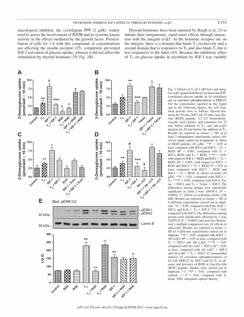
macological inhibitor, the cyclolignan PPP (2 �M), widelyused to assess the involvement of IGFIR and its tyrosine kinaseactivity in the effects mediated by the growth factor. Preincu-bation of cells for 1 h with this compound, at concentrationsnot affecting the insulin receptor (25), completely preventedIGF-I activation of glucose uptake, whereas it did not affect thestimulation by thyroid hormone (70; Fig. 2B).
Thyroid hormones have been reported by Bergh et al. (3) toinitiate their nongenomic, rapid-onset effects through interac-tion with the integrin �v�3. At the hormone receptor site onthe integrin, there is a domain that binds T3 exclusively and asecond domain that is responsive to T4 and also binds T3 but isless responsive to the latter (43). Because the inhibitory effectof T3 on glucose uptake in myoblasts by IGF-I was variably
7.5
10.0
A#
7.5
10.0
B**
0.0
2.5
5.0 ** ***§ °
0.0
2.5
5.0 ***
7.5
10.0
C
*7.5
10.0 #
D
0.0
2.5
5.0 **
0.0
2.5
5.0 ** ***§
Blot: pERK1/2
- pERK1- pERK240 kDa -
E
- Lamin B55 kDa -
250
300
**
50
100
150
200 **
++
I.O.D
.
0
2-D
eoxy
gluc
ose
upta
ke(p
mol
/wel
l × 1
0 m
in)
2-D
eoxy
gluc
ose
upta
ke(p
mol
/wel
l × 1
0 m
in)
2-D
eoxy
gluc
ose
upta
ke(p
mol
/wel
l × 1
0 m
in)
2-D
eoxy
gluc
ose
upta
ke(p
mol
/wel
l × 1
0 m
in)
T 4
Control
IGF-1
T 4 +
IGF-1
T 4 +
IGF-1
+ RGD
T 4 +
RGD
IGF-1
+ RGD
RGD T 4
Control
IGF-1
T 4 +
IGF-1
T 4 +
IGF-1
+ Tetr
ac
IGF-1
+ Tetr
acTetr
ac
T 4
Control
IGF-1
T 4 +
IGF-1
T 4 +
IGF-1
+ Ech
i
IGF-1
+ Ech
iEch
i T 4
Control
IGF-1
T 4 +
IGF-1
T 4 +
IGF-1
+ Ab αvβ3
IGF-1
+ Ab αvβ3
Ab αvβ3
T 4
Control
IGF-1
T 4 +
RGD
T 4 +
RGE
IGF-1
+ RGD
IGF-1
+ RGE
RGERGD
Fig. 3. Effects of T4 (0.1 nM free) and integ-rin �v�3 ligands/inhibitors on basal and IGF-I-mediated glucose uptake in L6 myoblastsand on activation (phosphorylation) of ERK1/2.For the experiments reported in this figureand in the following figures, the cell treat-ment periods were as follows: thyroid hor-mone for 30 min, IGF-I for 25 min, Arg-Gly-Asp (RGD) peptide, 3,3=,5,5=-tetraiodothy-roacetic acid (tetrac), and echistatin for 15min before addition of T4, and anti-�v�3integrin for 20 min before the addition of T4.Results are reported as means � SD of atleast 3 independent experiments, unless oth-erwise stated, carried out in duplicate. A: effectsof RGD peptide (10 �M). **P 0.05 atleast, compared with IGF-I and IGF-I � T4 �RGD; #P 0.001, compared with T4 �IGF-I, RGD, and T4 � RGD; ***P 0.05,with respect to IGF-I � RGD and IGF-I � T4 �RGD; §P 0.001, with respect to IGF-I �RGD and IGF-I � T4 � RGD;°P 0.01 atleast, compared with IGF-I � RGD andIGF-I � T4 � RGD. B: effects of tetrac (10�M). **P 0.01, compared with IGF-I �T4; ***P 0.05, compared with IGF-I, Tet-rac � IGF-I, and T4 � Tetrac � IGF-I. Thedifferences among groups were statisticallysignificant as from 1-way ANOVA (P 0.0002). C: effects of echistatin (Echi) (100nM). Results are reported as means � SD of4 different experiments carried out in dupli-cate. *P 0.05, compared with Echi, Echi �IGF-I, and Echi � T4 � IGF-I, **P 0.01compared with IGF-I. The differences amonggroups were significantly different by 1-wayANOVA (P 0.0001) and post hoc Bonfer-roni’s multiple comparison test. D: effects ofanti-�v�3. Results are reported as means �SD of 4 different experiments carried out induplicate. **P 0.05 compared with IGF-I �Ab �v�3; #P 0.05 at least, compared withT4 � IGF-I and Ab �v�3; ***P 0.01compared with Ab �v�3 � IGF-I; §P 0.01at least, compared with Ab �v�3 � IGF-Iand Ab �v�3 � T4 � IGF-I. E: immunoblotanalysis of activation (phosphorylation) ofL6 cell ERK1/2 by IGF-I and by T4 in ab-sence and presence of RGD or Arg-Gly-Glu(RGE) peptide. Studies were carried out induplicate �3. **P 0.01, compared withcontrol; ��P 0.01, compared with T4
alone. IOD, integrated optical density.
C153THYROXINE INHIBITS IGF-I EFFECTS THROUGH INTEGRIN �v�3
AJP-Cell Physiol • doi:10.1152/ajpcell.00308.2013 • www.ajpcell.org

demonstrated and, when present, less than that of T4 (resultsnot shown), we proceeded to study only the mechanism ofaction of T4 on glucose uptake. To confirm the involvement of�v�3 integrin in the actions of IGF-I and thyroid hormone inmyoblasts, we tested the ability of the RGD peptide, a smallligand that binds to the integrin RGD recognition site (20), toaffect the modulatory effects of T4. It is in fact known that theT4 site on the integrin is close to the RGD recognition site (20).The RGD peptide completely blocked the inhibition by T4 ofIGF-I-mediated glucose uptake and returned stimulation ofglucose uptake to the values obtained with IGF-I alone (Fig.3A). This result demonstrated that the integrin plays a role inthe T4 effect.
Tetrac has been shown to inhibit the short-term effects ofthyroid hormone at the plasma membrane level (14, 42) and isconsidered a probe for the involvement of the integrin �v�3 in
plasma membrane-initiated actions of T4; the agent also exhib-its pharmacological antitumor properties (19, 26, 44, 49). Asexpected, tetrac (10 �M) prevented the inhibitory effect of T4
on IGF-I-induced glucose uptake; surprisingly, tetrac itselfstimulated basal glucose uptake comparably to T4 (Fig. 3B).
Echistatin is a disintegrin protein of 49 amino acids (5.4kDa) obtained from the venom of the viper Echis carinatus. Itis an inhibitor of integrin �v�3 (2, 47). Disintegrins contain theRGD sequence; by binding to the RGD recognition site on theintegrins, they block ligand interaction with certain extracellu-lar matrix (ECM) proteins (27). Echistatin alone did not affectglucose uptake, but it significantly inhibited IGF-I-mediatedglucose uptake and to the same extent as T4. The combinationof echistatin, IGF-I, and T4 returned the glucose uptake to basallevel (Fig. 3C). An antibody to the �v�3 integrin, LM609, didnot affect the activation of glucose uptake by IGF-I but LM609
Blot: pAKT
- pERK2
Blot: pERK1/2- pERK1
A
- pAKT
- GAPDH- GAPDH
3.0
4.0
5.0 *
1.5
2.0
2.5
3.0
*
+
#
&
0.0
1.0
2.0+
Rel
ativ
e I.O
.D.
0.0
0.5
1.0
Rel
ativ
e I.O
.D.
CB
7.5
10.0 *7.5
10.0
**
0.0
2.5
5.0
0.0
2.5
5.0 * §
# °
2-D
eoxy
gluc
ose
upta
ke(p
mol
/wel
l × 1
0 m
in)
2-D
eoxy
gluc
ose
upta
ke(p
mol
/wel
l × 1
0 m
in)
T 4
Control
IGF-1
T 4 +
IGF-1
T 4 +
IGF-1
+ Wort
T 4 +
Wort
IGF-1
+ Wort
Wort T 4
Control
IGF-1
T 4 +
IGF-1
T 4 +
IGF-1
+ PD
T 4 +
PD
IGF-1
+ PDPD
T 4
Control
IGF-1
T 4 +
IGF-1
T 4 +
IGF-1
+ Wort
T 4 +
Wort
IGF-1
+ Wort
Wort T 4
Control
IGF-1
T 4 +
IGF-1
T 4 +
IGF-1
+ PD
T 4 +
PD
IGF-1
+ PDPD
Fig. 4. Roles of phosphoinositide 3-kinase (PI3K) and MAPK/ERK pathways in inhibition by T4 of IGF-I-stimulated glucose uptake in L6 myoblasts.Wortmannin (Wort), 100 nM (A, left; B), and PD 98059 (PD), 10 �M (A, right; C), were applied to cells 10 min before T4 treatment. A, left: immunoblots ofactivated Akt (pAkt) in lysates of L6 cells treated with T4, IGF-I, wortmannin, and combinations of the agents. Bar graphs are means � SD of blot densitiesin duplicate runs of 3 experiments. *P 0.01 compared with control; �P 0.01 compared with IGF-I alone. Right: immunoblots of activated ERK1/2(pERK1/2) in lysates of L6 cells treated with T4, IGF-I, PD 98059, and combinations of the agents. *P 0.05 vs. control; �P 0.01 compared with control;#P 0.01 compared with IGF-I alone; &P 0.05 vs. IGF-I alone. Results of glucose uptake studies in B and C are reported as means � SD of at least 3 differentexperiments carried out in duplicate. B: T4 inhibits the stimulatory effect of IGF-I on 2-deoxyglucose uptake. Wortmannin had no appreciable effect onunstimulated glucose uptake or on the action of T4 but completely inhibited the uptake stimulated by IGF-I either with or without T4. Results are reported asmeans � SD of 3 different experiments carried out in duplicate. *P 0.05 compared with all. C: MAPK activation inhibitor, PD 98059 (10 �M), suppressedthe inhibitory action of T4 on IGF-I stimulation of 2-deoxyglucose uptake. Results are reported as means � SD of at least 3 different experiments carried outin duplicate. *P 0.05 compared with IGF-I � T4 � PD, **P 0.05 at least compared with IGF-I � T4, PD, and PD � T4; #P 0.01 at least comparedwith IGF-I � PD and IGF-I � T4 � PD; °P 0.05 at least compared with IGF-I � PD and IGF-I � T4 � PD.
C154 THYROXINE INHIBITS IGF-I EFFECTS THROUGH INTEGRIN �v�3
AJP-Cell Physiol • doi:10.1152/ajpcell.00308.2013 • www.ajpcell.org
completely prevented the inhibition by T4 of IGF-I-mediatedglucose uptake (Fig. 3D). Echistatin thus appears to affectcrosstalk between the integrin and IGFIR more potently thanthe anti-�v�3 antibody (Fig. 3, C and D).
The functional contribution of �v�3 to the action of T4 wasadditionally examined by measurement of �v�3-dependentactivation (phosphorylation) of ERK1/2, a factor essential tothe interaction of T4 and IGF-I on glucose transport. L6 cellswere treated with T4 or IGF-I in the presence and absence ofRGD or RGE peptide, and pERK1/2 was measured (Fig. 3E).Both T4 and IGF-I increased pERK1/2, but RGD peptideaffected only the thyroid hormone-induced activation of theenzyme. Control RGE peptide was without effect. Thus themechanisms of ERK1/2 activation by T4 and IGF-I are dis-crete, and only the thyroid hormone effect requires the integrinin L6 cells. These results are consistent with glucose uptakestudies in Fig. 3A.
Short-term modulation of thyroid hormone on IGF-I-medi-ated glucose transport in L6 myoblasts: signal transduction byMAPK and Akt. The signal transduction pathways involved inthe effect of T4 on IGF-I action were studied by immunoblot-ting and with pharmacological probes (Fig. 4). Western blot-ting showed that IGF-I, but not T4, caused phosphorylation ofAkt (Fig. 4A, left). A PI3K inhibitor, wortmannin (100 nM),blocked the action of IGF-I on pAKT. Consistent with theseobservations, wortmannin eliminated the action of IGF-I onglucose transport and had no effect on glucose transport in thepresence of T4 (Fig. 4B).
Both IGF-I and T4 increased pERK1/2 (Fig. 4A, right), andthese actions were inhibited by PD 98059 (10 �M), an inhib-itor of the MAPK pathway at MEK. The PD compound did notaffect stimulation of glucose transport by IGF-I (Fig. 4C) butdid reduce glucose transport in the presence of T4 alone.Furthermore, PD 98059 eliminated the inhibitory effect of T4
on IGF-I-stimulated glucose uptake (Fig. 4C).These results supported a role for the PI3K/Akt pathway as
the principal mediator of glucose transport activation by IGF-Iand established that pERK1/2 is a critical contributor to theinhibitory action of thyroid hormone (T4) on IGF-I-enhancedglucose uptake. The T4 effect was shown above to require�v�3 and thus to be a nongenomic action of the hormone.
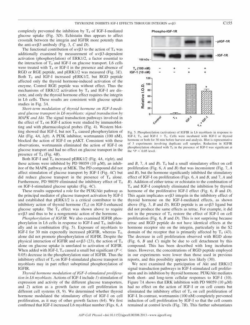
Phosphorylation of IGFIR. We also examined IGFIR phos-phorylation in L6 cells in response to IGF-I and T4, individu-ally and in combination (Fig. 5). Exposure of myoblasts toIGF-I for 30 min expectedly increased pIGFIR, whereas T4,alone did not promote phosphorylation of IGFIR. Despite thephysical interaction of IGFIR and �v�3 (23), the action of T4
alone on glucose uptake is unrelated to activation of IGFIR.When added with IGF-I, T4 caused a small but significant (P 0.05) decrease in the phosphorylation state of IGFIR. Thus theinhibitory effect of T4 on IGF-I-stimulated glucose transport inmyoblasts may in part reflect suboptimal phosphorylation ofIGFIR.
Thyroid hormone modulation of IGF-I-stimulated prolifera-tion in L6 myoblasts. Actions of IGF-I include 1) stimulation ofexpression and activity of the different glucose transporters,and 2) action as a growth factor on cell proliferation indifferent cell systems (8, 9). We determined whether thyroidhormone modulated the stimulatory effect of IGF-I on cellproliferation, as it may of other growth factors (64). We firstconfirmed that IGF-I increased L6 myoblast number (Figs. 6, A
and B, 7, A and B). T4 had a small stimulatory effect on cellproliferation (Fig. 6, A and B) that was inconsistent (Fig. 7, Aand B), but the hormone significantly inhibited the stimulatoryeffect of IGF-I on proliferation (Figs. 6, A and B, and 7, A andB). Addition of either tetrac or echistatin to the combination ofT4 and IGF-I completely eliminated the inhibition by thyroidhormone of the proliferative IGF-I effect (Fig. 6, B and D).This again implicates �v�3 integrin in the inhibitory effect ofthyroid hormone on the IGF-I-mediated effects, as shownabove (Fig. 3, B and D). RGD peptide is an �v�3 ligand butdid not produce the same effects as tetrac. For example, it didnot in the presence of T4 restore the effect of IGF-I on cellproliferation (Fig. 6, B and D). This is not surprising becausetetrac and RGD peptide do not have identical effects at thehormone receptor site on the integrin, particularly in the S2domain of the receptor that is primarily affected by T4 (43).The decrease in cell proliferation observed with RGD alone(Fig. 6, B and C) might be due to cell detachment by thiscompound. This has been described with long incubationtimes. However, the concentrations of RGD peptide employedin our experiments were lower than those used in previousreports, and this possibility appears less likely (30).
We next examined the participation of Akt and ERK1/2signal transduction pathways in IGF-I-stimulated cell prolifer-ation and its inhibition by thyroid hormone. PI3K/Akt mediatesboth short- and long-term cellular responses to IGF-I (10).Figure 7A shows that ERK inhibition with PD 98059 (10 �M)had no effect on the action of IGF-I or on cell counts buteliminated the inhibitory effect of T4 on cell proliferation ofIGF-I. In contrast, wortmannin (100 nM) completely preventedinduction of cell proliferation by IGF-I so that the cell countsremained at control levels (Fig. 7B). This further substantiates
Phospho-IGF-1R
- pIGF-1R
- IGF-1R100 kDa -
T4 (100 nM)IGF-1 (10 nM)
− −
++
− +
+−
100
120
*
20
40
60
80
0
Rel
ativ
e I.O
.D.
T 4
Control
IGF-1
T 4 +
IGF-1
Fig. 5. Phosphorylation (activation) of IGFIR in L6 myoblasts in response toIGF-I, T4, and IGF-I � T4. Cells were incubated with IGF-I or thyroidhormone or both for 30 min before harvest and analysis. Blot is representativeof 3 experiments involving duplicate cell samples. Reduction in IGFIRphosphorylation obtained with T4 in the presence of IGF-I was significant atthe *P 0.05 level.
C155THYROXINE INHIBITS IGF-I EFFECTS THROUGH INTEGRIN �v�3
AJP-Cell Physiol • doi:10.1152/ajpcell.00308.2013 • www.ajpcell.org

roles for the PI3K pathway in the mediation of the IGF-Iresponses that are both short term (glucose uptake; Fig. 4) andlong term (cell counts/cell proliferation at 24 h) (Fig. 7B).
We also examined the possibility that the effect of thyroidhormone on IGF-I-mediated cell proliferation might be theresult of a modification of different phases of cell cycle.Thyroid hormone is considered to be a regulator of the timecourse of cell differentiation, and this hormone-induced inhi-bition could represent in L6 myoblasts the starting point ofsuch an effect also for L6 myoblasts. Table 1 presents thepercentages of cells in the different phases of the cell cycleafter T4, IGF-I, or T4 � IGF-I treatment (n 3), and Fig. 8presents representative scans. T4 caused an increase in thenumber of dead cells (Table 1), but the hormone did notsignificantly affect the cell cycle. IGF-I promoted trends in thecell cycle (decreased cells in G1, increased cells in S andG2/M, compared with control cells). These results are consis-tent with the proliferative action of IGF-I (39). When thyroidhormone and IGF-I together were incubated with cells, therewas a small but significant reduction in the proliferative effectof IGF-I. In agreement with this, there was also, compared withIGF-I alone, a significant increase in cells in G0/G1 and adecrease of cells in G2/M in the presence of T4 � IGF-I. TheS phase was not significantly affected (Table 1), suggesting
that T4 in L6 myoblasts might slow the cell cycle and promotedifferentiation, as already reported for other cell lines (46).
DISCUSSION
The principal findings here are that thyroid hormone as T4
inhibits two actions of IGF-I in L6 myoblasts, glucose uptakeand proliferation. Glucose uptake is inhibited by T4 within ashort period of time (minutes), whereas the effect on stimula-tion by IGF-I on cell proliferation persists for a day or more.IGF-I treatment of rat myoblasts increased glucose uptakethree- to fourfold compared with basal levels. It has beenshown elsewhere that this effect of IGF-I is very fast andindependent of protein synthesis and apparently reflects trans-location of glucose transporters from intracellular stores to theplasma membrane (4, 38, 72). In the current studies, when T4
was added to cells together with IGF-I (10 nM), IGF-I-stimu-lated glucose uptake was significantly inhibited, suggesting theexistence of crosstalk between the T4 receptor on integrin�v�3 and IGFIR and consistent with the ternary complexformation described between the integrin and IGFIR (23).Acting alone, T4 variably increased glucose uptake. Studieselsewhere have shown that T3 consistently stimulated cellularglucose uptake (59, 61), probably attributable to an increase in
4000A
*2.0
*
B
*
2000
3000
1.0
1.5 **
***
0 24 48 72 960
1000
0.0
0.5
Time (h)
3000C
2.0D
1000
2000
1.0
1.5
0 24 48 72 960
1000
Time (h)
Num
ber o
f cel
ls (×
10−3
)N
umbe
r of c
ells
(× 1
0−3)
3 H-T
hym
idin
e in
corp
orat
ion
(rel
.)N
umbe
r of c
ells
(rel
.)
0.0
0.5
T 4
Control
IGF-1
T 4 +
IGF-1
T 4 +
IGF-1
+ Tetr
ac
T 4 +
IGF-1
+ RGD
T 4 +
IGF-1
+ Ech
iRGD
Echi
Tetrac
T 4
Control
IGF-1
T 4 +
IGF-1
T 4 +
IGF-1
+ Tetr
ac
T 4 +
IGF-1
+ RGD
T 4 +
IGF-1
+ Ech
i
Tetrac
T4
Control
RGD
Control
IGF-1T4 + IGF-1Fig. 6. Effects of T4 (0.1 nM free), RGD
peptide (10 �M), tetrac (10 �M), or echista-tin (100 nM) on cell proliferation stimulatedby IGF-I (10 nM) in L6 myoblasts. Resultsare reported as means � SD of at least 3different experiments, unless otherwisestated. A: effect of T4, IGF-I, and T4 �IGF-I on cell proliferation. Cells wereseeded on 60 � 15 mm Petri dishes andstimulated and counted every 24 h, as de-scribed in MATERIALS AND METHODS. *P 0.01 vs. all. At 96 h, all differences weresignificant. B: effect of tetrac, RGD peptide,echistatin, and T4 on cell proliferation acti-vated by IGF-I. *P 0.05 at least, com-pared with tetrac � T4 � IGF-I, RGD, andEchi � T4 � IGF-I; **P 0.001 comparedwith IGF-I, tetrac � T4 � IGF-I, and Echi �T4 � IGF-I. C: effect of RGD peptide (10�M) on cell proliferation assessed by cellcounting; the graph shows a representativeexperiment of 2 similar. D: effect of T4 andIGF-I, in the presence or absence of RGD,tetrac, echistatin, on 3H-thymidine incorpo-ration. Results are reported as means � SDof 3 different experiments carried out induplicate. 1-way ANOVA gave significantdifferences among the groups (P 0.0188).
C156 THYROXINE INHIBITS IGF-I EFFECTS THROUGH INTEGRIN �v�3
AJP-Cell Physiol • doi:10.1152/ajpcell.00308.2013 • www.ajpcell.org

activity of the glucose transporter. It may be noted that the T4
added in the current experiments resulted in a directly mea-sured near-physiological free T4 concentration of 10�10 M inthe cell culture medium.
The mechanisms by which T4 inhibits the effect of IGF-I onglucose transport and may increase glucose uptake are incom-pletely understood. IGF-I action in the present studies causedphosphorylation of IGFIR (Fig. 5), and results obtained withPPP (Fig. 2B) indicated that the activation of IGFIR indeedwas required for action of IGF-I on glucose uptake in L6 cells.Downstream of IGFIR, glucose uptake promoted by IGF-Ireflected Akt activation (phosphorylation) (Fig. 4A, left).ERK1/2 was also phosphorylated in response to IGF-I, but thiswas not linked to glucose uptake (Fig. 4C). In contrast, theinhibitory action of T4 on the stimulation of glucose uptake byIGF-I was ERK1/2 dependent (Fig. 4A, right, Fig. 4C). IGF-I
also can activate ERK1/2, but our results indicate that IGF-Iand T4 cause phosphorylation of ERK1/2 by different mecha-nisms upstream of the enzyme and with discrete consequencesdownstream of these kinases. As shown here, T4 does notaffect activation of Akt but does have a small but significantnegative effect on phosphorylation of IGFIR by IGF-I (Fig. 5),which may contribute to the blockade of IGF-I-induced glu-cose transport by thyroid hormone. Integrin �v�3 is expressedon the cell surface of various mammalian cells includingskeletal muscle (64).
The thyroid hormone-integrin interaction, particularly thatwith T4, activates the MAPK (ERK1/2) signal transductionpathway, promoting complex cellular responses such as angio-genesis (3, 44, 50). The extracellular domain of �v�3 includesan RGD recognition site, essential for the interaction withpolypeptide ECM ligands that contain the RGD sequence (73).The binding of T4 to the integrin and the consequent activationof the MAPK pathway and downstream physiological re-sponses are inhibited by the RGD peptide. This indicates thatthe T4 receptor on the integrin is proximal to the RGD recog-nition site (13, 21), but the actions of RGD peptide and tetracat the TR on �v�3 are not identical (44). Tetrac inhibits thenongenomic effects of thyroid hormone by competing with thehormone for the plasma membrane binding site (3, 14, 22, 43).In the present studies, RGD peptide and tetrac both blocked theeffect of thyroid hormone on IGF-I-stimulated glucose uptakein L6 myoblasts, demonstrating the involvement of the integrin�v�3 in the T4 effect. The RGD peptide was also effective inpreventing the inhibition by T3 of IGF-I-mediated glucoseuptake (not shown), confirming that T3 also binds to thisintegrin, as previously shown (13, 44). Very recently, short-term triiodothyronine (10�9 M) exposure was shown to nor-malize glucose transport in thyroid hormone-deprived L6 myo-tubes; the effect was additive to that of insulin. In these studies,there was no translocation of insulin-sensitive glucose trans-porters to the plasma membrane (69).
To further establish the role of integrin �v�3 in the inter-action of thyroid hormone and IGF-I effects on myoblastglucose transport, we employed a monoclonal antibody to the�v�3 integrin (LM 609) and a disintegrin, echistatin. Disin-tegrins have been considered to be antagonists of the receptor,although recent evidence indicates that they can have agonisteffects on integrins (2, 51). Our data suggest that echistatin isnot exclusively an antagonist of integrin �v�3 but that it mayhave a direct inhibitory effect on glucose uptake stimulated byIGF-I (Fig. 3C). In the proliferation assay, echistatin behavedlike RGD. In the presence of both IGF-I and T4, echistatin
Table 1. Distribution of L6 cells in the different phases ofcell cycle after treatment for 24 h with T4, IGF-I, or T4 �IGF-I
Dead Cells G0/G1 S G2/M
Control 10.3 � 3.5 73.0 � 3.4 2.2 � 2.5 14.8 � 3.5T4 18.3 � 5.0* 59.0 � 10.0 4.4 � 2.1 18.2 � 4.2IGF-I 8.7 � 0.2 54.8 � 3.5 8.7 � 2.0 27.7 � 2.5IGF-I� T4 7.4 � 0.9 62.4 � 3.6† 7.9 � 1.9 22.0 � 2.5†
Results are reported as means � SD of 3 different experiments. *P 0.02by Student’s t-test compared with insulin-like growth factor type I (IGF-I) andIGF-I � T4, same column. †P 0.05 by Student’s t-test compared with IGF-I,same column.
2000A
**
1000
1500
§ §***
0
500
2000B
*
1000
1500
0
500
Num
ber o
f cel
ls (×
10−3
)N
umbe
r of c
ells
(× 1
0−3)
T 4
Control
IGF-1
T 4 +
IGF-1
T 4 +
IGF-1
+ PD
T 4 +
PD
IGF-1
+ PDPD
T 4
Control
IGF-1
T 4 +
IGF-1
T 4 +
IGF-1
+ Wort
T 4 +
Wort
IGF-1
+ Wort
Wort
Fig. 7. Effects of inhibitors of MAPK (PD 98059) and PI3K (wortmannin)pathways on cell proliferation in L6 myoblasts. Results are reported asmeans � SD of 3 independent experiments carried out in duplicate.A: effect of MAPK pathway inhibitor, PD 98059 (10 �M). Results arereported as means � SD of 3 independent experiments carried out induplicate. **P 0.001 compared with IGF-I � T4, PD, and T4 � PD; ***P 0.001compared with IGF-I, IGF-I � PD, and IGF-I � T4 � PD; §P 0.001compared with IGF-I � PD and IGF-I � T4 � PD. B: effect of PI3K pathwayinhibitor, wortmannin (100 nM), on cell proliferation. Results are reported asmeans � SD of 3 different experiments carried out in duplicate. *P 0.001compared with all of the others.
C157THYROXINE INHIBITS IGF-I EFFECTS THROUGH INTEGRIN �v�3
AJP-Cell Physiol • doi:10.1152/ajpcell.00308.2013 • www.ajpcell.org

blocked the inhibitory effect of T4 on cell proliferation, mim-icking RGD and tetrac. To summarize, results obtained withRGD peptide, tetrac, echistatin, and the antibody for theintegrin �v�3 support the existence of crosstalk between theintegrin receptor for T4 and the IGFIR. Acting via �v�3,thyroid hormone increases phosphorylation of the IGFIR, con-sistent with clustering and physical interaction of the proteinson the cell surface and function of the integrin as a coreceptorfor IGF-I (9, 11). Experiments carried out by others on vascularsmooth muscle cells show that full expression of the activitiesof IGF-I requires activation of integrin �v�3 by ECM proteins,such as vitronectin (11, 36, 48, 53). Reported by Segal andIngbar in 1990 (61), the T3-induced increase in glucose trans-port in rat thymocytes was among the first nongenomic effectsof thyroid hormone to be identified. The effect was ascribed toactivation of transporters, rather than to transporter transloca-tion. Thyroid hormone stimulated the uptake of glucose with-out affecting transporter number or affinity, as indicated by thebinding of [3H]-cytochalasin B (61). In contrast to the obser-vations of Segal and Ingbar involving T3 and glucose uptake,we find that the effect of T4 on glucose transport in myoblastsis primarily modulation of IGF-I-stimulated glucose uptakeand secondarily or variably on sugar transport in the absence ofIGF-I. At the 10�10 M free T4 concentration employed, T4
opposed IGF-I-enhanced glucose uptake by an integrin �v�3-mediated mechanism distinct from the classical mechanism ofIGF-I action.
Signal transduction studies of thyroid hormone and IGF-Ithat we conducted on glucose uptake involved inhibitors ofPI3K/Akt and MAPK. Wortmannin, an inhibitor of PI3K,inhibited IGF-I stimulation of glucose uptake, as expected (8,
9). MAPK pathway inhibition with PD 98059 did not affectIGF-I action but did block the actions of T4. We confirmed thepharmacological inhibitor studies involving wortmannin andPD with immunoblots of activated Akt and ERK1/2. Thesefindings are in agreement with previous reports from ourlaboratories on several nongenomic effects of thyroid hor-mones that may involve these signal transduction pathways(14, 18, 21, 43). Figure 4, A and B, indicates that the additionof T4 does not significantly change the abundance of pAkt orpERK1/2 in IGF-I-stimulated cells. The upstream pathwaysources of pERK1/2 in the blot band phosphoprotein aredifferent in IGF-I-treated cells in the presence and absence ofT4, and the downstream consequences may be different (“tar-geted pools”). Our results, however, do suggest that the smalldecrease in activated IGFIR obtained with T4 in IGF-I-treatedL6 cells contributes to the decrease in glucose uptake seen withthyroid hormone. We do not yet know whether T4 is capable ofaffecting translocation of hormone-sensitive GLUT transport-ers from the cytoplasm to the plasma membrane or theirintrinsic activity (52, 66), an action that T3 appears to induce(61). Any stimulation of glucose uptake that T4 may achieve,e.g., that shown here in Fig. 3C, could reflect MAPK/ERK1/2-dependent stimulation of intrinsic activity of the glucosetransporter (52, 61, 66).
In addition to its effect on IGF-I-stimulated cell glucoseuptake, T4 impaired the action of IGF-I on myoblast prolifer-ation, as measured by cell counting, thymidine incorporationexperiments, and fluorescence-activated cell sorting experi-ments. The inhibitory effect is mediated by the MAPK path-way because PD 98059 reversed the inhibitory effect of T4 onproliferation. This is similar to the action of the PD compound
Control
G1 = 73.0S = 2.2G2/M = 14.8
T4
G1 = 56.7S = 6.2G2/M = 17.7
T4 + IGF-1
G1 = 65.4S = 7.1G2/M = 19.3
IGF-1
G1 = 56.3S = 7.7G2/M = 27.4
256
192
128
64
0
256
192
128
64
0
100 101 102 103 100 101 102 103
100 101
Fluorescence Fluorescence
Fluorescence Fluorescence
Eve
nts
Eve
nts
Eve
nts
Eve
nts
102 103 100 101 102 103
256
192
128
64
0
256
192
128
64
0
Fig. 8. Fluorescence-activated cell sorting anal-ysis of DNA in L6 myoblasts after incubationfor 24 h with T4 (0.1 nM free), IGF-I (10 nM),and T4 � IGF-I. The histogram shows a rep-resentative experiment and shows the numberof events vs. fluorescence emission. G1, S, andG2/M indicate the different cell cycle phases.Data from multiple analyses are presented inTable 1.
C158 THYROXINE INHIBITS IGF-I EFFECTS THROUGH INTEGRIN �v�3
AJP-Cell Physiol • doi:10.1152/ajpcell.00308.2013 • www.ajpcell.org

on the T4-IGF-I interaction on glucose uptake. Thus T4 is to beadded to a group of endogenous small molecules or peptidesthat may importantly modulate insulin or IGF-I activity inmuscle cells (29, 34, 35, 63).
Tetrac completely reversed the inhibitory effects of T4 oncell proliferation induced by IGF-I, implicating the iodothyro-nine receptor on �v�3 in this action of thyroid hormone. Theresults obtained with echistatin support those obtained withtetrac; that is, the disintegrin completely eliminated the inhi-bition of IGF-I-induced cell proliferation by T4. RGD peptidewas unable to reverse the inhibition by T4 of glucose uptake inIGF-I-treated L6 myoblasts. As we have already noted, theeffects of RGD peptide may overlap with, but are not identicalto, those of tetrac (44).
L6 myoblasts are customarily grown in the high-glucosemedium (4.5 g/l) used in the present studies (MATERIALS AND
METHODS). Increased ambient glucose may usefully retard myo-genesis (28) or confluency and myotube formation. Other celllines, such as neural stem cells (6), are also known to requirehigh-glucose medium. Apoptosis has been studied in neuralstem cells grown in high ambient glucose and has been shownto occur only at glucose concentrations above the 4.5 g/l levelused in the current study on myoblasts. Thus we believe it isunlikely that the medium glucose level used here influenced thecell proliferation results obtained.
Glucose intolerance is common in hyperthyroidism. De-creased peripheral insulin sensitivity with impaired insulinsecretion are factors contributing to the development of abnor-mal glucose tolerance in the hyperthyroid state (31, 67). How-ever, the mechanisms involved in this action of thyroid hor-mone are incompletely understood (54). An insight providedby the current studies is that thyroid hormone impairs glucoseuptake in L6 myoblasts that is promoted by IGF-I, functionallygiving rise to IGF-I resistance that can be considered as a partof a differentiation process involving L6 cells. One approach tointerpretation of these results is that impairment by thyroidhormone of IGF-I-stimulated glucose uptake in L6 myoblastsis antianabolic and might contribute to differentiation of thesecells.
ACKNOWLEDGMENTS
We thank Dr. Graziella Medori, Laboconsult s.r.l., Rome, for the assay offree T4.
GRANTS
We gratefully acknowledge the financial support from the Italian Ministryfor University and Research, General Management for Strategies and Devel-opment of Internationalization of Scientific and Technological Research; agrant from the Ordway Research Institute, Albany, NY; and a Lab Visit Grantfrom the Society of Endocrinology to Dr. R. G. Ahmed. Drs. F. B. and P. J.Davis appreciate the financial support of M. Frank and Margaret D. Rudy.
DISCLOSURES
No conflicts of interest, financial or otherwise, are declared by the authors.
AUTHOR CONTRIBUTIONS
Author contributions: S.I., P.D.V., R.G.A., P.L., and J.Z.P. conception anddesign of research; S.I., M.-T.H., H.-Y.L., G.-Y.C., P.D.V., A.M.F., R.S., E.C.,S.L., and P.J.D. analyzed data; S.I., M.-T.H., H.-Y.L., P.D.V., R.G.A., E.C.,S.L., P.L., J.Z.P., F.B.D., and P.J.D. interpreted results of experiments; S.I.,G.-Y.C., and A.M.F. prepared figures; S.I., J.Z.P., and P.J.D. drafted manu-script; S.I., P.L., J.Z.P., F.B.D., and P.J.D. edited and revised manuscript; S.I.,M.-T.H., H.-Y.L., G.-Y.C., P.D.V., A.M.F., R.G.A., R.S., E.C., S.L., P.L.,
J.Z.P., F.B.D., and P.J.D. approved final version of manuscript; M.-T.H.,H.-Y.L., G.-Y.C., P.D.V., A.M.F., R.S., E.C., and S.L. performed experiments.
REFERENCES
1. Ahmed RG, Davis PJ, Davis FB, De Vito P, Farias RN, Luly P,Pedersen JZ, Incerpi S. Nongenomic actions of thyroid hormones: frombasic research to clinical applications. An update. Immun Endoc MetabAgents Med Chem 13: 46–59, 2013.
2. Barja-Fidalgo C, Coelho AL, Saldanha-Gama R, Helal-Neto E,Mariano-Oliveira A, Freitas MS. Disintegrins: integrin selective ligandswhich activate integrin-coupled signaling and modulate leukocyte func-tion. Braz J Med Res 38: 1513–1520, 2005.
3. Bergh JJ, Lin HY, Lansing L, Mohamed SN, Davis FB, Mousa SA,Davis PJ. Integrin �V�3 contains a cell surface receptor site for thyroidhormone that is linked to activation of mitogen-activated protein kinaseand induction of angiogenesis. Endocrinology 146: 2864–2871, 2005.
4. Bilan PJ, Mitsumoto Y, Ramlal T, Klip A. Acute and long-term effectsof insulin-like growth factor 1 on glucose transporters in muscle cells.Translocation and biosynthesis. FEBS Lett 298: 285–290, 1992.
5. Blair AS, Hajduch E, Litherland GJ, Hundal HS. Regulation of glucosetransport and glycogen synthesis in L6 muscle cells during oxidativestress. Evidence for cross-talk between the insulin and SAPK2/p38 mito-gen-activated protein kinase signaling pathways. J Biol Chem 274: 36293–36299, 1999.
6. Chen J, Guo Y, Cheng W, Chen R, Liu T, Chen Z, Tan S. High glucoseinduces apoptosis and suppresses proliferation of adult rat neural stemcells following in vitro ischemia. BMC Neurosci 14: 24, 2013.
7. Cheng SY, Leonard JL, Davis PJ. Molecular aspects of thyroid hormoneactions. Endocr Rev 31: 139–170, 2010.
8. Clemmons DR. Involvement of insulin-like growth factor-1 in the controlof glucose homeostasis. Curr Opin Pharmacol 6: 620–625, 2006.
9. Clemmons DR. Modifying IGF1 activity: an approach to treat endocrinedisorders, atherosclerosis and cancer. Nat Rev Drug Discov 6: 821–833,2007.
10. Clemmons DR. Role of IGF-1 in skeletal muscle mass maintenance.Trends Endocrinol Metab 20: 349–356, 2009.
11. Clemmons DR, Maile LA. Integral membrane proteins that functioncoordinately with the insulin-like growth factor I receptor to regulateintracellular signaling. Endocrinology 144: 1664–1670, 2003.
12. Clemmons DR, Maile LA. Interaction between insulin-like growth fac-tor-1 receptor and �V�3 integrin linked signaling pathways: cellularresponses to changes in multiple signaling inputs. Mol Endocrinol 19:1–11, 2005.
13. Cody V, Davis PJ, Davis FB. Molecular modeling of the thyroid hormoneinteractions with �V�3 integrin. Steroids 72: 165–170, 2007.
14. D’Arezzo S, Incerpi S, Davis FB, Acconcia F, Marino M, Farias RN,Davis PJ. Rapid nongenomic effects of 3,5,3=-triiodo-L-thyronine onintracellular pH of L6 myoblasts are mediated by intracellular calciummobilization and kinase pathways. Endocrinology 145: 5694–5703, 2004.
15. Davis FB, Mousa SA, O’Connor L, Mohamed S, Lin HY, Cao HJ,Davis PJ. Proangiogenic action of thyroid hormone is fibroblast growthfactor-dependent and is initiated at the cell surface. Circ Res 94: 1500–1506, 2004.
16. Davis PJ, Davis FB. Nongenomic actions of thyroid hormone. Thyroid 6:497–504, 1996.
17. Davis PJ, Davis FB. Nongenomic actions of thyroid hormone on theheart. Thyroid 12: 459–466, 2002.
18. Davis PJ, Davis FB, Lin HY. Promotion by thyroid hormone of cyto-plasm-to-nucleus shuttling of thyroid hormone receptors. Steroids 73:1013–1017, 2008.
19. Davis PJ, Davis FB, Lin HY, Mousa SA, Zhou M, Luidens MK.Translational implications of nongenomic actions of thyroid hormoneinitiated at its integrin receptor. Am J Physiol Endocrinol Metab 297:E1238–E1246, 2009.
20. Davis PJ, Davis FB, Mousa SA, Luidens MK, Lin HY. Membranereceptor for thyroid hormone: physiologic and pharmacologic implica-tions. Annu Rev Pharmacol Toxicol 51: 98–115, 2011.
21. Davis PJ, Leonard JL, Davis FB. Mechanisms of nongenomic actions ofthyroid hormones. Front Neuroendocrinol 29: 211–218, 2008.
22. Davis PJ, Shih A, Lin HY, Martino LJ, Davis FB. Thyroxine promotesassociation of mitogen-activated protein kinase and nuclear thyroid hor-mone receptor (TR) and causes serine phosphorylation of TR. J Biol Chem275: 38032–38039, 2000.
C159THYROXINE INHIBITS IGF-I EFFECTS THROUGH INTEGRIN �v�3
AJP-Cell Physiol • doi:10.1152/ajpcell.00308.2013 • www.ajpcell.org

23. Fujita M, Takada YK, Takada Y. Insulin-like growth factor (IGF)signalling requires �v�3-IGF1-IGF type 1 receptor (IGF1R) ternary com-plex formation in anchorage independence, and the complex does notrequire IGF1R and Src activation. J Biol Chem 288: 3059–3069, 2013.
24. Furtado LM, Poon V, Klip A. GLUT4 activation: thoughts on possiblemechanisms. Acta Physiol Scand 178: 287–296, 2003.
25. Girnita A, Girnita L, del Prete F, Bartolazzi A, Larsson O, Axelson M.Cyclolignans as inhibitors of the insulin-like growth factor-1 receptor andmalignant cell growth. Cancer Res 64: 236–242, 2004.
26. Glinskii AB, Glinsky GV, Lin HY, Tang HY, Sun M, Davis FB,Luidens MK, Mousa SA, Hercbergs AH, Davis PJ. Modification ofsurvival pathway gene expression in human breast cancer cells by tetraio-dothyroacetic acid (tetrac). Cell Cycle 8: 3554–3562, 2009.
27. Gould RJ, Polokoff MA, Friedman PA, Huang TF, Holt JC, Cook JJ,Niewiarowski S. Distintegrins: a family of integrin inhibitory proteinsfrom viper venom. Proc Soc Exp Biol Med 195: 168–171, 1990.
28. Grzelkowska-Kowalczyk K, Wieteska-Skrzeczynska W, Grabiec K,Tokarska J. High glucose-mediated alterations of mechanisms importantin myogenesis of mouse C2C12 myoblasts. Cell Biol Int 37: 29–35, 2013.
29. Han XX, Bonen A. Epinephrine translocates GLUT-4 but inhibits insulin-stimulated glucose transport in rat muscle. Am J Physiol Endocrinol Metab274: E700–E707, 1998.
30. Hayman EG, Pierschbacher MD, Ruoslahti E. Detachment of cellsfrom culture substrate by soluble fibronectin peptides. J Cell Biol 100:1948–1954, 1985.
31. Holness MJ, Greenwood GK, Smith ND, Sugden MC. PPAR� activa-tion and increased dietary lipid oppose thyroid hormone signaling andrescue impaired glucose-stimulated insulin secretion in hyperthyroidism.Am J Physiol Endocrinol Metab 295: E1380–E1389, 2008.
32. Incerpi S, Luly P, De Vito P, Farias RN. Short-term effects of thyroidhormones on the Na�/H� antiport in L6 myoblasts: high molecularspecificity for 3,3=,5-triiodothyronine. Endocrinology 140: 683–689, 1999.
33. Incerpi S, Scapin S, D’Arezzo S, Spagnuolo S, Leoni S. Short-termeffects of thyroid hormone in prenatal development and cell differentia-tion. Steroids 70: 434–443, 2005.
34. Isenovic ER, Meng Y, Jamali N, Milivojevic N, Sowers JR. Ang IIattenuates IGF-1-stimulated Na�,K�-ATPase activity via PI3K/Akt invascular smooth muscle cells. Int J Mol Med 13: 915–922, 2004.
35. Jiang ZY, Zhou QL, Chatterjee A, Feener EP, Myers MG Jr, WhiteMF, King GL. Endothelin-1 modulates insulin signaling through phos-phatidylinositol 3-kinase pathway in vascular smooth muscle cells. Dia-betes 48: 1120–1130, 1999.
36. Jones JI, Prevette T, Gockerman A, Clemmons DR. Ligand occupancyof the �V�3 integrin is necessary for smooth muscle to migrate inresponse to insulin-like growth factor. Proc Natl Acad Sci USA 93:2482–2487, 1996.
37. Kewalramani G, Fink LN, Asadi F, Klip A. Palmitate-activated mac-rophages confer insulin resistance to muscle cells by a mechanism involv-ing protein kinase C � and . PLoS One 6: e26947, 2011.
38. Klip A. The many ways to regulate glucose transporter 4. Appl PhysiolNutr Metab 34: 481–487, 2009.
39. Kojima I, Mogami H, Shibata H, Ogata E. Role of calcium entry andprotein kinase C in the progression activity of insulin-like growth factor-1in Balb/c 3T3 cells. J Biol Chem 268: 10003–10006, 1993.
40. Lei J, Mariash CN, Ingbar DH. 3,3=,5-Triiodo-L-thyronine up-regula-tion of Na-K-ATPase activity and cell surface expression in alveolarepithelial cells is Src kinase- and phosphoinositide 3-kinase-dependent. JBiol Chem 279: 47589–47600, 2004.
41. Lei J, Wendt CH, Fan D, Mariash CN, Ingbar DH. Developmentalacquisition of T3-sensitive Na-K-ATPase stimulation by rat alveolarepithelial cells. Am J Physiol Lung Cell Mol Physiol 292: L6–L14, 2007.
42. Lin HY, Davis FB, Gordinier JK, Martino LJ, Davis PJ. Thyroidhormone induces activation of mitogen-activated protein kinase in cul-tured cells. Am J Physiol Cell Physiol 276: C1014–C1024, 1999.
43. Lin HY, Sun M, Tang HY, Luidens MK, Mousa SA, Incerpi S,Drusano GL, Davis FB, Davis PJ. L-Thyroxine vs. 3,5,3=-triiodothyro-nine and cell proliferation: Activation of mitogen activated protein kinaseand phosphatidylinositol 3-kinase. Am J Physiol Cell Physiol 296: C980–C991, 2009.
44. Lin HY, Tang HY, Davis FB, Mousa SA, Incerpi S, Luidens MK,Meng R, Davis PJ. Nongenomic regulation by thyroid hormone of plasmamembrane ion and small molecule pumps. Discov Med 14: 199–206,2012.
45. Lin HY, Thacore HR, Davis FB, Davis PJ. Potentiation by thyroxine ofinterferon-�-induced antiviral state requires PKA and PKC activities. AmJ Physiol Cell Physiol 271: C1256–C1261, 1996.
46. Lü HZ, Wang YX, Li Y, Fu SL, Hang Q, Lu PH. Proliferation anddifferentiation of oligodendrocyte progenitor cells induced from rat em-bryonic neural precursor cells followed by flow cytometry. Cytometry A73: 754–760, 2008.
47. Maile LA, Busby WH, Sitko K, Capps BE, Sergent T, Badley-ClarkeJ, Ling Y, Clemmons DR. The heparin binding domain of vitronectin isthe region that is required to enhance insulin-like growth factor-1 signal-ing. Mol Endocrinol 20: 881–892, 2006.
48. Marcinkiewicz C, Vijay-Kumar S, McLane MA, Niewiarowski S.Significance of RGD loop and C-terminal domain of echistatin for recog-nition of �IIb�3 and �v�3 integrins and expression of ligand-inducedbinding site. Blood 90: 1565–1575, 1997.
49. Meng R, Tang HY, Westfall J, London D, Cao JH, Mousa SA, LuidensM, Hercbergs A, Davis FB, Davis PJ, Lin HY. Crosstalk betweenintegrin �v�3 and estrogen receptor-� is involved in thyroid hormone-induced proliferation in human lung carcinoma cells. PLoS One 6: e27547,2011.
50. Mousa SA, Davis FB, Mohamed S, Davis PJ, Feng X. Pro-angiogenesisaction of thyroid hormone and analogs in a three-dimensional in vitromicrovascular endothelial sprouting model. Int Angiol 25: 407–413, 2006.
51. Neto EH, Coelho AL, Sampaio AL, Henriques MG, Marcinkiewicz C,De Freitas MS, Barja-Fidalgo C. Activation of human T lymphocytesvia integrin signaling induced by RGD-disintegrins. Biochim Biophys Acta1773: 176–184, 2007.
52. Niu W, Huang C, Nawaz Z, Levy M, Somwar R, Li D, Bilan PJ, KlipA. Maturation of the regulation of GLUT4 activity by p38 MAPK duringL6 cell myogenesis. J Biol Chem 278: 17953–17962, 2003.
53. Plow EF, Haas TA, Zhang L, Loftus J, Smith JW. Ligand binding tointegrins. J Biol Chem 275: 21785–21788, 2000.
54. Potenza M, Via MA, Yanagisawa RT. Excess thyroid hormone andcarbohydrate metabolism. Endocr Pract 15: 54–62, 2009.
55. Sasaki-Suzuki N, Arai K, Ogata T, Kasahara K, Sakoda H, Chida K,Asano T, Pessin JE, Hakuno F, Takahashi SI. Growth hormone inhi-bition of glucose uptake in adipocytes occurs without affecting GLUT-4translocation through an insulin receptor substrate-2-phosphatidylinositol3-kinase-dependent pathway. J Biol Chem 284: 6061–6070, 2009.
56. Scapin S, Leoni S, Spagnuolo S, Fiore AM, Incerpi S. Short-termeffects of thyroid hormones on the Na�-K�-ATPase activity of chickembryo hepatocytes during development: Focus on signal transduction.Am J Physiol Cell Physiol 296: C4–C12, 2009.
57. Scapin S, Leoni S, Spagnuolo S, Gnocchi D, De Vito P, Luly P,Pedersen JZ, Incerpi S. Short term effects of thyroid hormones duringdevelopment: focus on signal transduction. Steroids 75: 576–584, 2009.
58. Segal J, Ingbar SH. An immediate increase in calcium accumulation byrat thymocytes induced by triiodothyronine: its role in the subsequentmetabolic responses. Endocrinology 115: 160–166, 1984.
59. Segal J, Ingbar SH. In vivo stimulation of sugar uptake in rat thymocytes.An extranuclear action of 3,5,3=-triiodothyronine. J Clin Invest 76: 1575–1580, 1985.
60. Segal J, Ingbar SH. Evidence that an increase in cytoplasmic calcium isthe initiating event in certain plasma membrane-mediated responses to3,5,3=-triiodothyronine in rat thymocytes. Endocrinology 124: 1949–1955, 1989.
61. Segal J, Ingbar SH. 3,5,3=-Tri-iodothyronine enhances sugar transport inrat thymocytes by increasing intrinsic activity of the plasma membranesugar transporter. J Endocrinol 124: 133–140, 1990.
62. Selmi S, Samuels HH. Thyroid hormone receptor/and v-erbA. A singleamino acid difference in the C-terminal region influences dominant neg-ative activity and receptor dimer formation. J Biol Chem 266: 11589–11593, 1991.
63. Shemyakin A, Salehzadeh F, Bohm F, Al-Khalili L, Gonon A, WagnerH, Efendic S, Krook A, Pernow J. Regulation of glucose uptake byendothelin-1 in human skeletal muscle in vivo and in vitro. J ClinEndocrinol Metab 95: 2359–2366, 2010.
64. Shih AI, Zhang S, Cao HJ, Tang HY, Davis FB, Davis PJ, Lin HY.Disparate effects of thyroid hormone on actions of epidermal growthfactor and transforming growth factor-� are mediated by 3=,5=-cyclic-adenosine 5=-monophosphate-dependent protein kinase II. Endocrinology145: 1708–1717, 2004.
C160 THYROXINE INHIBITS IGF-I EFFECTS THROUGH INTEGRIN �v�3
AJP-Cell Physiol • doi:10.1152/ajpcell.00308.2013 • www.ajpcell.org

65. Sinanan ACM, Machell JRA, Wynne-Hughes GT, Hunt NP, LewisMP. �V�3 and �V�5 integrins and their role in muscle precursor cellsadhesion. Biol Cell 100: 465–477, 2008.
66. Somwar R, Kim DY, Sweeney G, Huang C, Niu W, Lador C, RamlalT, Klip A. GLUT4 translocation precedes the stimulation of glucoseuptake by insulin in muscle cells: potential activation of GLUT4 via p38mitogen-activated protein kinase. Biochem J 359: 639–649, 2001.
67. Styskal J, Van Remmen H, Richardson A, Salmon AB. Oxidative stressand diabetes: what can we learn about insulin resistance from antioxidantmutant mouse models? Free Radic Biol Med 52: 46–58, 2012.
68. Tang HY, Lin HY, Zhang S, Davis FB, Davis PJ. Thyroid hormonecauses mitogen-activated protein kinase-dependent phosphorylation of thenuclear estrogen receptor. Endocrinology 145: 3265–3272, 2004.
69. Teixeira SS, Tamrakar AK, Goulart-Silva S, Serrano-Nascimento C,Klip A, Nunes MT. Triiodothyronine acutely stimulates glucose transportinto L6 muscle cells without increasing surface GLUT4, GLUT1, orGLUT3. Thyroid 22: 747–754, 2012.
70. Vasilcanu R, Vasilcanu D, Sehat B, Yin S, Girnita A, Axelson M,Girnita L. Insulin-like growth factor type-I receptor-dependent phosphor-ylation of extracellular signal-regulated kinase 1/2 but not Akt (proteinkinase B) can be induced by picropodophyllin. Mol Pharmacol 73:930–939, 2008.
71. Watson RT, Pessin JE. Intracellular organization of insulin signaling andGLUT4 translocation. Recent Prog Horm Res 56: 175–193, 2001.
72. Wilson CM, Mitsumoto Y, Maher F, Klip A. Regulation of cell surfaceGLUT1, GLUT3 and GLUT4 by insulin and IGF-1 in L6 myotubes. FEBSLett 368: 19–22, 1995.
73. Xiong JP, Stehle T, Zhang R, Joachimiak A, Frech M, Goodman SL,Arnaout MA. Crystal structure of the extracellular segment of integrin�V�3 in complex with an Arg-Gly-Asp ligand. Science 296: 151–155,2002.
74. Yonkers MA, Ribera AB. Sensory neuron sodium current requiresnongenomic actions of thyroid hormone during development. J Neuro-physiol 100: 2719–2725, 2008.
C161THYROXINE INHIBITS IGF-I EFFECTS THROUGH INTEGRIN �v�3
AJP-Cell Physiol • doi:10.1152/ajpcell.00308.2013 • www.ajpcell.org