IGF-2 Is a Mediator of Prolactin-Induced Morphogenesis in the Breast
11
Developmental Cell, Vol. 3, 877–887, December, 2002, Copyright 2002 by Cell Press IGF-2 Is a Mediator of Prolactin-Induced Morphogenesis in the Breast genetically manipulated and because its mammary glands are readily accessible for experimental manipula- tion. Moreover, tissue recombination techniques make Cathrin Brisken, 1,2,5,6 Ayyakkannu Ayyannan, 1,6 Cuc Nguyen, 3 Anna Heineman, 2 Ferenc Reinhardt, 2 Tian Jan, 4 S.K. Dey, 4 G. Paolo Dotto, 3 and Robert A. Weinberg 2 it possible to distinguish between systemic and local 1 Department of Surgical Oncology effects of germline mutations. Thus, recombination be- Harvard Medical School tween mammary epithelial cells (MECs) of one genotype and Massachusetts General Hospital and mammary stroma of a second genotype within a Boston, Massachusetts 02114 wild-type host makes it possible to determine whether 2 Whitehead Institute for Biomedical Research the mammary gland phenotype of a mutant mouse is Nine Cambridge Center mediated by cell-autonomous processes that are intrin- Cambridge, Massachusetts 02142 sic to MECs, intrinsic to the mammary stroma, or sec- 3 Cutaneous Biology Research Center ondary to defects in other tissues that control breast Harvard Medical School development at a distance (Brisken et al., 1998). and Massachusetts General Hospital Prl blood levels are elevated during the luteal phase of Charlestown, Massachusetts 02129 the menstrual cycle (Franchimont et al., 1976), increase 4 Departments of Pediatrics during late pregnancy and lactation, and are highest and Cell and Developmental Biology following bouts of nursing (Horseman, 2001). The hor- Vanderbilt University Medical Center mone acts pleiotropically on a variety of target tissues Nashville, Tennessee 37232 (Bole-Feysot et al., 1998), explaining the observation that female mice lacking both copies of the Prl receptor gene (PrlR / mice) have multiple defects including a complex set of endocrinological abnormalities and infer- Summary tility (Binart et al., 2000; Ormandy et al., 1997). Recently, we described the behavior of PrlR / MECs The mechanisms by which prolactin controls prolifera- engrafted in the mammary fat pads of wild-type mice; tion of mammary epithelial cells (MECs) and morpho- these fat pads had previously been cleared of endoge- genesis of the breast epithelium are poorly under- nous epithelium (Brisken et al., 1999). When induced to stood. We show that cyclin D1 / MECs fail to proliferate become pregnant, these chimeras completed mammary in response to prolactin and identify IGF-2 as a down- ductal elongation and side branching but failed to devel- stream target of prolactin signaling that lies upstream op the alveoli that normally accompany late-stage preg- of cyclin D1 transcription. Ectopic IGF-2 expression re- stores alveologenesis in prolactin receptor / epithe- nancy. This behavior demonstrated that the defect in lium. Alveologenesis is retarded in IGF-2-deficient alveologenesis derived from the inability of PrlR / MECs MECs. IGF-2 and prolactin receptor mRNAs colocalize to respond to the high levels of circulating Prl that are in the mammary epithelium. Prolactin induces IGF-2 present late in pregnancy and hence reflected cell- mRNA and IGF-2 induces cyclin D1 protein in primary autonomous behavior of MECs. In addition, the en- MECs. Thus, IGF-2 is a mediator of prolactin-induced grafted PrlR / epithelium in these mice failed to differ- alveologenesis; prolactin, IGF-2, and cyclin D1, all of entiate properly into milk-secreting cells. Together, which are overexpressed in breast cancers, are com- these observations indicated that Prl signaling directly ponents of a developmental pathway in the mammary controls both the increase in MEC number during alveo- gland. logenesis and the differentiation of alveolar MECs into milk-secreting cells. Although the anterior lobe of the pituitary gland nor- Introduction mally produces the bulk of Prl in the female body, the hormone is also synthesized locally in the breast epithe- The systemic hormones estrogen, progesterone, and lium (Reynolds et al., 1997). Local Prl synthesis as well prolactin (Prl) control cell proliferation and morphogene- as expression of the PrlR is upregulated within breast sis in the breast (Nandi, 1958). The cellular and molecular carcinomas (Clevenger et al., 1995). While the physiolog- mechanisms underlying their growth-promoting func- ical relevance of the local Prl secretion remains unclear, tions remain poorly understood. These hormones act these observations suggest that localized deregulation directly on cells in the mammary gland and have indirect of Prl signaling within the breast may contribute to effects on this organ through their impact on other endo- breast carcinogenesis. Consistent with this notion are crine tissues. observations that blocking Prl signaling interferes with The mouse provides a suitable model system with the growth of various breast cancer cell lines (Fuh and which to study the regulation of growth and differentia- Wells, 1995; Ginsburg and Vonderhaar, 1995), and that tion of the breast, both because this animal can be mice lacking the Prl gene show a delay in polyoma mid- dle-T antigen-induced breast tumorigenesis (Vomachka 5 Correspondence: [email protected] et al., 2000). 6 Present address: Swiss Institute for Experimental Cancer Research, Ch. des Boveresses 155, 1066 Epalinges, Switzerland. Prl acts as a potent mitogen and morphogen on the
-
Upload
independent -
Category
Documents
-
view
0 -
download
0
Transcript of IGF-2 Is a Mediator of Prolactin-Induced Morphogenesis in the Breast
Developmental Cell, Vol. 3, 877–887, December, 2002, Copyright 2002 by Cell Press
IGF-2 Is a Mediator of Prolactin-InducedMorphogenesis in the Breast
genetically manipulated and because its mammaryglands are readily accessible for experimental manipula-tion. Moreover, tissue recombination techniques make
Cathrin Brisken,1,2,5,6 Ayyakkannu Ayyannan,1,6
Cuc Nguyen,3 Anna Heineman,2
Ferenc Reinhardt,2 Tian Jan,4 S.K. Dey,4
G. Paolo Dotto,3 and Robert A. Weinberg2 it possible to distinguish between systemic and local1Department of Surgical Oncology effects of germline mutations. Thus, recombination be-Harvard Medical School tween mammary epithelial cells (MECs) of one genotypeand Massachusetts General Hospital and mammary stroma of a second genotype within aBoston, Massachusetts 02114 wild-type host makes it possible to determine whether2Whitehead Institute for Biomedical Research the mammary gland phenotype of a mutant mouse isNine Cambridge Center mediated by cell-autonomous processes that are intrin-Cambridge, Massachusetts 02142 sic to MECs, intrinsic to the mammary stroma, or sec-3Cutaneous Biology Research Center ondary to defects in other tissues that control breastHarvard Medical School development at a distance (Brisken et al., 1998).and Massachusetts General Hospital Prl blood levels are elevated during the luteal phase ofCharlestown, Massachusetts 02129 the menstrual cycle (Franchimont et al., 1976), increase4Departments of Pediatrics during late pregnancy and lactation, and are highest
and Cell and Developmental Biology following bouts of nursing (Horseman, 2001). The hor-Vanderbilt University Medical Center mone acts pleiotropically on a variety of target tissuesNashville, Tennessee 37232 (Bole-Feysot et al., 1998), explaining the observation
that female mice lacking both copies of the Prl receptorgene (PrlR�/� mice) have multiple defects including acomplex set of endocrinological abnormalities and infer-Summarytility (Binart et al., 2000; Ormandy et al., 1997).
Recently, we described the behavior of PrlR�/� MECsThe mechanisms by which prolactin controls prolifera-engrafted in the mammary fat pads of wild-type mice;tion of mammary epithelial cells (MECs) and morpho-these fat pads had previously been cleared of endoge-genesis of the breast epithelium are poorly under-nous epithelium (Brisken et al., 1999). When induced tostood. We show that cyclin D1�/� MECs fail to proliferatebecome pregnant, these chimeras completed mammaryin response to prolactin and identify IGF-2 as a down-ductal elongation and side branching but failed to devel-stream target of prolactin signaling that lies upstreamop the alveoli that normally accompany late-stage preg-of cyclin D1 transcription. Ectopic IGF-2 expression re-
stores alveologenesis in prolactin receptor�/� epithe- nancy. This behavior demonstrated that the defect inlium. Alveologenesis is retarded in IGF-2-deficient alveologenesis derived from the inability of PrlR�/� MECsMECs. IGF-2 and prolactin receptor mRNAs colocalize to respond to the high levels of circulating Prl that arein the mammary epithelium. Prolactin induces IGF-2 present late in pregnancy and hence reflected cell-mRNA and IGF-2 induces cyclin D1 protein in primary autonomous behavior of MECs. In addition, the en-MECs. Thus, IGF-2 is a mediator of prolactin-induced grafted PrlR�/� epithelium in these mice failed to differ-alveologenesis; prolactin, IGF-2, and cyclin D1, all of entiate properly into milk-secreting cells. Together,which are overexpressed in breast cancers, are com- these observations indicated that Prl signaling directlyponents of a developmental pathway in the mammary controls both the increase in MEC number during alveo-gland. logenesis and the differentiation of alveolar MECs into
milk-secreting cells.Although the anterior lobe of the pituitary gland nor-Introduction
mally produces the bulk of Prl in the female body, thehormone is also synthesized locally in the breast epithe-The systemic hormones estrogen, progesterone, andlium (Reynolds et al., 1997). Local Prl synthesis as wellprolactin (Prl) control cell proliferation and morphogene-as expression of the PrlR is upregulated within breastsis in the breast (Nandi, 1958). The cellular and molecularcarcinomas (Clevenger et al., 1995). While the physiolog-mechanisms underlying their growth-promoting func-ical relevance of the local Prl secretion remains unclear,tions remain poorly understood. These hormones actthese observations suggest that localized deregulationdirectly on cells in the mammary gland and have indirectof Prl signaling within the breast may contribute toeffects on this organ through their impact on other endo-breast carcinogenesis. Consistent with this notion arecrine tissues.observations that blocking Prl signaling interferes withThe mouse provides a suitable model system withthe growth of various breast cancer cell lines (Fuh andwhich to study the regulation of growth and differentia-Wells, 1995; Ginsburg and Vonderhaar, 1995), and thattion of the breast, both because this animal can bemice lacking the Prl gene show a delay in polyoma mid-dle-T antigen-induced breast tumorigenesis (Vomachka5Correspondence: [email protected] al., 2000).6Present address: Swiss Institute for Experimental Cancer Research,
Ch. des Boveresses 155, 1066 Epalinges, Switzerland. Prl acts as a potent mitogen and morphogen on the
Developmental Cell878
Figure 1. Response of Cyclin D1�/� Mam-mary Epithelium to Progesterone and Pro-lactin
Cyclin D1�/� and Cyclin D1�/� MECs were en-grafted to the cleared fat pads of 3-week-old females. Eight weeks later, the recipientswere stimulated for 10 days with either pro-gesterone, left panels, or Prl, right panels.Shown are wholemount preparations of theengrafted mammary glands. Note that bothgrafts respond to progesterone stimulationwith extensive side branching but differ intheir response to Prl; the wt epithelium showsextensive alveolar development, which is ab-sent from the Cyclin D1�/� graft. Identical re-sults were obtained with six successfully en-grafted mice in each group. Left bar, 500 �m;right bar, 400 �m.
mammary epithelium in vivo (Nandi, 1958), but the mech- morphogenetic step and associated proliferation occurnormally in response to progesterone stimulation. Weanisms by which it induces cell proliferation remain un-
clear. To gain further insight into the mechanisms under- note here that in the course of conducting a large seriesof experiments, we have never observed alveologenesislying Prl function in vivo, we conducted a screen that
selects specifically for genes that function downstream when wt mice of this genetic background are treated inthis way. However, when stimulated with Prl, the wtof the PrlR. Our findings indicate that an extracellular
factor, IGF-2, acts as a downstream mediator of PrlR grafts (Figure 1, bottom right) developed alveolar struc-tures, which failed to form in the cyclin D1�/� graftssignaling and upstream regulator of cyclin D1 expres-
sion. These results provide a route by which Prl can (Figure 1, top right). We concluded that cyclin D1 func-tion is required to enable MECs to respond to the mor-act in a mitogenic fashion during alveologenesis and
carcinogenesis and links Prl signaling, IGF-2, and cyclin phogenetic effects of Prl. On the other hand, respon-siveness to progesterone is unaffected by the absenceD1 expression, all of which are deregulated during tu-
morigenesis, to a common signaling pathway. of cyclin D1 in MECs. With these observations in mind,we undertook to study the signaling pathway throughwhich Prl acts to trigger alveologenesis.Results
Response of cyclin D1�/� Epithelium Screening for Prl Target GenesWe initially sought to determine the signaling mecha-to Pregnancy Hormones
cyclin D1�/� female mice fail to lactate due to a develop- nism by which the ligand-activated PrlR is able to inducecyclin D1 synthesis in MECs. Control experiments (Fig-mental defect in the mammary gland (Fantl et al., 1995;
Sicinski et al., 1995), which is intrinsic to the mammary ure 2A) showed that direct application of Prl to culturedwt MECs induced relatively little cyclin D1 synthesisepithelium (Fantl et al., 1999). Both Prl and progesterone
are implicated in specific stages of mammary gland mor- within 8 hr when compared to the actions of a series ofgrowth factors such as epidermal growth factor (EGF),phogenesis during pregnancy, a time when the cyclin
D1�/�phenotype becomes apparent. To determine hepatocyte growth factor (HGF), and insulin. The pres-ence of an intact Prl-PrlR signaling system in these cellswhether the morphogenetic and mitogenic effects of
either of these hormones were dependent on cyclin D1 was indicated by the successful induction of �-caseinmRNA following exposure to Prl (data not shown). To-function, we established either cyclin D1�/� or wild-type
(wt) MECs as grafts in the mammary stroma of wt hosts gether, these observations suggested that the mecha-nisms controlling cyclin D1 transcription were intact incleared of the endogenous epithelium. Subsequently,
the recipients were treated with either Prl or progester- these cultured MECs and that it was unlikely that thePrlR signals directly to the controllers of cyclin D1 tran-one for 10 days, and the responses of the transplanted
cells were assessed by wholemount microscopy. Pro- scription via an intracellular signaling cascade.We reasoned that an alternative signaling mechanismgesterone was administered to ovariectomized female
mice by daily injections (Lydon et al., 1995). To achieve might involve the Prl-induced synthesis of some inter-mediary signaling molecule that, once produced, pro-stably elevated blood levels of the short-lived Prl, we
resorted to using transplants of pituitary glands from ceeds to induce cyclin D1 synthesis. Consistent withthis model were our observations that cyclin D1 protein,adult mice of the same genetic background engrafted
under the kidney capsule of the host. This ectopic local- while only weakly induced 8 hr after Prl exposure (Figure2A), was significantly induced 18 hr after stimulation ofization of the pituitary gland causes a selective increase
in Prl levels similar to those seen during pregnancy dur- growth factor-deprived primary MECs with Prl (Figure2B). To explore the possible involvement of such aning the first 10 days after transplant (Huseby et al., 1985).
As shown in Figure 1, left panels, the cyclin D1�/� and intermediary signaling molecule, we compared the tran-scription profiles of pairs of contralateral cleared mam-the wt MECs underwent extensive branching in re-
sponse to progesterone, indicating that this particular mary fat pads that had been reconstituted with PrlR�/�
IGF-2 Mediates Prolactin Function in the Mammary Gland879
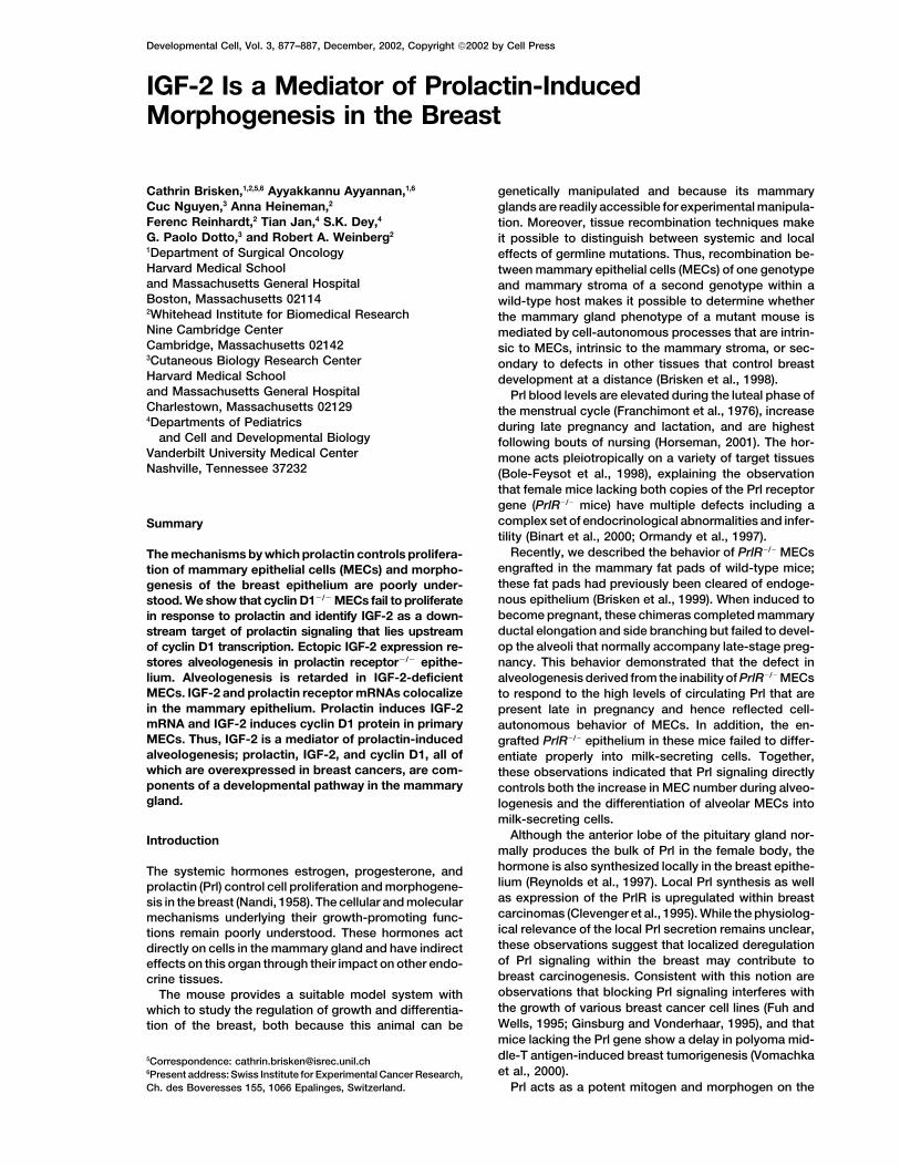
Figure 2. Establishing a Hierarchy betweenPrl, IGF-2, and cyclin D1
(A) Regulation of cyclin D1 protein expressionin primary MECs. Primary MECs were growthfactor deprived for 36 hr and subsequentlystimulated with various mammotropic factorsfor 8 hours: estradiol (20 nmol), R5020 (20nmol), hydrocortisone (1 �g/ml), Prl (5 �g/ml),insulin (10 �g/ml), HGF hepatocyte growthfactor (10 ng/ml), and EGF epidermal growthfactor (5ng/ml). Cyclin D1 protein levels as-sessed by Western blotting with anti-cyclinD1 antibodies.(B) Regulation of cyclin D1 protein expressionby Prl. Primary MECs were growth factor de-prived for 36 hr and subsequently stimulatedwith different concentrations of Prl for 18 hr.Cyclin D1 protein levels assessed by Westernblotting with anti-cyclin D1 antibodies.(C) PrlR�/� and Cyclin D1�/� mammary epithe-lia: morphogenetic block and IGF-2 mRNAexpression levels. Mammary epithelium fromPrlR�/�ROSA26�/� and Cyclin D1�/�ROSA26�/�
mice was engrafted to cleared inguinal fatpads of 3-week-old females. Eight weekslater, the recipients were mated, and at day16.5 of pregnancy, the engrafted glands andan unmanipulated gland were analyzed. Toppanels show glands subjected to X-gal stain-ing and wholemounted. PrlR�/�ROSA26�/�
(left) and Cyclin D1�/�ROSA26�/� epithelia(center) show fully branched ductal systemsbut lack alveoli. In contrast, the unmanipu-lated wt gland (right) shows extensive alveo-lar development. Bottom panels show IGF-2mRNA expression in the mutant transplants:RNA samples in three serial dilutions weresubjected to RT-PCR with IGF-2 (upperpanel) and �-gal (lower panel) specific prim-
ers. Densitometry revealed that the IGF-2 signal is 30-fold increased in the Cyclin D1�/�ROSA26�/�versus the PrlR�/�ROSA26�/� transplant. Thesame results were obtained with three further pairs of engrafted mammary glands. Right panel: IGF-2 mRNA expression levels in engraftedand control glands were also measured by Real Time PCR and normalized to mRNA levels of the epithelial marker Keratin 18. Results fromthree different experiments were averaged and plotted. Y-axis: arbitrary units.(D) IGF-2 expression in mammary glands engrafted with Cyclin D1 �/�ROSA26�/� and Cyclin D1�/�ROSA26�/� mammary epithelium harvestedat day 12.5 of pregnancy. Three serial dilutions of RNA from the engrafted glands were subjected to RT-PCR with IGF-2 (upper panel) and�-gal (lower panel) specific primers. The amplification products were subjected to agarose gel electrophoresis and visualized by ethidiumbromide staining. The same results were obtained with three further pairs of engrafted mammary glands.(E) Cyclin D1 induction by IGF-2 and RANKL. Primary MECs were growth factor deprived for 36 hr and subsequently stimulated with RANKL,IGF-2, or EGF and insulin for eight hours. Cyclin D1 protein levels assessed by Western blotting with anti-cyclin D1 antibodies.
MECs in one fat pad versus cyclin D1�/� MECs in the grafting experiments were both derived from mice thatwere also heterozygous for the ROSA26 transgeneother. The host mice were wt, and after engrafting, were
allowed to become pregnant. (Friedrich and Soriano, 1991) and hence expressed�-galactosidase (�-gal) constitutively. This enzyme andAs described above and illustrated in Figure 2C, top
panels, the morphogenetic block shown by MECs of its mRNA served as internal controls to ensure that simi-lar amounts of engrafted epithelia were being comparedthese two genotypes was very similar. Hence, these two
types of mammary glands, assayed at an identical day in these expression analyses. Serial dilutions of RNAderived from a group of engrafted recipient mice at dayof pregnancy, are presumed to carry comparable num-
bers of cells in comparable states of proliferation. We 16.5 of pregnancy were reverse-transcribed and levelsof �-gal expression were measured by semiquantitativereasoned that the only differences in gene expression
pattern between the mammary glands of these two ge- PCR. Three pairs of samples in which the contralateraltransplants showed comparable amounts of �-gal ex-notypes should involve those genes that lie downstream
of PrlR signaling and upstream of cyclin D1 expression. pression were pooled for cRNA probe preparation andhybridization on 6.5 K Affymetrix (B-chip) mouse expres-More specifically, we looked for genes that were ex-
pressed at high levels in the cyclin D1�/� but were ex- sion arrays. This analysis was performed in duplicate.Out of the 6,500 transcripts surveyed, 319 were ex-pressed at low or undetectable levels in the PrlR�/�
grafts. pressed at more than 3-fold higher levels in the cyclinD1�/� than in the PrlR�/� grafts; 430 transcripts wereThe donor cyclin D1�/� and PrlR�/� MECs in these
Developmental Cell880
downregulated more than 3-fold. Previous observations cyclin D1�/� and the wt counterpart becomes apparent.As shown in Figure 2D, the expression levels of IGF-2had indicated that, in contrast to the PrlR�/� epithelium,
the cyclin D1�/� epithelium retains its ability to undergo mRNAs were comparable when normalized to �-gal ex-pression levels in the two glands, indicating that cyclindifferentiation as manifested by its ability to synthesize
milk proteins late in pregnancy (Fantl et al., 1999). In- D1 does not act to repress IGF-2 expression. Takentogether, these various observations led us to concludedeed, many genes encoding proteins secreted with the
milk or involved in milk production such as metabolic that IGF-2 expression in the MECs of pregnant mice isstrongly dependent on signals emanating from the PrlR.enzymes, calcium transport, and intracellular vesicular
trafficking genes were found to be differentially ex-pressed, with their transcripts being found at far higher Effect of IGF-2 on Cyclin D1 Expressionlevels in the fat pads engrafted with cyclin D1�/� MECs While the above data indicated that the PrlR is requiredthan in the fat pads engrafted with PrlR�/� MECs. In for the induction of IGF-2 by MECs, it did not revealaddition, genes involved in signal transduction, the con- whether IGF-2, once synthesized, could proceed to in-struction of the cytoskeleton, and the extracellular ma- duce cyclin D1 synthesis. To address whether cyclintrix were preferentially expressed in the cyclin D1�/� re- D1 expression is under the direct control of IGF-2, wecombinants. stimulated primary MECs that had been deprived of
An overview of the genes whose expression levels growth factors with IGF-2 and assessed cyclin D1 pro-differed by more than 10-fold is given in Table 1. Among tein levels 8 hr later. We also stimulated the cells withthe secreted factors that were particularly highly ex- RANKL, a factor recently implied to be an importantpressed in the cyclin D1�/� grafts was insulin-like growth mediator of cyclin D1 expression in the mammary glandfactor-2 (IGF-2), which was found at a level 13.2-fold (Cao et al., 2001). We observed that the extent of cyclinhigher in the glands engrafted with cyclin D1�/� epithelia D1 induction is dependent on the concentration of IGF-2,than in the counterparts engrafted with PrlR�/� epithelia. amounting to a 6-fold increase at an IGF-2 concentrationThe expression levels of another growth factor, HB-EGF, of 50 ng/ml. However, RANKL failed to induce cyclin D1differed by 4.5-fold. protein synthesis in these cells (Figure 2E) when applied
To validate the initial findings, we assessed relative at the concentration that has been shown to activateIGF-2 and HB-EGF expression levels in four additional downstream signaling in these cells (Cao et al., 2001).matched pairs of engrafted glands by quantitative RT- This indicated that IGF-2 can directly upregulate cyclinPCR. After normalization for �-gal expression, IGF-2 D1 protein in MECs. Taken together, these observationsmRNA levels were consistently 12- to 30-fold higher in indicated that the PrlR is required for the synthesis ofthe cyclin D1�/� transplanted glands than in the PrlR�/� IGF-2 by MECs and that this growth factor, once synthe-counterparts (Figure 2C, bottom panels), whereas HB- sized and released from MECs, can act in an autocrineEGF levels differed by about 3-fold (data not shown). or paracrine fashion to elicit cyclin D1 synthesis.Other growth factors represented on the arrays such asFGF1, FGF15, HGF, EGF, and TGF� differed less than Role of IGF-2 in Mediating Prl Function3-fold between the two types of chimeric glands. We The scheme proposed above implies that IGF-2 acts asalso extended our analysis to additional growth factors an important mediator of PrlR signaling during alveolo-such as amphiregulin, IGF-1, and RANKL (receptor acti- genesis. In particular, these observations suggestedvator of NF-�b ligand) that are not represented on the that Prl induces IGF-2 and that the latter induces cyclinarrays but are developmentally regulated in the mam- D1 synthesis, enabling alveologenesis to proceed. Thismary gland. We found no difference in their expression thinking prompted us to determine the role of IGF-2 aslevels between contralateral grafts (data not shown). a possible mediator of Prl-induced alveologenesis. MoreThis indicated that the strongest upregulation of the specifically, we asked whether ectopic expression ofgrowth factors tested was that of IGF-2. Taken together, IGF-2 could rescue the defect in alveologenesis ob-these data provided evidence that IGF-2 synthesis in served in MECs lacking the PrlR and thus lacking PrlMECs is under the control of the PrlR and thus Prl. signaling.
To test this notion, we infected MECs derived from 10-week-old PrlR�/� female mice of 129SV/C57Bl6 mixedRole of Cyclin D1 in Regulating IGF-2 Expression
Our screen was based on the assumption that those genetic background with a retrovirus expressing IGF-2.The resulting cultures, a mixture of infected and nonin-genes that were expressed at higher levels in the fat
pads reconstituted with cyclin D1�/� epithelium than in fected MECs, were used to reconstitute cleared mam-mary fat pads of 3-week-old wt female mice. Previousthe fat pads reconstituted with PrlR �/� epithelium are
transcriptionally activated by Prl signaling. It was, how- work had shown that infection of MECs with a retrovirusexpressing �-gal does not affect in vivo morphogenesisever, formally possible that the difference in IGF-2 gene
expression levels was independent of Prl signaling and (Edwards et al., 1996). This indicated that use of a ret-roviral vector per se would have no effect on alveologen-resulted instead from a lack of repression in cyclin
D1�/�grafts. To address this possibility, we assessed esis. As a positive control, we determined whether thealveologenesis phenotype of the PrlR�/� MECs could beIGF-2 mRNA expression levels in the contralateral mam-
mary glands of a wild-type host that had been cleared rescued by infection with a retrovirus vector expressingthe PrlR. Eight weeks after injecting these MECs intoand reconstituted with either cyclin D1�/�ROSA26�/� epi-
thelium or cyclin D1�/�ROSA26�/� epithelium. We as- cleared fat pads, host mice were mated, and the mam-mary glands of the resulting pregnant mice were ex-sessed a sample at day 12.5 of pregnancy before the
time when the morphological difference between the amined.

IGF-2 Mediates Prolactin Function in the Mammary Gland881
Table 1. Putative Prolactin Target Genes
Genes Related to Milk Secretion
X93037 M. musculus mRNA for WDNM1 proteinW18308 Mouse ferritin heavy chain genex04673 AdipsinX61431 ACYL-COA-BINDING PROTEINW44201 Sim. to PROTEIN TRANSPORT PROTEIN SEC23Y00516 Mouse mRNA for aldolase A:FRUCTOSE-BISPHOSPHATE ALDOLASE AM32599 Mouse glyceraldehyde-3-phosphate dehydrogenase mRNAAA071776 Sim. to GLUCOSE-6-PHOSPHATE ISOMERASE (PHOSPHOGLUCOSE ISOMERASE)J05277 Mouse hexokinase mRNAx04490 Casein betaL09104 M. musculus glucose phosphate isomerase mRNA, 3� endM21285 Mouse stearoyl-CoA desaturase geneAA117004 Sim. to ER LUMEN PROTEIN RETAINING RECEPTOR (KDEL RECEPTOR) (P23)x02520 Lactate dehydrogenase 1, A chainW09506 Sim. to FATTY ACID-BINDING PROTEIN
Calcium Metabolism
X97991 CalcitoninM27844 ParvalbuminW20937 Sim. to CALCIUM-TRANSPORTING ATPASE SARCOPLASMIC RETICULUM TYPE
Cytoskeleton
x51438 VimentinAA168865 Sim. to ACTIN 1 (FRAGMENT)X13297 ACTIN, AORTIC SMOOTH MUSCLEJ04953 Mouse gelsolin geneX14425 Mouse mRNA for profilin:PROFILIN IU20365 M. musculus smooth muscle gamma actin mRNA
Growth Factors
AA002605 Mouse insulin-like growth factor II (IGF-II) gene, 5� flank
Extracellular Matrix
X04017 Mouse mRNA for cysteine-rich glycoprotein SPARCx14194 Nidogenx17069 Mouse COL1A2 mRNA for pro-alpha-2(I) collagenx65582 Procollagen, type VI, alpha 2U08020 M. musculus alpha 1 type I collagen gene, partial cds and 3� flanking regionET61037 Lectin, galactose binding, soluble 1x72862 M. musculus gene for beta-3-adrenergic receptor::Adrenergic receptor, beta 3X73523 Mouse mRNA for matrix Gla protein (MGP)W75072 Procollagen, type IX, alpha 2
Signal Transduction
x15358 Sim. to INSULIN-LIKE GROWTH FACTOR BINDING PROTEIN 4 PRECURSOR (IGFBP-4)W65899 Sim. to GUANINE NUCLEOTIDE-BINDING PROTEIN G(I)/G(S)/G(T) BETA SUBUNIT 2D10024 Mouse mRNA for protein-tyrosine kinase substrate p36 (calpactin I heavy chain)X58251 M. musculus mRNA for E-selectin ligand-1L23108 M. musculus mRNA for GTP-binding proteinX85788 M. musculus mRNA for DCC tumour suppressorL09192 Cathepsin DM16358 RAB1, member RAS oncogene family
Heat Shock
AA163643 Sim. to HEAT SHOCK COGNATE 71 KD PROTEINU73744 M. musculus heat shock 70 protein (Hsc70) geneAA105022 Sim. to HEAT SHOCK PROTEIN HSP 90-BETA (HSP 84) (HSP 90)
Miscellaneous
W41817 M. musculus cytochrome c oxidase subunit VIII precursor (Cox81) mRNAZ83368 M. musculus RPS3a geneM76131 Mouse elongation factor 2 (ef-2) mRNA, 3� endAA154007 Sim. to POL POLYPROTEIN; REVERSE TRANSCRIPTASEX05021 Murine mRNA with homology to yeast L29 ribosomal protein genex54691 Cytochrome C oxidase, subunit IVW88176 Sim. to THIOL-SPECIFIC ANTIOXIDANT PROTEIN (PRP)Z50159 M. musculus mRNA for Sui1M24263 Mouse testosterone 16-alpha-hydroxylase (CB) geneAA138107 Mouse COX7c1 mRNA for cytochrome c oxidase VIIc (EC 1.9.3.1)D00466 Mouse apolipoprotein E mRNAx82067 M. musculus thioredoxin-dependent peroxide reductase (tpx) mRNA
Genes expressed at more than 10-fold higher levels in mammary glands engrafted with cyclin DI�/� than with PrlR�/� mammary epithelium,as assessed by microarray analysis.
Developmental Cell882
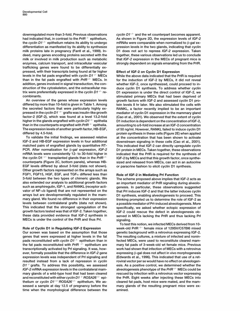
Figure 3. Rescuing the Morphogenetic Defect of PrlR�/� Epithelium by Ectopic Expression of IGF-2
MECs derived from PrlR�/�ROSA26�/� female mice were infected with retroviruses expressing either the PrlR (C) or IGF-2 (D,E,G, and I) ormock infected (B, F, and H) and subsequently engrafted into the cleared fat pads of 3-week-old females. The recipients were impregnated.At the end of pregnancy the engrafted glands and thoracic mammary gland were analyzed by X-gal staining and subsequent wholemountmicroscopy (top panels).(A) Unmanipulated gland displays extensive alveolar development.(B) Mock-infected PrlR�/� epithelium devoid of alveolar structures.(C) PrlR�/� epithelium infected with PrlR retrovirus showing a sector with alveolar development within a ductal system devoid of alveolarstructures. Note that the alveoli are as distended as in the wt control, indicating that the epithelium lining them displays secretory activity.(D and E) PrlR�/� epithelium infected with IGF-2 retrovirus showing a sector containing alveolar structures within a ductal system devoid ofalveoli (D), or showing more extensive alveolar sprouting (E). Note: outpouchings are not distended, suggesting that the epithelium lining themis not actively secreting.(F–I) H&E stained histological sections of mock-infected (F and H) and IGF-2-infected (G and I) PrlR�/� epithelium. Note the increase in MECnumber caused by ectopic IGF-2 expression (G versus F) and the similarity of the outpouchings formed (G and I) to alveoli seen at midpregnancy (Figure 4, bottom right panel).
Retrovirus-mediated expression of the PrlR in the PrlR�/�MECs could be rescued with an IGF-2-expressingretrovirus vector. As shown in Figures 3D and 3E, thePrlR�/�ROSA26 �/� MECs enabled alveolar development
to occur in discrete sectors of the reconstituted glands ectopic expression of IGF-2 in PrlR�/� MECs led to thedevelopment of multiple out-pouchings of the mammary(Figure 3C). In contrast, engrafted PrlR�/�ROSA26�/�
MECs that were not exposed to the PrlR retrovirus and ductal tree. Histological analysis of PrlR�/� grafts (Fig-ures 3F and 3H) and PrlR�/� epithelia ectopically ex-used as a negative control in this experiment (Figure
3B) developed into a highly branched ductal tree that pressing IGF-2 (Figures 3G and 3I) further illustrate theincrease in epithelial cell number caused by IGF-2 andcompletely lacked alveoli, as shown previously (Brisken
et al., 1999). Moreover, an unmanipulated thoracic gland show that the resulting structures closely resemble nor-mal alveoli at mid-pregnancy (see Figure 4, right bottomof these recipient mice (Figure 3A) used as a positive
control shows extensive alveolar development, typical panel). However, in contrast to the alveolar structuresseen in the mutant epithelium rescued by the PrlR, theseof late-stage pregnancy. As anticipated, the alveoli in
this unmanipulated gland are fully distended, indicating out-pouchings of the mammary ductal tree failed to ex-pand, indicating that the cells lining the alveoli werethat the cells lining them are differentiated and actively
secreting milk products. The alveolar structures whose not actively secreting milk products and were thus notterminally differentiated. We concluded that IGF-2 canformation has been rescued by the retroviral PrlR vector
were as dilated as the alveoli formed in the thoracic indeed function as an important downstream mediatorof Prl’s morphogenetic effects, whereas it does notgland of the host (Figure 3A), implying that these struc-
tures are secreting milk products. Thus, both the mor- make any discernible contribution to the differentiationof MECs leading to milk production and secretory ac-phogenetic as well as the differentiative effects of Prl
signaling could be rescued by ectopic expression of the tivity.When wt MECs infected with the IGF-2 retrovirus werewt PrlR in the previously PrlR�/�MECs.
We next determined whether the defects of the used to reconstitute cleared fat pads of wt hosts, no
IGF-2 Mediates Prolactin Function in the Mammary Gland883
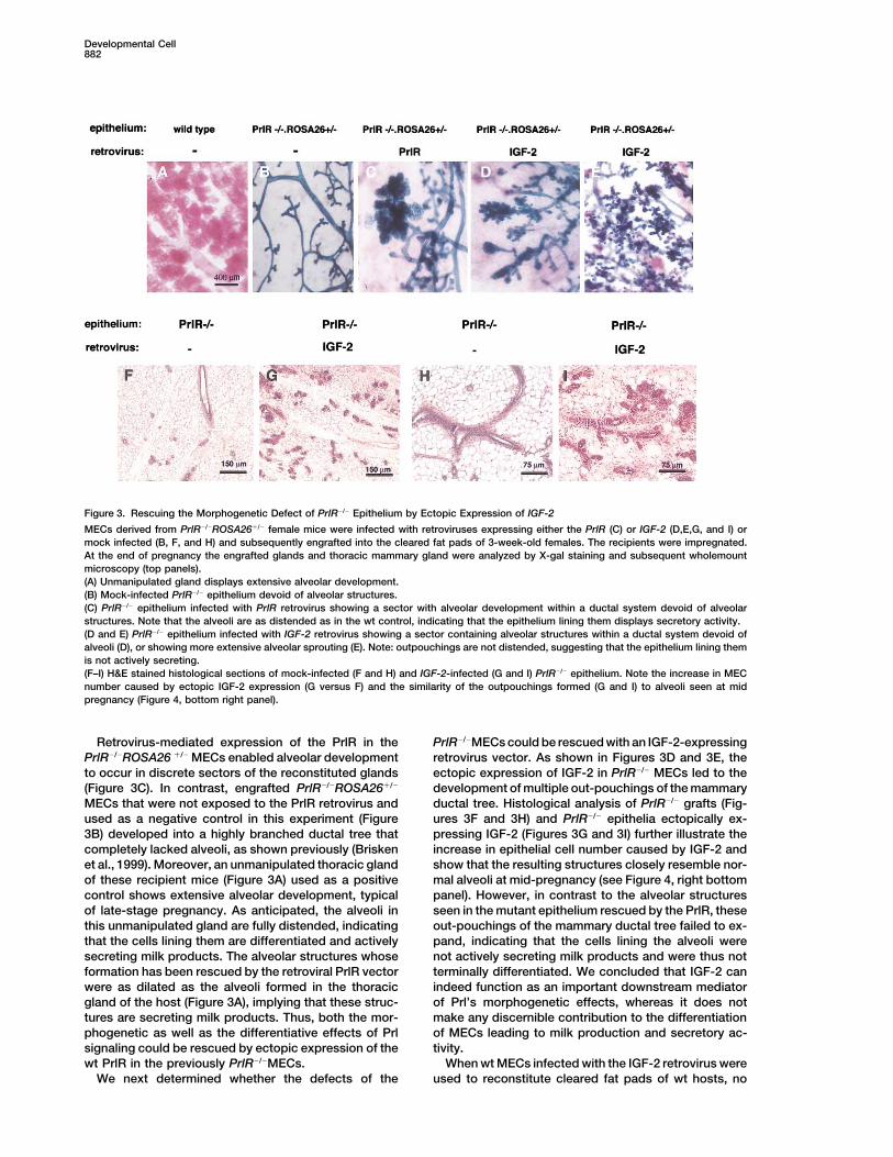
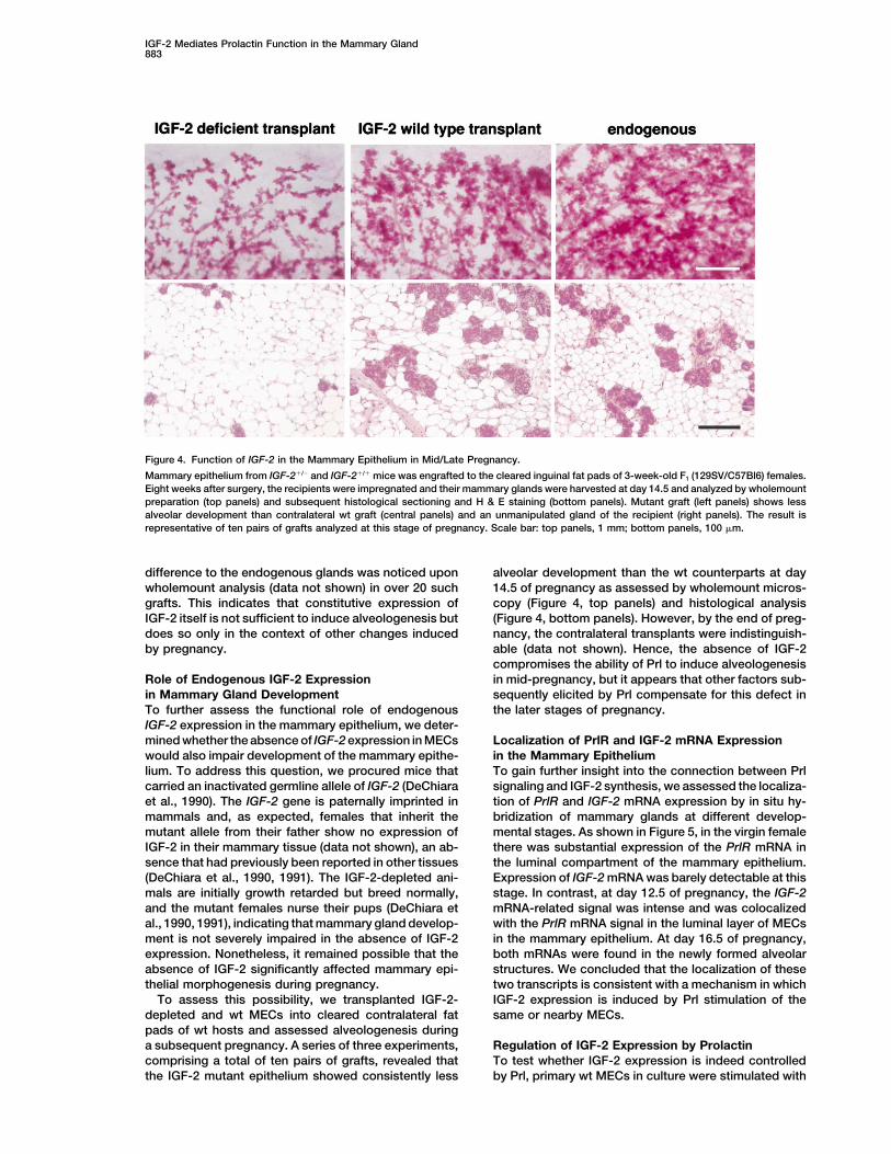
Figure 4. Function of IGF-2 in the Mammary Epithelium in Mid/Late Pregnancy.
Mammary epithelium from IGF-2�/� and IGF-2�/� mice was engrafted to the cleared inguinal fat pads of 3-week-old F1 (129SV/C57Bl6) females.Eight weeks after surgery, the recipients were impregnated and their mammary glands were harvested at day 14.5 and analyzed by wholemountpreparation (top panels) and subsequent histological sectioning and H & E staining (bottom panels). Mutant graft (left panels) shows lessalveolar development than contralateral wt graft (central panels) and an unmanipulated gland of the recipient (right panels). The result isrepresentative of ten pairs of grafts analyzed at this stage of pregnancy. Scale bar: top panels, 1 mm; bottom panels, 100 �m.
difference to the endogenous glands was noticed upon alveolar development than the wt counterparts at day14.5 of pregnancy as assessed by wholemount micros-wholemount analysis (data not shown) in over 20 such
grafts. This indicates that constitutive expression of copy (Figure 4, top panels) and histological analysis(Figure 4, bottom panels). However, by the end of preg-IGF-2 itself is not sufficient to induce alveologenesis but
does so only in the context of other changes induced nancy, the contralateral transplants were indistinguish-able (data not shown). Hence, the absence of IGF-2by pregnancy.compromises the ability of Prl to induce alveologenesisin mid-pregnancy, but it appears that other factors sub-Role of Endogenous IGF-2 Expression
in Mammary Gland Development sequently elicited by Prl compensate for this defect inthe later stages of pregnancy.To further assess the functional role of endogenous
IGF-2 expression in the mammary epithelium, we deter-mined whether the absence of IGF-2 expression in MECs Localization of PrlR and IGF-2 mRNA Expression
in the Mammary Epitheliumwould also impair development of the mammary epithe-lium. To address this question, we procured mice that To gain further insight into the connection between Prl
signaling and IGF-2 synthesis, we assessed the localiza-carried an inactivated germline allele of IGF-2 (DeChiaraet al., 1990). The IGF-2 gene is paternally imprinted in tion of PrlR and IGF-2 mRNA expression by in situ hy-
bridization of mammary glands at different develop-mammals and, as expected, females that inherit themutant allele from their father show no expression of mental stages. As shown in Figure 5, in the virgin female
there was substantial expression of the PrlR mRNA inIGF-2 in their mammary tissue (data not shown), an ab-sence that had previously been reported in other tissues the luminal compartment of the mammary epithelium.
Expression of IGF-2 mRNA was barely detectable at this(DeChiara et al., 1990, 1991). The IGF-2-depleted ani-mals are initially growth retarded but breed normally, stage. In contrast, at day 12.5 of pregnancy, the IGF-2
mRNA-related signal was intense and was colocalizedand the mutant females nurse their pups (DeChiara etal., 1990, 1991), indicating that mammary gland develop- with the PrlR mRNA signal in the luminal layer of MECs
in the mammary epithelium. At day 16.5 of pregnancy,ment is not severely impaired in the absence of IGF-2expression. Nonetheless, it remained possible that the both mRNAs were found in the newly formed alveolar
structures. We concluded that the localization of theseabsence of IGF-2 significantly affected mammary epi-thelial morphogenesis during pregnancy. two transcripts is consistent with a mechanism in which
IGF-2 expression is induced by Prl stimulation of theTo assess this possibility, we transplanted IGF-2-depleted and wt MECs into cleared contralateral fat same or nearby MECs.pads of wt hosts and assessed alveologenesis duringa subsequent pregnancy. A series of three experiments, Regulation of IGF-2 Expression by Prolactin
To test whether IGF-2 expression is indeed controlledcomprising a total of ten pairs of grafts, revealed thatthe IGF-2 mutant epithelium showed consistently less by Prl, primary wt MECs in culture were stimulated with

Developmental Cell884
Figure 5. Coexpression of PrlR and IGF-2 mRNAs in the Luminal Compartment of the Mammary Epithelium
Mammary glands were harvested from a virgin adult female mouse and from mice at days 12.5 and 16.5 of pregnancy. The glands wereprocessed for in situ hybridization; adjacent sections were hybridized with 35S-labeled antisense cRNA probes for PrlR or IGF-2 and exposedfor 6 days. H&E stained sections are shown next to the corresponding dark-field exposures. Both PrlR and IGF-2 mRNA expression localizesto the luminal epithelium. Light green areas represent the dense fibrous stroma surrounding the ducts. Scale bar: 150 �m.
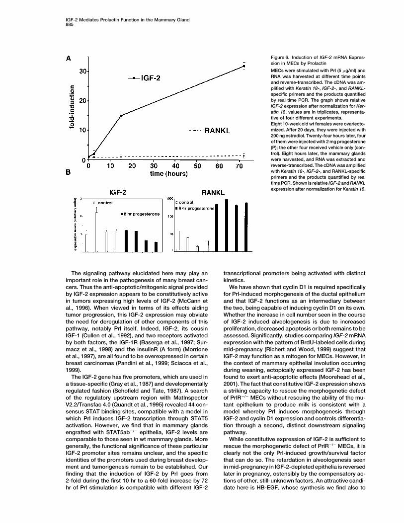
Prl, and IGF-2 mRNA expression levels were measured locally produced, secreted factor that appears to operatein a paracrine manner to encourage MEC proliferation.at various time points by real-time PCR. IGF-2 mRNA
levels increased 2-fold within 2 hr of Prl exposure, 15- These findings suggest a model in which the Prl-medi-ated induction of IGF-2 in PrlR-positive MECs providesfold at 16 hr, and were 30-fold elevated by 72 hr (Figure 6,
top). By contrast, expression of RANKL mRNA remained an amplification of the initial Prl signal by broadcastingit via paracrine signaling to nearby MECs. This may serveconstant. To test whether the induction is specific to Prl,
we analyzed IGF-2 and RANKL levels in progesterone- to coordinate proliferation among the MECs participat-ing in the formation of an alveolus and also may createstimulated mammary glands. RANKL was induced 80-
fold, whereas IGF-2 expression remained unchanged a means for sustaining the Prl stimulus over an extendedperiod of time, even when Prl levels have declined, as(Figure 6, bottom). Thus, Prl can specifically induce
IGF-2 expression in MECs through a direct mechanism often occurs because of pulsatile secretion of this hor-mone (Horseman, 2001).not involving other cell types present in the mammary
gland. In the same cells, RANKL expression is positively The closest relative of the PrlR—the growth hormonereceptor—responds to ligand by inducing IGF-1 secre-regulated by progesterone rather than Prl signaling.tion, and the latter serves to mediate some of its down-stream effects (Cohen and Rosenfeld, 1996). This cre-Discussionates a striking parallel with the presently observedbehavior of the PrlR and IGF-2. It remains to be seenAlthough the role of hormones in breast carcinogenesis
is widely recognized, in vitro studies are limited to breast whether IGF-2 mediates Prl signaling functions in vari-ous organs throughout the body or whether its role iscarcinoma cell lines (Brockman et al., 2002; Das and
Vonderhaar, 1996; Schroeder et al., 2002), and the pre- limited to the mammary gland.IGF-1, IGF-2, several IGF binding proteins and thecise mechanisms by which hormones control prolifera-
tion and differentiation of MECs in vivo remain largely IGF-1R have all been shown to be expressed in themammary gland (Wood et al., 2000). IGF-1 has beenunknown. Here, we identify IGF-2 as a downstream me-
diator of PrlR morphogenetic signaling and an upstream found to mediate the actions of growth hormone and tocooperate with estrogen to promote ductal develop-regulator of cyclin D1 expression. This links Prl signaling,
IGF-2, and cyclin D1 expression, all of which are deregu- ment (Kleinberg, 1998). In accord with such a role, IGF-1R�/� MECs transplanted to the cleared mammary fatlated during breast carcinogenesis in humans, to a com-
mon developmental signaling pathway, i.e., Prl → of a wt host shows impaired ductal outgrowth but anormal response to pregnancy hormones (Bonnette andIGF-2 → cyclin D1.
Our data have revealed that, unexpectedly, Prl does Hadsell, 2001). Hence this pathway is involved in a dis-tinct step of mammary morphogenesis that occurs be-not act directly on intracellular mitogenic regulatory
pathways but triggers instead proliferation through a fore those that are regulated by Prl and progesterone.
IGF-2 Mediates Prolactin Function in the Mammary Gland885
Figure 6. Induction of IGF-2 mRNA Expres-sion in MECs by Prolactin
MECs were stimulated with Prl (5 �g/ml) andRNA was harvested at different time pointsand reverse-transcribed. The cDNA was am-plified with Keratin 18-, IGF-2-, and RANKL-specific primers and the products quantifiedby real time PCR. The graph shows relativeIGF-2 expression after normalization for Ker-atin 18, values are in triplicates, representa-tive of four different experiments.Eight 10-week old wt females were ovariecto-mized. After 20 days, they were injected with200 ng estradiol. Twenty-four hours later, fourof them were injected with 2 mg progesterone(P); the other four received vehicle only (con-trol). Eight hours later, the mammary glandswere harvested, and RNA was extracted andreverse-transcribed. The cDNA was amplifiedwith Keratin 18-, IGF-2-, and RANKL-specificprimers and the products quantified by realtime PCR. Shown is relative IGF-2 and RANKLexpression after normalization for Keratin 18.
The signaling pathway elucidated here may play an transcriptional promoters being activated with distinctkinetics.important role in the pathogenesis of many breast can-
cers. Thus the anti-apoptotic/mitogenic signal provided We have shown that cyclin D1 is required specificallyfor Prl-induced morphogenesis of the ductal epitheliumby IGF-2 expression appears to be constitutively active
in tumors expressing high levels of IGF-2 (McCann et and that IGF-2 functions as an intermediary betweenthe two, being capable of inducing cyclin D1 on its own.al., 1996). When viewed in terms of its effects aiding
tumor progression, this IGF-2 expression may obviate Whether the increase in cell number seen in the courseof IGF-2 induced alveologenesis is due to increasedthe need for deregulation of other components of this
pathway, notably Prl itself. Indeed, IGF-2, its cousin proliferation, decreased apoptosis or both remains to beassessed. Significantly, studies comparing IGF-2 mRNAIGF-1 (Cullen et al., 1992), and two receptors activated
by both factors, the IGF-1R (Baserga et al., 1997; Sur- expression with the pattern of BrdU-labeled cells duringmid-pregnancy (Richert and Wood, 1999) suggest thatmacz et al., 1998) and the insulinR (A form) (Morrione
et al., 1997), are all found to be overexpressed in certain IGF-2 may function as a mitogen for MECs. However, inthe context of mammary epithelial involution occurringbreast carcinomas (Pandini et al., 1999; Sciacca et al.,
1999). during weaning, ectopically expressed IGF-2 has beenfound to exert anti-apoptotic effects (Moorehead et al.,The IGF-2 gene has five promoters, which are used in
a tissue-specific (Gray et al., 1987) and developmentally 2001). The fact that constitutive IGF-2 expression showsa striking capacity to rescue the morphogenetic defectregulated fashion (Schofield and Tate, 1987). A search
of the regulatory upstream region with MatInspector of PrlR�/� MECs without rescuing the ability of the mu-tant epithelium to produce milk is consistent with aV2.2/Transfac 4.0 (Quandt et al., 1995) revealed 44 con-
sensus STAT binding sites, compatible with a model in model whereby Prl induces morphogenesis throughIGF-2 and cyclin D1 expression and controls differentia-which Prl induces IGF-2 transcription through STAT5
activation. However, we find that in mammary glands tion through a second, distinct downstream signalingpathway.engrafted with STAT5ab�/� epithelia, IGF-2 levels are
comparable to those seen in wt mammary glands. More While constitutive expression of IGF-2 is sufficient torescue the morphogenetic defect of PrlR�/� MECs, it isgenerally, the functional significance of these particular
IGF-2 promoter sites remains unclear, and the specific clearly not the only Prl-induced growth/survival factorthat can do so. The retardation in alveologenesis seenidentities of the promoters used during breast develop-
ment and tumorigenesis remain to be established. Our in mid-pregnancy in IGF-2-depleted epithelia is reversedlater in pregnancy, ostensibly by the compensatory ac-finding that the induction of IGF-2 by Prl goes from
2-fold during the first 10 hr to a 60-fold increase by 72 tions of other, still-unknown factors. An attractive candi-date here is HB-EGF, whose synthesis we find also tohr of Prl stimulation is compatible with different IGF-2

Developmental Cell886
IGF-2 F, GTGCGGAGGGGAGCTTGTTGAC; IGF-2 R, GTGGbe dependent on PrlR action, albeit to a lesser extent.GCGTCTTTGGGTGGTA; RANKL F TGTACTTTCGAGCGCAGATG;The FGF signaling pathway has also been implicated toRANKL R, CCCACAATGTGTTGCAGTTC.play an important role in alveologenesis (Jackson et al.,
1997), and there is evidence that the Wnt pathway actingIn Situ Hybridizationvia cyclin D1 (Hsu et al., 2001; Imbert et al., 2001) may beIn situ hybridization was as described (Das et al., 1994). Frozen
involved. It is conceivable that certain genes encoding sections were hybridized with 35S-labeled antisense or sense cRNAmolecules linked to these regulatory pathways were probes for PrlR or IGF-2 for 4 hr at 45�C and exposed for 6 days.missed in our screen, which surveyed an arbitrary subsetof 6500 mouse genes. Acknowledgments
Recently, RANK signaling, which is important to boneWe thank G. Smith for stimulating discussions, A. Efstratiadis andremodeling and calcium homeostasis, has been linkedP. Scinski for providing the IGF-2 and cyclin D1 mutant mice, M.via IKK� to cyclin D1 expression in the mammary glandSocolovsky for the PrlR virus, and E. Reichmann for the � casein(Cao et al., 2001). Previously, RANKL, the ligand of theantibody. This work was supported by grants from Affymetrix, Inc.;
RANK receptor, was implicated as an important stimula- Millennium Pharmaceuticals, Inc.; and Bristol-Myers Squibb Com-tor of mammary gland development including alveolo- pany, Bristol-Myers Squibb Foundation. This work was also sup-
ported by grants from the NIH (NIH PPG 5 P01 CA80111) and HDgenesis (Fata et al., 2000). Indeed, the RANK �/� mam-12304 and HD 33994 (to S.K.D.). S.K.D. is a recipient of an NICHDmary glands show a dilated ductal system and cysticMERIT award.pouches (Fata et al., 2000), suggesting that the RANK/
RANKL signaling pathway is important for a step orReferencessteps in mammary gland development that are distinct
from those initiated by Prl signaling. This separation ofBaserga, R., Resnicoff, M., and Dews, M. (1997). The IGF-I receptor
function is now directly indicated by the present findings and cancer. Endocrine 7, 99–102.that mammary glands engrafted with PrlR�/� or cyclin
Binart, N., Helloco, C., Ormandy, C.J., Barra, J., Clement-Lacroix,D1�/� MECs show the same levels of RANKL expression P., Baran, N., and Kelly, P.A. (2000). Rescue of preimplantatoryand that Prl signaling specifically induces IGF-2 expres- egg development and embryo implantation in prolactin receptor-sion in MECs while having no effects on RANKL. RANKL deficient mice after progesterone administration. Endocrinology
141, 2691–2697.expression is linked by our work directly to progesteroneaction. Just as importantly, while IGF-2 treatment of Bole-Feysot, C., Goffin, V., Edery, M., Binart, N., and Kelly, P.A.
(1998). Prolactin (PRL) and its receptor: actions, signal transductionMECs is sufficient to directly induce cyclin D1 expres-pathways and phenotypes observed in PRL receptor knockout mice.sion, RANKL exposure has no such effect. We thereforeEndocr. Rev. 19, 225–268.conclude that RANKL is an important localized mediatorBonnette, S.G., and Hadsell, D.L. (2001). Targeted disruption of theof progesterone action and the morphogenetic stepsIGF-I receptor gene decreases cellular proliferation in mammaryexecuted by progesterone and RANKL are essential pre-terminal end buds. Endocrinology 142, 4937–4945.
cursors to the subsequent morphogenetic steps thatBrisken, C., Heineman, A., Chavarria, T., Elenbaas, B., Tan, J., Dey,
are triggered by Prl. S., McMahon, J., McMahon, A., and Weinberg, R. (2000). Essentialfunction of Wnt-4 in mammary gland development downstream of
Experimental Procedures progesterone signaling. Genes Dev. 14, 650–654.
Brisken, C., Kaur, S., Chavarria, T., Binart, N., Sutherland, R., Wein-Mice
berg, R., Kelly, P., and Ormandy, C. (1999). Prolactin controls mam-Mice were of C57Bl6x129SV background and genotyped as de-
mary gland development via direct and indirect mechanisms. Dev.scribed (Brisken et al., 1998; DeChiara et al., 1990; Ormandy et al.,
Biol. 210, 96–106.1997; Sicinski et al., 1995). For stimulation with progesterone, 10-
Brisken, C., Park, S., Vass, T., Lydon, J., O’Malley, B., and Weinberg,week-old female mice were ovariectomized. Twenty days later, theyR. (1998). A paracrine role for the epithelial progesterone receptorwere injected s.c. for 10 days either with 1 �g of 17-�-estradiol orin mammary gland development. Proc. Natl. Acad. Sci. USA 95,1 mg of progesterone in sesame oil per day (Lydon et al., 1995).5076–5081.Mammary reconstitutions, wholemounting, X-gal-, and immuno-
staining were as described (Brisken et al., 1998, 2000). Brockman, J.L., Schroeder, M.D., and Schuler, L.A. (2002). PRL acti-vates the cyclin d1 promoter via the jak2/stat pathway. Mol. Endocri-nol. 16, 774–784.Cell Culture
Primary MECs were prepared from 10-week-old virgin female mice Cao, Y., Bonizzi, G., Seagroves, T.N., Greten, F.R., Johnson, R.,as described (Kittrell et al., 1992). For retroviral infection, 293T cells Schmidt, E.V., and Karin, M. (2001). IKKalpha provides an essentialwere transiently transfected using Fugene (Roche) with plasmid link between RANK signaling and cyclin D1 expression during mam-DNAs. Conditioned medium was removed after 24 and 48 hr and mary gland development. Cell 107, 763–775.frozen and placed on MECs at day 3 of culture (Edwards, 1996).
Clevenger, C., Chang, W., Ngo, W., Pasha, T., Montone, K., andTomaszewski, J. (1995). Expression of prolactin and prolactin recep-
Retroviral Construct tor in human breast carcinoma. Evidence for an autocrine/paracrineIGF-2 was amplified from mammary gland polyA RNA using the loop. Am. J. Pathol. 146, 695–705.following primers: F GAATTCAAGATGGGGATCCCACATC, R GAA
Cohen, P., and Rosenfeld, R.G. (1996). Growth regulation. In Text-TTCCTGATGGTTGCTGGACATC. The amplified cDNA was clonedbook of Endocrine Physiology, J.E. Griffin and S.R. Ojeda, eds. (Newinto pCR-Blunt II-TOPO (Invitrogen), sequenced and subcloned intoYork: Oxford University Press), 244–259.MSCV2.2 (Hawley et al., 1994).Cullen, K., Allison, A., Martire, I., Ellis, M., and Singer, C. (1992).Insulin-like growth factor expression in breast cancer epitheliumQuantitative Real-Time PCR (rtPCR)and stroma. Breast Cancer Res. Treat. 22, 21–29.Quantitative rtPCR analysis was performed using the iCycler appa-
ratus (Bio-Rad) and SYBR Green PCR Core Reagents system (Per- Das, R., and Vonderhaar, B. (1996). Activation of raf-1, MEK, andMAP kinase in prolactin responsive mammary cells. Breast Cancerkin-Elmer Applied Biosystems). Results were evaluated with
ICYCLER IQ REAL TIME DETECTION SYSTEM SOFTWARE (Bio-Rad). Res. Treat. 40, 141–149.

IGF-2 Mediates Prolactin Function in the Mammary Gland887
Das, S.K., Wang, X.N., Paria, B.C., Damm, D., Abraham, J.A., Klags- Montgomery, C.A., Jr., Shyamala, G., Conneely, O.M., and O’Malley,B.W. (1995). Mice lacking progesterone receptor exhibit pleiotropicbrun, M., Andrews, G.K., and Dey, S.K. (1994). Heparin-binding EGF-
like growth factor gene is induced in the mouse uterus temporally reproductive abnormalities. Genes Dev. 9, 2266–2278.by the blastocyst solely at the site of its apposition: a possible McCann, A., Miller, N., O’Meara, A., Pedersen, I., Keogh, K., Gorey,ligand for interaction with blastocyst EGF-receptor in implantation. T., and Dervan, P. (1996). Biallelic expression of the IGF2 gene inDevelopment 120, 1071–1083. human breast disease. Hum. Mol. Genet. 5, 1123–1127.DeChiara, T.M., Efstratiadis, A., and Robertson, E.J. (1990). A Moorehead, R.A., Fata, J.E., Johnson, M.B., and Khokha, R. (2001).growth-deficiency phenotype in heterozygous mice carrying an in- Inhibition of mammary epithelial apoptosis and sustained phosphor-sulin-like growth factor II gene disrupted by targeting. Nature 345, ylation of Akt/PKB in MMTV-IGF-II transgenic mice. Cell Death Differ.78–80. 8, 16–29.DeChiara, T.M., Robertson, E.J., and Efstratiadis, A. (1991). Parental Morrione, A., Valentinis, B., Xu, S., Yumet, G., Louvi, A., Efstratiadis,imprinting of the mouse insulin-like growth factor II gene. Cell 64, A., and Baserga, R. (1997). Insulin-like growth factor II stimulates849–859. cell proliferation through the insulin receptor. Proc. Natl. Acad. Sci.
USA 94, 3777–3782.Edwards, P. (1996). Tissue reconstitution, or transgenic mammarygland, technique for modeling breast cancer development. Cancer Nandi, S. (1958). Endocrine control of mammary-gland developmentTreat. Res. 83, 23–36. in the C3H/He Crgl mouse. J. Natl. Cancer Inst. 21, 1039–1063.Edwards, P.A., Abram, C.L., and Bradbury, J.M. (1996). Genetic Ormandy, C.J., Camus, A., Barra, J., Damotte, D., Lucas, B., Buteau,manipulation of mammary epithelium by transplantation. J. Mam- H., Edery, M., Brousse, N., Babinet, C., Binart, N., and Kelly, P.A.mary Gland Biol. Neoplasia 1, 75–89. (1997). Null mutation of the prolactin receptor gene produces multi-
ple reproductive defects in the mouse. Genes Dev. 11, 167–178.Fantl, V., Edwards, P., Steel, J., Vonderhaar, B., and Dickson, C.(1999). Impaired mammary gland development in Cyl-1(�/�) mice Pandini, G., Vigneri, R., Costantino, A., Frasca, F., Ippolito, A., Fujita-during pregnancy and lactation is epithelial cell autonomous. Dev. Yamaguchi, Y., Siddle, K., Goldfine, I., and Belfiore, A. (1999). InsulinBiol. 212, 1–11. and insulin-like growth factor-I (IGF-I) receptor overexpression in
breast cancers leads to insulin/IGF-I hybrid receptor overexpres-Fantl, V., Stamp, G., Andrews, A., Rosewell, I., and Dickson, C.(1995). Mice lacking cyclin D1 are small and show defects in eye sion: evidence for a second mechanism of IGF-I signaling. Clin.
Cancer Res. 5, 1935–1944.and mammary gland development. Genes Dev. 9, 2364–2372.
Fata, J.E., Kong, Y.Y., Li, J., Sasaki, T., Irie-Sasaki, J., Moorehead, Quandt, K., Frech, K., Karas, H., Wingender, E., and Werner, T.(1995). MatInd and MatInspector: new fast and versatile tools forR.A., Elliott, R., Scully, S., Voura, E.B., Lacey, D.L., et al. (2000). The
osteoclast differentiation factor osteoprotegerin-ligand is essential detection of consensus matches in nucleotide sequence data. Nu-cleic Acids Res. 23, 4878–4884.for mammary gland development. Cell 103, 41–50.
Franchimont, P., Dourcy, C., Legros, J.J., Reuter, A., Vrindts-Gev- Reynolds, C., Montone, K., Powell, C., Tomaszewski, J., and Cle-venger, C. (1997). Expression of prolactin and its receptor in humanaert, Y., Van Cauwenberge, J.R., and Gaspard, U. (1976). Prolactin
levels during the menstrual cycle. Clin. Endocrinol. 5, 643–650. breast carcinoma. Endocrinology 138, 5555–5560.
Richert, M., and Wood, T. (1999). The insulin-like growth factorsFriedrich, G., and Soriano, P. (1991). Promoter traps in embryonicstem cells: a genetic screen to identify and mutate developmental (IGF) and IGF type I receptor during postnatal growth of the murine
mammary gland: sites of messenger ribonucleic acid expressiongenes in mice. Genes Dev. 5, 1513–1523.and potential functions. Endocrinology 140, 454–461.Fuh, G., and Wells, J. (1995). Prolactin receptor antagonists that
inhibit the growth of breast cancer cell lines. J. Biol. Chem. 270, Schofield, P.N., and Tate, V.E. (1987). Regulation of human IGF-IItranscription in fetal and adult tissues. Development 101, 793–803.13133–13137.
Ginsburg, E., and Vonderhaar, B. (1995). Prolactin synthesis and Schroeder, M.D., Symowicz, J., and Schuler, L.A. (2002). PRL modu-lates cell cycle regulators in mammary tumor epithelial cells. Mol.secretion by human breast cancer cells. Cancer Res. 55, 2591–2595.Endocrinol. 16, 45–57.Gray, A., Tam, A.W., Dull, T.J., Hayflick, J., Pintar, J., Cavenee, W.K.,
Koufos, A., and Ullrich, A. (1987). Tissue-specific and develop- Sciacca, L., Costantino, A., Pandini, G., Mineo, R., Frasca, F., Scalia,P., Sbraccia, P., Goldfine, I., Vigneri, R., and Belfiore, A. (1999).mentally regulated transcription of the insulin-like growth factor 2
gene. DNA 6, 283–295. Insulin receptor activation by IGF-II in breast cancers: evidence fora new autocrine/paracrine mechanism. Oncogene 18, 2471–2479.Hawley, R., Lieu, F., Fong, A., and Hawley, T. (1994). Versatile retrovi-
ral vectors for potential use in gene therapy. Gene Ther. 1, 136–138. Sicinski, P., Donaher, J., Parker, S., Li, T., Fazeli, A., Gardner, H.,Haslam, S., Bronson, R., Elledge, S., and Weinberg, R. (1995). CyclinHorseman, N.D. (2001). Prolactin. In Endocrinology, L.J.a.J.J.L. De-D1 provides a link between development and oncogenesis in theGroot, ed. (Philadelphia: W.B. Saunders), 209–220.retina and breast. Cell 82, 621–630.Hsu, W., Shakya, R., and Costantini, F. (2001). Impaired mammarySurmacz, E., Guvakova, M., Nolan, M., Nicosia, R., and Sciacca, L.gland and lymphoid development caused by inducible expression(1998). Type I insulin-like growth factor receptor function in breastof Axin in transgenic mice. J. Cell Biol. 155, 1055–1064.cancer. Breast Cancer Res. Treat. 47, 255–267.Huseby, R.A., Soares, M.J., and Talamantes, F. (1985). Ectopic pitu-Vomachka, A.J., Pratt, S.L., Lockefeer, J.A., and Horseman, N.D.itary grafts in mice: hormone levels, effects on fertility, and the(2000). Prolactin gene-disruption arrests mammary gland develop-development of adenomyosis uteri, prolactinomas, and mammaryment and retards T-antigen-induced tumor growth. Oncogene 19,carcinomas. Endocrinology 116, 1440–1448.1077–1084.Imbert, A., Eelkema, R., Jordan, S., Feiner, H., and Cowin, P. (2001).Wood, T.L., Richert, M.M., Stull, M.A., and Allar, M.A. (2000). TheDelta N89 beta-catenin induces precocious development, differenti-insulin-like growth factors (IGFs) and IGF binding proteins in postna-ation, and neoplasia in mammary gland. J. Cell Biol. 153, 555–568.tal development of murine mammary glands. J. Mammary GlandJackson, D., Bresnick, J., Rosewell, I., Crafton, T., Poulsom, R.,Biol. Neoplasia 5, 31–42.Stamp, G., and Dickson, C. (1997). Fibroblast growth factor receptor
signalling has a role in lobuloalveolar development of the mammarygland. J. Cell Sci. 110, 1261–1268.
Kittrell, F.S., Oborn, C.J., and Medina, D. (1992). Development ofmammary preneoplasias in vivo from mouse mammary epithelialcell lines in vitro. Cancer Res. 52, 1924–1932.
Kleinberg, D. (1998). Role of IGF-I in normal mammary development.Breast Cancer Res. Treat. 47, 201–208.
Lydon, J.P., DeMayo, F.J., Funk, C.R., Mani, S.K., Hughes, A.R.,