Adenosine: A mediator of the sleep-inducing effects of prolonged wakefulness
Upload
independentCategory
view
3download
0
Tollip Is a Mediator of Protein SumoylationAlessia Ciarrocchi1.¤a, Romina D’Angelo1., Chiara Cordiglieri1¤b, Ada Rispoli1, Spartaco Santi2, Massimo
Riccio2¤c, Simona Carone1, Anna Laura Mancia1¤d, Simone Paci1, Elena Cipollini1, Davide Ambrosetti1,
Marialuisa Melli1*
1 Department of Biology, Bologna University, Bologna, Italy, 2 Institute of Organ Transplantation and Immunocitology-ITOI, C.N.R, Bologna, Italy
Abstract
Tollip is an interactor of the interleukin-1 receptor involved in its activation. The endosomal turnover of ubiquitylated IL-1RIis also controlled by Tollip. Furthermore, together with Tom1, Tollip has a general role in endosomal protein traffic. Thiswork shows that Tollip is involved in the sumoylation process. Using the yeast two-hybrid technique, we have isolated newTollip partners including two sumoylation enzymes, SUMO-1 and the transcriptional repressor Daxx. The interactions wereconfirmed by GST-pull down experiments and immunoprecipitation of the co-expressed recombinants. More specifically,we show that the TIR domain of the cytoplasmic region of IL-1RI is a sumoylation target of Tollip. The sumoylated andunsumoylated RanGAP-1 protein also interacts with Tollip, suggesting a possible role in RanGAP-1 modification and nuclear-cytoplasmic protein translocation. In fact, Tollip is found in the nuclear bodies of SAOS-2/IL-1RI cells where it colocalizeswith SUMO-1 and the Daxx repressor. We conclude that Tollip is involved in the control of both nuclear and cytoplasmicprotein traffic, through two different and often contrasting processes: ubiquitylation and sumoylation.
Citation: Ciarrocchi A, D’Angelo R, Cordiglieri C, Rispoli A, Santi S, et al. (2009) Tollip Is a Mediator of Protein Sumoylation. PLoS ONE 4(2): e4404. doi:10.1371/journal.pone.0004404
Editor: Shukti Chakravarti, Johns Hopkins University, United States of America
Received October 2, 2008; Accepted December 14, 2008; Published February 9, 2009
Copyright: � 2009 Ciarrocchi et al. This is an open-access article distributed under the terms of the Creative Commons Attribution License, which permitsunrestricted use, distribution, and reproduction in any medium, provided the original author and source are credited.
Funding: This work was supported by Telethon grant GGP030248, partly by a grant from the Italian Ministry of Technology and Scientific Research and by a grantfrom FIRB Neuroscienze RBNE01WY7P. The funders had no role in study design, data collection and analysis, decision to publish, or preparation of the manuscript.
Competing Interests: The authors have declared that no competing interests exist.
* E-mail: [email protected]
¤a Current address: Molecular Biology laboratory, Arcispedale Santa Maria Nuova, Reggio Emilia, Italy¤b Current address: Neuroimmunology Laboratory-U.O. Neurologia IV, Istituto Neurologico Carlo Besta, Milano, Italy¤c Current address: Department of Anatomy and Histology, University of Modena and Reggio Emilia, Modena, Italy¤d Current address: Marine Biomedicine and Environmental Science Center, Medical University of South Carolina, Charleston, South Carolina, United States ofAmerica
. These authors contributed equally to this work.
Introduction
Tollip is an interactor of the Toll family of proteins, which are
highly conserved in evolution from Drosophila to Man, and include
the Toll-like and interleukin-1 (IL-1) receptors, all involved in the
inflammatory response. The sequence of Tollip, spanning 274
amino acids, contains some characterized functional domains. A
C2-like domain, lacking the two conserved Asp residues critical for
Ca2+ binding and present in numerous eukaryotic proteins as a
binding site for a variety of phospholipids [1]. A C-terminus
ubiquitin binding CUE domain identified by Shih et al. [2] as a
ubiquitin binding site that mediates intramolecular monoubiqui-
tylation. At the amino-terminus (aa 1–54), a GAT/TOM1
interacting domain is also described [3]. So far, Tollip has been
involved in two main functions. The first, proposed by Burns and
collaborators [1], indicates Tollip as an interactor of the IL-1
receptor TIR domain, mediating the binding of the serine/
threonine kinase IRAK-1 to the activated receptor complex, thus
being integral part of the IL-1RI signaling cascade. Later,
Yamakami & Yokosawa [4] proposed a negative regulatory role
of Tollip on the IL-1b and TNF-a signaling pathways, in
agreement with the inhibition of NF-kB activation observed
following Tollip overexpression [1,5]. The second function,
described by Yamakami et al. [3], concerns the interaction of
Tollip with Tom1, Ubiquitin and Clathrin in a high molecular
mass complex involved in protein sorting. In agreement with this
proposal, an endosomal function of the protein was suggested by
Katoh et al., [6,7]. In addition, Brissoni et al. [8] have recently
shown that Tollip is required in the sorting of the IL-1RI at late
endosomes, further clarifying its involvement in the IL-1
inflammatory pathway. Zhang and Ghosh [5] have shown that
Tollip associates not only with IL-1RI but also with the TLR2 and
TLR4 receptors when activated by LPS stimulation. Also this
interaction results in the suppression of TLR-mediated cellular
responses through the inhibition of phosphorylation and kinase
activity of IRAK1.
We show, for the first time, the involvement of Tollip in the
sumoylation process. Sumoylation is a post-translational modifi-
cation of proteins that conjugate SUMO-1 with a lysine of the
target peptide through the formation of a covalent bond. This
reaction is very similar to the ubiquitylation process and it is
catalyzed by three enzymes, that act in succession: 1) El, the
dimeric activation complex, composed of the proteins Aosl and
Uba2; 2) the single E2 conjugation enzyme named Ubc9; 3) at
least three types of E3 ligases. The first group of E3 ligases is
encoded by the protein inhibitor of activated signal transducer and
activator of transcription (PIAS) family and entails the 5
mammalian members: PIAS1, PIAS3, PIASxa/ARIP3 , PIASxb,
and PIASy. These ligases are characterized by the presence of a
RING-finger-like structure essential to their function. The second
PLoS ONE | www.plosone.org 1 February 2009 | Volume 4 | Issue 2 | e4404

group is represented by the RanBP2/Nup358 protein, which is a
component of the nucleopore complex and does not contain the
ring finger domain [9]. The third type is Pc2, a member of the
polycomb group of proteins (PcG) involved in gene silencing. Pc2
recruits the CtBP repressor to the PcG nuclear bodies and
enhances its sumoylation [10]. These ligases are not protein
specific and show overlapping targets. In fact, it is not clear how
the specificity of the sumoylation reaction is achieved. It seems
likely that more ligases or cofactors exist and contribute to the
specificity of the modification process.
In order to further characterize the function of Tollip, we have
screened a cDNA library by the yeast two-hybrid system, looking
for Tollip partners. In addition to the known interactors, TOM1,
Tollip itself and ubiquitin, we have isolated Daxx, SUMO-1, and
the E2 (Ubc9) and E3 (ARIP3) sumoylation enzymes. This work
shows that Tollip binds Ubc9, SUMO-1 and at least three ligases
of the Pias family and is itself sumoylated. The binding domain of
Tollip to the ligases of the PIAS family and of the ARIP3 ligase to
Tollip are identified. In particular, in a SAOS-2 clone enriched in
IL-1RI receptors (SAOS-2/IL1R), Tollip translocates into the
nucleus where it co-localizes with SUMO-1 and Daxx, in nuclear
bodies. The SAOS-2/IL1R clone was derived from the SAOS-2
osteoblastoma cell line which constitutively expresses IL-1RI
[11,12] and was kindly provided by Dr Sandra Marmiroli
(Modena and Reggio Emilia University). Our results may correlate
with the interaction of Tollip with the RanGAP-1 protein.
Furthermore, we identify the cytoplasmic domain of IL-1RI as a
target of Tollip sumoylation. We propose that Tollip is involved in
the nuclear translocation of proteins either as a sumoylation
cofactor or a ligase.
Results
Tollip interacting proteinsThe yeast two-hybrid mating assay was carried out as described
in material and methods. High stringency screening of a cDNA
library from rat brain hemispheres, allowed the isolation of 310
positive recombinant clones that were analyzed by PCR
amplification. 213 cDNA recombinants showed a single band of
amplification that was further characterized. Table 1 lists some of
the isolated cDNAs (column 1) with the corresponding aminoacid
regions (column 2) and the number of independent colonies for
each isolated recombinant clone (column 3). The majority of
cDNA recombinants were coding for ubiquitin or ubiquitin fused
to ribosomal RNA subunits. This is in agreement with the
presence of a ubiquitin binding CUE domain at the C-terminus of
Tollip and with the described cytoplasmic role of ubiquitinated
Tollip [8,2,3]. The already described binding of Tollip to TOM1
and its homodimerization are consistent with the published results
[3,5]. Interestingly, proteins involved in the sumoylation process
(SUMO-1, Ubc9, ARIP3) and the transcriptional repressor Daxx
are among the interactors that we have isolated. The interactions
of Tollip with some of the identified proteins were confirmed by
GST-pull down experiments. The B42 fused 35S-peptides were
incubated with GST-Tollip or GST beads and the interaction was
analyzed by SDS-PAGE and authoradiography (Fig. 1A). All the
peptides analyzed interact with Tollip but not with the GST
control. B42 alone does not interact with Tollip, showing the
specificity of the reaction. The cytoplasmic region of the IL-1RI
accessory protein (IL-1RAcP) and IL-1RI were used as positive
control. To confirm the interactions observed in yeast, 293T cells
were transfected with the pRK7-HA-Tollip cDNA together with
constructs coding for Ubc9 (Fig. 1B ) or the Flag-ARIP3 fusion
proteins (Fig. 1D). 293T cells expressing the unrelated protein HA-
Cystatin B, together with Ubc9 (Fig. 1C) and Flag-ARIP3 (Fig. 1E)
were used as control. The immunoprecipitation with anti-HA
antibodies (abs) shows that Ubc9 (B) and Flag-ARIP3 (D) co-
immunoprecipitate with HA-Tollip but not with the control
protein (C and E), confirming the specificity of the binding.
Tollip interaction with the PIAS family of proteinsThe site of interaction of HA-Tollip with ARIP3 is analyzed in
Fig. 2. The carboxyl- and amino-terminus deletion mutants
schematized in panel A were used to identify the Tollip domain(s)
interacting with ARIP3. Protein extracts from 293T cells
transfected with wild type or mutant HA-Tollip and Flag-ARIP3
were immunoprecipitated with anti-HA abs and analyzed by
western blot. Panel B shows the SDS-PAGE of the immunopre-
Table 1.
cDNA Isolated protein fragment Nu of independent colonies
Ubiquitin conjugating enzyme 9 (Ubc9) 1–159 aa (full length) 45
Death domain interacting protein (Daxx) 643–739 5
Target of Myb-1 homologue (TOM1) 3–492 5
Androgen receptor interacting protein 3 (ARIP3) 64–572 3
448–572 2
Small ubiquitin-like modifier 1 (SUMO-1) 1–101 aa (full length) 1
Tollip 141–274 1
Leucin Rich Protein 671–832 1
Brain expressed X-linked 2 (Bex1) 33–129 1
Receptor for Activated C Kinase (RACK 1) 241–317 1
ING 1 tumor suppressor 248–276 1
Ubiquitin family
Ubiquitin 1–76 (full length) 71
Ubiquitin/A52 fusion protein 72–105 7
Ubiquitin/S27 fusion protein 2–157 4
doi:10.1371/journal.pone.0004404.t001
Tollip and Sumoylation
PLoS ONE | www.plosone.org 2 February 2009 | Volume 4 | Issue 2 | e4404

cipitated samples (lanes 2) next to the respective protein extracts
(lanes 1) stained with anti-Flag abs. All Tollip mutants interact
with ARIP3 with the exception of D6 where the entire N-terminus
and C2 domain is removed. On the other hand, the D7 construct
spanning aa 54–179 (C2 domain) and D5 spanning aa 134–274
(C-terminus of the C2 plus the CUE regions), bind ARIP3. Thus,
the 134–179 peptide of the C2 region is sufficient for the
interaction and contains the ARIP3 binding site. The staining with
anti-HA abs (panel C) shows that all mutants are efficiently
expressed (lanes 1) and immunoprecipitated (lanes 2).
We have also analyzed the Tollip interacting sites of ARIP3
(Panels D and E). ARIP3 entails 572 amino acid with three
functionally characterized domains [13,14]: 1) The SAP (SAF-A/
B, Acinus PIAS) domain from aa 11–45 that may be a DNA
binding site; 2) The RING domain from aa 347–388, responsible
for the ligase function; 3) The androgen receptor binding site
spanning aa 443–548. We have generated the three carboxyl-
terminus deletion mutants shown in panel D. The wild type and
mutant pCDNA3.1-Flag-ARIP3 constructs were co-transfected
with pRK7-HA-Tollip cDNA. The cell extracts were immuno-
precipitated with anti-HA abs and analyzed by western blot as
shown in panel E. Lane 1 shows the protein extract and lane 2 the
immunoprecipitated samples. The Flag-ARIP3-Da and Db
mutants, deleted of the androgen receptor and ring domain
respectively, co-immunoprecipitate with HA-Tollip while the Dc
mutant, containing aa 1–169, is not detectable on the gel. This
shows that HA-Tollip interacts with a region of ARIP3 spanning
residues 169–347. This domain is highly conserved in the PIAS
family of proteins. Thus, we investigated the possible interaction of
Tollip with other members of the family. As expected, HA-Tollip
interacts with both PIAS1 and PIASxb (Fig. 2F and G).
Immunoprecipitation of Flag-PIAS1 and Flag-PIASxb with HA-
Tollip deletion mutants D5 and D6 shows that the carboxyl-
terminus region of the C2 domain represents the interaction site
with the E3 ligases of the PIAS family that we have examined.
Tollip triggers protein sumoylation and is itselfsumoylated
The interaction of Tollip with the sumoylation enzymes suggests
that Tollip is sumoylated and perhaps has a role in the
sumoylation process itself. The analysis of the rat amino acid
sequence (accession BC133067.1) indicates at least 3 putative
sumoylation sites within the C2 domain (the lysines at position 96,
143, and 162). However, the transfection of 293T cells with HA-
Tollip together with Flag-SUMO-1, Ubc9, and Flag-ARIP3 shows
that, under the conditions tested, Tollip migrates as a monomer at
the expected molecular weight of 29 kDa (Fig. 3A). No additional
slow migrating bands can be detected. This set of experiments
suggests that Tollip is not covalently modified. However, the
presence of ubiquitin-like protease activity (Ulp) in native cell
lysates combined with the short half-life of the SUMO-1
modification [15,16] may destabilize the process. Thus, we have
carried out an additional experiment were 293T cells were co-
transfected with HA-Tollip and SUMO-1 and the cells were lysed
under denaturing conditions, to block protease activity and
exclude non covalent association with interacting proteins. The
Figure 1. Tollip interactions. A) GST-Tollip interaction with 35S-methionine labelled proteins isolated by the yeast two hybrid technique. Lane 1contains 1/10 input of the 35S-methionine protein used for each interaction; lanes 2 and 3 contain the elution product from incubation of the 35S-proteins with the GST-Tollip and GST-protein alone, respectively. 35S-methionine labelled proteins were visualized by autoradiography. Western blotanalysis of 293T cells transfected with Ubc9 and HA-Tollip (B), Flag-ARIP3 and HA-Tollip (D), HA-Cystatin B and Ubc9 (C), HA-Cystatin B and Flag-ARIP3(E). HA-Cystatin B is a ubiquitous protein with antiprotease function, unrelated to Tollip, nor to the inflammatory pathway [28]. Lane 1 contains theprotein extract; lane 2 contains the proteins immunoprecipitated with anti-HA abs. In this and in the following figures, ‘‘pe.’’ refers to the proteinextract and ‘‘ip.’’ to the immunoprecipitated protein. Staining carried out as indicated under the figures.doi:10.1371/journal.pone.0004404.g001
Tollip and Sumoylation
PLoS ONE | www.plosone.org 3 February 2009 | Volume 4 | Issue 2 | e4404

control cells were transfected with SUMO-1 only. The staining of
the protein extracts with anti-SUMO-1 abs generates a similar
pattern of bands in the two samples (Fig. 3B, lanes 1 and 2). The
immunoprecipitation of the same protein extracts with anti-HA
abs is shown in panel B lanes 3–6. Both SUMO-1 and anti-HA abs
generate bands migrating in corresponding positions of the gel, at
a molecular weight higher than monomeric Tollip (compare lanes
3 and 5). This result confirms that Tollip is sumoylated, possibly in
more than one lysine residue. It should be noticed, however, that
Tollip may be dimeric as well as ubiquitinated [2,3] and this
makes it difficult to relate the molecular weight to the number of
SUMO-1 molecules covalently bound to it. We can conclude that
in 293T cells Tollip is transiently sumoylated. The low intensity of
the sumoylation bands detected in the imunoprecipitation blot
Figure 2. Tollip and ARIP3 interacting domains. A) Schematic representation of HA-Tollip deletion mutants. The black box represents the C2domain (aa 54–179), the grey box represents the CUE domain (aa 229–274). HA-Tollip wt: aa 1–274; D1: aa 1–229; D2; aa 1–179: D3: aa 54–274; D4: aa94–274; D5: aa 134–274; D6: aa 179–274; D7: aa 54–179. In all western blots described in this figure, Lane 1 contains the protein extract and lane 2 theimmunoprecipitated proteins. B) Western blot of protein extracts from 293T cells, transfected with Flag-ARIP3 cDNA and with the indicated HA-Tollipdeletion mutants, before (lane 1) and after (lane 2) immunoprecipitation with anti-HA abs. The staining is with anti-Flag abs. C) Western blot from thesame protein extracts as in B stained with anti-HA abs. D) Schematic representation of the Flag-ARIP3 deletion mutants. The circle represents the SAPdomain (aa 11–45), the white box with black stripes represents the RING domain (aa 347–388) and the black box with white stripes represents the AR-ID domain (aa 443–548). Flag-ARIP3 wt: aa 1–572; Da: aa 1–467; Db: aa 1–347; Dc: aa 1–169. E) Western blot of protein extracts from 293T cellstransfected with HA-Tollip and the indicated Flag-ARIP3 deletion mutants. The protein extract was immunoprecipitated with anti-HA abs and stainedwith anti-Flag antibodies. F) Western blot of protein extracts from 293T cells transfected with the Flag-PIAS-1 cDNA together with HA-Tollip wt andmutants as indicated. Immunoprecipitation with anti-HA abs; staining with anti-Flag abs. G) Western blot of protein extracts from 293T cellstransfected with the Flag-PIASxb cDNA together with the HA-Tollip wt and mutants as indicated. The protein extract was immunoprecipitated withanti-HA abs and stained with anti-Flag abs. H, I) Western blots of the same samples as in F and G respectively, stained with anti–HA abs.doi:10.1371/journal.pone.0004404.g002
Tollip and Sumoylation
PLoS ONE | www.plosone.org 4 February 2009 | Volume 4 | Issue 2 | e4404

may be due to the process of de-sumoylation of modified Tollip
that certainly occurs. To test the possible effect of Tollip on the
sumoylation process, 293T cells were transfected as indicated and
the protein extracts were analyzed by western blot with anti-Flag
abs (Fig. 3C). The sumoylation bands are not detectable in the
protein extracts of not transfected cells (lane 1). The transfection
with SUMO-1 cDNA shows a band ladder in the high molecular
weight range suggesting sumoylation of numerous proteins (lane
2). Cotransfection of SUMO-1 and Tollip increases the intensity of
the sumoylated bands, suggesting activation of the sumoylation
process (lanes 3 and 5). This trigger is specifically due to Tollip,
since overexpression of an unrelated cDNA does not activate the
sumoylation process (lane 4). The intensity of the actin bands
shows that the amount of proteins loaded on each lane is the same.
The IL-1 receptor is a Tollip sumoylation targetThe effect of Tollip on sumoylation has prompted the search for
sumoylation targets. Burns et al. [1] and Brissoni et al. [8] have
described the interaction of Tollip with the IL-1 receptors and its
involvement with the receptor intracellular traffic. We have
analyzed the possible modifications of the receptor co-transfected
with the Tollip and the SUMO-1 proteins. The transfection of the
full length IL-1RI receptor in 293T cells gave poor efficiency and
very low expression of the protein. As the cytoplasmic region of the
receptor interacts with Tollip (Fig. 1A), we have used this domain
for the transfection experiments. 293T cells were transfected as
indicated in Fig. 4A and the protein extracts were stained with anti
IL-1RI abs. Transfection of the cytoplasmic region of the IL-1RI
shows a single band migrating at about 25 KDa. Cotransfection of
the cells with plasmids expressing SUMO-1 (lane 2) and the
sumoylation enzymes (lane 3) only slightly modifies the banding
pattern of the receptor protein. In addition to the expected band of
the cytoplasmic receptor, a second band migrating at the
approximate molecular weight of 40 kDa can be detected.
However, the transfection of the cells with HA-Tollip together
with SUMO-1 triggers the formation of a banding pattern ranging
between 25 and 100 kDa molecular weight (lane 4). The western
blot of the cell extract immunoprecipitated with anti-IL1RI abs
and stained with anti-SUMO-1 abs shows that the band ladder
contains SUMO-1 (Fig. 4B lane 2). After immunoprecipitation, at
least four of the sumoylated bands stained by the abs are co-
migrating with the receptor bands (compare lanes 1 and 2). This
strongly suggests that sumoylation modifies the migration
characteristics of the cytoplasmic domain of the receptor. We
can conclude that the interaction of Tollip with the sumoylation
enzymes triggers the sumoylation of IL-1RI. Figures 4D and E
analyze the banding pattern of the cytoplasmic receptor in cells
transfected with the D1 and D2 receptor deletion mutants
indicated in C. Interestingly, the cytoplasmic mutant 355–503
(D2) is not sumoylated (Fig. 4E) while the fragment spanning
aminoacids 355–531 (D1) is sumoylated (Fig. 4D). The sumoylated
residues are one or more lysines present in the IL-1RI sequence
504–530 at the possible following positions: 504, 507, 509 and
527. These lysines are part of the phylogenetically conserved BOX
3 of the TIR homology region of the receptor. It should be noticed
Figure 3. Sumoylation of Tollip. A) Western blot analysis of proteinextracts from 293T cells transfected as indicated above the figure andstained for the proteins indicated on the right. B) Western blot analysisof protein extracts from 293T cells transfected with HA-Tollip andSUMO-1 (lane 1) and SUMO-1 only (lane 2) stained with anti-SUMO-1
abs. The same protein extracts were immunoprecipitated with anti-HAabs and stained with anti-SUMO-1 abs (lanes 3 and 4) and, afterstripping of the membrane, they were stained with anti-HA abs (lanes 5and 6). These samples were electrophoresed in 8% SDS-PAGE. C)Western blot of protein extracts from 293T cells transfected and stainedas indicated.doi:10.1371/journal.pone.0004404.g003
Tollip and Sumoylation
PLoS ONE | www.plosone.org 5 February 2009 | Volume 4 | Issue 2 | e4404

that the sumoylation of IL-1RI is stable and clearly detectable even
in cells lysed in non-denaturing buffer.
A well-known sumoylation target is the GTPase activated
protein RanGAP1, involved in the directional movement through
the nucleo-pore complex (NPC). RanGAP1 is constitutively
modified in vivo and remains modified even under conditions
where all other SUMO-1 targets are de-sumoylated [17]. Fig. 4F
shows a western blot of 293T cells transfected with HA-Tollip and
Flag-SUMO-1 stained with anti-SUMO-1 abs (lane 1). The
immunoprecipitation with anti-HA abs shows that Tollip interacts
with sumoylated proteins and one of them migrates at the position
of the sumoylated RanGAP1 (arrow) (lane 2). Fig. 4G shows the
immunoprecipitation with anti-HA abs of a sample transfected as
in Fig. 4F, confirming that Tollip co-immunoprecipitates with
sumoylated proteins (lane1). This interaction is specific, since the
immunoprecipitation of the unrelated protein HA-Cystatin B,
transfected into 293T cells together with SUMO-1, does not show
the same pattern (lane 2). To test whether Tollip interacts with
RanGAP1 we have immunoprecipitated with anti HA abs a
protein extract from 293T cells transfected with HA-Tollip or HA-
cystatin B. The western blot stained with anti-RanGAP1 abs is
shown in Fig. 4H. Two bands, migrating at the position of the
sumoylated (approximately 90 Kd) and unsumoylated (approxi-
mately 75 Kd) RanGAP1 protein, are present in the cell extract
(lane 1) and in the immunoprecipitate (lane 2). The same bands
are not detectable in the control experiment (Fig. 4I, lanes 1 and
2). We conclude that Tollip interacts with both the native and
sumoylated forms of RanGAP1.
The role of sumoylated TollipSo far, the function of Tollip has been linked to the endosomal
degradation machinery, as an adaptor molecule, operating
together with Tom1, on the ubiquitination system of protein
degradation. Recently, Brissoni et al. [8] have shown that
ubiquitinated Tollip is necessary to sort ubiquitinated IL-1RI at
late endosomes, acting as a ubiquitin receptor. SUMO-1 is a
Figure 4. IL-1RI and RanGAP-1 are Tollip targets. A) Western blot analysis of protein extracts from 293T cells transfected as indicated above thefigure. The term ‘‘cd’’ indicates the IL-1RI cytoplasmic domain. Staining carried out with anti-IL-1RI abs. B) Western blot analysis of protein extractsfrom 293T cells transfected with HA-Tollip, SUMO-1 and IL-1RI cd. Lane 1 contains the protein extract and lane 2 contains the proteinsimmunoprecipitated with anti-IL-1RI abs, stained as indicated. C) Schematic representation of the IL-1RI cd deletion mutants used in the experiments.The position of the TIR domain is indicated. The first and the last amminoacids present in each peptide are indicated. D) and E) Western blot analysisof protein extracts from 293T cells transfected with D1 and D2 IL-1RI mutants as indicated and stained with anti-IL-1RI abs. F) Western blot analysis ofprotein extract from 293T cells transfected with Flag-SUMO-1 together with HA-Tollip. The protein extract (lane 1) and the proteinsimmunoprecipitated with anti-HA abs (lane 2) are stained with anti-SUMO-1 abs. G) Western blot analysis of protein extracts from 293T cellstransfected with Flag-SUMO-1 together with HA-Tollip (lane 1) or HA-Cystatin B (lane 2) and immunoprecipitated with anti-HA abs. H) Western blotanalysis of protein extracts from 293T cells transfected as indicated above the figure, stained with anti-RanGAP-1 abs. Lanes 1 and 3 contain theprotein extract and lanes 2 and 4 contain the immunoprecipitated proteins. The asterisks indicate the position of the immunoglobuline band.doi:10.1371/journal.pone.0004404.g004
Tollip and Sumoylation
PLoS ONE | www.plosone.org 6 February 2009 | Volume 4 | Issue 2 | e4404
ubiquitin-like modifier that regulates several essential cellular
processes, i.e. nuclear-cytoplasmic signaling and transport, and
regulation of gene expression [18]. However, sumoylation does not
usually tag proteins to degradation but seems to enhance their
stability, modulate their nuclear compartimentalization, some-
times competing with ubiquitin for lysine modifications, with quite
a different outcome on cell function [19]. Perhaps, the most
studied role of SUMO-1 modification on proteins is that of nuclear
translocation [17,9]. As we have found that Tollip can be
sumoylated and interacts with Ran-GAP1, we have analyzed its
cellular localization both in cells that do not express the IL-1
receptor (HeLa cells) and in cells that constitutively express IL-1RI
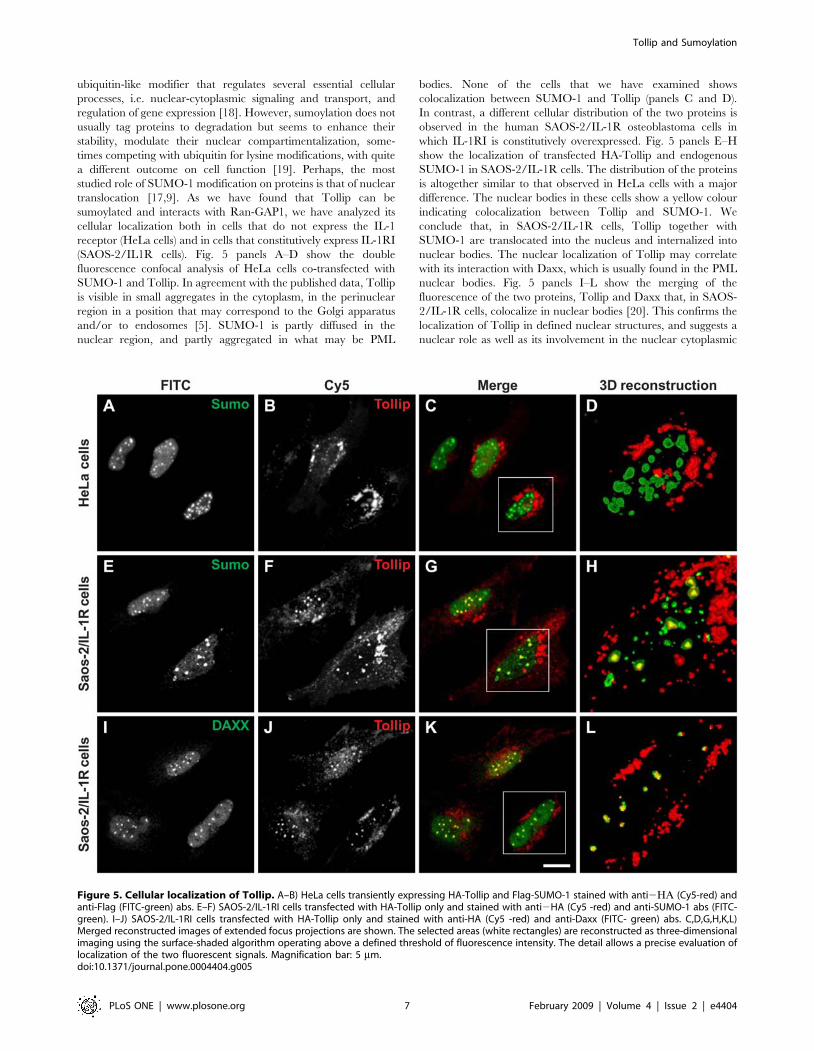
(SAOS-2/IL1R cells). Fig. 5 panels A–D show the double
fluorescence confocal analysis of HeLa cells co-transfected with
SUMO-1 and Tollip. In agreement with the published data, Tollip
is visible in small aggregates in the cytoplasm, in the perinuclear
region in a position that may correspond to the Golgi apparatus
and/or to endosomes [5]. SUMO-1 is partly diffused in the
nuclear region, and partly aggregated in what may be PML
bodies. None of the cells that we have examined shows
colocalization between SUMO-1 and Tollip (panels C and D).
In contrast, a different cellular distribution of the two proteins is
observed in the human SAOS-2/IL-1R osteoblastoma cells in
which IL-1RI is constitutively overexpressed. Fig. 5 panels E–H
show the localization of transfected HA-Tollip and endogenous
SUMO-1 in SAOS-2/IL-1R cells. The distribution of the proteins
is altogether similar to that observed in HeLa cells with a major
difference. The nuclear bodies in these cells show a yellow colour
indicating colocalization between Tollip and SUMO-1. We
conclude that, in SAOS-2/IL-1R cells, Tollip together with
SUMO-1 are translocated into the nucleus and internalized into
nuclear bodies. The nuclear localization of Tollip may correlate
with its interaction with Daxx, which is usually found in the PML
nuclear bodies. Fig. 5 panels I–L show the merging of the
fluorescence of the two proteins, Tollip and Daxx that, in SAOS-
2/IL-1R cells, colocalize in nuclear bodies [20]. This confirms the
localization of Tollip in defined nuclear structures, and suggests a
nuclear role as well as its involvement in the nuclear cytoplasmic
Figure 5. Cellular localization of Tollip. A–B) HeLa cells transiently expressing HA-Tollip and Flag-SUMO-1 stained with anti2GA (Cy5-red) andanti-Flag (FITC-green) abs. E–F) SAOS-2/IL-1RI cells transfected with HA-Tollip only and stained with anti2HA (Cy5 -red) and anti-SUMO-1 abs (FITC-green). I–J) SAOS-2/IL-1RI cells transfected with HA-Tollip only and stained with anti-HA (Cy5 -red) and anti-Daxx (FITC- green) abs. C,D,G,H,K,L)Merged reconstructed images of extended focus projections are shown. The selected areas (white rectangles) are reconstructed as three-dimensionalimaging using the surface-shaded algorithm operating above a defined threshold of fluorescence intensity. The detail allows a precise evaluation oflocalization of the two fluorescent signals. Magnification bar: 5 mm.doi:10.1371/journal.pone.0004404.g005
Tollip and Sumoylation
PLoS ONE | www.plosone.org 7 February 2009 | Volume 4 | Issue 2 | e4404

traffic. It should be noticed that the SAOS-2/IL-1RI cells used in
this experiment were transfected with HA-Tollip only, while both
Daxx and SUMO-1 were the endogenous cellular components.
Discussion
Tollip interaction with sumoylation enzymesThis work describes for the first time the involvement of Tollip
in the sumoylation process. Through the two-hybrid screening of a
rat cDNA library we have identified sequences coding for the
described Tollip partners: Tom1, ubiquitin and Tollip itself.
However, the presence of several recombinants coding for Ubc9,
Daxx, ARIP3, and one clone coding for SUMO-1 came as a
novelty. These interactors strongly suggest a role of Tollip in
sumoylation. The remaining clones were not further analyzed.
The identification of the binding site between HA-Tollip and the
Flag-tagged ligases shows that the carboxyl-terminus of the C2
domain contains the ligase binding region. In the ARIP3 ligase,
the binding region is just upstream the RING domain.
A direct interaction between Tollip and the E2/E3 sumoylation
enzymes suggests that the protein is sumoylated. Sumoylation is
not a stable reaction and a complex network of modifying and
demodifying enzymes is present both in the nucleus and in the
cytoplasm and controls the modification process [21]. In fact,
Tollip sumoylation is detectable only in cells lysed in 1% SDS
under conditions that block protease activity. It may be noticed
that the co-transfection of 293T cells with the SUMO-1 and Tollip
coding plasmids is sufficient to induce Tollip sumoylation
suggesting that, under these conditions, the sumoylating enzymes
are not limiting. The rat Tollip sequence contains 8 lysines as
potential sumoylation sites, and it is possible that the sumoylated
bands immunoprecipitated with Tollip are only due to SUMO-1
modification of the protein. However, different covalent modifi-
cations and, in particular, polyubiquitylation may occur in lysine
residues either competing with each other for the same site or
independently regulated by events that depend on the target of
interest. Although sumoylation does not usually target proteins to
destruction, it has been recently shown that sumoylated proteins
may become targets of E3 ubiquitylation ligases for proteosomal
degradation [18,22]. We cannot exclude that at least part of the
sumoylated Tollip has a proteosomal destiny. The strong increase
of the anti-SUMO-1 abs staining shown in figure 3C indicates that
the overexpression of Tollip activates the overall cellular
sumoylation of the proteins, thus suggesting a rather general role
of Tollip in this process. Tollip sumoylation and its interaction
with Ubc9 suggest a function of Tollip as a ligase. Conversely, the
interaction of Tollip with three different members of the PIAS
ligase family favours the idea of a sumoylation adaptor.
Tollip mediates IL-1RI sumoylationOur experiments identify at least one sumoylation substrate of
Tollip: the IL1-RI receptor. Here we show for the first time that
this receptor is sumoylated. IL-1RI sumoylation occurs within the
evolutionary conserved TIR domain, which is responsible for the
activation of the IL-1 signalling cascade. Furthermore, the TIR
domain is the site of ubiquitylation of IL-1RI mediated by Tollip.
The equilibrium between receptor ubiquitylation and sumoylation
is probably important in modulating the signaling cascade of IL-1,
establishing the number of receptors present on the cell surface.
Tollip may play a pivotal role in modulating the IL-1RI activity
through the different modifications. Hoeller et al [23] have shown
that monoubiquitylated sites do not interact with polyubiquiti-
nated proteins. It is possible that monoubiquitylated Tollip
interacts with the receptor stabilizing its sumoylation. Alternative-
ly, un-ubiquitinated Tollip interacts with the polyubiquitylated IL-
1 receptor accumulating it in the late endosomes for degradation.
Tollip has a nuclear functionIn contrast with the instability of Tollip sumoylation, its
expression in 293T cells seems to trigger stable modification of
proteins other than Tollip, suggesting a general activation of the
process following over-expression (Fig. 3C). One of the cellular
proteins constitutively modified by SUMO-1 is the RanGTPase
binding protein RanGAP-1, the binding partner of the RanBP2
ligase, a nucleoporine that serves as a docking site for the nuclear
import of proteins [17,9]. Our results show that transfected Tollip
co-immunoprecipitates with RanGAP-1, suggesting its role in
RanGAP-1 sumoylation and nuclear cytoplasmic traffic. Accord-
ingly, for the first time we show a nuclear localization of Tollip.
We do not know what the nuclear function of Tollip may be but
we can speculate on some hypothesis. Nuclear Tollip may be
important for the sumoylation of nuclear substrates. Alternatively,
Tollip could function as a carrier for nuclear localization of
sumoylated proteins, as suggested by the interaction with
RanGAP-1. Tollip may be involved in the translocation and
nuclear localization of the cytoplasmic portion of a number of
membrane receptors, among which the sumoylated cytoplasmic
IL-1RI [24]. Sumoylation regulates different cellular processes,
particularly transcriptional repression and activation [19]. The
regulation of transcription occurs mainly through the recruitment
of transcriptional co-repressors that cause downregulation. Daxx is
a transcriptional co-repressor interacting with many sumoylated
factors leading to SUMO-dependent repression and Daxx
interacts with Tollip. Generally, Daxx is localized in the
nucleoplasm and in defined nuclear structures, i.e. in the
SUMO-modified PML bodies, in the nucleolus and interacts with
CENP-C in the centromere, possibly exerting different functions in
the different compartments. In conclusion, here we show that
Daxx interacts and colocalizes with Tollip, thus suggesting the
existence of a nuclear function of Tollip, and a role of Tollip in
Daxx dependent transcriptional regulation.
Materials and Methods
AntibodiesAnti-HA, Ubc9, SUMO-1, Daxx and IL-1RI abs were
purchased from Santa Cruz. Anti-Flag and anti-Actin abs were
from Sigma-Aldrich, anti-RanGAP-1 abs were from Dr. A. Dasso
of the National Institute of Child Health and Development,
Betsda.
Rat cDNA Library Construction and Yeast Two hybridScreening
Total RNA was isolated from the brain of Sprague-Dawley rats
and poly(A)+ RNA was purified by affinity chromatography on
oligod(T)-cellulose (Amersham Bioscence) [25]. Equal amounts of
poly(A)+ RNA obtained from rat brain at 1, 2, 4, 8, 13 days of age
were pooled and used to generate cDNA according to standard
protocols based on the Gubler-Hoffman method [26]. The cDNA
was purified using a Chroma Spin1000 column (Clontech) to select
cDNAs 1 Kb long or more. The cDNA was cloned into the pJG4-
5 vector obtaining 16107 independent colonies.
For the two hybrid screening, Tollip cDNA fused to the DNA-
binding domain of LEX A was inserted into the pEG202 vector
(bait) and transformed into the yeast strain W303 (MATa) together
with the LacZ reporter plasmid pSH18-34. The cDNA library was
used to transform the EGY48 (mata) yeast strain to generate a pre-
transformed yeast library. The bait strain containing pEG202/
Tollip and Sumoylation
PLoS ONE | www.plosone.org 8 February 2009 | Volume 4 | Issue 2 | e4404

Tollip was crossed with an aliquot of the pre-transformed library
strain and 56107 diploids were screened for interactors [27].
Positive clones were confirmed in a second round of screening in
yeast and sequenced.
Ethical IssuesExperiments were authorized by the local bioethical Committee
and performed according to the Italian and European Community
legislation on the use of animals for experimental purposes.
In vitro transcription, translation, and GST pull-downassays
The Tollip cDNA sequence was inserted into the pGEX4T1
vector (Amersham Bioscence) in fusion with the Glutathione S-
Transferase (GST) protein. The GST-Tollip fusion protein was
expressed in E. coli cells and purified by binding to Glutathione-
Sepharose beads. In vitro transcription of the identified interactors
was carried out using the Riboprobe in vitro Transcription System
(Promega) according to the manufacturer instructions. In vitro
translation was carried out using the Rabbit Reticulocyte Lysate
System (Promega) in presence of 35S-methionine (Amersham
Bioscience). The GST pull-down assays were carried out by
incubating equal amounts of GST or GST-Tollip immobilised to
Gluthathione-Sepharose beads (Amersham Bioscence) with 5 ml of35S-labelled translation product in 100 ml final volume (binding
buffer: 20 mM sodium phosphate pH 6, 150 mM NaCl, 10%
glycerol, 1 mM PMSF, 1 mM DTT, 0.02% Nonidet P-40) The
mixtures were incubated 3 h at room temperature and washed
three times with binding buffer. Bound proteins were eluted with
26 SDS buffer, separated by SDS-PAGE and visualised by
autoradiography.
Cell lines and cDNA constructs293T and SAOS-2/IL-1RI (overexpressing IL-1RI) cells were
grown in Dulbecco’s Modified Eagle Medium (DMEM) supple-
mented with 10% fetal bovine serum (FBS) at 37uC and 5% CO2.
The cDNAs coding for rat Tollip wild type and its deletion
mutants were cloned into the pRK7-HA expression vector. The
cDNAs coding for rat Ubc9, the cytoplasmic region of the rat IL-
1RI receptor and its mutants were cloned into the pRK7
expression vector. The cytoplasmic portion of IL-1RI spans
aminoacid 355–569 of the rat cDNA sequence (accession
NP_037255). The cDNA coding for rat SUMO-1, PIAS-1,
PIASxb, ARIP3 and its deletion mutants were cloned into the
pcDNA3.1-Flag-C expression vector, which is a modified version
of the pcDNA3.1-His C plasmid (Clontech) where the hystidine
tag sequence was replaced with a Flag coding sequence. The
cDNA recombinant clones were amplified by PCR from the rat
cDNA library used for the two hybrid screening. Transfection of
293T cells with the expression vectors was performed using
Polyethylenimine (PEI) (Sigma Aldrich) according to manufacturer
instructions.
Immunoprecipitation and western blot analysis293T cells were lyzed, 24 hr after transfection, in RIPA buffer
(300 mM NaCl, 0.5% NP40, 20 mM Hepes KOH pH 8, 20%
Glycerol, 2 mM DTT, 0.4 mM EDTA pH 8) and sonicated. Lysis
under denaturing conditions was carried in 16 PBS containing
1% SDS. The samples were boiled 10 min and diluted 10 times in
PBS, containing a cocktail of anti-proteases and anti-phosphatases
(Sigma), to a final concentration of 0.1% SDS. The diluted
samples were sonicated. 400 mg protein extracts were immuno-
precipitated overnight with the specific abs and separated on
Protein A-Sepharose beads (Amersham Biosciences). 40 mg
protein extracts were loaded on the gel to check the efficiency of
the transfection.
Unless otherwise stated, the protein extracts and immunopre-
cipitates were analyzed in 12% SDS-PAGE. The western blots
were carried out using the BioRad apparatus according to the
manufacturer instructions. Staining was carried out with the ECL
Western Blot Detection Reagent (Amersham Biosciences) accord-
ing to the instructions.
ImmunofluorescenceSAOS-2/IL-1RI and HeLa cells were seeded onto collagen-
coated glass covers and, after 24 hr, fixed in 4% PFA for 209 at
4uC. Following 59 incubation in 0.1% Triton X-20, 3%BSA the
cells were stained with mouse anti-Tollip and rabbit anti-SUMO-1
abs followed by incubation with FITC conjugated anti-mouse and
Cy3 conjugated anti-rabbit secondary abs. After staining, the cells
were analyzed by confocal microscopy. Confocal images were
acquired using a Radiance 2000 confocal laser scanning
microscope (BioRad), equipped with a Nikon x60, oil immersion
1.4 N.A. objective and with krypton and red diode lasers. For
FITC and Cy5 double detection, the samples were simultaneously
excited with the 488-nm line of the krypton laser and with the 637-
nm line of the red diode laser. The emission signals from FITC
and Cy5 were separated by a dichroic mirror (DM; 560 nm) and
simultaneously detected by two photomultiplier tubes. Two barrier
filters (BP; 515/30 nm for FITC and LP; 660 nm for Cy5) were
placed before the two photomultiplier tubes to minimize the
overlap between the two signals, as previously described. Optical
sections were obtained at increments of 0.3 mm in the Z-axis and
were digitized with a scanning mode format of 5126512 pixels
and 256 grey levels. Image processing and volume rendering
(extended focus and surface-shaded) were carried out using the
ImageSpace software (Molecular Dynamics, Sunnyvale, CA)
running on a workstation Indigo (Silicon Graphics, Mountain
View, CA.).
Acknowledgments
We are very grateful to Dr. Dasso, theNational Institute of Child Health
and Development, Betsda, for providing the anti RanGap-1 antibodies; to
Dr. Marmiroli, University of Modena, for providing the SAOS-IL1R cell
line; to Dr. Elisabetta Ciani, University of Bologna for providing the PRK7
and PRK7-HA plasmids.
Author Contributions
Conceived and designed the experiments: AC RD DA MM. Performed the
experiments: AC RD CC AR SS MR SC AM SP EC. Analyzed the data:
AC RD MM. Wrote the paper: MM.
References
1. Burns K, Clatworthy J, Martin L, Martinon F, Plumpton C, et al. (2000) Tollip,
a new component of the IL-1RI pathway, links IRAK to the IL-1 receptor. Nat
Cell Biol 2: 346–51.
2. Shih SC, Prag G, Francis SA, Sutanto MA, Hurley JH, et al. (2003) A ubiquitin-
binding motif required for intramolecular monoubiquitylation, the CUE
domain. EMBO J 22: 1273–81.
3. Yamakami M, Yoshimori T, Yokosawa H (2003) Tom1, a VHS domain-
containing protein, interacts with Tollip, ubiquitin, and clathrin. J Biol Chem
278: 52865–72.
4. Yamakami M, Yokosawa H (2004) Tom1 (target of Myb 1) is a novel negative
regulator of interleukin-1- and tumor necrosis factor-induced signaling
pathways. Biol Pharm Bull 27: 564–6.
Tollip and Sumoylation
PLoS ONE | www.plosone.org 9 February 2009 | Volume 4 | Issue 2 | e4404

5. Zhang G, Ghosh S (2002) Negative regulation of toll-like receptor-mediated
signaling by Tollip. J Biol Chem 277: 7059–65.
6. Katoh Y, Shiba Y, Mitsuhashi H, Yanagida Y, Takatsu H, et al. (2004) Tollip
and Tom1 form a complex and recruit ubiquitin-conjugated proteins onto early
endosomes. J Biol Chem 279: 24435–43.
7. Katoh Y, Imakagura H, Futatsumori M, Nakayama K (2006) Recruitment of
clathrin onto endosomes by the Tom1-Tollip complex. Biochem Biophys Res
Commun 341: 143–9.
8. Brissoni B, Agostini L, Kropf M, Martinon F, Swoboda V, et al. (2006)
Intracellular trafficking of interleukin-1 receptor I requires Tollip. Curr Biol 16:
2265–70.
9. Pichler A, Gast A, Seeler JS, Dejean A, Melchior F (2002) The nucleoporin
RanBP2 has SUMO1 E3 ligase activity. Cell 108: 109–20.
10. Kagey MH, Melhuish TA, Wotton D (2003) The polycomb protein Pc2 is a
SUMO E3. Cell 113: 127–37.
11. Dedhar S (1989) Signal transduction via the beta 1 integrins is a required
intermediate in interleukin-1 beta induction of alkaline phosphatase activity in
human osteosarcoma cells. Exp Cell Res 183: 207–14.
12. Rodan SB, Wesolowski G, Chin J, Limjuco GA, Schmidt JA, et al. (1990) Il-1
binds to high affinity receptors on human osteosarcoma cells and potentiates
prostaglandin e2 stimulation of camp production. J Immunol 145: 1231–1237.
13. Moilanen AM, Karvonen U, Poukka H, Yan W, Toppari J, et al. (1999) A testis-
specific androgen receptor coregulator that belongs to a novel family of nuclear
proteins. J Biol Chem 274: 3700–4.
14. van den Akker E, Ano S, Shih HM, Wang LC, Pironin M, et al. (2005) FLI-1
functionally interacts with PIASxalpha, a member of the PIAS E3 SUMO ligase
family. J Biol Chem 280: 38035–46.
15. Johnson ES (2004) Protein modification by SUMO. Annu Rev Biochem 73:
355–82.
16. Hay RT (2005) SUMO: a history of modification. Mol Cell 18: 1–12.
17. Pichler A, Melchior F (2002) Ubiquitin-related modifier SUMO1 and
nucleocytoplasmic transport. Traffic 3: 381–7.18. Meulmeester E, Melchior F (2008) Cell biology: SUMO. Nature 452: 709–11.
19. Zhao J (2007) Sumoylation regulates diverse cellular processes. Cell Mol Life Sci
64: 3017–33.20. Shih HM, Chang CC, Kuo HY, Lin DY (2007) Daxx mediates SUMO-
dependent transcriptional control and subnuclear compartmentalization.Biochem Soc Trans 35: 1397–400.
21. Melchior F, Schergaut M, Pichler A (2003) SUMO: ligases, isopeptidases and
nuclear pores. Trends Biochem Sci 28: 612–8.22. Tatham MH, Geoffroy MC, Shen L, Plechanovova A, Hattersley N, et al. (2008)
RNF4 is a poly-SUMO-specific E3 ubiquitin ligase required for arsenic-inducedPML degradation. Nat Cell Biol 10: 538–46.
23. Hoeller D, Crosetto N, Blagoev B, Raiborg C, Tikkanen R, et al. (2006)Regulation of ubiquitin-binding proteins by monoubiquitination. Nat Cell Biol
8: 163–9.
24. Lin S, Makino K, Xia W, Matin A, Wen Y, et al. (2001) Nuclear localization ofEGF receptor and its potential new role as a transcription factor. Nat Cell Biol 3:
802–808.25. Edmonds M, Vaughan MH Jr, Nakazato H (1971) Polyadenylic acid sequences
in the heterogeneous nuclear RNA and rapidly-labeled polyribosomal RNA of
HeLa cells: possible evidence for a precursor relationship. Proc Natl AcadSci U S A 6: 1336–40.
26. Gubler U, Hoffman BJ (1983) A simple and very efficient method for generatingcDNA libraries. Gene 25: 263–9.
27. Kolonin MG, Zhong J, Finley RL (2000) Interaction mating methods in two-hybrid systems. Methods Enzymol 328: 26–46.
28. Di Giaimo R, Riccio M, Santi S, Galeotti C, Ambrosetti DC, et al. (2002) New
insights into the molecular basis of Progressive Myoclonus Epilepsy (EPM1): amultiprotein complex with cystatin B. Human molecular genetics 11:
2941–2950.
Tollip and Sumoylation
PLoS ONE | www.plosone.org 10 February 2009 | Volume 4 | Issue 2 | e4404
Copyright © 2022 FDOKUMEN