Sumoylation of p68 and p72 RNA Helicases Affects Protein Stability and Transactivation Potential
Upload
independentCategory
view
4download
0
Parallel SUMOylation-dependent pathways mediate gene- andsignal-specific transrepression by LXRs and PPARγ
Serena Ghisletti1, Wendy Huang1,2, Sumito Ogawa1,7, Gabriel Pascual1, Mu-En Lin2,Timothy M. Willson5, Michael G. Rosenfeld3,4, and Christopher K. Glass1,3,6
1 Department of Cellular and Molecular Medicine, University of California, San Diego 9500 Gilman Drive,La Jolla CA 92093
2 Biomedical Sciences Graduate Program, University of California, San Diego 9500 Gilman Drive, La JollaCA 92093
3 Department of Medicine, University of California, San Diego 9500 Gilman Drive, La Jolla CA 92093
4 Howard Hughes Medical Institute
5 GlaxoSmithKline, 5 Moore Drive, PO Box 13398 Research Triangle Park, NC 27709
SummaryTransrepression is widely utilized to negatively regulate gene expression, but the mechanisms bywhich different nuclear receptors effect gene- and signal-specific transrepression programs remainpoorly understood. Here, we report the identification of alternative SUMOylation-dependentmechanisms that enable PPARγ and LXRs to negatively regulate overlapping but distinct subsets ofpro-inflammatory genes. Ligand-dependent conjugation of SUMO2/3 to LXRs or SUMO-1 toPPARγ targets them to promoters of TLR target genes, where they prevent the signal-dependentremoval of NCoR corepressor complexes required for transcriptional activation. SUMO-1-PPARγand SUMO-2/3-LXRs inhibit distinct NCoR clearance mechanisms, allowing promoter- and TLR-specific patterns of repression. Mutational analysis and studies of naturally occurring oxysterolligands indicate that the transactivation and SUMOylation-dependent transrepression activities ofLXRs can be independently regulated. These studies define parallel but functionally distinctpathways that are utilized by PPARγ and LXRs to differentially regulate complex programs of geneexpression that control immunity and homeostasis.
INTRODUCTIONMembers of the nuclear-receptor family of ligand-dependent transcription factors influenceimmune responses by positively and negatively regulating gene expression in diverse celltypes, including macrophages, lymphocytes, and dendritic cells (Glass and Ogawa, 2006;Winoto and Littman, 2002). Recent studies of the glucocorticoid receptor (GR), liver Xreceptors (LXRs), peroxisome proliferator-activated receptor γ (PPARγ), retinoic acid receptor(RAR), and estrogen receptors (ERs) revealed that each nuclear receptor represses anoverlapping, but distinct, pattern of signal-dependent inflammatory gene expression (Galon et
6Correspondence: [email protected] Office: 858 534 6011 Fax: 858 822 2127.7Current address: Department of Geriatric Medicine Graduate School of Medicine, The University of Tokyo 7-3-1 Hongo, Bukkyo-ku,Tokyo, 113-8655 JapanPublisher's Disclaimer: This is a PDF file of an unedited manuscript that has been accepted for publication. As a service to our customerswe are providing this early version of the manuscript. The manuscript will undergo copyediting, typesetting, and review of the resultingproof before it is published in its final citable form. Please note that during the production process errors may be discovered which couldaffect the content, and all legal disclaimers that apply to the journal pertain.
NIH Public AccessAuthor ManuscriptMol Cell. Author manuscript; available in PMC 2008 January 12.
Published in final edited form as:Mol Cell. 2007 January 12; 25(1): 57–70.
NIH
-PA Author Manuscript
NIH
-PA Author Manuscript
NIH
-PA Author Manuscript
al., 2002; Joseph et al., 2003; Ogawa et al., 2005; Welch et al., 2003). While these observationsare consistent with the overlapping but distinct effects of specific ligands on inflammatoryprocesses at a biological level, the molecular mechanisms that are utilized to achieve thisspecificity remain poorly understood.
LXR α and β are members of the nuclear receptor superfamily that play essential roles incholesterol and fatty acid homeostasis (Janowski et al., 1996; Peet et al., 1998; Schultz et al.,2000). LXRs are required for normal cholesterol efflux from peripheral cells and cholesterolexcretion by the liver by positively regulating the expression of genes involved in cholesterolefflux (Costet et al., 2000; Repa et al., 2000) and genes involved in bile acid biosynthesis andtransport (Chiang et al., 2001; Lewis and Rader, 2005). The transcriptional activities of LXRsare thought to be regulated in vivo by oxysterols, oxygenated derivatives of cholesterol(Janowski et al., 1999; Janowski et al., 1996).
Recent studies have characterized LXRs as regulators of innate immunity and inflammatorysignalling in macrophages (Zelcer and Tontonoz, 2006). LXR agonists enhance macrophagesurvival during bacterial infection and LXRs are required for normal immunity to Listeriamonocytogenes (Castrillo et al., 2003; Joseph et al., 2004; Joseph et al., 2003; Valledor et al.,2004). In addition, LXR activation inhibits the lipopolysaccharide (LPS) induction of a numberof pro-inflammatory genes such as inducible nitric oxide synthase (iNOS), cytokines such asinterleukin-1β (IL-1β), and chemokines such as monocyte chemoattractant protein-1 (MCP-1)(Joseph et al., 2003). Repression of inflammatory mediators by LXR agonists is abolished inmacrophages lacking both LXRs, but not in macrophages containing either LXRα or LXRβ,indicating that both isoforms can mediate anti-inflammatory activity (Joseph et al., 2003). Themolecular mechanisms underlying the repression of inflammatory-response genes by LXRsremain to be established.
PPARγ, a nuclear receptor required for fat cell development and normal glucose homeostasis(Spiegelman, 1998; Willson et al., 2001), also exerts broad anti-inflammatory effects inmacrophages and other cell types (Delerive et al., 1999; Glass and Ogawa, 2006; Jiang et al.,1998; Ricote et al., 1998), and this activity has been correlated with insulin-sensitizing andanti-atherogenic activities of synthetic PPARγ agonists (Haffner et al., 2002; Li et al., 2000;Staels and Fruchart, 2005). Recent studies of the mechanism by which PPARγ repressesactivation of the iNOS gene by LPS led to the identification of a SUMOylation-dependenttransrepression pathway (Pascual et al., 2005). In this pathway, synthetic PPARγ agonistspromote PIAS1-dependent conjugation of SUMO-1 to the PPARγ ligand-binding domain. Thisin turn targets PPARγ to NCoR corepressor complexes that are normally bound to the iNOSpromoter under basal conditions and are required to mediate active repression in the absenceof an inflammatory stimulus. The recruitment of SUMO1-PPARγ was found to prevent thesignal-dependent ubiquitylation and clearance of the NCoR complex required for full geneactivation, resulting in the iNOS gene remaining in a repressed state (Pascual et al., 2005).These findings raised a series of questions, including whether this pathway is used by othernuclear receptors, whether it can be utilized to mediate repression in a gene or signal-specificmanner, and whether natural ligands can promote entry of their cognate nuclear receptors intothis pathway.
Here, we demonstrate that LXR transrepression of inflammatory target genes also utilizes aSUMOylation and NCoR-dependent pathway, indicating that this mechanism represents ageneral molecular strategy for transrepression by a subset of nuclear receptors that regulatemetabolism and immunity. In contrast to PPARγ, however, LXRs are SUMOylated by SUMO2and SUMO3 rather than SUMO1, and HDAC4 rather than PIAS1 functions as a requiredSUMO E3 ligase. As a consequence, transrepression by PPARγ and LXRs can beindependently regulated in a signal- and gene-specific manner.
Ghisletti et al. Page 2
Mol Cell. Author manuscript; available in PMC 2008 January 12.
NIH
-PA Author Manuscript
NIH
-PA Author Manuscript
NIH
-PA Author Manuscript
RESULTSLXR-dependent transrepression requires NCoR
Based on the requirement of NCoR for transrepression by PPARγ, we initially sought todetermine whether LXR-mediated repression is also NCoR-dependent. Experiments wereperformed in primary macrophages transfected with control non-specific or NCoR-specificsiRNAs (Fig. S1A), using GW3965 as an LXR-specific agonist. Reduction of NCoRexpression significantly reversed the LXR ligand-dependent repression of iNOS expressioninduced by LPS (Fig. 1A). To confirm this result, iNOS promoter activity was evaluated inRAW264.7 macrophages. As shown in Fig. 1B, knock down of NCoR reversed the LXRα andLXRβ-dependent transrepression of iNOS promoter activity induced by LPS. To determinewhether LXRs are recruited to the iNOS promoter and prevents NCoR clearance, chromatinimmunoprecipitation (ChIP) experiments were performed examining LXR and NCoRoccupancy on the iNOS promoter in primary LPS-stimulated macrophages. LXRs wererecruited to the iNOS promoter in a ligand-dependent manner in the presence or absence ofLPS and this recruitment was NCoR dependent (Fig. 1C). LXRs were not detected in doubleLXRα/LXRβ knockout macrophages, indicating that the LXR antibody specifically detectedLXRα/LXRβ bound to the iNOS promoter. As expected, NCoR was found to occupy the iNOSpromoter under basal conditions and to be dismissed by LPS (Pascual et al., 2005), whiletreatment with GW3965 prevented NCoR clearance(Fig.1D). These results suggest thatLXRα and LXRβ, in addition to PPARγ, repress LPS induction of the iNOS gene by preventingthe clearance of NCoR.
LXR-dependent transrepression is SUMOylation dependentThe identification of LXR transrepression as NCoR dependent raised the question of whethera SUMOylation-dependent pathway was also involved. The ligation of SUMO to targetproteins requires the E2-conjugating enzyme Ubc9 (Hay, 2005). We therefore knocked downUbc9 expression using a specific siRNA (Fig. S1B) in primary macrophages (Fig. 2A) and inRAW264.7 cells (Fig. 2B) transfected with LXRα and LXRβ expression plasmids. Knockdownof Ubc9 significantly impaired LXR ligand-dependent repression of LPS-induced iNOSexpression (Fig. 2A) and iNOS promoter activation (Fig. 2B). ChIP assays were next performedin primary macrophages to determine the role of SUMOylation in ligand-dependentrecruitment of LXRα and LXRβ to the iNOS promoter and in the prevention of NCoRclearance. Knockdown of Ubc9 expression prevented ligand-dependent recruitment of LXRto the iNOS promoter (Fig. 2C) and impaired the block in NCoR clearance from the iNOSpromoter (Fig. 2D). These results suggest that a SUMOylation-dependent step is critical forthe recruitment of LXR to the iNOS promoter to prevent corepressor clearance.
LXRs are modified by SUMO-2/3 dependent on HDAC4 E3 ligase activitySince a SUMOylation-dependent step is required for LXR transrepression, we tested whetherLXR itself was a target of SUMOylation. FLAG-tagged LXRβ was expressed in HeLa cellsin the presence or absence of Myc-tagged SUMO-1, SUMO-2 or SUMO-3. High-molecular-weight LXRβ, with an increased size expected for monoSUMOylation, was detected in thepresence of SUMO-2 and SUMO-3, but not in the presence of SUMO-1 (Fig. 3A) in a ligand-dependent manner. This high molecular weight species was detected by anti-Myc antibody,and was abolished by knockdown of Ubc9 (Fig. 3B), indicating that it corresponds toSUMOylated LXRβ. LXRα was also SUMOylated by SUMO-2 and SUMO-3 (data notshown). An additional LXR-dependent band indicated by asterisks in Fig. 3, was also observedin some experiments. This band likely represents an unknown posttranslational modificationof LXR that is not correlated with SUMOylation.
Ghisletti et al. Page 3
Mol Cell. Author manuscript; available in PMC 2008 January 12.
NIH
-PA Author Manuscript
NIH
-PA Author Manuscript
NIH
-PA Author Manuscript
Several SUMO E3 ligases have been identified that promote transfer of SUMO from the E2to specific substrates. To date, three types of SUMO E3 ligases have been well characterized:the protein inhibitor of activated signal transducer and activator of transcription (PIAS) family,the RanBP2/Nup358 protein and the human Polycomb member Pc2 (Melchior et al., 2003).Although SUMO-PPARγ mediated transrepression is PIAS1 dependent (Pascual et al.,2005), the systematic knockdown of all PIAS E3 ligases, RanBP2/Nup358, and PC2 did notreverse LXR ligand-dependent repression in primary macrophages (data not shown). Recently,HDAC4 and related members of class II deacetylases, such as HDAC5, have been describedas SUMO E3 ligases for MEF2 transcription factor (Gregoire and Yang, 2005; Zhao et al.,2005). Knocking down HDAC4 expression (Fig. S1C) resulted in a complete inhibition ofLXRβ SUMOylation (Fig. 3C). Consistent with these findings, a physical interaction betweenLXRβ and HDAC4 was revealed by co-immunoprecipitation assays (Fig. 3D). To determinewhether HDAC4 directly promotes LXRs SUMOylation, we performed an in vitroSUMOyation assay. Ligand-dependent LXRβ SUMOylation was significantly increased bythe addition of in vitro-translated HDAC4 protein in the presence of ligand (Fig. 3E), but notin the absence of ligand (data not shown). In contrast, an HDAC4 mutant (67-257), that doesnot have the coiled-coil domain required for the interaction with Ubc9 (Zhao et al., 2005),abolished LXRβ SUMOylation. Similar effects were observed in HeLa cells over-expressingWT HDAC4 or the HDAC4 mutant (67-257) (Fig. 3F). In concert, these findings stronglysuggest that HDAC4 functions as a SUMO E3 ligase for LXRs in vivo.
We next evaluated whether HDAC4 was required for LXRs-mediated transrepression inprimary macrophages. Unexpectedly, knockdown of HDAC4 prevented LPS-activation ofiNOS, precluding an assessment of its role in transrepression of this specific target (data notshown). Several recent reports suggest that activation of inflammatory response genes by IRFsis blocked by HDAC inhibitors (Chang et al., 2004; Nusinzon and Horvath, 2003; Sakamotoet al., 2004). As the iNOS gene is also a target of these factors, we speculate that HDAC4 isone of the key HDACs that are required for activation of this cohort of genes.
To identify alternative LPS-target genes that would be suitable for determining the role ofHDAC4 in LXR-dependent transrepression, microarray experiments were performed toidentify LPS-inducible, LXR-sensitive target genes that retained LPS-induction in the presenceof the HDAC inhibitor TSA (Table S1). MCP-1 was chosen as one of the most representativechemokine genes meeting these criteria. To determine whether the MCP-1 gene was anappropriate model, we performed transrepression experiments in primary macrophages andconfirmed that MCP-1 transrepression was NCoR and Ubc9-dependent (Fig. S2). ChIP assaysconfirmed that LXRs were recruited to the MCP-1 promoter in an NCoR and Ubc9-dependentmanner and prevented NCoR clearance (Fig. S3A and B). Knockdown of HDAC4 (Fig. S1C)had no effect on induction of MCP-1, as predicted by the microarray data, but reversed LXRtransrepression (Fig. 3G). Taken together, these results demonstrate that HDAC4 interacts withLXR and regulates LXR transrepression activity by promoting its SUMOylation. Furthermore,these findings indicate that, although both LXR and PPARγ transrepression are SUMOylation-dependent, LXR is SUMOylated in a ligand-dependent manner in vivo by SUMO-2 andSUMO-3 while PPARγ is modified by SUMO-1 (Floyd and Stephens, 2004;Ohshima et al.,2004;Pascual et al., 2005;Yamashita et al., 2004).
Mutation of LXR SUMO acceptor sites abolishes transrepression but not transactivationactivity
Two lysine residues in the human LXRβ primary amino acid sequence, the N-terminal Lys-31and the C-terminal Lys-448, conform with high probability to the proposed consensus motiffor SUMO-conjugation. In addition, a lower probability site was identified at Lys-410. Todetermine whether these lysine residues are targets for SUMOylation, mutants with lysine to
Ghisletti et al. Page 4
Mol Cell. Author manuscript; available in PMC 2008 January 12.
NIH
-PA Author Manuscript
NIH
-PA Author Manuscript
NIH
-PA Author Manuscript
arginine substitutions of K31, K410 and K448 were generated and assayed for SUMOylation.SUMOylation of LXRβ was reduced when the cells were transfected with mutants K410 andK448, and completely abolished with K410/448 double mutation, while mutation of K31 hadno effect (Fig. 4A).
To determine the functional significance of LXR SUMOylation, the ability of the LXRβmutants to transrepress the iNOS promoter and transactivate the ABCA1 promoter were testedin RAW264.7 cells. Individual mutations of K410 and K448 partially impaired transrepressionactivity, while the K410/448 double mutant fully abolished transrepression (Fig. 4B). Similarresults were observed for the MCP1 promoter (Fig. S3C). In contrast, these same lysinemutations did not impair ligand-dependent transactivation of the ABCA1 promoter. Parallelstudies of LXRα demonstrated ligand-dependent SUMOylation of lysine residues 328 and 434(corresponding to residue 448 of LXRβ). Mutation of these residues to arginine impairedLXRα transrepression but not transactivation (data not shown). In concert, these experimentsindicate that specific lysine residues in the ligand-binding domains of LXRα and LXRβ areSUMOylated by SUMO-2 and SUMO-3 and that their SUMOylation is required fortransrepression.
Endogenous LXRs ligands promote entry into the transrepression pathwayA number of naturally occurring oxysterols are potent transcriptional activators of LXRs(Janowski et al., 1999; Janowski et al., 1996). In macrophages, endogenous oxysterols arethought to be produced in proportion to intracellular cholesterol levels and induce theexpression of a number of genes involved in LXR-dependent cholesterol efflux, such asABCA1 (Repa et al., 2000). To investigate the relative abilities of naturally occurring LXRligands to both positively and negatively regulate gene expression their effects on ABCA1gene expression and LPS-dependent induction of the endogenous iNOS gene were evaluatedin primary macrophages. These experiments revealed that 22(R)-hydroxycholesterol (22R),24(S), 25 epoxycholesterol (EC) and 24-hydroxycholesterol (24HC) each repressed iNOSactivation and induced ABCA1 expression. This anti-inflammatory effect was LXR-dependentas it was not observed in LXR−/− macrophages (Fig. 5B). In contrast, 25-hydroxycholesterol(25HC) and 27-hydroxycholesterol (27HC) were able to activate ABCA1, but did not repressiNOS activation even at significantly higher concentrations than allow activation of theABCA1 gene (Fig. 5C), indicating that activation and transrepression activities can bechemically separated. Furthermore, the transrepression activities of 22(R)-hydroxycholesterol(22R), 24(S), 25epoxycholesterol (EC) and 24-hydroxycholesterol (24HC) are impaired byknocking down Ubc9, suggesting that a subset of the naturally occurring oxysterol ligands ofLXRs can promote their entry into the SUMOylation-dependent transrepression pathway.
Gene-specific utilization of parallel NCoR and SUMOylation-dependent pathways by LXRsand PPARγ
The finding that a SUMOylation/NCoR-dependent pathway parallel to that initially identifiedto mediate transrepression of inflammatory responses by PPARγ is utilized by LXRs to repressiNOS activation raised the questions of the extent to which these parallel pathways are utilizedto repress other LPS target genes and whether they can be utilized in a gene-specific manner.Previous expression profiling studies demonstrated that PPARγ and LXR agonists repressedoverlapping but distinct sets of genes induced in primary macrophages by LPS or the TLR3-activator poly I:C (Ogawa et al., 2005)(The complete data set is available atwww.lipidmaps.org). These relationships are illustrated in Fig. 6A, filtered for genes inducedat least 5-fold for each TLR agonist and exhibiting at least 50% repression with respect toactivation in the absence of PPARγ or LXR agonist. We initially evaluated several endogenousLPS-inducible target genes that were found to be transrepressed by both nuclear receptors inprimary macrophages. The majority of the LPS-inducible genes meeting these criteria,
Ghisletti et al. Page 5
Mol Cell. Author manuscript; available in PMC 2008 January 12.
NIH
-PA Author Manuscript
NIH
-PA Author Manuscript
NIH
-PA Author Manuscript
exemplified by MIP-1β, MCP-1, VCAM-1 and IL-15, were found to be transrepressed byPPARγ and LXRs by an NCoR and SUMOylation-dependent mechanism (Fig. S2). Thesefindings indicate that the NCoR and SUMOylation-dependent pathways are likely to be widelyutilized by LXRs and PPARγ to transrepress proinflammatory target genes.
We next evaluated subsets of LPS-inducible genes that were transrepressed only by the LXRagonist (represented in Fig. 6B by IL1β) or only by the PPARγ agonist (represented in Fig. 6Bby TNFα). Unexpectedly, although IL1β and TNFα were differentially transrepressed by LXRsand PPARγ ligands, both cases involved an NCoR-dependent transrepression mechanism (Fig.6B). This result was confirmed by ChIP experiments, showing that the LXR agonist, but notthe PPARγ agonist, was able to inhibit the clearance of NCoR from IL1β promoter (Fig. 6C,left panel). Conversely, NCoR was cleared from the TNFα promoter in the presence of theLXR agonist, but not following treatment with the PPARγ agonist (Fig. 6C, right panel). Wetherefore performed ChIP experiments in primary macrophages to determine whether theSUMOylated receptors were differentially recruited to these promoters. These assays indicatedthat PPARγ and LXRs were both recruited to the iNOS, IL1β and TNFα promoters in a ligand-dependent manner (Fig. S4A), suggesting that the SUMOylated receptors are interruptingdistinct signalling inputs that are responsible for NCoR clearance. This finding is supportedby the observation that saturating concentrations of PPARγ and LXRs agonists exerted additiverepressive effects on the iNOS promoter, which is sensitive to transrepression by bothreceptors, suggesting that they operate through independent mechanisms (Fig. S4 B).
To explore the roles of specific signalling adapter proteins in LPS activation andtransrepression, we investigated Tab2, which plays a cytoplasmic role in TLR4-dependentsignal transduction and is also a component of a subset of NCoR complexes (Baek et al.,2002; Jiang et al., 2004; Takaesu et al., 2000; Zhu et al., 2006). Knockdown of Tab2 inmacrophages (Fig. S1D) reduces the fold activation of iNOS in response to LPS by about 50%.Intriguingly, the residual activation of iNOS remained fully sensitive to repression by LXRsagonist, but was now completely resistant to repression by the PPARγ agonist (Fig. 6D).Furthermore, ChIP experiments demonstrated that Tab2 was present on the iNOS promoterunder basal conditions, was cleared in response to LPS treatment, but was retained in thepresence of LPS and PPARγ agonist. In contrast, Tab2 was not enriched above background(IgG control) levels on the IL1β promoter under any condition. In concert, these results areconsistent with Tab2 playing a promoter-specific role in mediating PPARγ-dependent, but notLXR-dependent, transrepression.
Signal-specific utilization of parallel NCoR and SUMOylation-dependent pathways by LXRsand PPARγ
Recent studies demonstrated that the ability of a particular nuclear receptor to repress specificsubsets of inflammatory response genes could be modulated in a signal specific manner, e.g.,a cohort of genes that was sensitive to GR-mediated repression when induced by LPS wereGR-resistant when activated by poly I:C (Ogawa et al., 2005). A similar relationship wasobserved for genes subject to transrepression by LXR and PPARγ agonists, illustrated in Figure7A. To investigate whether genes that exhibit signal-specific repression by LXRs andPPARγ utilize an NCoR-dependent mechanism, we evaluated the IL1β gene. While this geneis strongly repressed by LXR agonists in an NCoR-dependent manner when activated by LPS,it is almost completely resistant when activated by poly I:C (Fig. 7B). As a second example,we evaluated repression of the iNOS gene when activated by the TLR1/2 agonist Pam3CSK(Omueti et al., 2005).
Although repressed by both LXR and PPARγ agonists when induced by LPS, the ability of theLXR agonist to transrepress iNOS was selectively abolished when macrophages werestimulated by the TLR1/2 agonist (Fig. 7C). We therefore evaluated whether the ability of
Ghisletti et al. Page 6
Mol Cell. Author manuscript; available in PMC 2008 January 12.
NIH
-PA Author Manuscript
NIH
-PA Author Manuscript
NIH
-PA Author Manuscript
LXRs agonist to transrepress inflammatory responses is correlated with their ability to blockNCoR clearance induced by different TLR agonists. ChIP experiments revealed that LXRagonists inhibited NCoR clearance from the iNOS promoter in response to LPS treatment, butfailed to inhibit NCoR clearance when cells were treated with the TLR1/2 agonist Pam3CSK(Fig. S5A). In addition, poly I:C was found to promote clearance of NCoR from the IL1βpromoter even when the cells were treated with LXRs ligand, consistent with the failure ofGW3965 to transrepress IL1βactivation in response to this signal (Fig. S5B). In concert, theseresults indicate that the parallel SUMOylation-dependent transrepression pathways arethemselves subject to regulation, and can be over-ridden by specific signals in a gene specificmanner.
DISCUSSIONA general SUMO/NCoR transrepression mechanism
Many nuclear receptors can act in trans to repress inflammatory responses, with recent studiesindicating that ligand-dependent repression of inflammatory target genes occurs in a gene andsignal-specific manner (Ogawa et al., 2005). Although a large number of transrepressionmechanisms have been suggested from the analysis of artificial and transiently transfectedtarget genes, relatively few mechanisms have been established for endogenous target genes inprimary cells. The present studies demonstrate that ligand-dependent transrepression of anumber of endogenous inflammatory response genes in primary macrophages by LXRs isNCoR- and SUMOylation-dependent. Moreover, ChIP analysis revealed that LXRs arerecruited to the iNOS promoter in a SUMOylation-dependent manner and inhibit corepressorclearance induced by LPS. These observations extend the NCoR and SUMOylation-dependentpathway, initially identified for PPARγ (Pascual et al., 2005), to two additional members ofthe nuclear receptor family, LXRα and LXRβ. Thus, SUMOylation-dependent targeting of anuclear receptor to corepressor complexes to prevent their signal-dependent clearance is likelyto represent a general molecular strategy for transrepression of proinflammatory target genes,expanding upon previously described mechanisms by which SUMO proteins function tonegatively regulate gene expression (Gill, 2005).
Parallel SUMO-dependent transrepression pathwaysAlthough these studies indicate that LXRs and PPARγ both utilize a SUMOylation-dependentmechanism that converges on NCoR corepressor complexes, they also provide evidence fordistinct and parallel pathways that are used in a receptor-specific and gene-specific manner(Fig. 7). These conclusions are based on the findings that PPARγ and LXRs are conjugated todifferent SUMO proteins by different E3 ligases and differentially regulate proinflammatorytarget genes such as IL1β and TNFα. While PIAS1 has been described as the E3 ligase involvedin PPARγ SUMOylation (Ohshima et al., 2004;Pascual et al., 2005), the present studiesstrongly suggest that SUMOylation of LXRs requires HDAC4 E3 ligase activity. In the caseof PPARγ, a ligand-induced allosteric change in the position of the lysine residue that is thepoint of SUMO1 conjugation was proposed to be the basis for ligand-dependent SUMOylation(Pascual et al., 2005). In the case of LXRs, crystal structures of apo receptors are not available,so it is not yet possible to assess ligand-dependent changes in the lysine residues that representthe essential SUMO acceptor sites. However, association of LXRβ with HDAC4 does notappear to be ligand-dependent, suggesting that ligands alter the specific configurations of thelysine residues that serve as SUMO acceptor sites to provide the mechanism for ligand-dependence.
Since the conjugated forms of SUMO-2 and SUMO-3 form a distinct subfamily and are only50% identical in sequence to SUMO-1 (Hay, 2005), we propose that LXRs and PPARγ arefunctionally differentiated based on their differential SUMOylation and represent distinct,
Ghisletti et al. Page 7
Mol Cell. Author manuscript; available in PMC 2008 January 12.
NIH
-PA Author Manuscript
NIH
-PA Author Manuscript
NIH
-PA Author Manuscript
parallel transrepression pathways that can be utilized in a gene and signal-specific manner(Figure 7D). Gene-specific utilization of these parallel pathways is exemplified by the selectivetransrepression of IL1β by LXRs and TNFα by PPARγ, while signal-specific utilization isexemplified by the differential sensitivities of the IL1βand iNOS genes to LXR ligands whenactivated by TLR1/2, TLR3 and TLR4 agonists.
These findings thus raise a number of intriguing possibilities regarding the molecular targetsof SUMOylated PPARγ and LXRs within the NCoR complex and how the docking of theseproteins prevents NCoR clearance. ChIP experiments have now localized NCoR complexeson nearly all LPS-responsive genes we have examined. Furthermore, it appears thatSUMOylated PPARγ and LXRs localize to these complexes prior to an activating signal. Inall cases, whether or not a gene is sensitive or resistant to PPARγ or LXR agonists is strictlycorrelated with whether or not they prevent NCoR clearance. Previous studies suggest thatsignal-dependent clearance of NCoR complexes from target promoters involves the activationof the ubiquitin E3 ligase activities of the Tbl1 and TblR1 proteins that are core componentsof these complexes (Ogawa et al., 2004; Perissi et al., 2004), leading to the recruitment of theUbc5/19S proteosome machinery required for ubiquitylation of NCoR complexes and theirsubsequent dismissal. SUMOylation of PPARγ and its interaction with the NCoR complex hasbeen shown to block signal-dependent recruitment of UbcH5 to the iNOS promoter (Pascualet al., 2005), but whether this is due to inhibition of Tbl1/TblR1 phosphorylation, or somedistal step remains to be defined. The finding that Tab2 is required for transrepression of theiNOS gene by PPARγ, but not LXRs, suggests that the SUMOylated receptors inhibit distinctsignalling inputs that are involved in activating the ubiquitinligase activities of Tbl1/TblR1.Biochemical approaches have recently identified components of corepressor complexes asinteracting targets of SUMO2 (Rosendorff et al., 2006) and it will be of interest to utilizeSUMOylated LXRs and PPARγ as molecular probes to further elucidate the specific eventsthat allow NCoR corepressor complexes to be cleared from inflammatory response genes in asignal-specific manner.
Coordinate and independent regulation of transactivation and transrepressionIn macrophages, LXRs play critical roles in maintaining normal cholesterol homeostasis bypositively regulating the expression of genes involved in cholesterol storage and efflux. Theobservation that synthetic LXR agonists are also capable of potently repressing inflammatoryresponse genes raises the question of whether the production of endogenous oxysterols mightalso exert anti-inflammatory effects by binding to LXRs. Consistent with this possibility, wefind that 22R, 24(S), 25EC, and 24HC can both induce ABCA1 expression and repress iNOSactivation in primary macrophages. Intriguingly, 25HC and 27HC can activate ABCA1, butdo not repress iNOS activation. These findings suggest that the transactivation andtransrepression activities of LXRs may be independently regulated based on the specificligands that are produced under different contexts. Intriguingly, the oxysterols that inducetransrepression are not found at any significant levels in cultured macrophages, while theoxysterols that are made at high levels by cultured macrophages do not induce transrepression(Fu et al., 2001). It is possible that enzymes necessary for producing natural ligands that activatethis pathway are expressed in specific subsets of macrophages in vivo. Alternatively, thispathway may operate as a paracrine system, e.g., Kuppfer cells in liver may be exposed tohepatocyte-derived oxysterols that activate the transrepression pathway.
The ability of many ligand-dependent nuclear receptors to inhibit inflammatory geneexpression is also of considerable interest from the perspective of the development of newapproaches to modulate inflammatory diseases. This goal is often complicated by the fact thatpotent nuclear receptor agonists are associated with unwanted side effects: e.g., PPARγagonists promote weight gain and edema formation in diabetic patients (Staels and Fruchart,
Ghisletti et al. Page 8
Mol Cell. Author manuscript; available in PMC 2008 January 12.
NIH
-PA Author Manuscript
NIH
-PA Author Manuscript
NIH
-PA Author Manuscript
2005; Yki-Jarvinen, 2005), while LXR agonists have been shown to cause markedhypertriglyceridemia in animals, at least in part due to activation of SREBP1c in liver (Schultzet al., 2000). The present studies suggest that it should be possible to develop LXR ligands thatseparate transrepression from transactivation activities. Ligands that specifically promote entryof LXR into the SUMO-dependent transrepression pathway may be of therapeutic utility indiseases in which inflammation plays a significant pathogenic role.
EXPERIMENTAL PROCEDURESReagents and plasmids
TLR4 agonist (LPS), TLR3 agonist (poly I:C) were obtained from Sigma, TLR1/2 agonist(Pam3CysSK4) from ECM Microcollections, GW3965 and Rosiglitazone were provided byGlaxoSmithKline. 22(R)-hydroxycholesterol, 22(S)-hydroxycholesterol, 25-hydroxycholesterol were purchased from Sigma, 24(S),25epoxycholesterol and 24-hydroxycholesterol from Biomol, 27-hydroxycholesterol from Research Plus, Inc. Pointmutations in Flag-hLXRα and Flag-hLXRβ were made using the QuickChange side-directedmutagenesis kit (Stratagene). The reporter plasmid iNOS-luc, Ubc9 and SUMO-1 expressionvectors have been previously described (Pascual et al., 2005), the reporter plasmid ABCA1-luc was kindly donated by Dr. B. Wagner, the reporter plasmid MCP1-luc was kindly donatedby Dr. R. Natarajan and the HDAC4 mutant (67-257) was kindly donated by Dr.T.Yao.
Cell culture and transient transfectionThioglycollate-elicited macrophages were prepared as previously described (Pascual et al.,2005) from LXRα β+/+ and LXRα β−/− mice (Wagner et al., 2003) or C57BL/6 mice (CharlesRiver). For RNAi experiments in primary macrophages, cells were transfected with control orsmart-pool siRNAs (100 nM, Dharmacon) directed against NCoR, Ubc9, HDAC4 and TAB2using lipofectamine 2000 (Invitrogen). Cells were used for experiments after 48 h incubationand target gene knockdown was validated by QPCR (Supplemental Figure S1). RAW 264.7were transiently transfected with iNOS, ABCA1 or MCP1 promoters directing luciferaseexpression using Superfect reagent (Qiagen). A β-galactosidase expression vector wascotrasfected as an internal control. For siRNA experiments in RAW264.7 macrophages, cellswere transfected with siRNAs (40 nM) using Superfect reagent for 48 h.
ChIP assaysChIP assays were performed as previously described (Pascual et al., 2005). Anti-LXR(Calbiochem), anti- PPARβ (ActiveMotif) and anti-NCoR (Affinity Bioreagents) antibodieswere used. Rabbit preimmune serum was used as a control for non-specific binding. 150-bpregions of the iNOS, MCP1, IL1β and TNFα proximal promoters were amplified by real timePCR. Primer sequences are available upon request.
RNA isolation and QPCRTotal RNA (isolated by RNeasy kit, Qiagen) was prepared from primary macrophages. Oneμg of total RNA was used for cDNA synthesis, and 1 μl of cDNA was used for real time PCRusing inflammatory-gene-specific primers. Primer sequences are available upon request. PCR(SYBERgreen) analysis was performed on an Applied Biosystems 7300 Real-time PCRsystem. Values are normalized with GAPDH content.
Co-IP assayHeLa cells were transfected with Flag-LXRβ and Ha-HDAC4 and lysed in low salt lysis buffer.Ha-HDAC4 was immunoprecipitated using anti-HA beads (Covance) and Flag-LXRβ wasdetected using anti-FLAG antibody (Sigma).
Ghisletti et al. Page 9
Mol Cell. Author manuscript; available in PMC 2008 January 12.
NIH
-PA Author Manuscript
NIH
-PA Author Manuscript
NIH
-PA Author Manuscript
In vitro SUMOylation assayIn vitro SUMOylation reactions were carried out in 20 μl of reaction buffer by addingrecombinant SUMO, SAE-1, SAE-2, Ubc9 and in vitro translated HDAC4 or HDAC4 mutant(67-257) to the in vitro translated LXRβ. Reactions were incubated for 60 min at 37°Csupplemented with 2mM ATP and analyzed by SDS–PAGE and immunoblotted using anti-HA (Covance) antibody.
In vivo SUMOylation assayWhole protein extract was prepared in presence of NEM (N-Ethylmaleimide, Calbiochem)from HeLa cells transfected with FLAG-tagged wild-type LXRα and LXRβ, or lysine mutantsand Myc-tagged SUMO-1, SUMO-2 or SUMO-3, Ubc9 and HDAC4 or HDAC4mutant(67-257) expression vectors. Extracts were resolved by SDS–PAGE and immunoblotted usinganti-FLAG (Sigma) or anti-Myc (Santa Cruz) antibodies.
Supplementary MaterialRefer to Web version on PubMed Central for supplementary material.
Acknowledgements
We thank B. Wagner (X-ceptor Therapeutics) for the ABCA1-luc plasmid, R. Natarajan (City of Hope, Duarte) forthe MCP1-luc plasmid, T. Yao (Duke University, Durham) for the HDAC4 mutant (67-257) plasmid, A. Gamliel(University of California, San Diego) for the SUMO-2 and SUMO-3 plasmids. We thank the DERC/BIOGEM corefacility for assistance with microarray experiments and A. Howarth for assistance with manuscript preparation. Thesestudies were supported by an American Heart Association Postdoctoral Fellowship to S.G and NIH grants CA52599,HL56989, DK063491 and GM 069338 to CKG. MGR is an Investigator of the Howard Hughes Medical Institute.
ReferencesBaek SH, Ohgi KA, Rose DW, Koo EH, Glass CK, Rosenfeld MG. Exchange of N-CoR corepressor and
Tip60 coactivator complexes links gene expression by NF-kappaB and beta-amyloid precursor protein.Cell 2002;110:55–67. [PubMed: 12150997]
Castrillo A, Joseph SB, Vaidya SA, Haberland M, Fogelman AM, Cheng G, Tontonoz P. Crosstalkbetween LXR and toll-like receptor signaling mediates bacterial and viral antagonism of cholesterolmetabolism. Mol Cell 2003;12:805–816. [PubMed: 14580333]
Chang HM, Paulson M, Holko M, Rice CM, Williams BR, Marie I, Levy DE. Induction of interferon-stimulated gene expression and antiviral responses require protein deacetylase activity. Proc Natl AcadSci U S A 2004;101:9578–9583. [PubMed: 15210966]
Chiang JY, Kimmel R, Stroup D. Regulation of cholesterol 7alpha-hydroxylase gene (CYP7A1)transcription by the liver orphan receptor (LXRalpha). Gene 2001;262:257–265. [PubMed: 11179691]
Costet P, Luo Y, Wang N, Tall AR. Sterol-dependent transactivation of the ABC1 promoter by the liverX Receptor/Retinoid X receptor. J Biol Chem 2000;275:28240–28245. [PubMed: 10858438]
Delerive P, Martin-Nizard F, Chinetti G, Trottein F, Fruchart JC, Najib J, Duriez P, Staels B. Peroxisomeproliferator-activated receptor activators inhibit thrombin-induced endothelin-1 production in humanvascular endothelial cells by inhibiting the activator protein-1 signaling pathway. Circ Res1999;85:394–402. [PubMed: 10473669]
Floyd ZE, Stephens JM. Control of Peroxisome Proliferator-Activated Receptor {gamma}2 Stability andActivity by SUMOylation. Obes Res 2004;12:921–928. [PubMed: 15229330]
Fu X, Menke JG, Chen Y, Zhou G, MacNaul KL, Wright SD, Sparrow CP, Lund EG. 27-hydroxycholesterol is an endogenous ligand for liver X receptor in cholesterol-loaded cells. J BiolChem 2001;276:38378–38387. [PubMed: 11504730]
Galon J, Franchimont D, Hiroi N, Frey G, Boettner A, Ehrhart-Bornstein M, O'Shea JJ, Chrousos GP,Bornstein SR. Gene profiling reveals unknown enhancing and suppressive actions of glucocorticoidson immune cells. Faseb J 2002;16:61–71. [PubMed: 11772937]
Ghisletti et al. Page 10
Mol Cell. Author manuscript; available in PMC 2008 January 12.
NIH
-PA Author Manuscript
NIH
-PA Author Manuscript
NIH
-PA Author Manuscript
Gill G. Something about SUMO inhibits transcription. Curr Opin Genet Dev 2005;15:536–541. [PubMed:16095902]
Glass CK, Ogawa S. Combinatorial roles of nuclear receptors in inflammation and immunity. Nat RevImmunol 2006;6:44–55. [PubMed: 16493426]
Gregoire S, Yang XJ. Association with class IIa histone deacetylases upregulates the sumoylation ofMEF2 transcription factors. Mol Cell Biol 2005;25:2273–2287. [PubMed: 15743823]
Haffner SM, Greenberg AS, Weston WM, Chen H, Williams K, Freed MI. Effect of rosiglitazonetreatment on nontraditional markers of cardiovascular disease in patients with type 2 diabetesmellitus. Circulation 2002;106:679–684. [PubMed: 12163427]
Hay RT. SUMO: a history of modification. Mol Cell 2005;18:1–12. [PubMed: 15808504]Janowski BA, Grogan MJ, Jones SA, Wisely GB, Kliewer SA, Corey EJ, Mangelsdorf DJ. Structural
requirements of ligands for the oxysterol liver X receptors LXRalpha and LXRbeta. Proc Natl AcadSci U S A 1999;96:266–271. [PubMed: 9874807]
Janowski BA, Willy PJ, Devi TR, Falck JR, Mangelsdorf DJ. An oxysterol signalling pathway mediatedby the nuclear receptor LXRα. Nature 1996;383:728–731. [PubMed: 8878485]
Jiang C, Ting AT, Seed B. PPARγ agonists inhibit production of monocyte inflammatory cytokines.Nature 1998;391:82–86. [PubMed: 9422509]
Jiang Z, Mak TW, Sen G, Li X. Toll-like receptor 3-mediated activation of NF-kappaB and IRF3 divergesat Toll-IL-1 receptor domain-containing adapter inducing IFN-beta. Proc Natl Acad Sci U S A2004;101:3533–3538. [PubMed: 14982987]
Joseph SB, Bradley MN, Castrillo A, Bruhn KW, Mak PA, Pei L, Hogenesch J, O'Connell RM, ChengG, Saez E, et al. LXR-dependent gene expression is important for macrophage survival and the innateimmune response. Cell 2004;119:299–309. [PubMed: 15479645]
Joseph SB, Castrillo A, Laffitte BA, Mangelsdorf DJ, Tontonoz P. Reciprocal regulation of inflammationand lipid metabolism by liver X receptors. Nat Med 2003;9:213–219. [PubMed: 12524534]
Lewis GF, Rader DJ. New insights into the regulation of HDL metabolism and reverse cholesteroltransport. Circ Res 2005;96:1221–1232. [PubMed: 15976321]
Li A, Brown K, Silvestre M, Willson T, Palinski W, Glass C. Peroxisome proliferator-activated receptorγ ligands inhibit development of atherosclerosis in LDL receptor-deficient mice. J CIin Invest2000;106:523–531.
Melchior F, Schergaut M, Pichler A. SUMO: ligases, isopeptidases and nuclear pores. Trends BiochemSci 2003;28:612–618. [PubMed: 14607092]
Nusinzon I, Horvath CM. Interferon-stimulated transcription and innate antiviral immunity requiredeacetylase activity and histone deacetylase 1. Proc Natl Acad Sci U S A 2003;100:14742–14747.[PubMed: 14645718]
Ogawa S, Lozach J, Benner C, Pascual G, Tangirala RK, Westin S, Hoffmann A, Subramaniam S, DavidM, Rosenfeld MG, Glass CK. Molecular determinants of crosstalk between nuclear receptors andToll-like receptors mediating counter-regulation of inflammatory responses. Cell. 2005
Ogawa S, Lozach J, Jepsen K, Sawka-Verhelle D, Perissi V, Sasik R, Rose DW, Johnson RS, RosenfeldMG, Glass CK. A nuclear receptor corepressor transcriptional checkpoint controlling activatorprotein 1-dependent gene networks required for macrophage activation. Proc Natl Acad Sci USA2004;101:14461–14466. [PubMed: 15452344]
Ohshima T, Koga H, Shimotohno K. Transcriptional activity of peroxisome proliferator-activatedreceptor gamma is modulated by SUMO-1 modification. J Biol Chem 2004;279:29551–29557.[PubMed: 15123625]
Omueti KO, Beyer JM, Johnson CM, Lyle EA, Tapping RI. Domain exchange between human toll-likereceptors 1 and 6 reveals a region required for lipopeptide discrimination. J Biol Chem2005;280:36616–36625. [PubMed: 16129684]
Pascual G, Fong AL, Ogawa S, Gamliel A, Li AC, Perissi V, Rose DW, Willson TM, Rosenfeld MG,Glass CK. A SUMOylation-dependent pathway mediates transrepression of inflammatory responsegenes by PPAR-gamma. Nature 2005;437:759–763. [PubMed: 16127449]
Peet DJ, Janowski BA, Mangelsdorf DJ. The LXRs: a new class of oxysterol receptors. Curr Opin GenetDev 1998;8:571–575. [PubMed: 9794827]
Ghisletti et al. Page 11
Mol Cell. Author manuscript; available in PMC 2008 January 12.
NIH
-PA Author Manuscript
NIH
-PA Author Manuscript
NIH
-PA Author Manuscript
Perissi V, Aggarwal A, Glass CK, Rose DW, Rosenfeld MG. A corepressor/coactivator exchangecomplex required for transcriptional activation by nuclear receptors and other regulated transcriptionfactors. Cell 2004;116:511–526. [PubMed: 14980219]
Repa JJ, Turley SD, Lobaccaro JA, Medina J, Li L, Lustig K, Shan B, Heyman RA, Dietschy JM,Mangelsdorf DJ. Regulation of absorption and ABC1-mediated efflux of cholesterol by RXRheterodimers. Science 2000;289:1524–1529. [PubMed: 10968783]
Ricote M, Li AC, Willson TM, Kelly CJ, Glass CK. The peroxisome proliferator-activated receptor-γ isa negative regulator of macrophage activation. Nature 1998;391:79–82. [PubMed: 9422508]
Rosendorff A, Sakakibara S, Lu S, Kieff E, Xuan Y, DiBacco A, Shi Y, Shi Y, Gill G. NXP-2 associationwith SUMO-2 depends on lysines required for transcriptional repression. Proc Natl Acad Sci U S A2006;103:5308–5313. [PubMed: 16567619]
Sakamoto S, Potla R, Larner AC. Histone deacetylase activity is required to recruit RNA polymerase IIto the promoters of selected interferon-stimulated early response genes. J Biol Chem2004;279:40362–40367. [PubMed: 15194680]
Schultz JR, Tu H, Luk A, Repa JJ, Medina JC, Li L, Schwendner S, Wang S, Thoolen M, MangelsdorfDJ, et al. Role of LXRs in control of lipogenesis. Genes Dev 2000;14:2831–2838. [PubMed:11090131]
Spiegelman BM. PPARγ: adipogenic regulator and thiazolidinedione receptor. Diabetes 1998;47:507–514. [PubMed: 9568680]
Staels B, Fruchart JC. Therapeutic roles of peroxisome proliferator-activated receptor agonists. Diabetes2005;54:2460–2470. [PubMed: 16046315]
Takaesu G, Kishida S, Hiyama A, Yamaguchi K, Shibuya H, Irie K, Ninomiya-Tsuji J, Matsumoto K.TAB2, a novel adaptor protein, mediates activation of TAK1 MAPKKK by linking TAK1 to TRAF6in the IL-1 signal transduction pathway. Mol Cell 2000;5:649–658. [PubMed: 10882101]
Valledor AF, Hsu LC, Ogawa S, Sawka-Verhelle D, Karin M, Glass CK. Activation of liver X receptorsand retinoid X receptors prevents bacterial-induced macrophage apoptosis. Proc Natl Acad Sci USA2004;101:17813–17818. [PubMed: 15601766]
Wagner BL, Valledor AF, Shao G, Daige CL, Bischoff ED, Petrowski M, Jepsen K, Baek SH, HeymanRA, Rosenfeld MG, et al. Promoter-specific roles for liver X receptor/corepressor complexes in theregulation of ABCA1 and SREBP1 gene expression. Mol Cell Biol 2003;23:5780–5789. [PubMed:12897148]
Welch JS, Ricote M, Akiyama TE, Gonzalez FJ, Glass CK. PPARγ and PPARδ negatively regulatespecific subsets of lipopolysaccharide and IFNγ target genes in macrophages. Proc Natl Acad SciUSA 2003;100:6712–6717. [PubMed: 12740443]
Willson TM, Lambert MH, Kliewer SA. Peroxisome proliferator-activated receptor gamma andmetabolic disease. Annu Rev Biochem 2001;70:341–367. [PubMed: 11395411]
Winoto A, Littman DR. Nuclear hormone receptors in T lymphocytes. Cell 2002;109(Suppl):S57–66.[PubMed: 11983153]
Yamashita D, Yamaguchi T, Shimizu M, Nakata N, Hirose F, Osumi T. The transactivating function ofperoxisome proliferator-activated receptor gamma is negatively regulated by SUMO conjugation inthe amino-terminal domain. Genes Cells 2004;9:1017–1029. [PubMed: 15507114]
Yki-Jarvinen H. The PROactive study: some answers, many questions. Lancet 2005;366:1241–1242.[PubMed: 16214581]
Zelcer N, Tontonoz P. Liver X receptors as integrators of metabolic and inflammatory signaling. J ClinInvest 2006;116:607–614. [PubMed: 16511593]
Zhao X, Sternsdorf T, Bolger TA, Evans RM, Yao TP. Regulation of MEF2 by histone deacetylase 4-and SIRT1 deacetylase-mediated lysine modifications. Mol Cell Biol 2005;25:8456–8464. [PubMed:16166628]
Zhu P, Baek SH, Bourk EM, Ohgi KA, Garcia-Bassets I, Sanjo H, Akira S, Kotol PF, Glass CK, RosenfeldMG, Rose DW. Macrophage/cancer cell interactions mediate hormone resistance by a nuclearreceptor derepression pathway. Cell 2006;124:615–629. [PubMed: 16469706]
Ghisletti et al. Page 12
Mol Cell. Author manuscript; available in PMC 2008 January 12.
NIH
-PA Author Manuscript
NIH
-PA Author Manuscript
NIH
-PA Author Manuscript

Figure 1. LXR transrepression requires NCoRNCoR-specific siRNA abolishes LXR-dependent repression of LPS-induced endogenousiNOS gene expression in primary macrophages (A) and iNOS-luciferase reporter gene activityin RAW264.7 cells (B). After siRNA transfection, macrophages were stimulated with LPS(1μg/ml) for 6h in the absence or presence of LXR ligand (GW3965, 1μM). iNOS expressionin peritoneal macrophages was evaluated by QPCR and iNOS promoter activation inRAW264.7 cells was evaluated by luciferase assay. *p < 0.03 vs LPS+control siRNA; **p <0.02 vs GW3965+LPS control siRNA. C. ChIP assay reveals ligand-dependent and NCoR-dependent LXR recruitment to the iNOS promoter. Primary macrophages were treated withLPS (1μg/ml) with or without the LXR ligand (GW3965, 1μM) for 1h. ChIP assay wasperformed with antibody against LXR (both isoforms, α and α) or control IgG.Immunoprecipitated DNA was analyzed by QPCR using primers specific for the iNOS-promoter. Chromatin isolated from LXR−/−mice was used as a negative control for specificityof the LXR antibody. * p < 0.05 vs untreated or LPS alone; **p < 0.03 vs GW3965 or GW3965+LPS control siRNA. D. LXR ligand inhibits LPS-stimulated release of NCoR from the iNOSpromoter as shown by ChIP assay in peritoneal macrophages, treated as described in panel C.* p < 0.02 vs untreated; **p < 0.02 vs LPS. Results are expressed as the average of threeindependent experiments. Error bars represent standard deviations.
Ghisletti et al. Page 13
Mol Cell. Author manuscript; available in PMC 2008 January 12.
NIH
-PA Author Manuscript
NIH
-PA Author Manuscript
NIH
-PA Author Manuscript
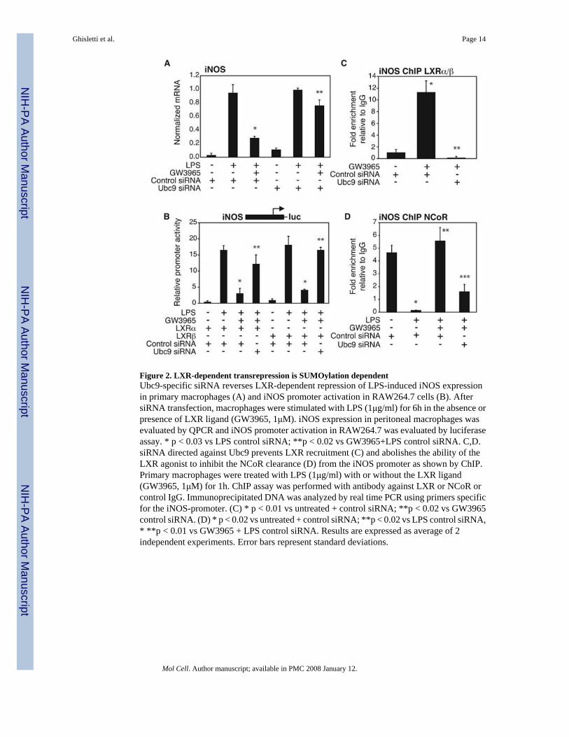
Figure 2. LXR-dependent transrepression is SUMOylation dependentUbc9-specific siRNA reverses LXR-dependent repression of LPS-induced iNOS expressionin primary macrophages (A) and iNOS promoter activation in RAW264.7 cells (B). AftersiRNA transfection, macrophages were stimulated with LPS (1μg/ml) for 6h in the absence orpresence of LXR ligand (GW3965, 1μM). iNOS expression in peritoneal macrophages wasevaluated by QPCR and iNOS promoter activation in RAW264.7 was evaluated by luciferaseassay. * p < 0.03 vs LPS control siRNA; **p < 0.02 vs GW3965+LPS control siRNA. C,D.siRNA directed against Ubc9 prevents LXR recruitment (C) and abolishes the ability of theLXR agonist to inhibit the NCoR clearance (D) from the iNOS promoter as shown by ChIP.Primary macrophages were treated with LPS (1μg/ml) with or without the LXR ligand(GW3965, 1μM) for 1h. ChIP assay was performed with antibody against LXR or NCoR orcontrol IgG. Immunoprecipitated DNA was analyzed by real time PCR using primers specificfor the iNOS-promoter. (C) * p < 0.01 vs untreated + control siRNA; **p < 0.02 vs GW3965control siRNA. (D) * p < 0.02 vs untreated + control siRNA; **p < 0.02 vs LPS control siRNA,* **p < 0.01 vs GW3965 + LPS control siRNA. Results are expressed as average of 2independent experiments. Error bars represent standard deviations.
Ghisletti et al. Page 14
Mol Cell. Author manuscript; available in PMC 2008 January 12.
NIH
-PA Author Manuscript
NIH
-PA Author Manuscript
NIH
-PA Author Manuscript
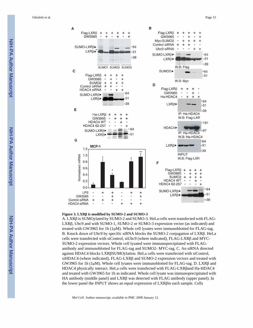
Figure 3. LXRβ is modified by SUMO-2 and SUMO-3A. LXRβ is SUMOylated by SUMO-2 and SUMO-3. HeLa cells were transfected with FLAG-LXRβ, Ubc9 and with SUMO-1, SUMO-2 or SUMO-3 expression vector (as indicated) andtreated with GW3965 for 1h (1μM). Whole cell lysates were immunoblotted for FLAG-tag.B. Knock down of Ubc9 by specific siRNA blocks the SUMO-2 conjugation of LXRβ. HeLacells were transfected with siControl, siUbc9 (where indicated), FLAG-LXRβ and MYC-SUMO-2 expression vectors. Whole cell lysated were immunoprecipitated with FLAG-antibody and immunoblotted for FLAG-tag and SUMO2- MYC-tag. C. An siRNA directedagainst HDAC4 blocks LXRβSUMOylation. HeLa cells were transfected with siControl,siHDAC4 (where indicated), FLAG-LXRβ and SUMO-2 expression vectors and treated withGW3965 for 1h (1μM). Whole cell lysates were immunoblotted for FLAG-tag. D. LXRβ andHDAC4 physically interact. HeLa cells were transfected with FLAG-LXRβand Ha-HDAC4and treated with GW3965 for 1h as indicated. Whole cell lysate was immunoprecipitated withHA antibody (middle panel) and LXRβ was detected with FLAG antibody (upper panel). Inthe lower panel the INPUT shows an equal expression of LXRβin each sample. Cells
Ghisletti et al. Page 15
Mol Cell. Author manuscript; available in PMC 2008 January 12.
NIH
-PA Author Manuscript
NIH
-PA Author Manuscript
NIH
-PA Author Manuscript
transfected with only LXRβ or HDAC4 (first and last lane) were used as negative control forthe IP. E. LXRβ is SUMOylated by HDAC4 in vitro. The in vitro SUMOylation assay wasperformed as described in Experimental Procedures, adding to the reaction HDAC4 or HDAC4mutant (67-257) as indicated. The proteins were immunoblotted for HA-tag. F. HDAC4modulates LXRβ SUMOylation in vivo. HeLa cells were transfected with FLAG-LXRβ, Ubc9,SUMO-2 and HDAC4 or HDAC4 mutant (67-257) expression vector (as indicated) and treatedwith GW3965 for 1h (1μM). Whole cell lysates were immunoblotted for FLAG-tag. G. Knockdown of HDAC4 blocks LXR transrepression of MCP1. Primary macrophages weretransfected with control or HDAC4 specific siRNA for 48h and then stimulated with LPS(1μg/ml) for 6h in the absence or presence of LXR ligand (GW3965, 1μM). MCP1 expressionwas evaluated by real time PCR. Results are expressed as average of two independentexperiments. Error bars represent standard deviations. * p < 0.02 vs LPS+control siRNA; **p< 0.03 vs GW3965+LPS+ control siRNA.
Ghisletti et al. Page 16
Mol Cell. Author manuscript; available in PMC 2008 January 12.
NIH
-PA Author Manuscript
NIH
-PA Author Manuscript
NIH
-PA Author Manuscript
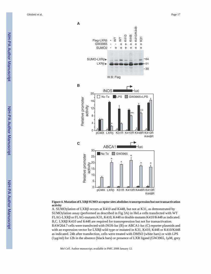
Figure 4. Mutation of LXRβ SUMO acceptor sites abolishes transrepression but not transactivationactivityA. SUMOylation of LXRβ occurs at K410 and K448, but not at K31, as demonstrated bySUMOylation assay (performed as described in Fig 3A) in HeLa cells transfected with WTFLAG-LXRβ or FLAG-mutants K31, K410, K448 or double mutants K410/K448 as indicated.B,C. LXRβ K410 and K448 are required for transrepression but not for transactivation.RAW264.7 cells were transfected with iNOS-luc (B) or ABCA1-luc (C) reporter plasmids andwith an expression vector for LXRβ wild type or mutated in K31, K410, K448 or K410/K448as indicated. 24h after transfection, cells were treated with DMSO (white bars) or with LPS(1μg/ml) for 12h in the absence (black bars) or presence of LXR ligand (GW3965, 1μM, grey
Ghisletti et al. Page 17
Mol Cell. Author manuscript; available in PMC 2008 January 12.
NIH
-PA Author Manuscript
NIH
-PA Author Manuscript
NIH
-PA Author Manuscript
bars). Results are expressed as average of three independent experiments. Error bars representstandard deviations. (B) * p < 0.01 vs LPS; **p < 0.05 vs GW3965+LPS+ LXRβ wild type.(C) * p < 0.03 vs pCMX.
Ghisletti et al. Page 18
Mol Cell. Author manuscript; available in PMC 2008 January 12.
NIH
-PA Author Manuscript
NIH
-PA Author Manuscript
NIH
-PA Author Manuscript

Figure 5. Endogenous LXRs ligands promote entry into transrepression pathwayA,B. Natural LXRs ligands induce ABCA1 expression and repress iNOS activation. Primarymacrophages from WT (panel A and B, grey bars) and LXR−/− mice (panel B, black bars) weretreated with GW3965 (1μM) or 5μM oxysterols 22R, 22S, 24(S),25EC, 24HC, 25HC and 27HC(A) for 18 hours. In the B panel cells were pretreated with the indicated ligands and then treatedwith LPS (1μg/ml) for 6h. Expression of ABCA1 (A) and iNOS (B) was evaluated by QPCR.(A) * p < 0.03 vs untreated; (B) * p < 0.01 vs LPS. C. Dose-response experiment of 25HC and27HC. Primary macrophages were pretreated with GW3965(1μM), 25HC and 27HC atincreasing concentrations (1μM, 5 μM, 10 μM, 50 μM) and then treated with LPS (1μg/ml) for6h. Expression of iNOS was evaluated by QPCR. * p < 0.01 vs LPS. D. Natural LXRs ligandsrequire Ubc9 to transrepress iNOS expression. Primary macrophages were transfected withUbc9 siRNA (black bars) or non specific siRNA (white bars) for 48h and then stimulated withLPS (1μg/ml) for 6h in the absence or presence of LXR ligands as indicated (5μM). iNOSexpression in peritoneal macrophages was evaluated by QPCR. * p < 0.05 vs control siRNA.Results are expressed as average of two independent experiments. Error bars represent standarddeviations.
Ghisletti et al. Page 19
Mol Cell. Author manuscript; available in PMC 2008 January 12.
NIH
-PA Author Manuscript
NIH
-PA Author Manuscript
NIH
-PA Author Manuscript
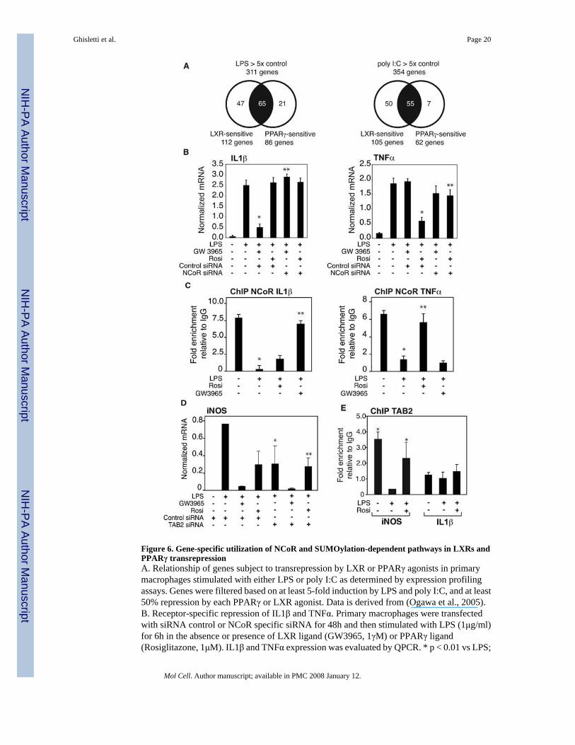
Figure 6. Gene-specific utilization of NCoR and SUMOylation-dependent pathways in LXRs andPPARγ transrepressionA. Relationship of genes subject to transrepression by LXR or PPARγ agonists in primarymacrophages stimulated with either LPS or poly I:C as determined by expression profilingassays. Genes were filtered based on at least 5-fold induction by LPS and poly I:C, and at least50% repression by each PPARγ or LXR agonist. Data is derived from (Ogawa et al., 2005).B. Receptor-specific repression of IL1β and TNFα. Primary macrophages were transfectedwith siRNA control or NCoR specific siRNA for 48h and then stimulated with LPS (1μg/ml)for 6h in the absence or presence of LXR ligand (GW3965, 1γM) or PPARγ ligand(Rosiglitazone, 1μM). IL1β and TNFα expression was evaluated by QPCR. * p < 0.01 vs LPS;
Ghisletti et al. Page 20
Mol Cell. Author manuscript; available in PMC 2008 January 12.
NIH
-PA Author Manuscript
NIH
-PA Author Manuscript
NIH
-PA Author Manuscript
** p < 0.01 vs GW3965 (left panel) or Rosi (right panel) +LPS control siRNA. C. Differentialinhibition of NCoR clearance from IL1β and TNFα promoters by PPARγ and LXR ligands.Primary macrophages were treated with LPS (1μg/ml) with or without PPARγ (Rosiglitazone,1μM) or LXR ligand (GW3965, 1μM) for 1h. ChIP assays were performed with antibodyagainst NCoR and or control IgG. * p < 0.01 vs untreated; ** p < 0.08 vs LPS. D. PPARγtransrepression is TAB2-dependent. Primary macrophages were transfected with siRNAcontrol or TAB2 specific siRNA for 48h and then stimulated with LPS (1μg/ml) for 6h in theabsence or presence of LXR ligand (GW3965, 1μM) or PPARγ ligand (Rosiglitazone, 1μM).iNOS expression was evaluated by QPCR. * p < 0.01 vs LPS; ** p < 0.01 vs Rosi +LPS controlsiRNA. E. Tab2 is present in the basal state on the iNOS promoter, but not on the IL1β promoter.ChIP assays were performed with antibody against Tab2 or control IgG. * p<0.01 vs controlIgG. Results are expressed as the average of 2 independent experiments. Errors bars representstandard deviations.
Ghisletti et al. Page 21
Mol Cell. Author manuscript; available in PMC 2008 January 12.
NIH
-PA Author Manuscript
NIH
-PA Author Manuscript
NIH
-PA Author Manuscript
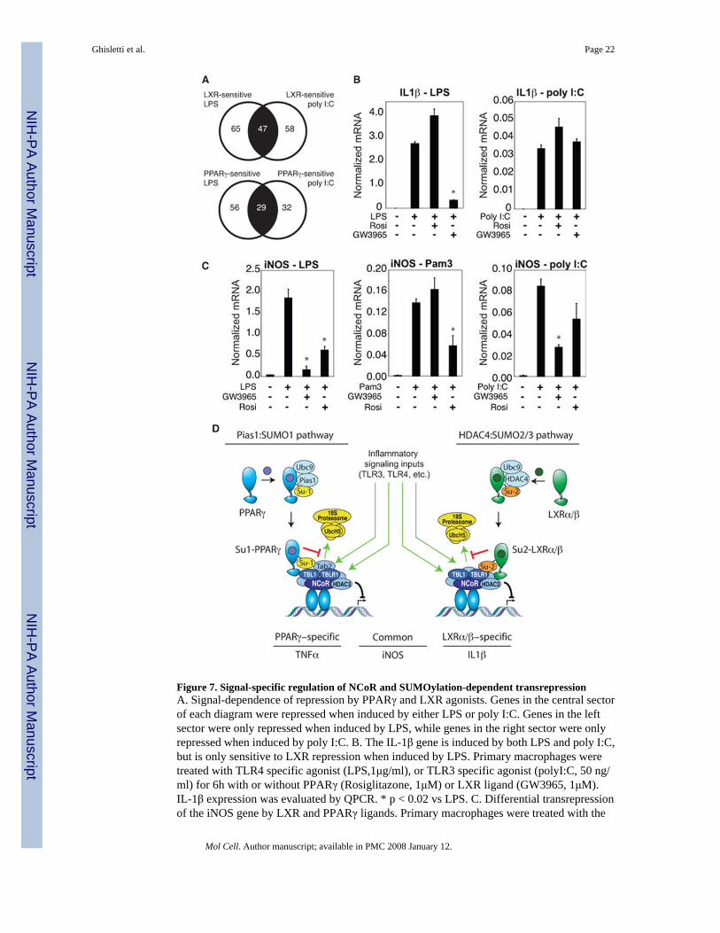
Figure 7. Signal-specific regulation of NCoR and SUMOylation-dependent transrepressionA. Signal-dependence of repression by PPARγ and LXR agonists. Genes in the central sectorof each diagram were repressed when induced by either LPS or poly I:C. Genes in the leftsector were only repressed when induced by LPS, while genes in the right sector were onlyrepressed when induced by poly I:C. B. The IL-1β gene is induced by both LPS and poly I:C,but is only sensitive to LXR repression when induced by LPS. Primary macrophages weretreated with TLR4 specific agonist (LPS,1μg/ml), or TLR3 specific agonist (polyI:C, 50 ng/ml) for 6h with or without PPARγ (Rosiglitazone, 1μM) or LXR ligand (GW3965, 1μM).IL-1β expression was evaluated by QPCR. * p < 0.02 vs LPS. C. Differential transrepressionof the iNOS gene by LXR and PPARγ ligands. Primary macrophages were treated with the
Ghisletti et al. Page 22
Mol Cell. Author manuscript; available in PMC 2008 January 12.
NIH
-PA Author Manuscript
NIH
-PA Author Manuscript
NIH
-PA Author Manuscript
LPS (1 μg/ml), TLR2-specific agonist (PAM3, 300 ng/ml) or poly I:C (50 ng/ml) for 6h withor without PPARγ (Rosiglitazone, 1μM) or LXR ligand (GW3965, 1μM). iNOS expressionwas evaluated by real time PCR. * p < 0.02 vs LPS (left), Pam 3 (middle), poly I:C (right).Results are expressed as the average of 2 independent experiments performed in duplicate.Error bars represent standard deviations. D. Model depicting parallel transrepression pathwaysutilized by PPARγ and LXRs. See text for discussion.
Ghisletti et al. Page 23
Mol Cell. Author manuscript; available in PMC 2008 January 12.
NIH
-PA Author Manuscript
NIH
-PA Author Manuscript
NIH
-PA Author Manuscript
Copyright © 2022 FDOKUMEN