Unraveling DNA helicases : Motif, structure, mechanism and function
Upload
independentCategory
view
1download
0
pubs.acs.org/BiochemistryPublished on Web 12/08/2009r 2009 American Chemical Society
Biochemistry 2010, 49, 1–10 1
DOI: 10.1021/bi901263m
Sumoylation of p68 and p72 RNA Helicases Affects Protein Stabilityand Transactivation Potential†
Steven M. Mooney,‡ Joseph P. Grande,§ Jeffrey L. Salisbury,‡ and Ralf Janknecht*,‡, )
‡Departments of Biochemistry and Molecular Biology, and §Laboratory Medicine and Pathology, Mayo Clinic, Rochester,Minnesota 55905, and )Department of Cell Biology, University of Oklahoma Health Sciences Center, Oklahoma City,
Oklahoma 73104
Received July 23, 2009; Revised Manuscript Received November 12, 2009
ABSTRACT: The p68 (DDX5) and p72 (DDX17) proteins are members of the DEAD-box (DDX) family ofRNA helicases. We show that both p68 and p72 are overexpressed in breast tumors. Bioinformatical analysisrevealed that the SUMO pathway is upregulated in breast tumors and that both p68 and p72 contain oneconsensus sumoylation site, implicating that sumoylation of p68 and p72 increases during breast tumo-rigenesis and potentially contributes to their overexpression. We determined that p68 and p72 are indeedsumoylated at a single, homologous site. Importantly, sumoylation significantly increased the stability ofp68 and p72. In contrast to p72 and consistent with an ∼3-fold lesser half-life, p68 was found to bepolyubiquitylated, and mutation of the sumoylation site increased polyubiquitylation, suggesting thatsumoylation increases p68 half-life by reducing proteasomal degradation. Moreover, whereas p68 robustlycoactivated transcription from an estrogen response element, its sumoylation mutant showed a drasticallyreduced coactivation potential. In contrast, the p68 sumoylation status did not affect the ability to enhancep53-mediatedMDM2 transcription. On the contrary, preventing sumoylation of p72 caused an increase in itsability to transactivate both estrogen receptor and p53. Furthermore, sumoylation promoted the interactionof p68 and p72 with histone deacetylase 1 but had no effect on binding to histone deacetylases 2 and 3, thecoactivator p300, or estrogen receptor and also did not affect homo/heterodimerization of p68/p72. Inconclusion, sumoylation exerts pleiotropic effects on p68/p72, which may have important implications inbreast cancer by modulating estrogen receptor and p53 activity.
RNA helicases are necessary for ribosome biogenesis, pre-mRNA splicing, mRNA export from the nucleus, translationinitiation, and RNA decay. In fact, RNA helicases are indispens-able for all cellular functions that involveRNA (1). A hallmarkofRNA helicases is their ability to hydrolyze ATP to dissociateRNA-RNA or RNA-DNA double strands or to modulateRNA-protein interactions. One subfamily of RNA helicases iscomposed of the DEAD-box (DDX)1 proteins that are char-acterized by a conserved Asp-Glu-Ala-Asp sequence involved inATP hydrolysis. Whereas the catalytic domains are highlyconserved, flanking sequences are variable among the DDXRNA helicases and are thought to contain distinguishing ele-ments that control cellular localization, catalytic activity, andbinding to other proteins (2).
The DDX RNA helicase p68 (also called DDX5) and itsparalogue, p72 (DDX17), are 92% similar within their centralcatalytic domains and share 71% and 44% homology in theirN- and C-termini, respectively. Each of these two proteins isessential for development, since corresponding knockout mousemodels display embryonic lethality (3). Both proteins possessRNA helicase activity and are involved in splicing, but alsofunction in the regulation of gene transcription (4). Indeed,
p68 and p72 RNA helicases interact with and thereby activatevarious transcription factors, including hormone binding pro-teins (estrogen receptor R (ERR) and androgen receptor), p53,MyoD, and Runx2 and the cofactors CBP/p300, PCAF, andβ-catenin (5-15).
Protein expression of p68 and p72 is upregulated in colorectal,prostate, and, as shown in this study, breast cancer, but evidencesuggests that this is not due to enhanced mRNA levels (7, 14, 16).This led us to hypothesize that posttranslational modificationsmight play a role in stabilizing p68/p72 at the protein level intumor cells. In particular, since our analysis of the p68 and p72amino acid sequence revealed one consensus sumoylation site, wefocused in this report on the covalent attachment of SUMO(small ubiquitin-like modifier) to lysine residues within p68 andp72 and how this may affect protein stability.
Four distinct SUMO proteins exist in humans that resembleubiquitin in their three-dimensional structure. SUMO1-SUMO3are ubiquitously expressed and can be covalently attached to lysineresidues in target proteins. This sumoylation is reversible andhighly dynamic and can have profound effects, including regulat-ing the intracellular localization, stability, and activity of targetproteins (17, 18). In particular, many transcription factors havebeen found to be sumoylated, resulting in repression of theiractivity (19). However, a few transcription factors, including thetumor suppressor p53 andheat shock transcription factors 1 and 2,are activated upon sumoylation (20-23). Interestingly, estrogen-dependent transcription is stimulated by the SUMOpathway (24).In part, this may be due to the reported sumoylation of ERR (25),
†Research partially supported by a grant from the Department ofDefense Breast Cancer Research Program (W81XWH-06-1-0492).*To whom correspondence should be addressed. Phone: 405-271-
2377. Fax: 405-271-3548. E-mail: [email protected]: DDX, DEAD box; ERR, estrogen receptor R; ERE,
estrogen response element; HDAC, histone deacetylase; HEK, humanembryonic kidney; SUMO, small ubiquitin-like modifier.

2 Biochemistry, Vol. 49, No. 1, 2010 Mooney et al.
but we speculated and therefore tested here whether sumoylationof the ERR interaction partners, p68 and p72 RNA helicases, isalso involved.
EXPERIMENTAL PROCEDURES
Tumor Stainings. Breast tissue microarrays were purchasedfrom Petagen (AccuMax Array) and then stained as describedbefore (26). Antibodies directed against p68 amino acids501-524 (14) and p72 amino acids 632-650 (12) were employed.In Vivo Sumoylation. HEK293T cells were grown in 6 cm
dishes and transiently transfected with 2 μg of 6Myc-p68/p72 orempty vector pCS3þ-6Myc and 4 μg of the indicated SUMOexpression vector or empty vector pEV3S (27). Lysates weregenerated 36 h after transfection in 10 mM Tris-HCl, 30 mMNa4P2O7 (pH 7.1), 200 mMNaCl, 50 mMNaF, 0.1% SDS, 1%Triton X-100, 10 μg/mL leupeptin, 2 μg/mL aprotinin, 1 μg/mLpepstatin A, 1 mM phenylmethanesulfonyl fluoride, and 10 mMN-ethylmaleimide. Where indicated, immunoprecipitations wereperformed with anti-Myc 9E10 monoclonal antibodies as des-cribed (28). Alternatively, 1 μg of Flag-tagged p68/p72 wascoexpressed with 2 μg of His-tagged yeast ubiquitin expres-sion vector, and cells were treated with 20 μM MG-132 for 6 hbefore lysis in 8 M urea, 0.1 M Na2HPO4 (pH 8), and 10 mMN-ethylmaleimide. His-tagged proteins were then affinity puri-fied on Ni2þ-NTA agarose (Qiagen) according to standardprocedures.Pulse-Chase Experiments. HEK293T cells grown in 6 cm
dishes were transiently transfected by the calcium phosphatecoprecipitationmethod (29, 30) with 8 μg of 6Myc-p68 (wild typeand K53R) or 6Myc-p72 (wild type and K50R). Thirty-six hoursafter transfection, cells were pulsed for 2 h with 100 μCi of[35S]methionine followed by a chase with nonradioactive methio-nine (31). At various time points the cells were harvested andlysed in 10 mM Tris-HCl, 30 mM Na4P2O7 (pH 7.1), 50 mMNaCl, 50 mMNaF, 1% sodium deoxycholate, 1%Triton X-100,10 μg/mL leupeptin, 2 μg/mL aprotinin, 1 μg/mL pepstatin A,1 mM phenylmethanesulfonyl fluoride, 0.5 mM Na3VO4, and0.2 mM DTT. Then, immunoprecipitations were performed asdescribed (32) with anti-p68 antibodies directed against aminoacids 501-524 (14) or anti-Myc 9E10 monoclonal antibodies inthe case of p72 (33). Quantitation of incorporation of radio-activity was done with a PhosphorImager.Coimmunoprecipitation. HEK293T cells grown in 6 cm
dishes were transiently transfected with plasmids encoding for6Myc-p68 or 6Myc-p72 (8 μg) and Flag-ERR (1 μg) (34). Cellswere lysed 36 h after transfection in 2.5 mM Tris-HCl, 7.5 mMNa4P2O7 (pH 7.1), 12.5 mMNaCl, 12.5 mMNaF, 0.25%TritonX-100 supplemented with 10 μg/mL leupeptin, 2 μg/mL aproti-nin, 1 μg/mLpepstatinA, 1mMphenylmethanesulfonyl fluoride,0.5 mMNa3VO4, and 0.2 mMDTT. Immunoprecipitations withanti-Myc 9E10 antibody were then essentially performed asdescribed before (35). Coimmunoprecipitated Flag-ERR wasdetected by Western blotting utilizing anti-Flag M2 anti-bodies (36). Alternatively, HEK293T cells were transfected with3 μg of p300-HA and 1 μg of 6Myc-p68 or 6Myc-p72 constructs,immunoprecipitations were performed with anti-Myc antibodiesas described (37), and coprecipitated p300 was detected by anti-HA Western blotting (38). Similarly, HEK293T cells weretransfected with plasmids encoding HA-p68 (8 μg) and Flag-HDAC (1 μg), and immunoprecipitations with anti-Flag M2antibody were then performed followed by Western blotting
employing anti-HA antibodies. In the case of p72, HEK293Tcells grown in 10 cm dishes were transfected with 20 μg ofHA-p72 and 5 μg of Flag-HDAC expression plasmids.Luciferase Assays. MDA-MB-231 cells grown in 12 wells
were transiently transfected using the calcium phosphate co-precipitation method (39, 40). As reporter plasmid, 500 ng ofERE-luc (41) or 200 ng of MDM2-luc (42) were employed.Where indicated, 30 ng of pSG5-ERR or empty vector pSG5 and10 ng of pcDNA3-p53 or empty vector pcDNA3 were cotrans-fected. HA-tagged p68 or p72 (1600 ng) or empty vectorpEV3S (43) was employed, with the exception of wild-type p72where 500 ng of expression vector plus 1100 ng of pEV3S wascotransfected. In the case of ERE-luc, media were replaced aftertransfection with one containing 5% charcoal-stripped serumwith or without 1 nM estradiol. Thirty-six hours after transfec-tion, cells were lysed, and luciferase activity was measured in aBerthold Lumat (44, 45). Alternatively, protein extracts wereprepared and assayed by Western blotting (46).
Similarly, HCT116 cells were transiently transfected utilizing200 ng of Gal42-tk80-luc reporter plasmid (47), 400 ng of pAB-GAL4-linker (48), which encodes the DNA binding domain ofthe yeast protein GAL4 or the corresponding GAL4-β-cateninplasmid (14), 400 ng of pEV3S or HA-tagged p68 expressionvector, and 25 ng of HA-SUMO1 or 50 ng of ARIP3 (49)expression plasmids. Further, HEK293T cells were transientlytransfected by the calciumphosphate coprecipitationmethod (50)utilizing 200 ng of Gal42-tk80-luc reporter plasmid, 30 ng ofpAB-GAL4-linker or GAL4-p72 (12), and 100 ng of SuPr-1expression vector (51) or empty vector pcDNA3.
RESULTS
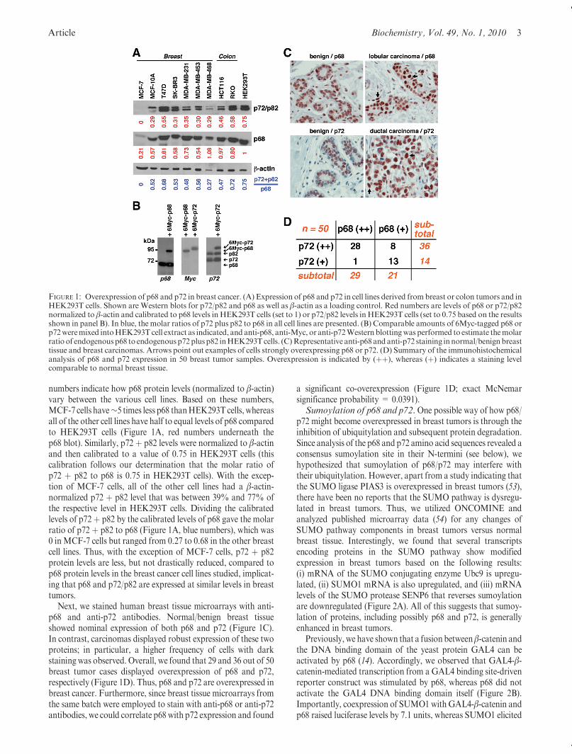
Overexpression of p68 and p72 in Breast Tumors. Insti-gated by the facts that ERR plays an oncogenic role in manyhuman breast tumors and that p68 and p72 are coactivators ofERR (5, 6), we assessed the level of their expression in breasttumors. To this end, we first determined the expression of p68and p72 in established breast cancer cell lines.With the exceptionof MCF-7 cells, all of the other six breast cancer cell linesdisplayed robust expression of p68 and p72 that was comparableto two colon cancer cell lines (RKO and HCT116) and trans-formed human embryonic kidney (HEK) 293T cells (Figure 1A);note that due to an alternative start codon in the p72mRNA (52),a slightly longer p82 isoformwas also detectablewith the anti-p72antibody. Since RKO and HCT116 colon cancer cells havepreviously been shown to overexpress p68 and p72 (14), thesedata suggested that p68 and p72 might also be overexpressed inbreast tumors.
To estimate the molar ratio of p68 to p72/p82 in the variouscell lines shown inFigure 1A,we employed 6Myc-tagged versionsof p68 and p72 that have an∼20 kDa larger apparent molecularmass than respective endogenous proteins. We mixed compar-able amounts of 6Myc-p68 or 6Myc-p72, as determined by anti-MycWestern blotting (Figure 1B, middle panel), into HEK293Tcell extract and then probed with anti-p68 (Figure 1B, left panel)or anti-p72 antibodies (Figure 1B, right panel). This allowed usto estimate that the molar ratio of p72 þ p82 to p68 is 0.75in HEK293T cells. Then, we normalized p68 protein levels toβ-actin levels by densitometric scanning of the respectiveWesternblots in Figure 1A. The resulting unitless numbers were thencalibrated to the one from HEK293T cells, where the β-actin-normalized p68 protein level was arbitrarily set to 1. The resulting
Article Biochemistry, Vol. 49, No. 1, 2010 3
numbers indicate how p68 protein levels (normalized to β-actin)vary between the various cell lines. Based on these numbers,MCF-7 cells have∼5 times less p68 thanHEK293T cells, whereasall of the other cell lines have half to equal levels of p68 comparedto HEK293T cells (Figure 1A, red numbers underneath thep68 blot). Similarly, p72 þ p82 levels were normalized to β-actinand then calibrated to a value of 0.75 in HEK293T cells (thiscalibration follows our determination that the molar ratio ofp72 þ p82 to p68 is 0.75 in HEK293T cells). With the excep-tion of MCF-7 cells, all of the other cell lines had a β-actin-normalized p72 þ p82 level that was between 39% and 77% ofthe respective level in HEK293T cells. Dividing the calibratedlevels of p72þ p82 by the calibrated levels of p68 gave the molarratio of p72 þ p82 to p68 (Figure 1A, blue numbers), which was0 inMCF-7 cells but ranged from 0.27 to 0.68 in the other breastcell lines. Thus, with the exception of MCF-7 cells, p72 þ p82protein levels are less, but not drastically reduced, compared top68 protein levels in the breast cancer cell lines studied, implicat-ing that p68 and p72/p82 are expressed at similar levels in breasttumors.
Next, we stained human breast tissue microarrays with anti-p68 and anti-p72 antibodies. Normal/benign breast tissueshowed nominal expression of both p68 and p72 (Figure 1C).In contrast, carcinomas displayed robust expression of these twoproteins; in particular, a higher frequency of cells with darkstaining was observed. Overall, we found that 29 and 36 out of 50breast tumor cases displayed overexpression of p68 and p72,respectively (Figure 1D). Thus, p68 and p72 are overexpressed inbreast cancer. Furthermore, since breast tissue microarrays fromthe same batch were employed to stain with anti-p68 or anti-p72antibodies, we could correlate p68 with p72 expression and found
a significant co-overexpression (Figure 1D; exact McNemarsignificance probability = 0.0391).Sumoylation of p68 and p72. One possible way of how p68/
p72 might become overexpressed in breast tumors is through theinhibition of ubiquitylation and subsequent protein degradation.Since analysis of the p68 and p72 amino acid sequences revealed aconsensus sumoylation site in their N-termini (see below), wehypothesized that sumoylation of p68/p72 may interfere withtheir ubiquitylation. However, apart from a study indicating thatthe SUMO ligase PIAS3 is overexpressed in breast tumors (53),there have been no reports that the SUMO pathway is dysregu-lated in breast tumors. Thus, we utilized ONCOMINE andanalyzed published microarray data (54) for any changes ofSUMO pathway components in breast tumors versus normalbreast tissue. Interestingly, we found that several transcriptsencoding proteins in the SUMO pathway show modifiedexpression in breast tumors based on the following results:(i) mRNA of the SUMO conjugating enzyme Ubc9 is upregu-lated, (ii) SUMO1 mRNA is also upregulated, and (iii) mRNAlevels of the SUMO protease SENP6 that reverses sumoylationare downregulated (Figure 2A). All of this suggests that sumoy-lation of proteins, including possibly p68 and p72, is generallyenhanced in breast tumors.
Previously, we have shown that a fusion betweenβ-catenin andthe DNA binding domain of the yeast protein GAL4 can beactivated by p68 (14). Accordingly, we observed that GAL4-β-catenin-mediated transcription from a GAL4 binding site-drivenreporter construct was stimulated by p68, whereas p68 did notactivate the GAL4 DNA binding domain itself (Figure 2B).Importantly, coexpression of SUMO1withGAL4-β-catenin andp68 raised luciferase levels by 7.1 units, whereas SUMO1 elicited
FIGURE 1: Overexpression of p68 and p72 in breast cancer. (A) Expression of p68 and p72 in cell lines derived frombreast or colon tumors and inHEK293T cells. Shown are Western blots for p72/p82 and p68 as well as β-actin as a loading control. Red numbers are levels of p68 or p72/p82normalized to β-actin and calibrated to p68 levels in HEK293T cells (set to 1) or p72/p82 levels in HEK293T cells (set to 0.75 based on the resultsshown in panel B). In blue, the molar ratios of p72 plus p82 to p68 in all cell lines are presented. (B) Comparable amounts of 6Myc-tagged p68 orp72weremixed intoHEK293T cell extract as indicated, and anti-p68, anti-Myc, or anti-p72Western blottingwas performed to estimate themolarratio of endogenousp68 to endogenousp72plusp82 inHEK293Tcells. (C)Representative anti-p68and anti-p72 staining innormal/benignbreasttissue and breast carcinomas. Arrows point out examples of cells strongly overexpressing p68 or p72. (D) Summary of the immunohistochemicalanalysis of p68 and p72 expression in 50 breast tumor samples. Overexpression is indicated by (þþ), whereas (þ) indicates a staining levelcomparable to normal breast tissue.

4 Biochemistry, Vol. 49, No. 1, 2010 Mooney et al.
only a small increase (1.6 units) of GAL4-β-catenin luciferaseactivity in the absence of overexpressed p68, implicating thatsumoylation may activate p68. Accordingly, overexpression ofARIP3, a SUMO ligase also called PIASxR (49), synergized withp68 and GAL4-β-catenin, whereas the ARIP3-W383A mutantthat is impaired in its SUMO ligase activity had no effect(Figure 2B).
Further, we previously reported that GAL4-p72 exerts arepressive effect on GAL4 binding site-driven transcription (12).Surprisingly, overexpression of a SUMO protease, SuPr-1 (51),alleviated this repression mediated by GAL4-p72, whereasSuPr-1 had no effect on the GAL4 moiety itself (Figure 2C).These data suggested that sumoylation of p72 might repress itstranscriptional function.
Prompted by the aforementioned results raising the possibilitythat p68 and p72 are sumoylated and thereby regulated in theiractivities, we wished to determine whether p68 and p72 are indeedposttranslationally modified by SUMO. Thus, we expressedMyc-tagged p68 and p72 with and without Flag-tagged SUMO1in HEK293T cells. Anti-Myc Western blotting revealed an addi-tional band at higher molecular mass (see asterisks in Figure 2D,left panel) only in the presence of coexpressed Flag3-SUMO1,suggesting that both p68 and p72 become sumoylated. Indeed,anti-Myc immunoprecipitation followed by anti-Flag Westernblotting proved that these higher molecular mass bands aresumoylated forms of p68 and p72 (Figure 2D, right panel).
To identify the sumoylation sites in p68/p72, we searched forthe sumoylation consensus motif ψKxE, where ψ is an aliphaticbranched amino acid and x any amino acid (18). p68 and p72
were found to have only one such site, which was located withintheir N-termini at K53 in p68 and K50 in p72 (Figure 3A). Thus,we mutated K53 to arginine in p68 and assessed how this wouldaffect sumoylation. As shown in Figure 3B (left panel), therespective K53Rmutant did not display a higher molecular massband in the presence of HA-SUMO1 or Flag3-SUMO1, indicat-ing that indeed K53 is the attachment site for SUMO in p68.Similarly, we assessed whether the homologous K50 is thesumoylation site in p72. Indeed, p72-K50R lost the highermolecular mass band in the presence of HA-SUMO1 or Flag3-SUMO1 (Figure 3B, right panel), demonstrating that K50 is thesite for SUMO attachment in p72. While this work was inprogress, another laboratory also reported that p68 becomessumoylated on K53 but did not assess sumoylation of p72 (55).
We also tested whether SUMO2 is capable of sumoylating p68and p72. SUMO2 was expressed at a similar level as SUMO1 inHEK293T cells (Figure 3C). However, we were unable to detectsignificant sumoylation with SUMO2 on p68, and also SUMO2attachment to p72 was less efficient than attachment of SUMO1(Figure 3C). This indicates that SUMO1, rather than SUMO2, isthe main SUMO variant modifying p68 and p72.Sumoylation Increases the Half-Life of p68 and p72.
Previously, it was reported that p68 is polyubiquitylated (16) andits stability may therefore be regulated through the ubiquitinpathway. Indeed, we found that p68 was significantly poly-ubiquitylated in HEK293T cells (Figure 4A). In contrast, verylittle monoubiquitylation and no polyubiquitylation were ob-servable in the case of p72. Thus, we focused on p68 and foundthat sumoylation of p68 appears to suppress ubiquitylation, since
FIGURE 2: Changes in transcripts encoding for sumoylation pathway components in breast cancer and evidence for sumoylated p68 and p72. (A)Analysis ofmicroarray data from 40 tumor (T) and 7 normal (N) breast tissues. Normalization was done by log 2 transformingmRNAdata sets,with themedian set to 0 and the standard deviation set to 1. Shown aremedians and 25th/75th percentile ranges. Significancewas determinedwithStudent’s t test. (B) SUMO1or the SUMO ligaseARIP3 stimulate p68.As indicated,GAL4orGAL4-β-cateninwere cotransfectedwith p68 intoHCT116 cells and luciferase activities from a GAL4 binding site-driven reporter construct measured. (C) The SUMO protease SuPr-1 alleviatesp72-dependent transcriptional repression. GAL4 or GAL4-p72 were expressed with or without SuPr-1 in 293T cells, and repression of thecotransfected GAL4 binding site-driven luciferase reporter was measured. (D) 6Myc-tagged p68 or p72 were coexpressed with Flag3-SUMO1 inHEK293T cells. The left panel shows an anti-Myc Western blot of cell lysates and the right panel an anti-Flag Western blot after anti-Mycimmunoprecipitation. Asterisks mark higher molecular mass species representing sumoylated p68/p72. The bracket indicates unmodifiedp68/p72.

Article Biochemistry, Vol. 49, No. 1, 2010 5
the K53R mutant was approximately twice as much ubiquity-lated as wild-type p68 (Figure 4B); this difference was alsopresent when no ectopic ubiquitin was expressed (Figure 4C).These data suggest that sumoylation enhances the stability ofp68 by preventing proteasomal degradation.
To test this hypothesis, we performed pulse-chase experi-ments with [35S]methionine and then determined the decay ofradioactively labeled p68 over time; please note that levels ofnonmodified 35S-labeled p68 were measured, since the amountsof both sumoylated and ubiquitylated p68 were below the
FIGURE 3: Mapping of sumoylation sites. (A) Diagrams of p68 and p72. The sequences surrounding the predicted sumoylation sites K53 in p68and K50 in p72 are presented. (B) HEK293T cells were transfected with indicated expression constructs. The top shows a longer exposure of ananti-Myc Western blot, with asterisks marking the sumoylated proteins. The bottom panels are shorter exposures to show that comparableamounts of respectiveMyc-tagged proteins were expressed. (C) HA-tagged SUMO1 or SUMO2 were coexpressed with 6Myc-tagged p68 or p72as indicated. The top panel shows a longer exposure of an anti-MycWestern blot and the middle panel a shorter exposure to reveal comparableloading of p68 and its K53Rmutant or of p72 and its K50Rmutant. The bottom panel is an anti-HA blot showing input levels of SUMO1 andSUMO2.
FIGURE 4: Ubiquitylation and half-lives of p68 and p72. (A) Flag-tagged p68 or p72 were coexpressed with His-tagged ubiquitin in HEK293Tcells. After pull-down of His-tagged proteins with Ni2þ-NTA, ubiquitylated p68 and p72 were found by anti-FlagWestern blotting. The bottompanel shows the input levels of p68 and p72 at a shorter exposure. (B) Analogous comparison of the ubiquitylation of wild-type p68 and its K53Rmutant. (C) HA-tagged indicated p68 and p72 proteins were expressed in HEK293T cells and their ubiquitylated forms detected by anti-HAWestern blotting of cell extracts. The bottomblot is a shorter exposure showing themutated proteins to be expressed at the same level as respectivewild-type ones. (D) Representative pulse-chase experiment with [35S]methionine. The decay of radioactive p68 (wild-type and K53R mutant)over time is displayed.Half-lives correspond to the hours of chasewhere 50%of radioactivity (plotted ona logarithmic scale) remains.Meanhalf-lives with standard deviations are given in brackets. (E) Similar for p72.

6 Biochemistry, Vol. 49, No. 1, 2010 Mooney et al.
detection limit in this experiment. Whereas wild-type p68 had ahalf-life of 20.2 h, its K53Rmutant displayed a markedly shorterhalf-life of 11.5 h (Figure 4D). Consistent with being much lessubiquitylated than p68, p72 displayed a >3-fold longer half-lifecompared to p68 (Figure 4E). Again, the sumoylation sitemutantwas less stable than wild-type p72 (54.4 versus 67.7 h half-life),although the difference was less pronounced than that of p68.Altogether, these results indicate that sumoylation increases thehalf-lives of p72 and especially p68.Impact of Sumoylation on Binding to Partner Proteins.
Posttranslational modifications can affect protein-protein inter-actions. Several interactants of p68 andp72 have been uncovered,including the coactivator p300 and ERR (5, 6, 11, 12). Thus, wetested whether p68/p72 and their respective sumoylation sitemutants differentially bind to these two proteins in coimmuno-precipitation experiments. Compared to wild-type p68/p72, therewas no difference in the ability of the K53R/K50R mutants tobind to p300 or ERR (Figure 5A,B). Since p68 and p72 formbothhomo- and heterodimers (56), we also assessed the interaction of6Myc-tagged p68/p72 with endogenous p68/p72. Note that6Myc-tagged p68 and p72 electrophorese at ∼20 kDa aboveand are therefore easily separated from endogenous p68 and p72.Again, we did not find a difference in the ability of the 6Myc-p68-K53R/6Myc-p72-K50Rmutants to form homo- or heterodimerswith endogenous p68 or p72 compared to wild-type 6Myc-p68/p72 (Figure 5C).
Previously, it was shown that p68 and p72 also interact withthe histone deacetylase (HDAC) 1 (57). Thus, we addition-ally tested the ability of p68 and p72 to interact with variousHDACs. p68 bound expectedly to HDAC1 and also to HDAC2
and HDAC3 as revealed by coimmunoprecipitation assays(Figure 5D). Importantly, the sumoylation site mutant of p68bound lesswell toHDAC1 thanwild-type p68, whereas therewasno difference in the case of HDAC2 or HDAC3. Similarly,mutation of K50 in p72 resulted in drastically diminshed bindingto HDAC1 but did not affect binding to HDAC2 or HDAC3(Figure 5E). These data indicate that sumoylation selectivelypromotes the interaction of p68 and p72withHDAC1.While thiswork was in progress, another report also demonstrated thatp68 sumoylation enhances binding to HDAC1 (55).Sumoylation Modulates Transcriptional Coactivation.
Both p68 and p72 have been reported to coactivate ERR (5, 6).Thus, we studied how sumoylation might affect this property ofp68/p72 by utilizing MDA-MB-231 breast cancer cells that areERR-negative and a luciferase reporter that is driven by anestrogen response element (ERE). Expectedly, in the absence ofcotransfected ERR or in the absence of estrogen stimulation, noluciferase activity was measurable. However, when ERR wasstimulated with estrogen, robust luciferase activity was observed(Figure 6A). When wild-type p68 was coexpressed, it stimulatedERR activity >6-fold. In contrast, the K53R mutant showed amuch reduced ability to coactivate (∼2-fold). These data indicatethat sumoylation enhances the ability of p68 to coactivate ERR.
Another promoter we investigated wasMDM2, a target of thetumor suppressor p53 andwhose transcription has been shown tobe enhanced by p68 and p72 (8, 12). We employed an MDM2luciferase construct in the p53-negative MDA-MB-231 cells todetermine the impact of p68 sumoylation on this promoter. p68synergized with p53 in the activation of the MDM2 promoter(Figure 6B). However, there was no difference in this abilitywhen
FIGURE 5: Impact of sumoylation on protein interactions. (A) Interaction with p300. Myc-tagged p68/p72 were coexpressed with HA-taggedp300. After anti-Myc immunoprecipitation, coprecipitated p300 was revealed by anti-HAWestern blotting. The bottom two panels show inputlevels of p300-HA and Myc-tagged p68/p72. (B) Similar interaction of Myc-tagged p68/p72 with Flag-tagged ERR in the presence and absenceof estrogen. (C) Homo- and heterodimerization of Myc-tagged p68/p72 with endogenous p68 or p72/p82. (D) Coimmunoprecipitation ofHA-tagged p68 with Flag-tagged HDACs. (E) Similar interaction of p72 with HDACs.

Article Biochemistry, Vol. 49, No. 1, 2010 7
utilizing the K53R mutant. Thus, sumoylation affects the trans-activation potential of p68 in a promoter-specific context.
Similarly, we tested the impact of sumoylation on the p72coactivation potential. Surprisingly, the p72-K50R mutant wasmore active than wild-type p72 at both the ERE and MDM2promoters (Figure 6C,D). This suggests that sumoylation of p72represses its coactivation function and marks an unexpecteddifference between p68 and p72. These findings are consistentwith our results shown in Figure 2B,C, where sumoylationappeared to stimulate the coactivation potential of p68 and toenhance p72-mediated transcriptional repression.
Sumoylation has been shown to influence the intracellularlocalization of several proteins (17, 18). Thus, it is conceivablethat the altered coactivation potential of the p68 and p72sumoylation mutants is simply a reflection of a changed intra-cellular localization. However, we did not find any evidence thatthe p68-K53R or p72-K50R mutants localize differently withincells compared to the respective wild-type proteins (SupportingInformation Figure 1).
DISCUSSION
About 70% of breast tumors are ERR-positive, and therapiestargeting estrogen or ERR have been developed and proveneffective to fight early stage breast cancer (58). However,
acquired or inherent endocrine resistance is a major problem inthe clinic. One mechanism by which ERR may become indepen-dent of estrogen or insensitive to antiestrogens is throughenhanced stimulation by cofactors (59). This report is the firstdemonstration that the ERR cofactors p68 and p72 are over-expressed in the majority of human breast tumors and maythereby cause aberrant activation of ERR and contribute tocarcinogenesis.
In addition, we demonstrated that p68 and p72 can besumoylated on one particular lysine residue, K53 or K50,respectively, which are at homologous sites in the N-termini ofthese RNA helicases. While this work was in progress, anotherreport established similarly that K53 is the sumoylation site ofp68 but did not address sumoylation of p72 (55). Our resultsshowed that SUMO1 was more efficiently added onto p68 andp72 than SUMO2 in HEK293T cells, but this may be cell-typedependent since the opposite preference was reported for p68 inCOS-7 cells (55).
The SUMO pathway is hyperactivated in breast cancer basedon published data indicating that the SUMO ligase PIAS3 isoverexpressed (53) and our bioinformatical analysis revealingthat SUMO1 and the SUMO conjugating enzyme Ubc9 areupregulated whereas the SUMO protease SENP6 is downregu-lated in breast tumors. Importantly, overexpression of Ubc9 inMCF-7 breast cancer cells led to larger tumors in a xenograftmouse model, while a dominant-negative Ubc9 molecule sup-pressed tumor growth (60). These data suggest that a hyper-activated SUMO pathway contributes to breast tumor growth,possibly by stimulating estrogen-dependent transcription that isactivated by the SUMOpathway (24). Therefore, sumoylation ofthe ERR cofactors, p68 and p72, is likely to be relevant in breastcarcinogenesis.
Mutation of the sumoylation site in p68markedly decreased itshalf-life, and the mutation of K50 in p72 also resulted in a smallbut significant decrease of protein stability. This indicates thatsumoylation stabilizes p68/p72 and provides one explanation ofhow a hyperactivated SUMOpathway causes, at least in part, theobserved overexpession of p68/p72 in breast tumors. Sumoyla-tion may increase the stability of a protein by simply preventingubiquitylation at the same lysine residue as shown, for instance,for IκBR orMDM2 (61, 62). This is not the case for p68, since theK53Rmutant, which can neither be sumoylated nor be ubiquity-lated at position 53, is even more ubiquitylated than wild-typep68. Rather, sumoylation appears to prevent ubiquitylation at alysine residue(s) other than K53 in p68, possibly by stericallyinhibiting the docking of ubiquitin ligases to p68.
Sumoylation also affected the transcriptional activity of p68and p72. Our data revealed a significant enhancement of ERR-mediated transcription by sumoylated p68, which demonstrateshow sumoylation of p68 may promote breast tumorigenesis.However, the opposite held true for p72: sumoylation appearedto repress the ERE luciferase reporter. Thus, enhanced sumoyla-tion may dampen the activating effect of p72 overexpression onERR in breast tumors.
In the case of the MDM2 promoter, a prominent target of thetumor suppressor p53 (63), sumoylation of p68 had no impact onits ability to coactivate p53, which indicates that the importanceof sumoylation on p68 is promoter specific. In contrast to ourdata, a previous report showed that the K53R mutant of p68activated a luciferase gene driven by multiple synthetic p53 sites1.5 times more efficiently than wild-type p68 (55), furthersuggesting that sumoylation of p68 has promoter-dependent
FIGURE 6: Sumoylation of p68/p72 modulates transcription. (A)HA-tagged p68 (wild type or K53R mutant) was expressed inMDA-MB-231 cells. ERR or empty expression vector pSG5 werecotransfected and cells stimulated with estrogen as indicated. Resul-tant luciferase activities derived from the cotransfected ERE-lucreporter are depicted. The inset shows an anti-HA blot indicatingthat comparable amounts of wild-type andK53Rp68were expressedupon ERR cotransfection and estrogen stimulation. (B) Similarstimulation of the MDM2 promoter by p68. Where indicated, p53or the empty pcDNA3 expression vectorwas cotransfected. The insetis an anti-HA blot showing the comparable expression of wild-typeand K53R p68 upon p53 coexpression. (C, D) Analogous activationof the ERE-luc reporter andMDM2-luc promoter by p72 (wild typeor K50R mutant).

8 Biochemistry, Vol. 49, No. 1, 2010 Mooney et al.
effects on p53 and may be neutral or repressing. Of note,advanced, chemotherapy-resistant breast cancer is characterizedby loss of p53 transcriptional activity (64), indicating that p68and p72 overexpression may play an oncogenic role by upregu-lating MDM2 transcription and thereby facilitating MDM2-mediated destruction of the tumor suppressor p53. Unlike p68,mutation of the p72 sumoylation site enhanced the ability ofp72 to coactivate the MDM2 promoter, suggesting that sumoy-lation of p72 could suppress tumor formation by reducingMDM2 transcription and thereby stabilizing p53.
How can one explain the impact of sumoylation on thecoactivation potential? One property that could be influencedby sumoylation is the recruitment of other cofactors. However,we found no impact of p68/p72 sumoylation on the recruitmentof the coactivator p300, the histone deacetylases HDAC2 andHDAC3, or ERR and also no change in the ability to homo/heterodimerize. But we observed that HDAC1 recruitment issuppressed when the sumoylation site in p68 or p72 was mutated.This clearly represents a proof of principle that sumoylation canmodulate p68/p72 by selectively regulating the binding to anotherprotein (HDAC1) and should instigate further studies on howassociation with other p68/p72 interaction partners is modulatedby sumoylation.
Consistent with many reports describing the sumoylation ofvarious proteins (17-19), only a very small fraction of p68 orp72 is sumoylated (see Figure 2D), raising the issue of how thismay affect p68 and p72 function. In particular, when wedetermined the ability of p68 or p72 to coimmunoprecipitatewith HDAC1, we did not measure how much sumoylated p68/p72 coimmunoprecipitated (this was below our detection limit)but howmuch nonsumoylated p68/p72did, and thiswas differentbetween wild type and sumoylation mutants. Thus, it appearsthat sumoylation of p68/p72 positively affects binding of non-modified p68/p72 to HDAC1.
Several models have been put forward to explain such a fact,all taking into account the high dynamics of intracellularsumoylation guaranteeing any target protein to be modified bySUMO for at least a very short time span (18, 65). One modelsuggests that sumoylation induces a conformational change in atarget protein that persists after desumoylation. Accordingly,such an irreversible conformational change may be required forp68/p72 to interact with HDAC1. A second model proposes thatsumoylation increases the rate constant for complex associationbut does not affect the dissociation constant. If so, sumoylationof p68/p72 is predicted to greatly increase the association withHDAC1, whereas subsequent desumoylation of p68/p72 will notaffect the release of HDAC1 from p68/p72 complexes. Again,p68/p72 sumoylation, albeit transient and at a low steady-statelevel, will thereby greatly promote the complex formation withHDAC1. These models also explain how sumoylation increasesthe half-lives of nonsumoylated p68 and p72: an irreversibleconformational change or sequestration into a protein complexinduced by transient sumoylation conceivably precludes ubi-quitylation and thus proteasomal destruction.
Histone deacetylation is commonly thought to suppress genetranscription. In line with this, reduced HDAC1 binding of theK50R mutant of p72 correlated with an enhanced coactivationpotential. However, a few studies indicate that HDAC1 can alsoactivate transcription, for instance, at certain interferon targetgenes (66, 67). Thus, it remains to be studied whether enhancedinteraction with HDAC1 is one of the underlying causes of whysumoylation of p68 leads to increased coactivation of ERR.
In conclusion, our study has revealed that p68 and p72 areoverexpressed in breast tumors and may therefore contribute tobreast carcinogenesis by augmenting ERR. In addition, hyper-activation of the SUMO pathway and the resultant enhancedsumoylation of p68 and p72 may be an underlying cause of theiroverexpression in breast cancer. Moreover, p68 sumoylation in-creases its coactivation potential and will thereby further stimulateERR function. By contrast, sumoylation of p72 reduces estrogen-dependent transcription and MDM2 promoter activity. It issuspected that the balance of all these effects determines whethersumoylation of p68 and p72 promotes breast tumorigenesis.
SUPPORTING INFORMATION AVAILABLE
One figure as described in the text. This material is availablefree of charge via the Internet at http://pubs.acs.org.
REFERENCES
1. Bleichert, F., and Baserga, S. J. (2007) The long unwinding road ofRNA helicases. Mol. Cell 27, 339–352.
2. Rocak, S., and Linder, P. (2004) DEAD-box proteins: the drivingforces behind RNAmetabolism.Nat. Rev. Mol. Cell Biol. 5, 232–241.
3. Fukuda, T., Yamagata, K., Fujiyama, S., Matsumoto, T., Koshida,I., Yoshimura, K., Mihara, M., Naitou, M., Endoh, H., Nakamura,T., Akimoto, C., Yamamoto, Y., Katagiri, T., Foulds, C., Takezawa,S.,Kitagawa,H., Takeyama,K.,O’Malley, B.W., andKato, S. (2007)DEAD-box RNA helicase subunits of the Drosha complex arerequired for processing of rRNA and a subset of microRNAs. Nat.Cell Biol. 9, 604–611.
4. Fuller-Pace, F. V. (2006) DExD/H box RNA helicases: multifunc-tional proteins with important roles in transcriptional regulation.Nucleic Acids Res. 34, 4206–4215.
5. Endoh, H., Maruyama, K., Masuhiro, Y., Kobayashi, Y., Goto, M.,Tai, H., Yanagisawa, J., Metzger, D., Hashimoto, S., and Kato, S.(1999) Purification and identification of p68 RNA helicase acting as atranscriptional coactivator specific for the activation function 1 ofhuman estrogen receptor alpha. Mol. Cell. Biol. 19, 5363–5372.
6. Watanabe,M.,Yanagisawa, J., Kitagawa,H., Takeyama,K., Ogawa,S., Arao, Y., Suzawa, M., Kobayashi, Y., Yano, T., Yoshikawa, H.,Masuhiro, Y., and Kato, S. (2001) A subfamily of RNA-bindingDEAD-box proteins acts as an estrogen receptor alpha coactivatorthrough the N-terminal activation domain (AF-1) with an RNAcoactivator, SRA. EMBO J. 20, 1341–1352.
7. Clark, E. L., Coulson, A., Dalgliesh, C., Rajan, P., Nicol, S. M.,Fleming, S., Heer, R., Gaughan, L., Leung, H. Y., Elliott, D. J.,Fuller-Pace, F. V., andRobson, C. N. (2008) The RNAhelicase p68 isa novel androgen receptor coactivator involved in splicing and isoverexpressed in prostate cancer. Cancer Res. 68, 7938–7946.
8. Bates, G. J., Nicol, S. M., Wilson, B. J., Jacobs, A. M., Bourdon, J. C.,Wardrop, J., Gregory,D. J., Lane, D. P., Perkins,N.D., andFuller-Pace,F. V. (2005) The DEAD box protein p68: a novel transcriptionalcoactivator of the p53 tumour suppressor. EMBO J. 24, 543–553.
9. Caretti, G., Schiltz, R. L., Dilworth, F. J., Di Padova, M., Zhao, P.,Ogryzko, V., Fuller-Pace, F. V., Hoffman, E. P., Tapscott, S. J., andSartorelli, V. (2006) The RNA helicases p68/p72 and the noncodingRNA SRA are coregulators of MyoD and skeletal muscle differentia-tion. Dev. Cell 11, 547–560.
10. Jensen, E. D., Niu, L., Caretti, G., Nicol, S. M., Teplyuk, N., Stein,G. S., Sartorelli, V., van Wijnen, A. J., Fuller-Pace, F. V., andWestendorf, J. J. (2008) p68 (Ddx5) interacts with Runx2 andregulates osteoblast differentiation. J. Cell. Biochem. 103, 1438–1451.
11. Rossow, K. L., and Janknecht, R. (2003) Synergism between p68RNA helicase and the transcriptional coactivators CBP and p300.Oncogene 22, 151–156.
12. Shin, S., and Janknecht, R. (2007) Concerted activation of the Mdm2promoter by p72RNAhelicase and the coactivators p300 and P/CAF.J. Cell. Biochem. 101, 1252–1265.
13. Yang, L., Lin, C., Zhao, S., Wang, H., and Liu, Z. R. (2007)Phosphorylation of p68 RNA helicase plays a role in platelet-derivedgrowth factor-induced cell proliferation by up-regulating cyclin D1and c-Myc expression. J. Biol. Chem. 282, 16811–16819.
14. Shin, S., Rossow, K. L., Grande, J. P., and Janknecht, R. (2007)Involvement of RNA helicases p68 and p72 in colon cancer. CancerRes. 67, 7572–7578.

Article Biochemistry, Vol. 49, No. 1, 2010 9
15. Yang, L., Lin, C., and Liu, Z. R. (2006) P68 RNA helicase mediatesPDGF-induced epithelial mesenchymal transition by displacing axinfrom beta-catenin. Cell 127, 139–155.
16. Causevic, M., Hislop, R. G., Kernohan, N. M., Carey, F. A., Kay,R. A., Steele, R. J., and Fuller-Pace, F. V. (2001) Overexpression andpoly-ubiquitylation of theDEAD-boxRNAhelicase p68 in colorectaltumours. Oncogene 20, 7734–7743.
17. Johnson, E. S. (2004) Protein modification by SUMO. Annu. Rev.Biochem. 73, 355–382.
18. Geiss-Friedlander, R., and Melchior, F. (2007) Concepts in sumoyla-tion: a decade on. Nat. Rev. Mol. Cell. Biol. 8, 947–956.
19. Girdwood, D.W., Tatham,M. H., and Hay, R. T. (2004) SUMO andtranscriptional regulation. Semin. Cell Dev. Biol. 15, 201–210.
20. Gostissa, M., Hengstermann, A., Fogal, V., Sandy, P., Schwarz,S. E., Scheffner, M., and Del Sal, G. (1999) Activation of p53 byconjugation to the ubiquitin-like protein SUMO-1. EMBO J. 18,6462–6471.
21. Rodriguez, M. S., Desterro, J. M., Lain, S., Midgley, C. A., Lane,D. P., and Hay, R. T. (1999) SUMO-1 modification activates thetranscriptional response of p53. EMBO J. 18, 6455–6461.
22. Goodson, M. L., Hong, Y., Rogers, R., Matunis, M. J., Park-Sarge,O. K., and Sarge, K. D. (2001) Sumo-1 modification regulates theDNA binding activity of heat shock transcription factor 2, a pro-myelocytic leukemia nuclear body associated transcription factor.J. Biol. Chem. 276, 18513–18518.
23. Hong, Y., Rogers, R., Matunis, M. J., Mayhew, C. N., Goodson,M. L., Park-Sarge, O. K., and Sarge, K. D. (2001) Regulation of heatshock transcription factor 1 by stress-induced SUMO-1modification.J. Biol. Chem. 276, 40263–40267.
24. Chauchereau, A., Amazit, L., Quesne,M., Guiochon-Mantel, A., andMilgrom, E. (2003) Sumoylation of the progesterone receptor and ofthe steroid receptor coactivator SRC-1. J. Biol. Chem. 278, 12335–12343.
25. Sentis, S., Le Romancer,M., Bianchin, C., Rostan,M. C., and Corbo,L. (2005) Sumoylation of the estrogen receptor alpha hinge regionregulates its transcriptional activity. Mol. Endocrinol. 19, 2671–2684.
26. Fuchs, B., Inwards, C. Y., and Janknecht, R. (2004) Vascularendothelial growth factor expression is up-regulated by EWS-ETSoncoproteins and Sp1 and may represent an independent predictor ofsurvival in Ewing’s sarcoma. Clin. Cancer Res. 10, 1344–1353.
27. Kim, T. D., Shin, S., and Janknecht, R. (2008) Repression of Smad3activity by histone demethylase SMCX/JARID1C.Biochem. Biophys.Res. Commun. 366, 563–567.
28. Shin, S., and Janknecht, R. (2007) Activation of androgen receptor byhistone demethylases JMJD2A and JMJD2D.Biochem. Biophys. Res.Commun. 359, 742–746.
29. Bosc, D. G., Goueli, B. S., and Janknecht, R. (2001) HER2/Neu-mediated activation of the ETS transcription factor ER81 andits target gene MMP-1. Oncogene 20, 6215–6224.
30. Janknecht, R. (2003) Regulation of the ER81 transcription factor andits coactivators by mitogen- and stress-activated protein kinase 1(MSK1). Oncogene 22, 746–755.
31. Goel, A., and Janknecht, R. (2003) Acetylation-mediated transcrip-tional activation of the ETS protein ER81 by p300, P/CAF, andHER2/Neu. Mol. Cell. Biol. 23, 6243–6254.
32. Papoutsopoulou, S., and Janknecht, R. (2000) Phosphorylation ofETS transcription factor ER81 in a complex with its coactivatorsCREB-binding protein and p300. Mol. Cell. Biol. 20, 7300–7310.
33. Shin, S., and Janknecht, R. (2007)Diversity within the JMJD2histonedemethylase family. Biochem. Biophys. Res. Commun. 353, 973–977.
34. Goueli, B. S., and Janknecht, R. (2003) Regulation of telomerasereverse transcriptase gene activity by upstream stimulatory factor.Oncogene 22, 8042–8047.
35. Goel, A., and Janknecht, R. (2004) Concerted activation of ETSprotein ER81 by p160 coactivators, the acetyltransferase p300 andthe receptor tyrosine kinase HER2/Neu. J. Biol. Chem. 279, 14909–14916.
36. Knebel, J., De Haro, L., and Janknecht, R. (2006) Repression oftranscription by TSGA/Jmjd1a, a novel interaction partner of theETS protein ER71. J. Cell. Biochem. 99, 319–329.
37. Rossow, K. L., and Janknecht, R. (2001) The Ewing’s sarcoma geneproduct functions as a transcriptional activator. Cancer Res. 61,2690–2695.
38. Janknecht, R. (2001) Cell type-specific inhibition of the ETS tran-scription factor ER81 by mitogen-activated protein kinase-activatedprotein kinase 2. J. Biol. Chem. 276, 41856–41861.
39. Shin, S., Bosc, D. G., Ingle, J. N., Spelsberg, T. C., and Janknecht, R.(2008) Rcl is a novel ETV1/ER81 target gene upregulated in breasttumors. J. Cell. Biochem. 105, 866–874.
40. Bosc, D. G., and Janknecht, R. (2002) Regulation of HER2/Neupromoter activity by the ETS transcription factor, ER81. J. Cell.Biochem. 86, 174–183.
41. Paech, K., Webb, P., Kuiper, G. G., Nilsson, S., Gustafsson, J.,Kushner, P. J., and Scanlan, T. S. (1997) Differential ligand activationof estrogen receptors ERalpha and ERbeta at AP1 sites. Science 277,1508–1510.
42. Ries, S., Biederer, C., Woods, D., Shifman, O., Shirasawa, S.,Sasazuki, T., McMahon, M., Oren, M., and McCormick, F. (2000)Opposing effects of Ras on p53: transcriptional activation of mdm2and induction of p19ARF. Cell 103, 321–330.
43. Janknecht, R. (1996) Analysis of the ERK-stimulated ETS transcrip-tion factor ER81. Mol. Cell. Biol. 16, 1550–1556.
44. Wu, J., and Janknecht, R. (2002) Regulation of the ETS transcriptionfactor ER81 by the 90-kDa ribosomal S6 kinase 1 and protein kinaseA. J. Biol. Chem. 277, 42669–42679.
45. Dowdy, S. C.,Mariani, A., and Janknecht, R. (2003)HER2/Neu- andTAK1-mediated up-regulation of the transforming growth factorbeta inhibitor Smad7 via the ETS protein ER81. J. Biol. Chem. 278,44377–44384.
46. Goueli, B. S., and Janknecht, R. (2004) Upregulation of the catalytictelomerase subunit by the transcription factor ER81 and oncogenicHER2/Neu, Ras, or Raf. Mol. Cell. Biol. 24, 25–35.
47. Janknecht, R., and Hunter, T. (1997) Convergence of MAP kinasepathways on the ternary complex factor Sap-1a. EMBO J. 16, 1620–1627.
48. De Haro, L., and Janknecht, R. (2002) Functional analysis of thetranscription factor ER71 and its activation of the matrix metallo-proteinase-1 promoter. Nucleic Acids Res. 30, 2972–2979.
49. Kotaja, N., Karvonen, U., Janne, O. A., and Palvimo, J. J. (2002)PIAS proteins modulate transcription factors by functioning asSUMO-1 ligases. Mol. Cell. Biol. 22, 5222–5234.
50. De Haro, L., and Janknecht, R. (2005) Cloning of the murine ER71gene (Etsrp71) and initial characterization of its promoter. Genomics85, 493–502.
51. Best, J. L., Ganiatsas, S., Agarwal, S., Changou, A., Salomoni, P.,Shirihai, O., Meluh, P. B., Pandolfi, P. P., and Zon, L. I. (2002)SUMO-1 protease-1 regulates gene transcription through PML.Mol.Cell 10, 843–855.
52. Uhlmann-Schiffler, H., R€ossler, O. G., and Stahl, H. (2002) ThemRNA of DEAD box protein p72 is alternatively translated into an82-kDa RNA helicase. J. Biol. Chem. 277, 1066–1075.
53. Wang, L., and Banerjee, S. (2004) Differential PIAS3 expression inhuman malignancy. Oncol. Rep. 11, 1319–1324.
54. Richardson, A. L., Wang, Z. C., De Nicolo, A., Lu, X., Brown, M.,Miron, A., Liao, X., Iglehart, J. D., Livingston, D. M., and Ganesan,S. (2006) X chromosomal abnormalities in basal-like human breastcancer. Cancer Cell 9, 121–132.
55. Jacobs, A. M., Nicol, S. M., Hislop, R. G., Jaffray, E. G., Hay,R. T., and Fuller-Pace, F. V. (2007) SUMO modification ofthe DEAD box protein p68 modulates its transcriptional acti-vity and promotes its interaction with HDAC1. Oncogene 26, 5866–5876.
56. Ogilvie, V. C., Wilson, B. J., Nicol, S. M., Morrice, N. A., Saunders,L. R., Barber, G. N., and Fuller-Pace, F. V. (2003) The highly relatedDEAD box RNA helicases p68 and p72 exist as heterodimers in cells.Nucleic Acids Res. 31, 1470–1480.
57. Wilson, B. J., Bates, G. J., Nicol, S.M.,Gregory,D. J., Perkins,N.D.,and Fuller-Pace, F. V. (2004) The p68 and p72 DEAD box RNAhelicases interact with HDAC1 and repress transcription in apromoter-specific manner. BMC Mol. Biol. 5, 11.
58. Yager, J. D., and Davidson, N. E. (2006) Estrogen carcinogenesis inbreast cancer. N. Engl. J. Med. 354, 270–282.
59. Normanno, N., Di Maio, M., De Maio, E., De Luca, A., de Matteis,A., Giordano, A., and Perrone, F. (2005) Mechanisms of endocrineresistance and novel therapeutic strategies in breast cancer. Endocr.Relat. Cancer 12, 721–747.
60. Mo, Y. Y., Yu, Y., Theodosiou, E., Ee, P. L., and Beck, W. T. (2005)A role for Ubc9 in tumorigenesis. Oncogene 24, 2677–2683.
61. Desterro, J. M., Rodriguez, M. S., and Hay, R. T. (1998) SUMO-1modification of IkappaBalpha inhibits NF-kappaB activation. Mol.Cell 2, 233–239.
62. Buschmann, T., Fuchs, S. Y., Lee, C. G., Pan, Z. Q., and Ronai, Z.(2000) SUMO-1 modification of Mdm2 prevents its self-ubiquitina-tion and increases Mdm2 ability to ubiquitinate p53. Cell 101,753–762.
63. Bond,G. L., Hu,W., andLevine, A. J. (2005)MDM2 is a central nodein the p53 pathway: 12 years and counting.Curr. Cancer Drug Targets5, 3–8.

10 Biochemistry, Vol. 49, No. 1, 2010 Mooney et al.
64. Lacroix, M., Toillon, R. A., and Leclercq, G. (2006) p53 and breastcancer, an update. Endocr. Relat. Cancer 13, 293–325.
65. Hay, R. T. (2005) SUMO: a history of modification. Mol. Cell 18,1–12.
66. Nusinzon, I., and Horvath, C. M. (2003) Interferon-stimulatedtranscription and innate antiviral immunity require deacetylase
activity and histone deacetylase 1. Proc. Natl. Acad. Sci. U.S.A.100, 14742–14747.
67. Zupkovitz, G., Tischler, J., Posch, M., Sadzak, I., Ramsauer, K., Egger,G., Grausenburger, R., Schweifer, N., Chiocca, S., Decker, T., andSeiser, C. (2006) Negative and positive regulation of gene expression bymouse histone deacetylase 1.Mol. Cell. Biol. 26, 7913–7928.
Copyright © 2022 FDOKUMEN