Truncated Total Least Squares: A New Regularization Method for the Solution of ECG Inverse Problems
Upload
independentCategory
view
3download
0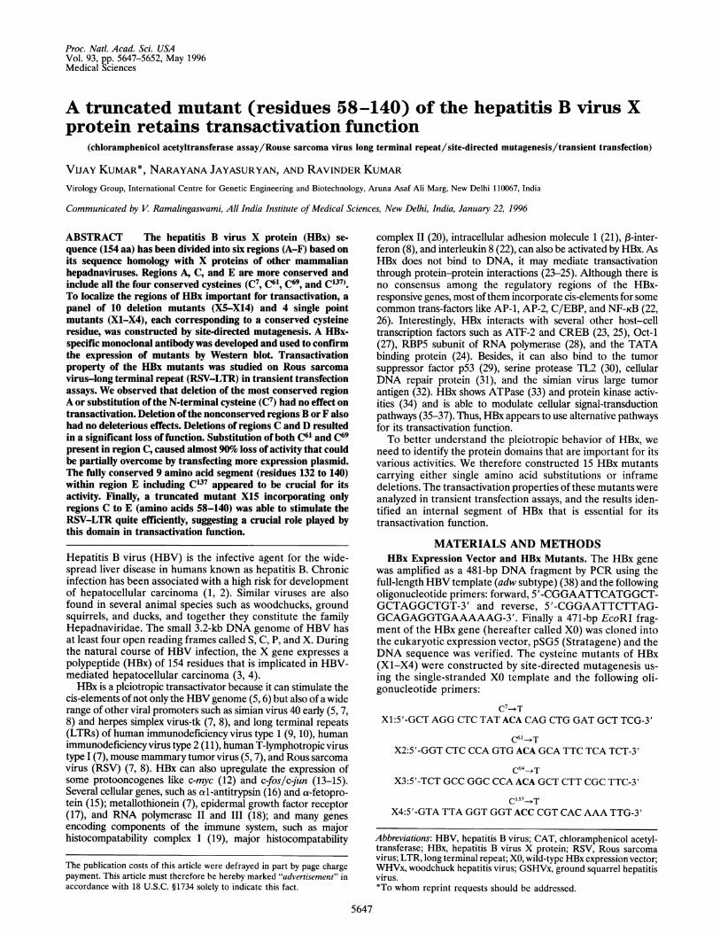
Proc. Natl. Acad. Sci. USAVol. 93, pp. 5647-5652, May 1996Medical Sciences
A truncated mutant (residues 58-140) of the hepatitis B virus Xprotein retains transactivation function
(chloramphenicol acetyltransferase assay/Rouse sarcoma virus long terminal repeat/site-directed mutagenesis/transient transfection)
VIJAY KUMAR*, NARAYANA JAYASURYAN, AND RAVINDER KUMARVirology Group, International Centre for Genetic Engineering and Biotechnology, Aruna Asaf Ali Marg, New Delhi 110067, India
Communicated by V Ramalingaswami, All India Institute of Medical Sciences, New Delhi, India, January 22, 1996
ABSTRACT The hepatitis B virus X protein (HBx) se-quence (154 aa) has been divided into six regions (A-F) based onits sequence homology with X proteins of other mammalianhepadnaviruses. Regions A, C, and E are more conserved andinclude all the four conserved cysteines (C7, C61, C69, and C137).To localize the regions of HBx important for transactivation, apanel of 10 deletion mutants (X5-X14) and 4 single pointmutants (X1-X4), each corresponding to a conserved cysteineresidue, was constructed by site-directed mutagenesis. A HBx-specific monoclonal antibody was developed and used to confirmthe expression of mutants by Western blot. Transactivationproperty of the HBx mutants was studied on Rous sarcomavirus-long terminal repeat (RSV-LTR) in transient transfectionassays. We observed that deletion of the most conserved regionA or substitution of the N-terminal cysteine (C7) had no effect ontransactivation. Deletion ofthe nonconserved regions B or F alsohad no deleterious effects. Deletions of regions C and D resultedin a significant loss of function. Substitution of both C6' and C69present in region C, caused almost 90%0o loss of activity that couldbe partially overcome by transfecting more expression plasmid.The fully conserved 9 amino acid segment (residues 132 to 140)within region E including C137 appeared to be crucial for itsactivity. Finally, a truncated mutant X15 incorporating onlyregions C to E (amino acids 58-140) was able to stimulate theRSV-LTR quite efficiently, suggesting a crucial role played bythis domain in transactivation function.
Hepatitis B virus (HBV) is the infective agent for the wide-spread liver disease in humans known as hepatitis B. Chronicinfection has been associated with a high risk for developmentof hepatocellular carcinoma (1, 2). Similar viruses are alsofound in several animal species such as woodchucks, groundsquirrels, and ducks, and together they constitute the familyHepadnaviridae. The small 3.2-kb DNA genome of HBV hasat least four open reading frames called S, C, P, and X. Duringthe natural course of HBV infection, the X gene expresses apolypeptide (HBx) of 154 residues that is implicated in HBV-mediated hepatocellular carcinoma (3, 4).HBx is a pleiotropic transactivator because it can stimulate the
cis-elements of not only the HBVgenome (5, 6) but also of a widerange of other viral promoters such as simian virus 40 early (5, 7,8) and herpes simplex virus-tk (7, 8), and long terminal repeats(LTRs) of human immunodeficiency virus type 1 (9, 10), humanimmunodeficiency virus type 2 (11), human T-lymphotropic virustype 1 (7), mouse mammary tumor virus (5, 7), and Rous sarcomavirus (RSV) (7, 8). HBx can also upregulate the expression ofsome protooncogenes like c-myc (12) and c-fos/c-jun (13-15).Several cellular genes, such as al-antitrypsin (16) and a-fetopro-tein (15); metallothionein (7), epidermal growth factor receptor(17), and RNA polymerase II and III (18); and many genesencoding components of the immune system, such as majorhistocompatability complex I (19), major histocompatability
complex 11 (20), intracellular adhesion molecule 1 (21), ,3-inter-feron (8), and interleukin 8 (22), can also be activated by HBx. AsHBx does not bind to DNA, it may mediate transactivationthrough protein-protein interactions (23-25). Although there isno consensus among the regulatory regions of the HBx-responsive genes, most of them incorporate cis-elements for somecommon trans-factors like AP-1, AP-2, C/EBP, and NF-KB (22,26). Interestingly, HBx interacts with several other host-celltranscription factors such as ATF-2 and CREB (23, 25), Oct-1(27), RBP5 subunit of RNA polymerase (28), and the TATAbinding protein (24). Besides, it can also bind to the tumorsuppressor factor p53 (29), serine protease TL2 (30), cellularDNA repair protein (31), and the simian virus large tumorantigen (32). HBx shows ATPase (33) and protein kinase activ-ities (34) and is able to modulate cellular signal-transductionpathways (35-37). Thus, HBx appears to use alternative pathwaysfor its transactivation function.To better understand the pleiotropic behavior of HBx, we
need to identify the protein domains that are important for itsvarious activities. We therefore constructed 15 HBx mutantscarrying either single amino acid substitutions or inframedeletions. The transactivation properties of these mutants wereanalyzed in transient transfection assays, and the results iden-tified an internal segment of HBx that is essential for itstransactivation function.
MATERIALS AND METHODSHBx Expression Vector and HBx Mutants. The HBx gene
was amplified as a 481-bp DNA fragment by PCR using thefull-length HBV template (adw subtype) (38) and the followingoligonucleotide primers: forward, 5'-CGGAATTCATGGCT-GCTAGGCTGT-3' and reverse, 5'-CGGAATTCTTAG-GCAGAGGTGAAAAAG-3'. Finally a 471-bp EcoRI frag-ment of the HBx gene (hereafter called XO) was cloned intothe eukaryotic expression vector, pSG5 (Stratagene) and theDNA sequence was verified. The cysteine mutants of HBx(X1-X4) were constructed by site-directed mutagenesis us-ing the single-stranded XO template and the following oli-gonucleotide primers:
C7--TX1:5'-GCT AGG CTC TAT ACA CAG CTG GAT GCT TCG-3'
X2:5'-GGT CTC CCA GTG ACA GCA TTC TCA TCT-3'
C69-*TX3:5'-TCT GCC GGC CCA ACA GCT CTT CGC TTC-3'
C137T
X4:5'-GTA TTA GGT GGT ACC CGT CAC AAA TTG-3'
Abbreviations: HBV, hepatitis B virus; CAT, chloramphenicol acetyl-transferase; HBx, hepatitis B virus X protein; RSV, Rous sarcomavirus; LTR, long terminal repeat; XO, wild-type HBx expression vector;WHVx, woodchuck hepatitis virus; GSHVx, ground squarrel hepatitisvirus.*To whom reprint requests should be addressed.
The publication costs of this article were defrayed in part by page chargepayment. This article must therefore be hereby marked "advertisement" inaccordance with 18 U.S.C. §1734 solely to indicate this fact.
5647
5648 Medical Sciences: Kumar et al.
The 10 deletion mutants of HBx (X5-X14) were alsoconstructed by site-directed mutagenesis by using the single-stranded XO template according a published procedure (39).The truncated form X15 was constructed by PCR using thefollowing primers: forward, 5'-CGGAATTCCATATGCTC-CCCGTCTGTGCCTTC-3' and reverse, 5'-CGGAATTCG-GATCCTTATTTGTGCCTACAGCCTCCTAA-3'. Finally,a 270-bp EcoRI fragment of X15 was cloned into pSG5.Monoclonal Antibody, Western Blot Analysis, and Immu-
noprecipitation. A monoclonal antibody was developedagainst HBx by immunizing BALB/c mice with the purifiedantigen expressed in Escherichia coli (40). Spleen cells from theimmunized animals were fused with myeloma cells(P3x63Ag8.653; ATCC no. CRL 1580) by using polyethyleneglycol (41), and the anti-HBx secreting hybridomas werescreened by ELISA on wells coated with purified HBx. Culturesupernatant of one of the positive clones, B-8/2/8, was used toanalyze the expression of HBx mutants.For expression analysis, the wild-type and the mutant HBx
expression vectors were transfected (1 ,tg DNA per 60-mmculture dish at 30% confluence) in COS 1 cells (ATCC no.CRL 1650) by using the lipofectin reagent (GIBCO/BRL).Cells were harvested 48 h after transfection, and the cellextracts were processed for Western blot analysis (40). Forimmunoprecipitation, the 48-h posttransfected COS 1 cellswere metabolically radiolabeled in the presence of [35S]me-thionine (Amersham) for 5 h, processed as described (39), andautoradiographed. B-8/2/8 is specific for HBx and does notrecognize any other cellular protein (see Fig. 3A lane 4 and 3Blane 3).DNA Transfection and Chloramphenicol Acetyltransferase
(CAT) Assay. Functional analysis of the HBx mutants wasperformed on RSV-LTR in transient transfection assays. Thehuman hepatoma Hep G2 cells (ATCC no. HB 8065) at 50%confluence in a 60-mm culture dish were cotransfected withthe wild-type or mutant HBx expression vectors, and theRSV-CAT reporter plasmid was cotransfected by either thecalcium phosphate method (42) or by using lipofectin reagent(GIBCO/BRL). Cell extracts were prepared 48 h posttrans-fection, and CAT activity was measured as described byGorman et al. (43) by using equal amounts of protein. Afterthin layer chromatography and autoradiography, radioactivityof the substrate and product spots was measured by liquidscintillation counting.
RESULTS
Sequence Homology of HBx. Fig. 1 shows the homology inamino acid sequence of X proteins of the human hepatitis Bvirus (HBx), the ground squirrel hepatitis virus (GSHVx), andthe woodchuck hepatitis virus (WHVx). The overall homologyamong the three viral proteins is about 35% with nine con-served basic amino acids and four conserved cysteines (C7, C61,C69, and C137). Further, HBx can be subdivided into at least sixregions (A-F) of which the more conserved regions, A, C, andE, are interspersed with relatively variable regions B, D, andF.Region A, containing the N-terminal 20 aa, is the most
conserved segment of HBx (80% identity) including threearginines and one cysteine (C7). Rich in serine and glycineresidues, region B (21-57) is poorly conserved (only 5.4%identity). With about 55% homology and rich in hydrophobicamino acids, region C spans residues 58-84 and is character-ized by two conserved cysteines, C61 and C69. Located betweenamino acids 85 and 119, region D shows poor homology (17%),with two conserved basic amino acids. Region E, betweenresidues 120 and 140, is well conserved (-67%) and ischaracterized by a stretch of nine fully conserved amino acidsincluding a cysteine (CI37) and three basic amino acids. With
A
HBx
GSHVx
WHVx
A B C D E F
I I I I I I I
-z n .KNEO _~ . .__, ...,=,I
0 20 40 60 80 100 120 140 160sequence position
B11K I Y yQMPSPDVa 20GSHVx 1 C SS L 20WHVx I c SA L 20
***** **** **** ***
1E 21 GFRPI S57ASPS MADHMNUALR; 57GOHVx 21 zmQS PSVSGSrSPSSA AUSSNDIPV 57WHVz 21 FGPES PPIPRASA SPSPSDKSDLPIG 57
* *
0 0~~~~~HIBx 58 IWVCAFSSAUCAIRFTS.--4UTTVN 84OSHYx 58 A Y SR C G C M TDS 86WHVx 58 ANAAS CV CDlTDS 86
** * * *** * ** * * * ***
00H3x 85 AHQIL IA3 l DLK&YF 119GSHVx 87 ----FVPWM Q IeQ-W--WhA 1K QLLTL 115WHVx 87 ----FVaS Q WS1-XDI-WP IX =ILTK 115
** ** * *
O 00 0EM 120 IMaI 140GSHVx 116 GIDP KL 136WHVx 116 GSIDP SI 136
*** ** **0**0***
Ex 141 LVAPACNUTS& 154GSHVx 137 ----------- 138WHVx 137 ---------CRLL 141
154
138
141
A(80%)
B(5.4%)
C(55.2%)
D(17.1%)
E(66.7%)
F(nil)
FIG. 1. Sequence homology of HBx (38), GSHVx (44), and WHVx(45). (A) Schematic alignment of the three X proteins were made byusing MACAW software (46). Identical residues are represented as darkbands. Segments of higher homology-i.e., A, C, and E-are promi-nently shown while gaps in C and D regions are introduced to havemaximal alignment. (B) Alignment of amino acid sequences of thethree X proteins using the CLUSTAL program of PC/GENE. Only thenonidentical amino acids are shown in the GSHVx and WHVxsequences. Gaps (-) are introduced in order have maximum align-ment of the identical amino acid residues (*). The HBx sequence (154amino acids) was divided into six regions (A-F) based on its homologywith GSHVx and WHVx and the percent homology of each region isshown in parenthesis on the right. The three most conserved regions(A, amino acids 1-20; C, 58-84 and E, 120-140) are boxed, and thefour conserved cysteines (0) and the nine conserved basic amino acids(co) are indicated.
no apparent homology, region F is highly variable in length andis longest (14 aa) in HBV.HBx Expression Vector, HBx Mutagenesis, and Expression
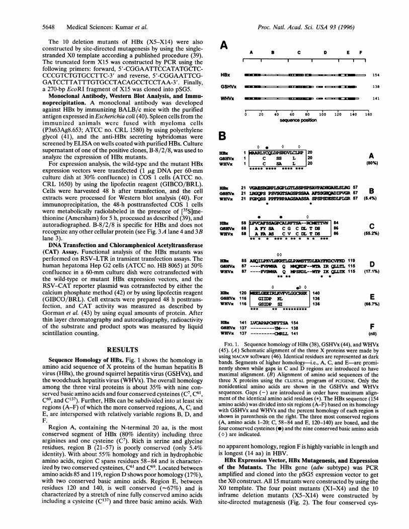
of the Mutants. The HBx gene (adw subtype) was PCRamplified and cloned into the pSG5 expression vector to getthe XO construct. All 15 mutants were constructed by using theXO template. The four point mutants (X1-X4) and the 10inframe deletion mutants (X5-X14) were constructed bysite-directed mutagenesis (Fig. 2). The four conserved cys-
Proc. Natl. Acad. Sci. USA 93 (1996)

Proc. Natl. Acad. Sci. USA 93 (1996) 5649
HOMOLOGYlD./GSHVXlWHV-
XO (WT)xi
X2
X3
X4
to X5
zf X6
X X7
z X8x 9
xio
xiiX12X12X13
X14
XIS
20
A B58 84
U11 71%
120 140 154
;S-I !E'IC( n *E 1 z u m
=m l l1l-|-
l-
S-|
- i--,_t ,.t
C7 T
1,T
C69 T
c1 3 7 T
A 2-20
A 21-57
A 58-84
A 71-84
A 85-119A 120-140
A 120-131
A 128-140
A 132-140
A 141-154
A 1-57 &141-154
FIG. 2. Scheme of point and deletion mutants of HBx. The154-aa-long HBx sequence has been divided into six regions (A-F)based on its homologywith GSHVx and WHVx. Figures in parenthesesrepresent extent of homology among X proteins of the three viralstrains in individual regions (for details see Fig. 1). The point (X1-X4)and deletion (A) mutants (X5-X15) of HBx are shown schematically.The positions of point mutations in X1-X4 are shown by asterisks. Indeletion mutants, a solid bar represents the portion of HBx present inthe mutant, with the deleted portion represented as a gap. The brokenvertical lines represent the boundaries of the six regions of homology(A-F).
teines (C7, C61, C69, and C137) were individually replaced bythreonine residue in mutants X1-X4, respectively. Similarly,the six regions of HBx (i.e., A-F) were deleted in mutants X5,X6, X7, X9, X10, and X14, respectively. For a finer mappingof the transactivation domain of HBx, additional mutants wereconstructed for regions C (X8) and E (X11-X13). In mutantX8, the segment 71-84 of region C was deleted withoutaffecting the two conserved cysteines C61 and C69. In mutantXll, all the acidic amino acids of region E were deleted,whereas in X12 the three conserved basic amino acids alongwith C137 were deleted. The fully conserved stretch of nineamino acids in region E encompassing C137 was deleted inmutant X13. Finally, to explore if the regions of HBx identifiedby the above mutational study alone were sufficient for trans-
activation, a truncated mutant with only regions C, D, and E(X15) was constructed by PCR amplification.
Expression of the HBx mutants X1-X14 in transientlytransfected COS 1 cells was verified by Western blot analysisby using the HBx-specific monoclonal antibody B-8/2/8 (Fig.3A). All the mutants except X9 (lane 13) expressed a specificband of the expected size. The wild-type HBx (XO) constructexpressed a protein of 16.5 kDa (lane 4) with a mobility similarto the X protein expressed in E. coli (lane 2). As expected, allthe point mutants (X1-X4) showed a size similar to XO (lanes5-8) while all the deletion mutants (X5-X14) had smaller geneproducts (lanes 9-12 and 14-18). Mutant protein X6 with thelargest deletion of 37 aa, showed the highest mobility (lane 10).No band was observed for X9 in lane 13 probably due todeletion of the epitope for the monoclonal antibody. However,expression of X9 was confirmed using an anti-X polyclonalrabbit serum (data not shown). Expression of mutant X15could be verified only after metabolically labeling the trans-fected cells with [35S]methionine followed by immunoprecipi-tation (Fig. 3B, lane 4). Higher steady-state level of XO underidentical experimental conditions (compare lanes 3 and 4)suggests the labile nature of X15 when expressed in mamma-lian cells. No such problem was observed when this mutant wasexpressed in E. coli (data not shown).
Localization of Region(s) Essential for Transactivation.Functional analysis of the HBx mutants was done by transientlytransfecting them in Hep G2 cells together with the reporterconstruct, RSV-CAT that contains the LTR ofRSV (43). Thesensitivity of the assay was determined by transfecting increas-ing amounts of the reporter in the absence or presence of afixed amount (1 ,ug) of XO plasmid. X-mediated stimulation ofCAT activity could be detected even when 0.1 ,ug of thereporter construct was used (Fig. 4A). Maximum stimulation(-3-to 6-fold) of RSV-LTR by XO was seen with 0.5 ,ug ofRSV-CAT, which plateaued at higher concentrations of thereporter. Therefore, in all subsequent experiments, only 0.5 ,ugof RSV-CAT plasmid was used. Further, we also determinedthe activation curve for XO and worked out the conditionsunder which functional analysis of the HBx mutants could bedone without having a squelching effect. It was observed thatwith 0.5 ,g of the transfecting RSV-CAT plasmid, the enzyme
0 T-
CO xx
0 lq- C4J r'qqe t co r co e )n- - v- i-i-
X X X X X XX X X X X X X. . . . * i.. .. N ,., , , _.. , ;. . ........ , . .E.... . ..... ...... : I.. l
.{ ' : .^. ....... . , . : ....... . . . i B Cu
200 x x
4 6. -
30.0 -
21.5- __
i 4.3 -
1 2 3 4 5 6 7 8 9 10 11 12 13 14 15 16 17 18 1 2 3 4
FIG. 3. Western blot analysis of the HBx mutants. (A) COS 1 cells were transfected (1 ,ugper 60-mm culture dish) with the wild-type (XO) andmutant HBx expression vectors (X1-X14) and total protein extracts were resolved by SDS/15% PAGE. Samples were electrotransferred ontonitrocellulose membrane and western blot analysis was performed using a monoclonal antibody. Purified X protein (expressed in E. coli) is shownin lane 2. Lane 3 (control) shows extract prepared from the pSG5-transfected cells. Cell extracts of the HBx constructs are run in lanes 4-18. Lane1 shows Bio-Rad prestained markers (lane M). (B) COS 1 cells transfected with pSG5, XO or X15 constructs were metabolically labeled with[35S]methionine. Cell extracts were immunoprecipitated, electrophoresed in a 15% polyacrylamide/SDS gel, and autoradiographed as described.Lane 2 (control) represents cells that were transfected with pSG5. Position of the X15 gene product is shown by an arrowhead. 14C-Labeledmethylated markers (lane M; from Amersham) are shown in lane 1.
A
106 -80 -
49.5 -
32.527.5 -
18.5
Medical Sciences: Kumar et al.
UI.-%

5650 Medical Sciences: Kumar et al.
activity peaked (-11-fold stimulation) in the presence of 5 ,ugof the cotransfecting XO plasmid, but declined when 10 jig ofthe plasmid was used (Fig. 4B). Therefore, functional analysisof all the HBx mutants was carried out at three submaximalplasmid concentrations of 0.5, 1, and 2.5 ,ug.The results of mutational analyses are presented in Table 1.
We observed that deletion of the most conserved N-terminalregion A (X5) had no effect on the HBx-mediated transacti-vation. Interestingly, deletion of the nonconserved regions Band F in mutants X6 and X14, respectively, also did not alter
A6
*xo
5Xo
4.
0
0.1 0.25 0.5 1 2.5 5 10
RSV-CAT (fig)
B
x
10-
OE
HBx construct (jig)
FIG. 4. Experimental conditions for the functional analysis of HBxmutants on RSV-LTR. (A) Amount of the reporter construct requiredto give optimum signal was measured by cotransfecting Hep-G2 cellswith XO (1 jig) and increasing amounts (0.1-10 jig) of the RSV-CATplasmid. CAT assay was performed using cell extracts and the radio-activity was measured. Mean value of three observations is shown as
bars. (B) Activation curves for XO and X15 on RSV-LTR was
determined by cotransfecting Hep G2 cells with RSV-CAT (0.5 jig)and increasing amounts (0.1-10 i,g as indicated inA) of either XO or
X15 plasmids. After CAT assay, radioactivity was measured andplotted. Each point in the figure represents mean of two independentobservations.
the levels of transactivation. However, a significant decrease intransactivation of about 50-80% was seen when the entireregion C (X7) or its portion (X8) were deleted. Similarly,deletion of the poorly conserved region D (X9) also showednearly a 50% drop in the transactivation level. The fullyconserved nine amino acid segment (residues 132-140) ofregion E appeared crucial for the activity because all deletionsinvolving this segment (X10, X12, and X13) resulted in a 5- to10-fold decrease in activity. Surprisingly, deletion of the seg-ment 120-128, which is rich in acidic residues (Xll), resultedin only a marginal decline (up to 35%) in transactivation. Theseresults suggest that the HBx segment spanning regions C to E(i.e., residues 58-140) is important for its transactivationfunction. Interestingly, in cotransfection experiments none ofthe inactive or poorly active mutants was able to suppress theXO activity (data not shown).The C-Terminal Cysteine (C'37) Is Important for Transac-
tivation Function. Involvement of the four conserved cysteinesin the HBx-mediated transactivation was studied by using thepoint mutants X1-X4. We observed that substitution of theN-terminal conserved cysteine C7 (X1) had no effect on theability of the mutant to transactivate RSV-LTR (Table 1).Point mutations of the other three conserved cysteines-i.e.,C61, C69 and C137 (in X2-X4, respectively)-however, resultedin a dramatic loss (- 10-fold) in the transactivation property ofHBx when 0.5 jig of the expression vectors were transfected.For X2 and X3, this effect was, however, partially overcome bytransfecting more (2.5 jig) mutant plasmid. Mutant X4 re-mained inactive under similar conditions (see Table 1). Thus,the integrity of the conserved C-terminal cysteine C'37 of HBxwas essential for its transactivation function.A Mutant with Only C, D, and E Regions Is an Efficient
Transactivator. After identifying the regions of HBx that wereinvolved in transactivation, we planned to explore the possi-bility of having a functional mutant with just the minimalactivation domain of HBx. Thus, using PCR, we constructed atruncated mutant X15 with only regions C, D, and E (residues58-140) and determined a transactivation curve for X15 byusing the experimental protocols described for XO. Analysis ofthe curves (Fig. 4B) as well as subsequent comparative muta-tional analyses (Table 1) showed that X15 was able to stimulatethe RSV-LTR quite efficiently. Although the level of stimu-lation was about 60% of the wild type when 0.5 ,ug of the X15plasmid was used, the stimulation values were at par with eachother when plasmid dosages were 1 and 2.5 ,ug, respectively.Cotransfection of XO and X15 showed an additive effect ontransactivation (data not shown). Apparently, the target in-teraction and the transactivation functions of HBx converge inthe same domain represented by X15.
DISCUSSIONWe have investigated the regions of HBx that are important forits transactivation function. Our mutational analysis has iden-tified a segment in the C terminus of HBx corresponding toregions C, D, and E that is essential for its activity. We foundthat deletions involving either the entire E region (X10) or itsfully conserved segment of nine amino acids (X13) as well assubstitution of the conserved cysteine (C'37) of this region(X4) resulted in a drastic loss of function. The difference in thetransactivation property is unlikely to be related to the expres-sion levels because all the mutants appeared to be expressedwith equal efficiency. Thus, HBx region E is crucial for itstransactivation property. Likewise, deletion of the entire Cregion (X7) or its portions (X8) as well as substitution of theconserved cysteines C61 and C69 of this region (X2 and X3)were equally deleterious. There was, however, one importantdifference about the C-region mutants in that their effectcould be partially overcome by transfecting more expressionplasmids. Therefore it is unlikely that region C with two
Proc. Natl. Acad. Sci. USA 93 (1996)

Proc. Natl. Acad. Sci. USA 93 (1996) 5651
Table 1. Relative ability of the HBx mutants to stimulate expression of RSV-CAT
% CAT activity by the amount ofHBx constructs transfected, jig
HBx construct 0.5 (n = 5) 1.0 (n = 3) 2.5 (n = 4)XO (wt) 100 100 100Xl (C7 -> T) 101.3 ± 8.4 102.0 ± 3.8 110.1 ± 9.0X2 (C61 -+ T) 12.6 ± 3.7a 5.5 ± 2.6a 48.6 ± 6.1aX3 (C69 -+ T) 5.4 ± 2.0a 8.0 ± 3.5a 33.4 ± 9.1aX4 (C137 -- T) 6.7 ± 3.7a 10.3 ± 3.2a 14.6 ± 6.9aX5 (A2-20) 105.1 ± 10.6 105.5 ± 3.9 98.8 ± 12.7X6 (A21-57) 94.8 ± 8.9 100.2 ± 17.3 88.0 ± 9.2X7 (A58-84) 56.2 ± 16.1d 18.3 ± 8.4b 37.8 ± 11.1bX8 (A71-84) 19.5 ± 8.3a 21.7 ± 8.3b 26.3 ± 8.7aX9 (A85-119) 51.2 ± 12.8b 11.7 ± 3.8a 38.3 ± 16.8cX10 (A120-140) 9.6 ± 4.0a 13.7 ± 4.7a 20.8 ± 10.4aXll (A120-131) 76.7 ± 27.1 66.3 ± 3.5a 91.3 ± 8.9X12 (A128-140) 8.3 ± 3.6a 10.1 ± 5.5a 12.6 ± 4.7aX13 (A132-140) 9.9 ± 2.9a 9.6 ± 2.9a 15.9 ± 4.9aX14 (A141-154) 103.6 ± 7.7 106 ± 13.9 104.5 ± 9.4X15 (A1-57 and 141-154) 57.2 ± 9.2b 85.0 ± 6.7 94.7 ± 3.5
The table is a compilation of data from 12 independent experiments. In each experiment, a subset ofmutants was compared to XO. Hep G2 cells were cotransfected with 0.5 jig RSV-CAT and three differentamounts of the HBx mutant expression plasmids. Cells were cultured for 48 h, and CAT activity wasmeasured. The values are the percentage CAT activity obtained against XO and are expressed as mean± SEM. The number of independent observations is shown in parenthesis against each concentration ofthe mutant plasmids tested. The levels of significance were determined by using the unpaired t test: a,P < 0.001; b, P < 0.005; c, P < 0.01; and d, P < 0.025.
conserved cysteines plays a crucial role in transactivation.Using the E. coli-expressed X protein, a disulfide linkage hasbeen assigned between residues C6' of region C and C137 ofregion E (47). The structural and functional relevance of thisintramolecular linkage remains to be established. The poorlyconserved region D may not play an important role in trans-activation because deletion of this region (X9) did not oblit-erate the activity. Although, the ATPase activity of HBx mapsto this region (33), its relevance in transcriptional regulation isnot known. Nevertheless, the epitope for the monoclonalantibody used in this study appears to be localized in region Dbecause it failed to recognize mutant X9. It was puzzling,however, to observe that deletion of the most conserved regionA (X5) or mutation of the N-terminal conserved cysteine (X1)had no bearing on the transactivation function of HBx. Recentreports suggest that region A may have a role in transrepres-sion (48-50). The poorly conserved region B (X6) and non-conserved region F (X14) are unlikely to have a major role intransactivation.
Mutational analyses of HBx using different rationales andapproaches have been attempted by several groups. Usinglinker insertion mutagenesis, Runkel et al. (51) were able tolocalize two separate regions of HBx that were important fortransactivation (one around amino acid 68 and the otherbetween segments 110 and 139). Other reports on the muta-tional studies of HBx (13, 48, 49, 52) suggest that its transac-tivation region is localized toward the C-terminal region.However most of these analyses are based primarily on the lossof reporter function. In the present study, we have mapped the"minimum functional domain" of HBx that could show trans-activation function. On the basis of our results of deletion andpoint mutagenesis, a highly truncated mutant X15 (with onlyregions C, D, and E) was constructed. Although the geneproduct was relatively unstable in mammalian cell lines, it wasable to show a transactivation property like the wild-type HBx.Thus, segments 58-140 or regions C-E of HBx is the minimumdomain involved in transactivation function. Interestingly, twoimportant sequence regions of HBx (i.e., C and E, identifiedby us) also map to the regions that interact the RPB5 subunitof RNA polymerase (28) as well as a serine protease (30).These interactions have been proposed to influence the trans-
activation function of HBx. The role of region E in transacti-vation is further substantiated by the fact that the TATA-binding protein, which is considered central to transcriptionregulation, also interacts with HBx through this region (24).
Further, we find an interesting correlation of our work withthe naturally occurring variants ofHBV (usually sero-negativeby ELISA). DNA sequence analysis of some of these variants hasshown that very often their genomes have deletions or pre-mature "stop" codons around region E of the X gene (6,53-55). The presence of a mutated X protein in those casesmight be responsible for a subdued expression of the HBVgenes. Yet another interesting possibility could be the inte-gration of a minimum transactivation region (similar to X15)of the HBx gene in HBV carriers. Analysis of patient sera usinga sensitive ELISA based on this region and/or PCR amplifi-cation of this region of the X gene should provide interestingresults and more insight into HBV-related pathogenesis.
We thank D. Sahal and V. L. Kumar for helpful discussions, S.Jameel for plasmids pET-X and HBV (adw subtype) and criticalreading of the manuscript, and J. A. Wolff for the RSV-CATconstruct. Technical assistance was provided by Honey V. Reddi, andA. Bhardwaj synthesized the oligonucleotides. This work was sup-ported by an internal fund of the International Centre for GeneticEngineering and Biotechnology.
1. Tiollais, P., Pourcell, C. & Dejean, A. (1985) Nature (London)317, 489-495.
2. Robinson, W. S. (1994) Annu. Rev. Med. 45, 297-323.3. Lo, S. J., Chien, M. L. & Wu Lee, Y. H. (1988) Virology 167,
289-292.4. Kim, C.-M., Koike, K., Saito, I., Miyamura, T. & Gilbert, J. (1991)
Nature (London) 351, 317-320.5. Wollersheim, M., Debelka, U. & Hofschneider, P. H. (1988)
Oncogene 3, 545-552.6. Nakatake, H., Chisaka, O., Yamamoto, S., Matsubara, K. &
Koshy, R. (1993) Virology 195, 305-314.7. Zahm, P., Hofschneider, P. H. & Koshy, R. (1988) Oncogene 3,
169-177.8. Twu, J. S. & Schloemer, R. H. (1987) J. Virol. 61, 3448-3453.9. Seto, E., Bendict-Yen, T. S., Matija Peterlin, B. & Ou, J.-H.
(1988) Proc. Natl. Acad. Sci. USA 85, 8286-8290.
Medical Sciences: Kumar et aL

5652 Medical Sciences: Kumar et al.
10. Siddiqui, A., Gaynor, R., Srinivasan, A., Mapoles, J. & Farr,R. W. (1989) Virology 169, 479-484.
11. Levrero, M, Balsano, C., Natoli, G., Avantaggiati, M. L. &Elfassi, E. (1990) J. Virol. 64, 3082-3086.
12. Balsano, C., Avantaggiati, M. L., Natoli, G., De Marzio, E., Will,H., Perricaudet, M. & Levrero, M. (1991) Biochem. Biophys. Res.Commun. 176, 985-992.
13. Avantaggiati, M. L., Natoli, G., Balsano, C., Chirillo, P., Artini,M., De Marzio, E., Collepardo, D. & Levrero, M. (1993) Onco-gene 8, 1567-1574.
14. Twu, J. S., Lai, M. Y., Chen, D. S. & Robinson, W. S. (1993)Virology 192, 346-350.
15. Zhou, M. X., Watabe, M. & Watabe, K. (1994)Arch. Virol. 134,369-378.
16. Lee, T.-H., Ginegold, M. J., Shen, R.-F., DeMayo, J. L., Woo,S. L. C. & Butel, J. S. (1990) J. Virol. 64, 5939-5947.
17. Menzo, S., Clementi, M., Alfani, E., Bagnarelli, P., lacovacci, S.,Manzin, A., Dandri, M., Natoli, G., Levrcro, M. & Carloni, G.(1993) Virology 196, 878-882.
18. Aufiero, B. & Schneider, R. J. (1990) EMBO J. 9, 497-504.19. Zhou, D.-X., Taraboulos, A., Ou, J.-H. & Bencdict Yen, T. S.
(1990) J. Virol. 64, 4025-4028.20. Hu, K.-Q., Vierling, J. M. & Siddiqui, A. (1990) Proc. Natl. Acad.
Sci. USA 87, 7140-7144.21. Hu, K.-Q., Yu, C.-H. & Vierling, J. M. (1992) Proc. Natl. Acad.
Sci. USA 89, 11441-11445.22. Mahe, Y., Mukaida, N., Kuno, K., Akiyama, M., Ikeda, N.,
Matsushima, K. & Murakami, S. (1991) J. Biol. Chem. 266,13759-13763.
23. Maguire, H. F., Hoeffler, J. P. & Siddiqui, A. (1991) Science 252,842-844.
24. Qadri, I., Maguire, H. F. & Siddiqui, A. (1995) Proc. Nat!. Acad.Sci. USA 92, 1003-1007.
25. Williams, J. S. & Andrisani, 0. M. (1995) Proc. Natl. Acad. Sci.USA 92, 3819-3823.
26. Seto, E., Mitchell, P. J. & Benedict-Yen, T. S. (1990) Nature(London) 344, 72-74.
27. Antunovic, J., Lexieux, N. & Cromlish, J. A. (1993) Cell. Mol.Biol. Res. 39, 463-482.
28. Cheong, J., Yi, M., Lin, Y. & Murakami, S. (1995) EMBO J. 14,143-150.
29. Wang, X. W., Forrester, K., Yeh, H., Feitelson, M. A., Gu, J. R.& Harris, C. C. (1994) Proc. Natl. Acad. Sci. USA 91, 2230-2234.
30. Takada, S., Kido, H., Fukutomi, A., Mori, T. & Koike, K. (1994)Oncogene 9, 341-348.
31. Lee, T.-H. Elledge, S. J. & Butel, J. S. (1995) J. Virol. 69,1107-1114.
32. Seto, E. & Bcnedict-Yen, T. S. (1991) J. Virol. 65, 2351-2356.33. De-Medina, T., Haviv, I., Noiman, S. & Shaul, Y. (1994) Virology
202, 401-407.34. Wu, J. Y., Zhou, Y., Judd, A., Cartwright, C. A. & Robinson,
W. S. (1990) Cell 63, 687-695.35. Kekule, A. S., Lauer, U., Wciss, L., Lubcr, B. & Hofschneider,
P. H. (1993) Nature (London) 361, 742-745.36. Cross, J. C., Weng, P. & Rutter, W. J. (1993) Proc. Natl. Acad.
USA 90, 8078-8082.37. Benn, J. & Schneider, R. J. (1994) Proc. Natl. Acad. Sci. USA 91,
10350-10354.38. Ono, Y., Onda, H., Sasada, R., Igarashi, K., Sugino, Y. &
Nishioka, K. (1983) Nucleic Acids Res. 11, 1747-1757.39. Kumar, V., Green, S., Stack, G., Berry, M., Jin, J. R. & Chambon
P. (1987) Cell 51, 941-951.40. Jameel, S., Siddiqui, A., Maguire, H. F. & Rao, K. V. S. (1990)
J. Virol. 64, 3963-3966.41. Harlow, E. & Lane, D. (1988) Antibodies: A Laboratory Maniual
(Cold Spring Harbor Lab. Press, Plainview NY), pp. 139-243.42. Wigler, M., Pellicer, A., Silverstein, S. & Axel, R. (1978) Cell 14,
725-731.43. Gorman, C. M., Merlino, G. T., Willingham, M. C., Pastan, I. &
Howard, B. H. (1982) Proc. Natl. Acad. Sci. USA 79, 6777-6781.44. Seeger, C. M., Ganem, D. & Varmus, H. E. (1984) J. Virol. 51,
367-375.45. Entiemble, J., Moroy, T., Trepo, C., Tiollais, P. & Buendia, M. A.
(1986) Gene 50, 207-214.46. Schuler, G. D., Altschul, S. F. & Lipman, D. J. (1991) Proteins 9,
180-190.47. Gupta, A., Mal, T. K., Jayasuryan, N. & Chauhan, V. S. (1995)
Biochem. Biophys. Res. Commun. 212, 919-924.48. Ritter, S. E., Whitten, T. M., Quets, A. T. & Schloemer, R. H.
(1991) Virology 182, 841-845.49. Arii, M., Takada, S. & Koike, K. (1992) Oncogene 7, 397-403.50. Murakami, S., Cheong, J. H. & Kaneko, S. (1994) J. Biol. Chem.
269, 15118-15123.51. Runkel, L., Fischer, M. & Schaller, H. (1993) Virology 197,
529-536.52. Kim, Y.-H., Kang, S.-K. & Lee, Y. I. (1993) Biochemn. Biophys.
Res. Commun. 197, 894-903.53. Takada, S. & Koike, K. (1990) Proc. Natl. Acad. Sci. USA 87,
5628-5632.54. Repp, R., Keller, C., Borkhardt, A., Csecke, A., Schaefer, S.,
Gerlich, W. H. & Lampert, F. (1992) Arch. Virol. 125, 299-304.55. Uchida, T., Shimojima, M., Gotoh, K., Shikata, T., Tanaka, E. &
Kiyosawa, K. (1994) Microbiol. Immunol. 38, 281-285
Proc. Natl. Acad. Sci. USA 93 (1996)
Copyright © 2022 FDOKUMEN