Mouse Retinoid X Receptor Contains a Separable Ligand-Binding and Transactivation Domain in Its E...
10
1995, 15(1):255. Mol. Cell. Biol. X Leng, J Blanco, S Y Tsai, K Ozato, B W O'Malley and M J Tsai its E region. ligand-binding and transactivation domain in Mouse retinoid X receptor contains a separable http://mcb.asm.org/content/15/1/255 Updated information and services can be found at: These include: CONTENT ALERTS more» cite this article), Receive: RSS Feeds, eTOCs, free email alerts (when new articles http://journals.asm.org/site/misc/reprints.xhtml Information about commercial reprint orders: http://journals.asm.org/site/subscriptions/ To subscribe to to another ASM Journal go to: on November 15, 2014 by guest http://mcb.asm.org/ Downloaded from on November 15, 2014 by guest http://mcb.asm.org/ Downloaded from
-
Upload
independent -
Category
Documents
-
view
0 -
download
0
Transcript of Mouse Retinoid X Receptor Contains a Separable Ligand-Binding and Transactivation Domain in Its E...

1995, 15(1):255. Mol. Cell. Biol.
X Leng, J Blanco, S Y Tsai, K Ozato, B W O'Malley and M J Tsai its E region.ligand-binding and transactivation domain in Mouse retinoid X receptor contains a separable
http://mcb.asm.org/content/15/1/255Updated information and services can be found at:
These include:
CONTENT ALERTS more»cite this article),
Receive: RSS Feeds, eTOCs, free email alerts (when new articles
http://journals.asm.org/site/misc/reprints.xhtmlInformation about commercial reprint orders: http://journals.asm.org/site/subscriptions/To subscribe to to another ASM Journal go to:
on Novem
ber 15, 2014 by guesthttp://m
cb.asm.org/
Dow
nloaded from
on Novem
ber 15, 2014 by guesthttp://m
cb.asm.org/
Dow
nloaded from

MOLECULAR AND CELLULAR BIOLOGY, Jan. 1995, p. 255–263 Vol. 15, No. 10270-7306/95/$04.0010Copyright q 1995, American Society for Microbiology
Mouse Retinoid X Receptor Contains a Separable Ligand-Bindingand Transactivation Domain in Its E RegionXIAOHUA LENG,1 JORGE BLANCO,2 SOPHIA Y. TSAI,1 KEIKO OZATO,2
BERT W. O’MALLEY,1 AND MING-JER TSAI1*
Department of Cell Biology, Baylor College of Medicine, Houston, Texas 77030,1 and Laboratoryof Molecular Growth Regulation, National Institute of Child Health and Human
Development, National Institutes of Health, Bethesda, Maryland 208922
Received 18 May 1994/Returned for modification 17 August 1994/Accepted 20 October 1994
Steroid, thyroid, and retinoid hormones exert their biological functions by interacting with their cognatenuclear receptors. Upon binding receptors, hormones induce a protease-resistant structural change in thereceptor ligand-binding domain and subsequently activate the receptors. Utilizing partial proteolysis, we havebeen able to delineate a region in the mouse retinoid X receptor b (mRXRb) required for ligand binding. Aseparable activation domain within the mRXRb E region has been identified. The activation domain, which is21 amino acids in length, is located at the extreme C terminus of mRXRb. This domain is not required forligand binding since removal of this sequence neither eliminates the ligand-induced, protease-resistant con-formational change nor alters the ligand-enhanced DNA binding. Furthermore, deletion of this activationdomain converts the receptor into a transcriptional silencer. Finally, a further truncation of 9 amino acids (fora total of 30 amino acids) from the C terminus results in a mutant which does not undergo the protease-resistant conformational change and cannot bind DNA as a homodimer. Nevertheless, this mutant is still ableto form a heterodimer with the thyroid hormone receptor. Therefore, homodimerization and heterodimeriza-tion can be distinguished by this nine-amino-acid sequence.
Steroid and thyroid hormone receptors are nuclear recep-tors which are ligand-activated transcription factors (12, 38).They play a key role in many biological functions such ashomeostasis, reproduction, development, and differentiation.They link extracellular or intracellular signals directly to tran-scriptional responses. Nuclear receptors regulate gene expres-sion by binding to specific response elements in the promoterregion of target genes. On the basis of structural homologies,this family of nuclear receptors has been classically divided intotwo general subfamilies (6, 39). One subfamily includes recep-tors for steroid hormones (glucocorticoids, progestins, andro-gens, estrogens, and mineralocorticoids), and the other con-sists of the thyroxine receptor (TR), retinoic acid receptor(RAR), retinoid X receptor (RXR), and vitamin D3 receptor.The receptors in the latter subfamily can be functionally dis-tinguished from the former ones in the following aspects: (i)they heterodimerize with a common coregulator, RXR (23, 28,32, 51, 52); (ii) they do not associate with heat shock proteinsand can bind to DNA in the absence of hormones (9, 17, 43);and (iii) they all bind to various arrays of the PuGGTCA coremotif (34, 49). Nevertheless, both of these groups of nuclearreceptors require their cognate ligands for activation of targetgenes.It is a general belief that transcription factors have a mod-
ular structure (15). Likewise, nuclear receptors are all com-posed of at least four domains which are differentially con-served among members within each subfamily. The A-Bdomain, which has been implicated in gene activation andpromoter selection, and the hinge domain (D) are highly vari-able and poorly conserved. The DNA-binding domain (C),which consists of two zinc modules, is the most conservedregion among all the receptors. The C-terminal E domain is
the largest and has a complex structure. It mediates ligandbinding, dimerization, repression, and transactivation.These domains of nuclear receptors can usually act as func-
tional units independent of their natural structural context.Intramolecular and intermolecular domain swapping experi-ments have been carried out without affecting the function ofindividual domains (18, 40). Such an approach led to the iden-tification of the DNA-binding domain (DBD) and activationdomains t1 and t2 of glucocorticoid receptors (20). One of theroutine procedures to identify a functional domain within areceptor is to generate receptor fusions with a heterologousDBD such as the yeast transcription factor Gal4 (Gal4-DBD)(3, 20, 46). Specifically, the entire C termini of various recep-tors have been fused to the Gal4-DBD, resulting in hormone-dependent transcription factors that bind to a Gal4 binding site(5, 50). Utilizing Gal4-receptor fusions, we recently have iden-tified three functional activation domains within human TRb(4).Despite the knowledge that nuclear receptors are composed
of functional domains, it is often difficult to precisely localizethem. The problem is more obvious with the ligand-bindingdomain (LBD) because the conservation among receptors inthis region is relatively low. Recently, using protease digestionas a probe of conformation, we have demonstrated that uponbinding to a hormone, a receptor undergoes a ligand-inducedconformational change that produces a distinct proteolyticcleavage pattern (1, 30). The observed structural change cen-ters upon the LBD and is independent of other domains of thereceptor (30). This conformational change renders the LBDmore resistant to protease digestion. This conclusion was fur-ther confirmed by other recent publications (21, 27, 48).RXR is rather unique among the nuclear receptors. While
RXR homodimers appear to bind and function on specificRXR response elements (53), RXR can also act as a het-erodimeric coregulator with other members of the subfamily,including TR, RAR, and the vitamin D3 and peroxisome pro-
* Corresponding author. Phone: (713) 798-6253. Fax: (713) 798-8227.
255
on Novem
ber 15, 2014 by guesthttp://m
cb.asm.org/
Dow
nloaded from

liferator-activated receptors (22, 24). The heterodimers bindDNA elements more efficiently than do the homodimers of TRor RAR and result in increased transcriptional activities. More-over, while TR and RAR can actively repress basal promoteractivity (3, 13), RXR has no detectable silencing function.Thus, we believe there must be some structural characteristicsthat distinguish RXR from the other nuclear receptors.In this report, utilizing partial proteolysis, we have defined a
200-amino-acid region in the putative E domain that was ableto bind 9-cis-retinoic acid (9-cis-RA) and attain a protease-resistant conformation. However, this region alone does nothave ligand-dependent transactivation activity. Consequently,we found a stretch of 21 amino acids at the C terminus ofRXRb which acts as a separable functional activation domain.This region is conserved in other members of the nuclearreceptor superfamily and has been implicated in receptor AF-2function (10, 41). Deletion of this region did not abolish thereceptor ligand-binding ability but, interestingly, convertedRXRb into a transcriptional repressor. Thus, we have beenable to functionally separate the ligand-binding function fromthe ligand-dependent activation domain of RXRb.
MATERIALS AND METHODS
Plasmids. pT7bRXR has been described before (30). All RXR mutants for invitro transcription and translation were constructed in pT7bSal (36). pT7bRXR16-148, pT7bRXRD16-227, pT7bRXRD16-263, pT7bRXRDc2, and pT7bRXRDc3 were generated by inserting the EcoNI-EcoRI fragments of pBS-H2RIIBPDN1, pBS-H2RIIBPDN2, pBS-H2RIIBPDN3, pBS-H2RIIBPDc2, and pBS-H2RIIBPDc3, respectively, into the HindII-EcoRI site of pT7bSal. The pBSconstructs have been described previously (19, 32). pT7bRXRDc4 was generatedby introducing the HindIII-MaeII fragment of pT7bRXR into the HindIII-AccIsite of pT7bSal. pT7bRXR156, pT7bRXR173, and pT7bRXR189 were gener-ated by isolating the HindII-EcoRI, Bsu36I-EcoRI, and EarI-EcoRI fragmentsfrom pT7bRXR and subcloning them into the HindII-EcoRI, AccI-EcoRI, andHindII-EcoRI sites of pT7bSal, respectively. The expression vectors for Gal94-RXR156, Gal94-RXRDc2, Gal94-RXRDc3, and Gal94-RXRDc4 fusion proteinswere created by subcloning HindII-EcoRI fragments of the correspondingpT7bSal constructs into the AccI site of pABGal94 (4). Likewise, pABGal94-RXR173 and pABGal94-RXR189 were subcloned by inserting the Bsu36I-EcoRI and NcoI-NcoI fragments, respectively, of the cognate pT7bSal constructsinto the SmaI site of pABGal94. BglII-EcoRI fragments of pT7bRXRD16-227and pT7bRXRD16-263 were subcloned into the PvuII site of pABGal94 toproduce pABGal94-RXR227 and pABGal94-RXR263, respectively. Expressionvectors for RXR (pCNXRXRb) and RXRDc2 (pCNXRXRbDc2) were gener-ated by inserting the NcoI fragments of pExpressRXRb and pExpressRXRbDc2into the XhoI site of the pCNX2 expression vector (35). pExpress constructs weredescribed previously (37). If necessary, overhangs were blunt-ended with Klenowenzyme. To generate pABGal94-C21, oligonucleotides (MT672 and MT673)corresponding to the last 21 amino acids of mouse RXRb (mRXRb) with aBamHI overhang at the 39 end were synthesized and subcloned into the PvuII-BamHI site of pABGal94. The MT672 sequence is 59-CGGATTGGCGACACCCCCATTGACACCTTCCTCATGGAGATGCTTGAGGCTCCCCACCAGCTGGCCG-39, and the MT673 sequence is 59-GATCCGGCCAGCTGGTGGGGAGCCTCAAGCATCTCCATGAGGAAGGTGTCAATGGGGGTGTCGCCAATCCG-39. The reading frames of all expression vectors were verified bysequencing. 17mer tkCATDH/N and (17mer)32 tkCATDH/N were describedpreviously (3). CRBPII32 tkLuc was described previously (33).Gel mobility shift assays. In vitro-translated RXR or its mutants were utilized
in DNA binding assays in the presence or absence of 9-cis-RA. The experimentswere performed as described previously except that the protein was preincubatedwith 1027 M 9-cis-RA for 15 min at room temperature where specified (30).In vitro transcription and translation and partial proteolytic analysis. [35S]
methionine-labeled protein was synthesized with a Promega translation kit ac-cording to the manufacturer instructions, and the protease digestion assay wascarried out as described previously (30).Transient transfection and transactivation assay. CV1, Lmtk2, and P19 cells
were grown in Dulbecco’s modified Eagle’s medium supplemented with 10%charcoal-stripped fetal bovine serum, 100 U of penicillin per ml, and 100 mg ofstreptomycin per ml. For the transfection assay with Gal-receptor chimeras, 5 mgof reporter (17mer [or 32] tkCATDH/N) and 2 mg of expression vector wereused per 100-mm dish. A total of 1027 M 9-cis-RA was added after glycerolshock. Transfections were carried out as described previously (8). The chloram-phenicol acetyltransferase (CAT) activity was determined as previously de-scribed (44). Transfection assays in P19 cells were done as described previously,except that 300 ng of CRBPII32 tkLuc, 200 ng of pCH110 (Pharmacia), 20 to 40
ng of expression vectors, and 1 mM 9-cis-RA were used (29). Luciferase activitywas determined as described previously (45).
RESULTS
9-cis-RA alters the kinetics of RXR proteolytic digestion.Partial protease digestion has been used widely to identifystructural domains within proteins. Recently, we have shownthat it also can be used to detect ligand-induced conforma-tional changes within steroid hormone receptors (1, 30). Thisconformational change renders the LBD resistant to proteasedigestion. Our results convincingly indicate that the protease-resistant fragment contains the region sufficient for receptorligand binding. Thus, we felt this method could be used toidentify the LBD of RXR. In vitro-synthesized, [35S]methi-onine-labeled mRXRb was digested with increasing concen-trations of trypsin. The products were analyzed by sodiumdodecyl sulfate-polyacrylamide gel electrophoresis (SDS-PAGE) (Fig. 1). Although a weak protease-resistant fragmentwas generated from the aporeceptor, it was clear that theresistant fragment was enhanced upon addition of 9-cis-RA(Fig. 1; compare lanes 7 to 12 with 1 to 6). This was not due tononspecific effects of retinoids, since all-trans-RA did not affectthe kinetics of proteolysis (Fig. 1; compare lanes 13 to 18 with1 to 6). Similar patterns were observed when chymotrypsin orV8 protease was used (data not shown). Since there is a strongcorrelation of ligand binding and protease resistance, the pro-tease-resistant fragment was used to define the boundaries ofthe LBD of RXR.A protease-resistant fragment is located in the C-terminal
half of RXR. To precisely localize the region of RXR thatharbors the protease resistance, we constructed a series ofdeletion mutants (Fig. 2). Mutants were examined by trypsindigestion, and the results were summarized in Fig. 2. Analo-gous to other receptors that we have tested, the resistant frag-ment localized to the C-terminal half of RXR. Removal of theN-terminal sequence of RXR resulted in a receptor (i.e.,RXRD16-148) which exhibited the same protease-resistantfragment as contained in the wild-type receptor (Fig. 3a).When we compared the trypsin digestion patterns of RXR156and RXR173 with that of RXRD16-148, we found that theaddition of 9-cis-RA altered the kinetics of the protease diges-
FIG. 1. 9-cis-RA alters the kinetics of proteolysis of in vitro-translatedmRXRb. 35S-labeled mRXRb was treated with 100 nM 9-cis-RA (lanes 7 to 12),100 nM all-trans RA (lanes 13 to 18), or carrier control (lanes 1 to 6) prior todigestion with the indicated levels of trypsin. Digestion products were analyzedby SDS-PAGE. Resistant fragments (asterisk) and the undigested proteins (ar-row) are indicated as shown.
256 LENG ET AL. MOL. CELL. BIOL.
on Novem
ber 15, 2014 by guesthttp://m
cb.asm.org/
Dow
nloaded from

tion of these mutants (data not shown) and rendered all threemutants resistant to trypsin digestion (Fig. 3a). Since the pro-tease-resistant fragment of RXR173 was slightly smaller thanthe resistant fragment of RXRD16-148, we deduced that theN-terminal boundary of the resistant fragment of the full-length receptor was located at one of the three putative trypsincleavage sites in this region (amino acid 158, 160, or 162).Although the protease-resistant fragment is sufficient for li-gand binding, the minimal domain required for ligand bindingmay be smaller. A further deletion from the N-terminal end(RXR16-227) destroyed the protease resistance (Fig. 3b; com-pare lanes 3 and 4 to 11 and 12). Thus, these results suggestedthat the N-terminal border of the RXR LBD lies betweenamino acids 211 and 265. These data also imply that the ex-treme N-terminal sequence of the E domain is not importantfor RXR ligand binding.The C-terminal truncation mutant RXRDc2, but not
RXRDc3, can undergo the hormone-induced conformationalchange. In order to define the C-terminal boundary of theRXR LBD, we constructed RXR C-terminal truncation mu-tants and tested them in the proteolytic digestion assay. Re-moval of 21 amino acids from the C terminus (RXRDc2) didnot affect the ligand-induced, protease-resistant conformation,suggesting that these 21 amino acids are not required for hor-mone binding (Fig. 4a). Similar protease-resistant conforma-tions were also detected with chymotrypsin and V8 protease(data not shown). Since the protease-resistant fragment fromRXRDc2 was clearly smaller than that from wild-type RXR(Fig. 4a; compare lanes 1 to 4 with 9 to 12), the protease-
resistant fragment of the full-length receptor must encompassthe entire sequence downstream of amino acids 158 to 162.Interestingly, another mutant (RXRDc3) bearing a furthernine-amino-acid deletion from the C terminus yielded no de-tectable protease-resistant fragment even in the presence of ahigh concentration of 9-cis-RA (Fig. 4a). The ability ofRXRDc2 to retain the protease-resistant fragment in the pres-ence of the ligand was intriguing because removal of the sim-ilar region in other receptors, i.e., TR and RAR, destroys theligand-binding ability of these receptors as well as the proteaseresistance (30).To verify that RXRDc2 could bind 9-cis-RA, we employed a
gel shift assay. It has been shown previously that 9-cis-RA caninduce RXR to bind as a homodimer to its response elementwith a higher affinity than that seen in the absence of theligand. If RXRDc2 is able to bind 9-cis-RA, we should be ableto detect stronger DNA binding in the presence of the ligand.As expected, RXRDc2 displayed a higher DNA-binding affinityin the presence of the ligand (Fig. 4b). In contrast, we did notdetect any DNA binding by RXRDc3 even though twofoldmore RXRDc3 was used in the binding assay (Fig. 4b). Thisobservation was surprising since both RXRDc2 and RXRDc3still contained the intact C-terminal heptad repeats and wereable to heterodimerize with TR as efficiently as the wild-typeRXR (Fig. 4c).These data indicate that the boundaries of the RXR LBD
are located between amino acids 173 and 227 at the N terminusand 380 and 389 at the C terminus. We conclude that theextreme C-terminal end of the E region is not important for
FIG. 2. A schematic representation of various deletion mutants of RXR and Gal94-RXR fusions. Full-length RXR in our constructs consists of 448 amino acids.With reference to other steroid receptors, the sequences from amino acids 1 to 81, 82 to 146, 147 to 170, and 171 to 410 are designated as domains A-B, C, D, andE, respectively (31). The plus sign indicates that the protein retained the protease-resistant fragment (left panel) and the hormone-dependent activation of thecorresponding Gal fusion (right panel). The asterisk indicates that the fusion was active in the absence of the ligand. NE, not examined. The names of the expressionplasmids were derived according to those of the deletion mutants.
VOL. 15, 1995 FUNCTIONAL DISSECTION OF mRXRb 257
on Novem
ber 15, 2014 by guesthttp://m
cb.asm.org/
Dow
nloaded from

ligand binding. Moreover, we presented evidence that differentregions in the receptor are involved in receptor homodimer-ization and heterodimerization functions.A C-terminal truncation converts RXR to a transcriptional
repressor. In addition to hormone binding, the LBD containsregions important for dimerization and transactivation. Sinceproteolytic analysis has enabled us to dissect the minimal re-gion of RXR for ligand binding, it was of interest to determinewhether this region is sufficient for transactivation. Variousfragments of the C terminus of RXR were then fused to theyeast Gal4-DBD (amino acids 1 to 94) (Fig. 2). The Gal4-DBDcontains dimerization and nuclear localization functions, thuseliminating interpretation problems with receptor mutationsaffecting these functions. The Gal4 receptor deletion mutantswere cotransfected into CV1 cells together with a reportercontaining a single Gal4 binding site upstream of the tk pro-moter CAT reporter. As expected, fusion of the complete Cterminus of mRXRb to the Gal4-DBD (Gal94-RXR156) re-
sults in a protein that activated reporter gene expression uponaddition of 9-cis-RA (Fig. 5). This confirms that the E domainof RXR is a functional and independent transcriptional mod-ule.As expected, Gal94-RXR173 exhibited ligand-dependent
activation, whereas Gal94-RXR227 and Gal94-RXRDc3 couldnot activate transcription (Fig. 5a). In fact, as summarized inFig. 2, there was a close correlation between the transactiva-tion activity of a fusion protein and hormone-binding ability, asdetermined by the protease resistance of the receptor frag-ment. The only exception, however, was the RXRDc2 mutant;Gal94-RXRDc2 had no hormone-dependent transactivationalactivity (Fig. 5a), even if higher concentrations of hormone(1025 M) were used (data not shown). Since removal of 21amino acids from the receptor C terminus did not hamper theligand-binding ability, the inability of Gal94-RXRDc2 to trans-activate suggested that the 21 C-terminal amino acids must beimportant for transactivation. Therefore, we were able to sep-arate the LBD from the ligand-dependent transactivation.Interestingly, on closer examination, we found that the CAT
activity derived from Gal94-RXRDc2 and Gal94-RXRDc3 inthe absence of the ligand was actually lower than the reporterconstruct transfected with a control vector containing only theGal4-DBD (Gal94). In order to confirm this repression, weused tkCAT linked to two copies of the Gal4 binding site as areporter to increase the silencing effect. As shown in Fig. 5b,Gal94-RXRDc2 and Gal94-RXRDc3 actively silenced thebasal promoter activity about fivefold. The full-length con-struct, Gal94-RXR156, on the other hand, had no effect (Fig.5b). The repression activity was abolished when 42 aminoacids were truncated from the C terminus of RXR (Gal94-RXRDc4). These results suggest that the 21 C-terminal aminoacids of the receptor suppress an intrinsic repression activitywithin the receptor and that deletion of this sequence convertsthe receptor to a negative regulator.Since RXRDc2 can still form homo- or heterodimers, one
would expect, given its intrinsic repression function, that thismutant may function in a dominant negative fashion whencoexpressed with other receptors into cells. In order to exam-ine the repression function in the context of the wild-typereceptor and a natural response element, RXR and RXRDc2were cotransfected with CRBPII32 tkLuc into P19 cells. Aspredicted, cotransfection of increasing amounts of RXRDc2gradually diminished the ligand-induced activation of wild-typeRXR on a reporter driven by a natural RXRE (Fig. 5c; com-pare results with and without cotransfection with RXRbDc2).For unknown reasons, the effects of RXRDc2 on RAR- andTR-mediated transactivation were far more complicated. Al-though it can potentiate T3 responsiveness on the myosinheavy-chain (MHC) TRE (29), RXRDc2 blocks RAR- andTR-dependent activation of reporter genes driven by bRAREand ME TRE, respectively (unpublished data). Nevertheless,the fact that RXRDc2 can function as a powerful dominantnegative receptor substantiates the identification of themasked intrinsic repression function of RXR and strongly sug-gests that the C-terminal amino acids play an important role inthe transcriptional activation function.A conserved stretch in the last 21 amino acids of RXR is a
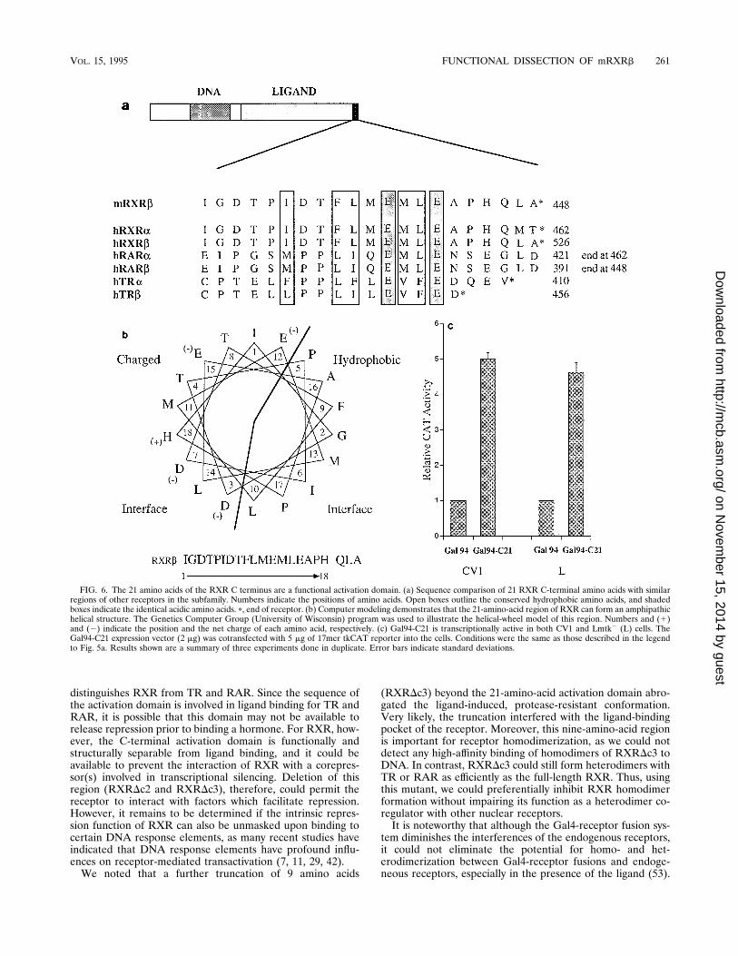
functional activation domain. The results presented above sug-gested that the 21 C-terminal amino acids of RXR are impor-tant for transactivation. Alignment of these 21 amino acidswith the C-terminal ends of other members of the TR-RARnuclear hormone receptor subfamily revealed a high homologywithin the sequence (Fig. 6a). Computer modeling of these 21amino acids predicted an amphipathic helical structure (Fig.6b). This is reminiscent of the C-terminal regions of TR and
FIG. 3. Protease-resistant fragments of RXR encompass the entire C termi-nus of the receptor. (a) RXRD16-148, RXR156, and RXR173 were all treatedwith 100 nM 9-cis-RA and digested as described in the legend to Fig. 1. Theasterisk indicates the resistant fragments. (b) Details are similar to those forpanel a, except that RXR and RXRD16-227 were used in the analysis.
258 LENG ET AL. MOL. CELL. BIOL.
on Novem
ber 15, 2014 by guesthttp://m
cb.asm.org/
Dow
nloaded from

the receptors for estrogens and glucocorticoids, which are alsocapable of forming a putative amphipathic a-helix (10, 41).Substitution of one of the conserved hydrophobic residues witha proline residue destroys the helical structure as well as theligand-dependent transactivation of TR (41). Consequently,this putative a-helical region, known as AF-2, was implicatedin the C-terminal ligand-dependent activation function. In or-der to prove directly that these 21 amino acids can serve as atransactivation domain, the region was fused to the Gal4-DBD. Again, cotransfection experiments were performed inCV1 cells and mouse fibroblast Lmtk2 cells. Gal94-C21 led toa protein that was active in both cell lines (Fig. 6c). Theseresults suggest that although the last 21 amino acids are func-tional alone, the sequence is subject to hormonal regulation ifembedded in its natural context of the RXR LBD. We believeit corresponds to the activation domain referred to as AF-2 inRARa or t4 in TRb (4, 47). Complete removal of this activa-tion domain from RXR does not abolish the ligand-bindingfunction of RXR. Thus, RXR differs from RAR and TR in thatthe AF-2 and t4 activation functions are not separable fromthe ligand-binding function (4, 47). Removal of AF-2 or aportion of t4 eliminates 9-cis-RA or T3 binding to RARa andTRb, respectively.
DISCUSSION
The C terminus of a nuclear receptor, which encompassesthe D and E-F regions, has a complex structure with multiplefunctions. Since the D region is highly variable in length andpoorly conserved in sequence, it has been very difficult toseparate the D region from the E region. Taking advantage ofthe protease-resistant conformation change in the LBD uponligand binding, we have been able to localize a region essentialfor ligand binding (30). Using limited proteolysis, we defined a200-amino-acid region in the E region of RXR that is essentialfor hormone binding (Fig. 2).It has been suggested that, at least for the TR-RAR sub-
family, both the N- and C-terminal ends of the E domain arenecessary for ligand binding (14, 16). We have demonstratedpreviously that the removal of eight amino acids from theextreme C terminus of the human TRb E region or part of theD region of human RARa eliminates the ligand-dependentconformational change in TR and RAR (30). In the ‘‘regula-tory zipper’’ model presented by Forman and Samuels (14), thecentral part of the E region is devoted to dimerization andcontains a so-called ti domain implicated in the ligand-depen-dent transactivation function. For RXR, however, this is notthe case. Our studies clearly show that the truncation of 18(RXR189) and 21 (RXRDc2) amino acids from the N and Ctermini of RXR’s E region, respectively, did not abolish theligand-binding ability of the receptor. RXR is more distinctfrom other members of the subfamily when compared withRARa. For RARa, the determinant for high-affinity 9-cis-RAbinding resides in a stretch of 15 amino acids (corresponding toamino acids 393 to 408 of mRXRb) (47). Deletion of thisregion from RARa completely destroys the 9-cis-RA-inducedconformational change and its ligand binding. Thus it has been
FIG. 4. RXRDc2 but not RXRDc3 can still bind the ligand. (a) mRXRb,RXRDc2 (Dc2), and RXRDc3 (Dc3) were treated and digested as described inthe legend to Fig. 1. Equal volumes of translation product for each protein wereused since the translation efficiencies for RXR, RXRDc2, and RXRDc3 weresimilar. The asterisks indicate the resistant fragments. (b) Addition of 9-cis-RAenhanced the binding affinity of in vitro-translated RXR and RXRDc2 to a DR1element. One microliter of RXR or RXRDc2 was used in total 10-ml gel shift
reactions, whereas 3 ml of RXRDc3 was added. A total of 100 nM 9-cis-RA wasincluded as indicated. B and F indicate the DNA-protein complex and the freeprobe, respectively. (c) Both RXRDc2 and RXRDc3 can heterodimerize withTR. One microliter of TR and/or 0.5 ml of RXR, RXRDc2, and RXRDc3 wereincubated with 32P-labeled MHC TRE elements in a gel shift assay. DR1 andMHC TRE oligonucleotides were described previously (8). B and F indicate theDNA-protein complex and the free probe, respectively.
VOL. 15, 1995 FUNCTIONAL DISSECTION OF mRXRb 259
on Novem
ber 15, 2014 by guesthttp://m
cb.asm.org/
Dow
nloaded from

suggested that the conserved amino acid sequence within thisregion, Asp-Met-Leu-Asp, may be important for 9-cis-RAbinding (47). Our results show that, at least for mRXRb, wecan remove the Asp-Met-Leu-Asp residues (amino acids 401to 404) by truncating up to amino acid 389 without abolishing9-cis-RA binding by the receptor. Thus, we believe there is aseparate determinant(s) in the RXR LBD that directs 9-cis-RA binding.The fact that the 21 extreme C-terminal amino acids were
not essential for ligand binding does not suggest that they arethe F region. Although the exact function of the F region is notclear, deletion of the F region from estrogen receptor does notaffect any known function of the receptor (25). Removal ofthese 21 amino acids from RXR, however, abolished the li-gand-dependent transactivation. In fact, when fused to theGal4-DBD, these 21 amino acids functioned as an activationdomain. These results are consistent with previous findingswhich suggest that similar regions in TR and RAR also act asactivation domains (t4 and AF-2, respectively) (4, 47). Thedifference between the 21 amino acids in RXR and those in TRor RAR is that t4 and AF-2 are not separable from the recep-tor ligand-binding function. Nonetheless, it is feasible that thisregion is important for ligand-dependent activation in manyother nuclear receptors because similar regions of other recep-tors share significant conservation in the primary sequence.Although deletion of the last 21 amino acids (Gal94-
RXRDc2) eliminated ligand-dependent activation of RXR, itis quite possible that RXR has other ligand-dependent activa-tion domains. In recent studies with human TRb, three acti-vation domains have been identified in the TR LBD (4). Sim-ilarly, a small deletion into the t4 region (homologous to the 21amino acids in RXR) renders the thyroid hormone receptorcompletely inactive. It is likely that this 21-amino-acid se-quence may serve as an interaction site for an important cel-lular factor(s), which, when deleted, renders the receptor in-active.Unlike other members in the subfamily (i.e., TR and RAR),
full-length RXR has not been reported to contain a silencingfunction on any known RXRE. Thus, it was interesting todiscover that Gal4-RXRDc2 and Gal4-RXRDc3 were activetranscriptional repressors and that RXRDc2 can function in adominant negative fashion. Recent studies suggest that (i) therepression function of a nuclear receptor (i.e., TR or RAR) ismediated though an interaction with a corepressor(s) and thegeneral transcriptional factor TFIIB, and (ii) hormone-depen-dent derepression involves the release of these interactions (2,4). We also suggest that at least the C-terminal activationdomain of TR is required for the hormone-dependent releaseof a corepressor(s) since a small truncation of this activationdomain results in a constitutive silencer which still retains thehormone-binding ability (4). Since RXR displays significanthomology with TR and RAR, it is likely that RXR could havea similar domain(s) required for these interactions. Consider-ing that the C-terminal activation domain is essential for hor-mone-dependent release of repression, we believe this is what
FIG. 5. Transcriptional activation and repression by RXR mutants. (a) Com-parison of 9-cis-RA induction of transactivation of 17mer-tkCAT by RXR de-letion mutants in CV1 cells. Transfections were performed with equal amountsof expression vectors and reporters. After transfection, the cells were incubatedin the absence (2) or presence (1) of 9-cis-RA (1027 M) for 36 to 42 h. Cellextracts were subsequently prepared and assayed for protein concentration andCAT activity. CAT activities of all mutants were normalized to that of Gal94.Error bars indicate standard deviations. (b) Gal94-RXRDc2 and Gal94-RXRDc3can actively repress basal promoter activity. The reporter 17mer 32 tkCAT wascotransfected with the receptor deletion mutants. Conditions were the same asthose described for panel a. CAT activity of Gal94 served as basal promoteractivity. Results shown are a summary of at least two experiments done in
duplicate. Error bars indicate standard deviations. (c) RXRbDc2 can reduce the9-cis-RA-dependent activation of wild-type RXR. Undifferentiated P19 cellswere transfected with CRBPII32 tkLuc, pCH110, and expression vectors formRXRb and/or mRXRbDc2 (20 ng for each1). Cells were incubated with 1 mM9-cis-RA for 24 h before being harvested in lysis buffer. Transfection efficiencywas normalized against control bGal activities. The relative luciferase activity ofeach assay was normalized to that of control pCNX2 vector in the absence of theligand. Results shown are a summary of three independent experiments. Errorbars indicate standard deviations.
260 LENG ET AL. MOL. CELL. BIOL.
on Novem
ber 15, 2014 by guesthttp://m
cb.asm.org/
Dow
nloaded from
distinguishes RXR from TR and RAR. Since the sequence ofthe activation domain is involved in ligand binding for TR andRAR, it is possible that this domain may not be available torelease repression prior to binding a hormone. For RXR, how-ever, the C-terminal activation domain is functionally andstructurally separable from ligand binding, and it could beavailable to prevent the interaction of RXR with a corepres-sor(s) involved in transcriptional silencing. Deletion of thisregion (RXRDc2 and RXRDc3), therefore, could permit thereceptor to interact with factors which facilitate repression.However, it remains to be determined if the intrinsic repres-sion function of RXR can also be unmasked upon binding tocertain DNA response elements, as many recent studies haveindicated that DNA response elements have profound influ-ences on receptor-mediated transactivation (7, 11, 29, 42).We noted that a further truncation of 9 amino acids
(RXRDc3) beyond the 21-amino-acid activation domain abro-gated the ligand-induced, protease-resistant conformation.Very likely, the truncation interfered with the ligand-bindingpocket of the receptor. Moreover, this nine-amino-acid regionis important for receptor homodimerization, as we could notdetect any high-affinity binding of homodimers of RXRDc3 toDNA. In contrast, RXRDc3 could still form heterodimers withTR or RAR as efficiently as the full-length RXR. Thus, usingthis mutant, we could preferentially inhibit RXR homodimerformation without impairing its function as a heterodimer co-regulator with other nuclear receptors.It is noteworthy that although the Gal4-receptor fusion sys-
tem diminishes the interferences of the endogenous receptors,it could not eliminate the potential for homo- and het-erodimerization between Gal4-receptor fusions and endoge-neous receptors, especially in the presence of the ligand (53).
FIG. 6. The 21 amino acids of the RXR C terminus are a functional activation domain. (a) Sequence comparison of 21 RXR C-terminal amino acids with similarregions of other receptors in the subfamily. Numbers indicate the positions of amino acids. Open boxes outline the conserved hydrophobic amino acids, and shadedboxes indicate the identical acidic amino acids. p, end of receptor. (b) Computer modeling demonstrates that the 21-amino-acid region of RXR can form an amphipathichelical structure. The Genetics Computer Group (University of Wisconsin) program was used to illustrate the helical-wheel model of this region. Numbers and (1)and (2) indicate the position and the net charge of each amino acid, respectively. (c) Gal94-C21 is transcriptionally active in both CV1 and Lmtk2 (L) cells. TheGal94-C21 expression vector (2 mg) was cotransfected with 5 mg of 17mer tkCAT reporter into the cells. Conditions were the same as those described in the legendto Fig. 5a. Results shown are a summary of three experiments done in duplicate. Error bars indicate standard deviations.
VOL. 15, 1995 FUNCTIONAL DISSECTION OF mRXRb 261
on Novem
ber 15, 2014 by guesthttp://m
cb.asm.org/
Dow
nloaded from

Since such dimers are very unlikely to bind to the 17mer DNAelement (4, 28), potential dimerization between Gal4-receptorchimeras and endogenous receptors will not affect our conclu-sions about the repression function of Gal4-RXR mutants.Nevertheless, the ability of endogenous receptors to form non-DNA-binding dimers with the Gal4-receptor fusions in thepresence of the ligand could explain the ligand-induced partialrelease of the silencing function of Gal4-RXR chimeras (Fig.5a). We also wish to point out that using a heterologous DBD(Gal4) and response element (17mer) may overlook the im-portant contribution of the hormone response element in thereceptor activation of target genes (26, 42).During the review process for this paper, we learned that
Zhang et al. identified an almost identical region in humanRXRa that determines the homo- and heterodimeric forma-tion (54). In terms of dimerization and DNA binding, theirmutants DRXR2, DRXR3, and DRXR4 (which have deletionsof 19, 29, and 49 amino acids from the C terminus, respec-tively) function essentially the same as our RXRDc2, RXRDc3,and RXRDc4, respectively (which have deletions of 21, 30, and42 amino acids from the C terminus, respectively). Sequencecomparison indicates that the regions separating the homo-and heterodimerization are identical. In a manner similar tothat of their human RXRa counterparts, RXRDc2 andRXRDc3 could also potentiate the T3 responsiveness of theMHC TRE in cotransfection assays (29, 54). However, thescenario is more complicated with these RXRb deletion mu-tants, since we could show that their exact function as het-erodimeric partners is at least partially mediated by the specificsequence of a response element (29). Moreover, because of theintrinsic repression function of RXRb, RXRDc2 can evenfunction as a powerful dominant negative receptor on certainresponse elements when cotransfected with RXR, TR, RAR,and the vitamin D3 receptor (unpublished data). Thus, al-though DRXR2 of human RXRa and RXRDc2 of mRXRbshare an identity in their E regions of more than 90%, they mayfunction differently as heterodimeric partners on certain re-sponse elements. This is reminiscent of the recent studies bySaatcioglu et al., in which they have identified a novel responseelement that can discriminate the function of TR isoforms (42).In summary, we have delineated the boundaries of the LBD
and have subsequently isolated and characterized an activationdomain of mRXRb. These two regions can be physically sep-arated without destroying either of their functions. Thus wehave functionally dissected the ligand-binding and transactiva-tion functions of the nuclear receptor RXRb. Removal of thisactivation domain, however, converts the receptor into a re-pressor. We also observed that the deletion of 30 amino acidsfrom the extreme C terminus impaired RXRb homodimerformation and DNA binding but left intact the capacity forRXRb heterodimerization with other receptors.
ACKNOWLEDGMENTS
We thank K. Jackson for excellent technical assistance and membersof our laboratories for critically reading the manuscript. We thank J.Grippo and S. Minucci for 9-cis-RA and CRBPII32 tkLuc.This work is supported by NIH grants to M.-J.T. and B.W.O.
REFERENCES
1. Allan, G. F., X. Leng, S. Y. Tsai, N. L. Weigel, D. P. Edwards, M. J. Tsai, andB. W. O’Malley. 1992. Hormone and antihormone induce distinct confor-mational changes which are central to steroid receptor activation. J. Biol.Chem. 267:19513–19520.
2. Baniahmad, A., I. Ha, D. Reinberg, S. Tsai, M.-J. Tsai, and B. W. O’Malley.1993. Interaction of human thyroid hormone receptor b with transcriptionfactor TFIIB may mediate target gene derepression and activation by thyroidhormone. Proc. Natl. Acad. Sci. USA 90:8832–8836.
3. Baniahmad, A., A. C. Kohne, and R. Renkawitz. 1992. A transferable silenc-ing domain is present in the thyroid hormone receptor, in the v-erbA onco-gene product and in the retinoic acid receptor. EMBO J. 11:1015–1023.
4. Baniahmad, A., X. Leng, T. P. Burris, S. Y. Tsai, M.-J. Tsai, and B. W.O’Malley. 1995. The t4 activation domain of the thyroid hormone receptoris required to release a corepressor(s) necessary for transcriptional silencing.Mol. Cell. Biol. 15:76–86.
5. Baniahmad, A., S. Y. Tsai, B. W. O’Malley, and M.-J. Tsai. 1992. Kindred Sthyroid hormone receptor is an active and constitutive silencer and a repres-sor for thyroid hormone and retinoic acid responses. Proc. Natl. Acad. Sci.USA 89:10633–10637.
6. Beato, M. 1989. Gene regulation of steroid hormones. Cell 56:335–344.7. Carlberg, C., I. Bendik, A. Wyss, E. Meier, L. J. Sturzenbecker, J. F. Grippo,and W. Hunziker. 1993. Two nuclear signalling pathways for vitamin D.Nature (London) 361:657–660.
8. Cooney, A. J., X. Leng, S. Y. Tsai, B. W. O’Malley, and M.-J. Tsai. 1993.Multiple mechanisms of chicken ovalbumin upstream promoter transcrip-tion factor-dependent repression of transactivation by the vitamin D, thyroidhormone, and retinoic acid receptors. J. Biol. Chem. 268:4152–4160.
9. Damm, K., C. C. Thompson, and R. M. Evans. 1989. Protein encoded byv-erbA functions as a thyroid-hormone receptor antagonist. Nature (Lon-don) 339:593–597.
10. Danielian, P. S., R. White, J. A. Lees, and M. G. Parker. 1992. Identificationof a conserved region required for hormone dependent transcriptional acti-vation by steroid hormone receptors. EMBO J. 11:1025–1033.
11. Durand, B., M. Saunders, P. Leroy, M. Leid, and P. Chambon.1992. All-trans and9-cis retinoic acid induction of CRABPII transcription is mediated by RAR-RXRheterodimers bound to DR1 and DR2 repeated motifs. Cell 71:73–85.
12. Evans, R. M. 1990. The steroid and thyroid hormone receptor superfamily.Science 240:889–895.
13. Fondell, J. D., A. L. Roy, and R. G. Roeder. 1993. Unliganded thyroidhormone receptor inhibits formation of a functional preinitiation complex:implications for active repression. Genes Dev. 7:1400–1411.
14. Forman, B. M., and H. H. Samuels. 1990. Interactions among a subfamily ofnuclear hormone receptors: the regulatory zipper model. Mol. Endocrinol.4:1293–1301.
15. Frankel, A. D., and P. S. Kim. 1991. Modular structure of transcriptionfactors: implications for gene regulation. Cell 65:717–719.
16. Glass, C. K., S. M. Lipkin, O. V. Devary, and M. G. Rosenfeld. 1989.Structure and functional expression of a cloned Xenopus thyroid hormonereceptor. Nucleic Acids Res. 17:9395–9405.
17. Graupner, G., K. N. Wills, M. Tzukerman, X. K. Zhang, and M. Pfahl. 1990.Dual regulatory roles for thyroid-hormone receptors allows the control ofretinoic-acid receptor activity. Nature (London) 340:653–656.
18. Green, S., and P. Chambon. 1989. Chimeric receptors used to probe theDNA-binding domain of the estrogen and glucocorticoid receptors. CancerRes. 49(Suppl. 8):2282s–2285s.
19. Hamada, K., S. L. Gleason, B.-Z. Levi, S. Hirschfeld, E. Appella, and K.Ozato. 1989. H-2RIIBP, a member of the nuclear hormone receptor super-family that binds to both the regulatory element of major histocompatibilityclass I genes and the estrogen response element. Proc. Natl. Acad. Sci. USA86:8289–8293.
20. Hollenberg, S. M., and R. M. Evans. 1988. Multiple and cooperative trans-activation domains of the human glucocorticoid receptor. Cell 55:899–906.
21. Keidel, S., P. LeMotte, and C. Apfel. 1994. Different agonist- and antagonist-induced conformational changes in retinoic acid receptors analyzed by pro-tease mapping. Mol. Cell. Biol. 14:287–298.
22. Keller, H., C. Dreyer, J. Medin, A. Mahfoudi, K. Ozato, and W. Wahli. 1993.Fatty acids and retinoids control lipid metabolism through activation ofperoxisome proliferator-activated receptor-retinoid X receptor het-erodimers. Proc. Natl. Acad. Sci. USA 90:2160–2164.
23. Kliewer, S. A., K. Umesono, D. J. Mangelsdorf, and R. M. Evans. 1992.Retinoid X receptor interacts with nuclear receptors in retinoic acid, thyroidhormone and vitamin D3 signalling. Nature (London) 355:446–449.
24. Kliewer, S. A., K. Umesono, D. J. Noonan, R. A. Heyman, and R. M. Evans.1992. Convergence of 9-cis retinoic acid and peroxisome proliferator signal-ling pathways through heterodimer formation of their receptors. Nature(London) 358:771–774.
25. Kumar, V., S. Green, G. Stack, M. Berry, J. Jin, and P. Chambon. 1987.Functional domains of the human estrogen receptor. Cell 51:941–951.
26. Kurokawa, R., J. DiRenzo, M. Boehm, J. Sugarman, B. Gloss, M. G. Rosen-feld, R. A. Heyman, and C. K. Glass. 1994. Regulation of retinoid signallingby receptor polarity and allosteric control of ligand binding. Nature (Lon-don) 371:528–531.
27. Leid, M. 1994. Ligand-induced alteration of the protease sensitivity of reti-noid X receptor alpha. J. Biol. Chem. 269:14175–14181.
28. Leid, M., P. Kastner, R. Lyons, H. Nakshatri, M. Saunders, T. Zacharewski,J. Y. Chen, A. Staub, J. M. Garnier, S. Mader, and P. Chambon. 1992.Purification, cloning, and RXR identity of the HeLa cell factor with whichRAR or TR heterodimerizes to bind target sequences efficiently. Cell 68:377–395.
29. Leng, X., J. Blanco, S. Y. Tsai, K. Ozato, B. W. O’Malley, and M.-J. Tsai.
262 LENG ET AL. MOL. CELL. BIOL.
on Novem
ber 15, 2014 by guesthttp://m
cb.asm.org/
Dow
nloaded from

Mechanisms for synergistic activation of thyroid hormone receptor and ret-inoid X receptor on different response elements. J. Biol. Chem., in press.
30. Leng, X., S. Y. Tsai, B. W. O’Malley, and M. J. Tsai. 1993. Ligand-dependentconformational changes in thyroid hormone and retinoic acid receptors arepotentially enhanced by heterodimerization with retinoic X receptor. J. Ste-roid Biochem. Mol. Biol. 46:643–661.
31. Mangelsdorf, D. J., U. Borgmeyer, R. A. Heyman, J. Y. Zhou, E. S. Ong, A. E.Oro, A. Kakizuka, and R. M. Evans. 1992. Characterization of three RXRgenes that mediate the action of 9-cis retinoic acid. Genes Dev. 6:329–344.
32. Marks, M. S., P. L. Hallenbeck, T. Nagata, J. H. Segars, E. Appella, V. M.Nikodem, and K. Ozato. 1992. H-2RIIBP (RXR beta) heterodimerizationprovides a mechanism for combinatorial diversity in the regulation of reti-noic acid and thyroid hormone responsive genes. EMBO J. 11:1419–1435.
33. Minucci, S., D. J. Zand, A. Dey, M. S. Marks, T. Nagata, J. F. Grippo, andK. Ozato. 1994. Dominant negative retinoid X receptor b inhibits retinoicacid-responsive gene regulation in embryonal carcinoma cells. Mol. Cell.Biol. 14:360–372.
34. Naar, A. M., J. M. Boutin, S. M. Lipkin, V. C. Yu, J. M. Hollow, C. K. Glass,and M. G. Rosenfeld. 1991. The orientation and spacing of core DNA-binding motifs dictate selective transcriptional responses of three nuclearreceptors. Cell 65:1267–1279.
35. Niwa, H., K. Yamamura, and J.-I. Miyasaki. 1991. Efficient selection forhigh-expression transfectants with a novel eukaryotic vector. Gene 91:378–389.
36. Norman, C. M., M. Runswick, R. Pollock, and R. Treisman. 1988. Isolationand properties of cDNA clones encoding SRF, a transcription factor thatbinds to the c-fos serum response element. Cell 55:989–1003.
37. Nunez, S., J. Medin, H. Keller, K. Wang, K. Ozato, W. Wahli, and J. Segars.Retinoid X receptor beta and peroxisome proliferator-activated receptoractivate an estrogen response element. Recent Prog. Horm. Res., in press.
38. O’Malley, B. W. 1990. The steroid receptor superfamily: more excitementpredicted for the future. Mol. Endocrinol. 4:363–369.
39. Parker, M. G. 1990. Structure and function of nuclear hormone receptors.Semin. Cancer Biol. Ser. 1:81–87.
40. Picard, D., S. J. Salser, and K. R. Yamamoto. 1988. A moveable and regu-latable inactivation function within the steroid binding domain of the glu-cocorticoid receptor. Cell 54:1073–1080.
41. Saatcioglu, F., P. Bartunek, T. Deng, M. Zenke, and M. Karin. 1993. Aconserved C-terminal sequence that is deleted in v-ErbA (the thyroid hor-mone receptor). Mol. Cell. Biol. 13:3675–3685.
42. Saatcioglu, F., T. Deng, and M. Karin. 1993. A novel cis element mediatingligand-independent activation by c-ErbA: implications for hormonal regula-
tion. Cell 75:1095–1105.43. Sap, J., A. Munoz, J. Schnitt, H. Stunnenberg, and B. Vennstrom. 1989.
Repression of transcription mediated at a thyroid hormone response ele-ment by the v-erb-A oncogene product. Nature (London) 340:242–244.
44. Seed, B., and J. Y. Sheen. 1988. A simple phase-extraction assay for chlor-amphenicol acetyltransferase activity. Gene 67:271–277.
45. Segars, J. H., T. Nagata, V. Bours, J. A. Medin, G. Franzoso, J. C. G. Blanco,P. D. Drew, K. G. Becker, J. An, T. Tang, D. A. Stephany, B. Neel, U.Siebenlist, and K. Ozato. 1993. Retinoic acid induction of major histocom-patibility complex class I genes in NTera-2 embryonal carcinoma cells in-volves induction of NF-kB (p50-p65) and retinoic acid receptor b-retinoid Xreceptor b heterodimers. Mol. Cell. Biol. 13:6157–6169.
46. Tasset, D., L. Tora, C. Fromental, E. Scheer, and P. Chambon. 1990. Distinctclasses of transcriptional activating domains function by different mecha-nisms. Cell 62:1177–1187.
47. Tate, B. F., G. Allenby, R. Janocha, S. Kazmer, J. Speck, L. J. Sturzenbecker,P. Abarzua, A. A. Levin, and J. F. Grippo. 1994. Distinct binding determi-nants for 9-cis retinoic acid are located within AF-2 of retinoic acid receptora. Mol. Cell. Biol. 14:2323–2330.
48. Toney, J. H., L. Wu, A. E. Summerfield, G. Sanyal, B. M. Forman, and H. H.Samuels. 1993. Conformational changes in chicken thyroid hormone recep-tor alpha 1 induced by binding to ligand or to DNA. Biochemistry 32:2–6.
49. Umesono, K., K. K. Murakami, C. C. Thompson, and R. M. Evans. 1991.Direct repeats as selective response elements for the thyroid hormone,retinoid acid, and vitamin D receptors. Cell 65:1255–1266.
50. Webster, N. J. G., S. Green, J. Jin, and P. Chambon. 1988. The hormone-binding domains of the estrogen and glucocorticoid receptors contain aninducible transcription activation function. Cell 54:199–207.
51. Yu, V. C., C. Delsert, B. Andersen, J. M. Holloway, O. V. Devary, A. M. Naar,S. Y. Kim, J. M. Boutin, C. K. Glass, and M. G. Rosenfeld. 1992. RXRb: acoregulator that enhances binding of retinoic acid, thyroid hormone, andvitamin D receptors to their cognate response elements. Cell 67:1251–1266.
52. Zhang, X. K., B. Hoffmann, P. B. Tran, G. Graupner, and M. Pfahl. 1992.Retinoid X receptor is an auxiliary protein for thyroid hormone and retinoicacid receptors. Nature (London) 355:441–446.
53. Zhang, X. K., J. Lehmann, B. Hoffmann, M. I. Dawson, J. Cameron, G.Graupner, T. Hermann, P. Tran, and M. Pfahl. 1992. Homodimer formationof retinoid X receptor induced by 9-cis retinoic acid. Nature (London) 358:587–591.
54. Zhang, X.-K., G. Salbert, M.-O. Lee, and M. Pfahl. 1994. Mutations thatalter ligand-induced switches and dimerization activities in the retinoid Xreceptor. Mol. Cell. Biol. 14:4311–4323.
VOL. 15, 1995 FUNCTIONAL DISSECTION OF mRXRb 263
on Novem
ber 15, 2014 by guesthttp://m
cb.asm.org/
Dow
nloaded from