Evolution and Function of Innate Immune Receptors - Karger ...
Upload
independentCategory
view
3download
0
Retinoid X receptor α controls innate inflammatoryresponses through the up-regulation ofchemokine expressionVanessa Núñeza,1, Daniel Alamedaa,1, Daniel Ricoa,2, Rubén Motab, Pilar Gonzalob, Marta Cedenillaa, Thierry Fischerc,Lisardo Boscád, Christopher K. Glasse, Alicia G. Arroyob, and Mercedes Ricotea,3
Departments of aRegenerative Cardiology and bVascular Biology and Inflammation, Centro Nacional de Investigaciones Cardiovasculares, Madrid 28029,Spain; cDepartment of Immunology and Oncology, Centro Nacional de Biotecnología, Madrid 28049, Spain; dInstituto de Investigaciones Biomédicas“Alberto Sols” (CSIC-UAM), Madrid 28029, Spain; and eDepartment of Medicine, Department of Cellular and Molecular Medicine, University of California,La Jolla, CA 92093
Edited* by Daniel Steinberg, University of California, La Jolla, CA, and approved April 19, 2010 (received for review November 25, 2009)
The retinoid X receptor α (RXRα) plays a central role in the regulationof many intracellular receptor signaling pathways and can mediateligand-dependent transcription by forming homodimers or hetero-dimers with other nuclear receptors. Although several members ofthe nuclear hormone receptor superfamily have emerged as impor-tant regulators ofmacrophagegeneexpression, the existence in vivoof an RXR signaling pathway in macrophages has not been estab-lished. Here, we provide evidence that RXRα regulates the transcrip-tion of the chemokines Ccl6 and Ccl9 in macrophages independentlyof heterodimeric partners. Mice lacking RXRα in myeloid cells exhibitreduced levels of CCL6 and CCL9, impaired recruitment of leukocytesto sites of inflammation, and lower susceptibility to sepsis. Thesestudies demonstrate that macrophage RXRα plays key roles in theregulation of innate immunity and represents a potential target forimmunotherapy of sepsis.
nuclear hormone receptors | macrophages | innate immunity | sepsis
Nuclear receptors are ligand-dependent transcription factorsthat regulate diverse aspects of development and homeostasis
(1, 2). Several members of the nuclear receptor superfamily haveemerged recently as key regulators of inflammation and immuneresponses (2, 3). Retinoid X Receptors (RXRs) occupy a centralposition in the nuclear receptor superfamily because they formheterodimers with many other family members and hence areinvolved in the control of a variety of physiologic processes (4, 5).RXRs are also able to activate transcription from cognate reportergenes as homodimers (6, 7). There are three RXR isotypes, RXRα(NR2B1), RXRβ (NR2B2), and RXRγ (NR2B3), which showtissue-specific differences in expression (4, 8). Previous studiessuggest that the most abundant RXR in myeloid cells, or at leastthemost functionally important, is RXRα (9). RXRs are receptorsfor ligands such as 9-cis-retinoic acid and endogenous fatty acids(10, 11), and for a variety of synthetic agonists (called rexinoids),such as LG100268 (12). Selective RXR ligands are being de-veloped for cancer therapy and are promising agents for thetreatment of metabolic diseases (13).Innate immunity is an ancient form of host defense that is acti-
vated rapidly to enable, through a multiplicity of effector mecha-nisms, defense against a broad spectrum of foreign substances (14).Inflammation, one of the first responses of the immune system toinfection, is mediated by immune system cells, whose accumulationin injured tissues triggers the removal of the foreign agent, preventssubsequent infections, and promotes tissue repair (15). Normally,inflammation is self-controlled in intensity and duration (16). How-ever, when this process is dysregulated, as in sepsis, excessiveproinflammatory mediators are released into the bloodstream, re-sulting in multiple organ failure. Sepsis-induced multiorgan failurehas a high death rate in humans and is one of the leading causes ofdeath in intensive care units (17).
Chemokines and their receptors have been implicated in themodulation of leukocyte trafficking, immune/inflammatory respon-ses, sepsis, and multiorgan failure (18, 19). Clinical studies have alsoidentified elevated levels of chemokines associated with humansepsis and acute lung injury (20).We have examined the role of RXRα in the innate immune
system by conditionally disrupting RXRα in myeloid cells. Weshow that chemokines Ccl6 and Ccl9 are novel target genes forRXRα in primary peritoneal macrophages. RXRα deletion alsoresults in decreased levels of CCL6 and CCL9 in vivo, correlatingwith impaired leukocyte recruitment to inflammatory sites andprolonged survival in sepsis induced by cecal ligation and puncture(CLP) or lipopolysaccharide (LPS). These results establish thatRXR is involved in the regulation of the innate immune responseand provide evidence for the existence of RXR signaling inmacrophages in vivo.
ResultsRXRα Controls Chemokine Gene Expression in Macrophages. To in-vestigate the role of RXRα in inflammation and in macrophagefunction, we generated mice lacking RXRα specifically in myeloidcells (RXRαKO) (Fig. S1A–D). Gene expression profiling ofWTand RXRα KO peritoneal macrophages showed that Ccl6 (C10,Mrp-1, Scya6) andCcl9 (Mip-1γ,Mrp-2, Scya9) are down-regulatedin the KO mice. Reduction in Ccl6 and Ccl9 gene expression inRXRα KO macrophages was confirmed by real-time quantitativePCR (Q-PCR) (Fig. 1A). Q-PCR further showed that the RXRligands 9-cis-retinoic acid (RA) and LG100268 induced Ccl6 andCcl9 gene expression in WTmacrophages, but expression was notinduced byLG100754 (Fig. 1B). LG100268 and 9-cis-RAareRXRpan-agonists, whereas LG100754 is an agonist of PPAR/RXR andRAR/RXR heterodimers but an antagonist of RXR homodimers(12). Ccl6 and Ccl9 mRNA levels were maximally induced after24 h stimulation with 9-cis-RA (Fig. S2A), and the induction ofCcl6 and Ccl9 by LG100268 and 9-cis-RA was inhibited byLG100754 (Fig. S2B). The effect of RXR ligands on protein ex-pressionwas examined byELISA.Treatment ofmacrophages with9-cis-RA or LG100268 significantly increased CCL6 and CCL9
Author contributions: T.F., L.B., C.K.G., A.G.A., and M.R. designed research; V.N., D.A.,D.R., R.M., P.G., and M.C. performed research; T.F. contributed new reagents/analytictools; V.N., D.A., D.R., R.M., P.G., T.F., L.B., A.G.A., and M.R. analyzed data; and C.K.G.,A.G.A., and M.R. wrote the paper.
The authors declare no conflict of interest.
*This Direct Submission article had a prearranged editor.1V.N. and D.A. contributed equally to this work.2Present address: Structural Biology and Biocomputing Programme, Centro Nacional deInvestigaciones Oncológicas, Madrid 28029, Spain.
3To whom correspondence should be addressed. E-mail: [email protected].
This article contains supporting information online at www.pnas.org/lookup/suppl/doi:10.1073/pnas.0913545107/-/DCSupplemental.
10626–10631 | PNAS | June 8, 2010 | vol. 107 | no. 23 www.pnas.org/cgi/doi/10.1073/pnas.0913545107

accumulation in the culturemedium (Fig. 1C). The ligand-inducedmRNA expression of these chemokines was substantially lower inRXRα KO macrophages (Fig. 1D); the residual effect of theligands is consistent with the presence of RXRβ.
Ccl6 and Ccl9 Are Transcriptionally Regulated by RXRα. The in-duction of Ccl6 and Ccl9 mRNA by treatment with RXRαligands raised the possibility that these genes might be directtargets of RXRα. To test this, we generated luciferase reporterconstructs (pGL3 vector) driven by the proximal 1.1 kb of themouse Ccl6 or Ccl9 promoter. These constructs were separatelycotransfected into the mouse macrophage cell line RAW 264.7together with empty (pCMX) or pCMX-RXRα expression vec-tors. After transfection, cells were treated with vehicle or theRXR-specific ligands 9-cis-RA or LG100268. RXRα activatedtranscription from the proximal promoters of Ccl6 and Ccl9genes in a ligand-dependent manner (Fig. 2 A and B). The re-quirement for transfected RXRα suggests that these cells do notexpress sufficient endogenous RXRα to support transactivation(Fig. 2 A and B). Ligands specific for PPARs and LXRs couldnot activate these promoters (Fig. S2C), suggesting that RXRαcould regulate Ccl6 and Ccl9 gene transcription independently ofthese heterodimeric partners.Analysis of the two promoter regions identified sequences, be-
tween −67 and −54 (Ccl6) and between −80 and −67 (Ccl9), withhomology to a DR-1 (direct repeat with one nucleotide spacer)type hormone response element. To determine the contribution ofthe putative DR-1 sites to RXRα-dependent transactivation, wegenerated shorter (∼200 bp) promoter-luciferase reporter con-structs spanning these sites (−200 to +14 for Ccl6 and −187 to +3for Ccl9), together with versions containing point mutations (Fig.2C). The 200-bp Ccl6 and Ccl9 promoter constructs both retainedthe ability to be activated by RXRα ligands, whereas the mutantversions showed minimal responsiveness to RXRα, showing thatthese DR-1-like sites are rexinoid response elements (RXREs)(Fig. 2 D and E). Ligand-dependent binding of RXRα to the DR-1-like RXREs in macrophages in vivo was shown by chromatinimmunoprecipitation (ChIP) assays (Fig. 2F). Gel-shift analysisconfirmed the ability of RXRα homodimers to bind to oligonu-cleotides spanning the Ccl6 and Ccl9 DR-1-like RXREs, but notto mutated versions (Fig. 2G).
Of the 49 nuclear receptors encoded by the mouse genome,systematic quantitative PCRanalysis has demonstrated that 28 areexpressed in macrophages (21). Of these, PPARγ, PPARδ, andNurr1 have been demonstrated to form heterodimers with RXRson DR-1 elements. RXR also forms heterodimers with LXRs,TRs, RARs, and VDR on DR-4, DR-2/5, and DR-3 elements, re-spectively (4). To investigate whether RXR induces Ccl6 and Ccl9transcription as a homodimer or by forming a heterodimer, we testedtheability of potential heterodimeric partners to influence expressionof these genes in macrophages. Expression of Ccl6 and Ccl9 was up-regulated in cells treated with theRXR ligand LG100268 but not thePPAR ligands rosiglitazone, GW327647, or GW610742; moreover,we observed no additive or synergistic effect when PPAR and RXRligands were both present (Fig. 2H). PPAR ligands were, however,able to induce the expression of the PPAR- and RXR-target genesAbcg1 and Adrp (Fig. S2D). Furthermore, no effect was observedupon treatment with the Nurr1/RXR selective ligand XCT0135980,theLXR ligandT1317, theRAR ligandTTNPB, theVDRligandVitD3, or the TR ligand T3 (Fig. S2E). Collectively, these findingsprovide evidence for an RXRα signaling pathway in macrophagesthat is independent of RXR heterodimeric partners.
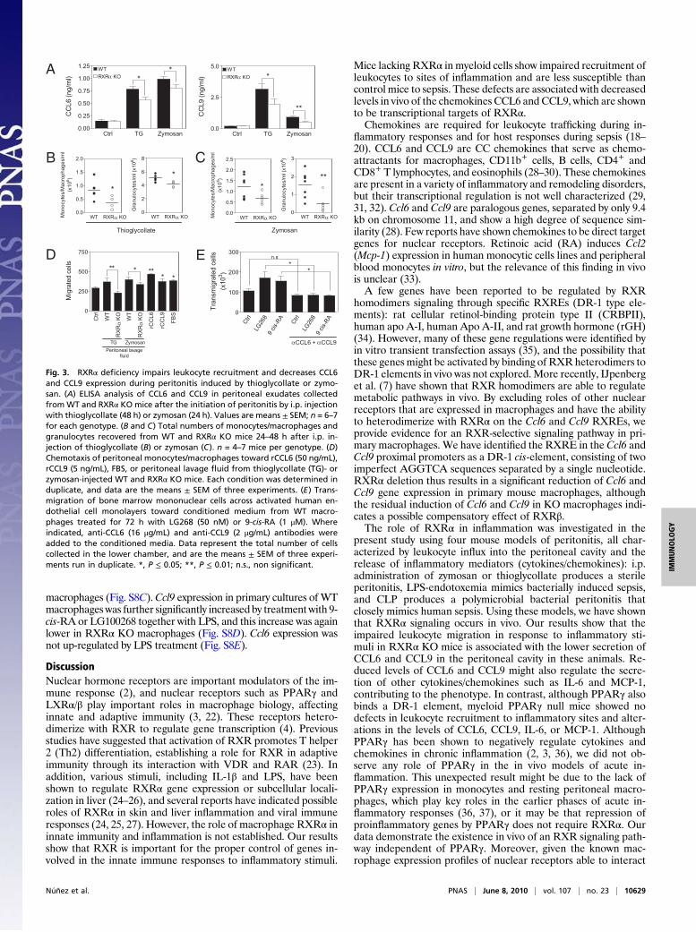
Decreased CCL6 and CCL9 Expression Impairs Leukocyte Recruitmentin Myeloid RXRα Knockout Mice. We next investigated the func-tional consequences of RXRα-mediated chemokine regulation inan in vivo model of acute inflammation.We induced peritonitis byinjecting thioglycollate or zymosan into the mouse peritonealcavity andmonitored the accumulation of CCL6 andCCL9 in cell-free peritoneal lavage fluid. Levels of CCL6 and CCL9 peaked at24 h and remained high 48 h after injection (Fig. S3A). Peritonealexudates collected from RXRα KO mice 24 or 48 h after the ini-tiation of peritonitis contained significantly less CCL6 and CCL9than did time-matched WT exudates (Fig. 3A). Similarly, the ex-udate levels of IL-6 and MCP-1 in response to thioglycollate- orzymosan-induced peritonitis, respectively, were lower in RXRαKO mice (Fig. S3B). There were no significant differences in thequantity of MIP-1α, GROα, Rantes, IL-12, and TNFα (Fig. S3B).Because chemokines are chemoattractants that direct leuko-
cytes to inflammation sites, we next investigated whether de-creased CCL6 and CCL9 levels in peritoneal exudates had animpact on leukocyte recruitment. Thioglycollate or zymosan in-jection induced strong recruitment of monocytes/macrophagesand granulocytes from 24 h up to 48 h into the abdominal cavityof WT mice (Fig. S3 C and D). In contrast, the influx induced bythese agents in RXRα KO mice was significantly lower (Fig. 3 Band C). The number of resident monocytes/macrophages andgranulocytes was, however, unaffected by deletion of RXRα (Fig.S4A). The impaired ability of RXRα KO mice to mount a properleukocytic response was not due to any overt leukocyte defect, asshown by the similar sizes and composition of leukocyte sub-populations in blood and bone marrow from WT and RXRα KOmice (Table S1 and Fig. S4 B and C). In addition, RXRα KOmice challenged with i.p. thioglycollate showed normal leukocytepopulation profiles in bone marrow and peripheral blood (Fig.S4D), strongly indicating that the low number of leukocytes in-filtrating the inflamed peritoneum in the absence of RXRα is theresult of impaired leukocyte migration.To explore the mechanistic link between decreased peritoneal
exudate levels of CCL6 and CCL9 and impaired leukocyte migra-tion, we conducted a series of in vitro migration assays. Peritonealexudates fromWTmicewere as effective as recombinantCCL6 andCCL9 inducing monocyte/macrophage migration; in contrast, exu-dates from RXRα KOmice were significantly less chemoattractive(Fig. 3D). We next tested the chemoattractant potential of theconditioned media obtained from peritoneal macrophages stimu-lated in culture with the RXRα ligands LG100268 or 9-cis-RA for72 h. These conditioned media significantly induced the migrationof bonemarrowmononuclear cells across activated endothelial cells
Ctrl
9 cis-
RA
LG26
8
LG75
40
2
4
6
8
**
CC
L6
Rela
tive e
xpre
ssio
n
Ctrl
9cis-
RA
LG26
8
LG75
40
1
2
3
4
**
CC
L9
Rela
tive
Expre
ssio
n
Ctrl
9 cis-
RA
LG26
8
LG75
40
1
2
**
CC
L9 (
ng/m
l)
Ctrl
9 cis-
RA
LG26
8
LG75
40
1
2
3
4
* *
*
CC
L6 (
ng/m
l)
WT
KOα
RXR
0.00
0.25
0.50
0.75
1.00
1.25
*
CC
L9
Rela
tive
Expre
ssio
n
WT
KOα
RXR
0.0
0.5
1.0
*CC
L6
Rela
tive E
xpre
ssio
n
Ctrl
9 cis-
RA
LG26
80
1
2
3
4
5
*
RXRα KO
WT
*
**
CC
L9
Rela
tive
Expre
ssio
n
Ctrl
9 cis-
RA
LG26
80
1
2
3
4
5
6
7
8
*
RXRα KO
WT
**
CC
L6
Rela
tive E
xpre
ssio
n
A B C D
Fig. 1. Loss of RXRα in macrophages reduces Ccl6 and Ccl9 chemokine ex-pression. (A) Q-PCR analysis of Ccl6 and Ccl9 mRNA expression in RXRα KOperitoneal macrophages. *, P ≤ 0.05 compared with WT. (B) Q-PCR analysis ofCcl6 and Ccl9 gene expression in peritoneal macrophages treated for 24 hwith the RXR ligands 9-cis-RA (1 μM), LG268 (50 nM), or LG754 (1 μM). *, P ≤0.05 compared with control (Ctrl). (C) ELISA of CCL6 and CCL9 protein inmacrophages treated as in C for 48 h. *, P ≤ 0.05 compared with control (Ctrl).(D) Q-PCR analysis of Ccl6 and Ccl9 mRNA expression in WT and RXRα KOmacrophages treated with LG268 or 9-cis-RA for 24 h. *, P ≤ 0.05 comparedwith WT. Data are means ± SEM of at least three independent experiments.
Núñez et al. PNAS | June 8, 2010 | vol. 107 | no. 23 | 10627
IMMUNOLO
GY

(Fig. 3E); this transmigration was significantly inhibited by pre-incubation of themedia with anti-CCL6 and anti-CCL9 antibodies,indicating that CCL6 and CCL9 are key chemoattractants in thissetting. Further experiments showed thatWT andRXRαKObonemarrow mononuclear cells have the same capacity to transmigratetoward recombinant CCL6 and CCL9 (Fig. S5A). Consistently,therewas nodifference between the two genotypes in the expressionof CCR1 mRNA (the receptor for CCL6 and CCL9) in peritonealmacrophages or in the expression ofCCR1protein in bonemarrow,blood leukocytes, and peritoneal macrophages (Fig. S5B).To exclude the possibility that the defects in leukocyte migra-
tion involved RXRα heterodimerization with PPARγ, we per-formed thioglycollate-induced peritonitis experiments in myeloidPPARγ KOmice. In contrast to RXRα KOmice, the PPARγ KOmice showed no defect in leukocyte recruitment (Fig. S6A and B),no significant differences in the expression levels of CCL6, CCL9,MCP-1, or IL-6 in peritoneal exudates after thioglycollate in-jection (Fig. S6C), and no changes in Ccl6 and Ccl9 gene ex-pression in peritoneal macrophages in vitro (Fig. S6D).Theseresults suggest that the regulation of chemokine production andleukocyte migration by RXRα is independent of PPARγ.
Absence of RXRα from Macrophages Prolongs Mouse Survival in CLP-and LPS-Induced Sepsis. To investigate the roleofRXRα in the innateimmune response toexperimental sepsis,weperformedcecal ligationand puncture (CLP) inWT andRXRαKOmice. After 24 h, plasma
levels of CCL6 and CCL9 and peritoneal levels of CCL6 in RXRαKO mice were significantly lower than in WT animals (Fig. 4A). Inaddition, levels of MCP-1 were lower in RXRα KO peritoneal exu-date, whereas they were unaltered in plasma, and there was also nosignificant difference in peritoneal GRO-α, MIP-1α, or Rantes, or inthe peritoneal and plasma concentrations of IL-6, IL-1α, IL-12, andTNFα (Fig. S7 A and B). We also found that RXRα KO micesurvivedCLP-induced sepsis for significantly longer thanWT (Fig.4B). Consistently, the severity of CLP, scored by quantification ofliver histological parameters (hemorrhage, fibrin accumulation,inflammatory infiltrate, and degenerated tissue), was lower inRXRαKOmice (Fig. 4 C and D).RXRαKOmice were also less susceptible thanWTmice to septic
shock induced by LPS (Fig. 4E), and the increase in plasma CCL9after LPS challenge (90 min) was significantly lower in RXRα KOmice (Fig. 4F). The absence of RXRα did not, however, affect theplasmaconcentrationsof IL-6, IL-1α, IL-12,TNFα, andMCP-1 (Fig.S7C). We did not find significant differences in the plasma levels ofCCL9,MCP-1, and IL-6afterLPS injection inPPARγKOmice (Fig.S7D). Next, we studied the effect of RXR signaling on LPS-inducedCCL9 production by peritoneal macrophages in vitro. In WT mac-rophages,LPS induced theexpressionofCcl9mRNA,peakingat 4h,and a strong accumulation of CCL9 protein in culture supernatants;in contrast, RXRαKOmacrophages showed significantly lowerCcl9mRNAexpression andCCL9protein secretion (Fig. S8A andB).Nochanges in LPS-induced Ccl9 expression were found in PPARγ KO
C
0
10
20
30
40
50
60
9 cis-RA
LG268 -
- -+
- -
-
+
-
-
+
- RXRα + RXRα
+
Rela
tive
Lucifera
se
activity
D
0
10
20
30
40
9 cis-RA +- +- +
CCL61kb
CCL6200bp
CCL6200bp MUT
-
Rela
tive L
ucifera
se
activity
0
10
20
30
40
9 cis-RA +- +- +
CCL91kb
CCL9200bp
CCL9200bp MUT
-
Rela
tive L
ucifera
se
activity
0
5
10
15
20
25
9 cis-RA
LG268 -
- -+
- -
-
+
-
-
+
- RXRα + RXRα
+
Rela
tive L
ucifera
se
activity
B
E
A
0 30 90 1200.00
0.01
0.02
0.03
0.04
0.05
0.06
0.07
Time (min)
IgG
RXRα
CC
L9
Chro
matin in
put (%
)
0 30 90 1200.00
0.01
0.02
0.03
0.04
0.05
0.06
Time (min)
IgG
RXRα
CC
L6
Chro
matin input(%
)F
G
0
2
4
6
RosiGW647
GW742
LG268
- - - +-
- + + --
- - ---
- -
- -
- -
+ +
- +++-
-
+
-
+
**
***
CC
L9
Rela
tive E
xpre
ssio
n
0
1
2
3
4
5
6
RosiGW647
GW742
LG268
- - - -- +-
- + - -+ --
- - + +---
- - - +++-
-
+
-
+
* **
***CC
L6
Re
lativ
e E
xp
ressio
nH
Fig. 2. Transcription of Ccl6 and Ccl9 is regulated by RXRα via DR-1 rexinoid response elements. (A and B) RAW 264.7 macrophages were cotransfected witha luciferase reporter plasmid under the transcriptional control of either the Ccl6 (pGL3-CCL6-luc) or the Ccl9 (pGL3-CCL9-luc) promoters together with an RXRαexpression plasmid (pCMX-RXRα) or empty vector, as indicated. Cells were treated with LG268 (50 nM) or 9-cis-RA (1 μM) and analyzed for reporter activity 24h later. (C) Mutations introduced into the Ccl6 and Ccl9 DR-1 motifs. (D and E) RAW 264.7 cells were transfected with 1 kb, 200 bp, or DR-1-mutated 200 bp(MUT) proximal sequences from the Ccl6 or Ccl9 promoters. Cells were treated with LG268 (50 nM) or 9-cis-RA (1 μM) and analyzed for reporter activity 24 hlater. Values are means ± SEM from at least three experiments. (F) ChIP analysis of the binding of RXRα to Ccl6 and Ccl9 proximal promoter regions inmacrophages treated for the indicated times with LG268 (50 nM). ChIP assays were performed with antibodies against RXRα and control IgG. Immunopre-cipitated DNA was analyzed by Q-PCR. Values are means ± SEM. (G) Recombinant GST-labeled RXRα homodimers were bound to 32P-labeled oligonucleotidescorresponding to the RXRE from the Ccl6 and Ccl9 promoter or a version mutated as in C (mut). Competition assays were conducted with unlabeled WT andmutant RXRE oligonucleotides. (H) Q-PCR analysis of mRNA expression in peritoneal macrophages treated with ligands for PPARα (GW7647, 0.1 μM), PPARβ(GW0742, 0.1 μM), PPARγ (Rosiglitazone, 1μM), or RXR (LG268, 50 nM), alone or in the indicated combinations. Values are means ± SEM *, P ≤ 0.05; **, P ≤0.01; ***, P ≤ 0.001.
10628 | www.pnas.org/cgi/doi/10.1073/pnas.0913545107 Núñez et al.
macrophages (Fig. S8C).Ccl9 expression in primary cultures of WTmacrophages was further significantly increased by treatment with 9-cis-RA or LG100268 together with LPS, and this increase was againlower in RXRα KO macrophages (Fig. S8D). Ccl6 expression wasnot up-regulated by LPS treatment (Fig. S8E).
DiscussionNuclear hormone receptors are important modulators of the im-mune response (2), and nuclear receptors such as PPARγ andLXRα/β play important roles in macrophage biology, affectinginnate and adaptive immunity (3, 22). These receptors hetero-dimerize with RXR to regulate gene transcription (4). Previousstudies have suggested that activation of RXR promotes T helper2 (Th2) differentiation, establishing a role for RXR in adaptiveimmunity through its interaction with VDR and RAR (23). Inaddition, various stimuli, including IL-1β and LPS, have beenshown to regulate RXRα gene expression or subcellular locali-zation in liver (24–26), and several reports have indicated possibleroles of RXRα in skin and liver inflammation and viral immuneresponses (24, 25, 27). However, the role of macrophage RXRα ininnate immunity and inflammation is not established. Our resultsshow that RXR is important for the proper control of genes in-volved in the innate immune responses to inflammatory stimuli.
Mice lacking RXRα in myeloid cells show impaired recruitment ofleukocytes to sites of inflammation and are less susceptible thancontrol mice to sepsis. These defects are associated with decreasedlevels in vivo of the chemokines CCL6 and CCL9, which are shownto be transcriptional targets of RXRα.Chemokines are required for leukocyte trafficking during in-
flammatory responses and for host responses during sepsis (18–20). CCL6 and CCL9 are CC chemokines that serve as chemo-attractants for macrophages, CD11b+ cells, B cells, CD4+ andCD8+ T lymphocytes, and eosinophils (28–30). These chemokinesare present in a variety of inflammatory and remodeling disorders,but their transcriptional regulation is not well characterized (29,31, 32). Ccl6 and Ccl9 are paralogous genes, separated by only 9.4kb on chromosome 11, and show a high degree of sequence sim-ilarity (28). Few reports have shown chemokines to be direct targetgenes for nuclear receptors. Retinoic acid (RA) induces Ccl2(Mcp-1) expression in human monocytic cells lines and peripheralblood monocytes in vitro, but the relevance of this finding in vivois unclear (33).A few genes have been reported to be regulated by RXR
homodimers signaling through specific RXREs (DR-1 type ele-ments): rat cellular retinol-binding protein type II (CRBPII),human apo A-I, human Apo A-II, and rat growth hormone (rGH)(34). However, many of these gene regulations were identified byin vitro transient transfection assays (35), and the possibility thatthese genes might be activated by binding of RXRheterodimers toDR-1 elements in vivo was not explored.More recently, IJpenberget al. (7) have shown that RXR homodimers are able to regulatemetabolic pathways in vivo. By excluding roles of other nuclearreceptors that are expressed in macrophages and have the abilityto heterodimerize with RXRα on the Ccl6 and Ccl9 RXREs, weprovide evidence for an RXR-selective signaling pathway in pri-mary macrophages. We have identified the RXRE in the Ccl6 andCcl9 proximal promoters as a DR-1 cis-element, consisting of twoimperfect AGGTCA sequences separated by a single nucleotide.RXRα deletion thus results in a significant reduction of Ccl6 andCcl9 gene expression in primary mouse macrophages, althoughthe residual induction of Ccl6 and Ccl9 in KO macrophages indi-cates a possible compensatory effect of RXRβ.The role of RXRα in inflammation was investigated in the
present study using four mouse models of peritonitis, all char-acterized by leukocyte influx into the peritoneal cavity and therelease of inflammatory mediators (cytokines/chemokines): i.p.administration of zymosan or thioglycollate produces a sterileperitonitis, LPS-endotoxemia mimics bacterially induced sepsis,and CLP produces a polymicrobial bacterial peritonitis thatclosely mimics human sepsis. Using these models, we have shownthat RXRα signaling occurs in vivo. Our results show that theimpaired leukocyte migration in response to inflammatory sti-muli in RXRα KO mice is associated with the lower secretion ofCCL6 and CCL9 in the peritoneal cavity in these animals. Re-duced levels of CCL6 and CCL9 might also regulate the secre-tion of other cytokines/chemokines such as IL-6 and MCP-1,contributing to the phenotype. In contrast, although PPARγ alsobinds a DR-1 element, myeloid PPARγ null mice showed nodefects in leukocyte recruitment to inflammatory sites and alter-ations in the levels of CCL6, CCL9, IL-6, or MCP-1. AlthoughPPARγ has been shown to negatively regulate cytokines andchemokines in chronic inflammation (2, 3, 36), we did not ob-serve any role of PPARγ in the in vivo models of acute in-flammation. This unexpected result might be due to the lack ofPPARγ expression in monocytes and resting peritoneal macro-phages, which play key roles in the earlier phases of acute in-flammatory responses (36, 37), or it may be that repression ofproinflammatory genes by PPARγ does not require RXRα. Ourdata demonstrate the existence in vivo of an RXR signaling path-way independent of PPARγ. Moreover, given the known mac-rophage expression profiles of nuclear receptors able to interact
αCCL6 + αCCL9
Ctrl
LG26
89
cis-
RA
Ctrl
LG26
89
cis-
RA
0
100
200
300n.s
**
Tra
nsm
igra
ted c
ells
(x10
3)
**
Ctr
l
WT
KO
αR
XR
WT
KO
αR
XR rC
CL6
rCC
L9
FB
S
0
250
500
750
*
Peritoneal lavagefluid
TG Zymosan
***
*
Mig
rate
d c
ells
Ctrl TG Zymosan0.00
0.25
0.50
0.75
1.00
1.25
**
RXRα KO
WTC
CL6 (
ng/m
l)
Ctrl TG Zymosan0.0
2.5
5.0
*
**
RXRα KO
WT
CC
L9 (
ng/m
l)
WT RXRα KO0.0
0.5
1.0
1.5
2.0
*
Mo
no
cyte
s/M
acro
ph
ag
es/m
l
(x1
06)
WT RXRα KO0
2
4
6
8
*
Gra
nulo
cyte
s/m
l (x1
06)
WT RXRα KO0.0
0.5
1.0
1.5
2.0
2.5
*
Mo
nocyte
s/M
acro
pha
ge
s/m
l
(x1
06)
WT RXRα KO0
1
2
3
**
Gra
nu
locyte
s/m
l (x10
6)
ZymosanThioglycollate
A
B C
D E
Fig. 3. RXRα deficiency impairs leukocyte recruitment and decreases CCL6and CCL9 expression during peritonitis induced by thioglycollate or zymo-san. (A) ELISA analysis of CCL6 and CCL9 in peritoneal exudates collectedfrom WT and RXRα KO mice after the initiation of peritonitis by i.p. injectionwith thioglycollate (48 h) or zymosan (24 h). Values are means ± SEM; n = 6–7for each genotype. (B and C) Total numbers of monocytes/macrophages andgranulocytes recovered from WT and RXRα KO mice 24–48 h after i.p. in-jection of thioglycollate (B) or zymosan (C). n = 4–7 mice per genotype. (D)Chemotaxis of peritoneal monocytes/macrophages toward rCCL6 (50 ng/mL),rCCL9 (5 ng/mL), FBS, or peritoneal lavage fluid from thioglycollate (TG)- orzymosan-injected WT and RXRα KO mice. Each condition was determined induplicate, and data are the means ± SEM of three experiments. (E) Trans-migration of bone marrow mononuclear cells across activated human en-dothelial cell monolayers toward conditioned medium from WT macro-phages treated for 72 h with LG268 (50 nM) or 9-cis-RA (1 μM). Whereindicated, anti-CCL6 (16 μg/mL) and anti-CCL9 (2 μg/mL) antibodies wereadded to the conditioned media. Data represent the total number of cellscollected in the lower chamber, and are the means ± SEM of three experi-ments run in duplicate. *, P ≤ 0.05; **, P ≤ 0.01; n.s., non significant.
Núñez et al. PNAS | June 8, 2010 | vol. 107 | no. 23 | 10629
IMMUNOLO
GY

with RXRs on DR-1 elements (4, 21), our findings provide evi-dence supporting an action of RXR homodimers. However,the variety of potential heterodimers formed by RXRs meansthat the possibility that a different heterodimer contributes tothe observed effects cannot be completely ruled out at present.Whether other nuclear receptors are also involved in the controlof acute inflammatory responses in vivo will require conditionalmacrophage-specific deletion of each candidate RXR partner.Sepsis is a very complex syndrome in which the underlying in-
flammatory response involves the interplay of several biologicalsystems (the complement, coagulation, and fibrinolytic cascadesand the autonomic nervous system) and cell types, resulting in animbalance of the inflammatory network (17, 38). Recent strategiesto identify potential therapeutic targets have focused on thesesystems but with little clinical success. The finding that survivalafter LPS- or CLP-induced sepsis is prolonged in RXRα KOmiceis therefore of possible clinical interest. Interestingly, mice de-ficient for CCR1, the receptor for CCL6 and CCL9, are also sig-nificantly protected against CLP-induced lethality (39). However,immunoneutralization of CCL6 enhances sepsis-related mortality(31). This discrepancy might arise because the immunoneutrali-zation only interferes with CCL6, potentially allowing a compen-satory effect of CCL9. It is also possible that the decreasedMCP-1levels in the peritoneum of RXRα KO mice after CLP-inducedsepsis might contribute to the phenotype.RXR is activated in vitro by the vitamin A metabolite 9-cis-
RA, which binds with high affinity to the RXR ligand bindingdomain; however, 9-cis-RA has been difficult to detect in vivo(40). Recently, it has been shown that RXR can be activated bypolyunsaturated long chain fatty acids (PUFA) (11), althoughthe nature of the physiologically relevant agonists remains to beestablished. Interestingly, in response to LPS, RAW 264.7 cellsand thioglycollate-elicited or BM-derived macrophages producePUFA (www.lipidmaps.org), which might be important for theregulation of RXR-mediated inflammatory responses. Ccl9 isalso induced by LPS and this response is lost in RXRα KOmacrophages, suggesting that natural ligands for RXR are pro-
duced in response to LPS treatment. Further studies are re-quired to determine whether endogenous ligands for RXR areproduced during inflammatory processes.Our data support a model in which peritoneal macrophages re-
spond to inflammatory stimuli by secreting cytokines and endoge-nous RXRα ligands. The activation of the RXRα-transcriptionalprogram inmacrophageswill result in theproductionof chemokinessuch as CCL6 and CCL9, leading to increased leukocyte recruit-ment and an inflammatory response; excessive activation of thisprogramwill have deleterious consequences, as in the case of sepsis.This study outlines a previously unrecognized role for RXRα in theregulation of leukocyte migration and sepsis, and supports the ex-istence of an RXR signaling pathway in vivo. Our data suggest thatRXRs are potential targets for immunotherapy in sepsis patientsand in chronic inflammatory diseases.
Materials and MethodsCell Culture. Peritoneal macrophages were harvested from WT and RXRα KOmice as previously described (37). Further information is provided in SIMaterials and Methods.
RNA Analysis and Chemokine Quantification. Total RNA isolation and Q-PCRwere performed as described in ref. 9. Further information is provided in SIMaterials and Methods. CCL9 (R&D Systems) and CCL6 (Antigenix) werequantified with ELISA kits.
ChIP Assays. Macrophages were cross-linked with 1% formaldehyde at roomtemperature for 10 min. 100-bp regions of the Ccl6 and Ccl9 proximal pro-moters were amplified by Q-PCR. Primer sequences are available upon re-quest. Anti-RXRα (D-20, sc-553) and anti-RXRDN-197 (sc-774) (Santa CruzBiotechnology) antibodies were used in combination. IgG was used as a con-trol of nonspecific binding.
Gel Shift and Transient Transfection Assays. Oligonucleotides were annealedand labeled using Klenow enzyme (Roche). Purified bacterially expressedRXRα protein was incubated with labeled DNA, and protein–DNA complexeswere electrophoresed and visualized by autoradiography. For competitionstudies, a 5- to 100-fold molar excess of unlabeled oligonucleotide wasadded. Oligonucleotides corresponding to RXR binding sites in Ccl6 and Ccl9
0 12 24 36 48 60 72 84 96 1080
20
40
60
80
100 WT
RXRα KO
* p<0.05
Time after LPS inoculation (hours)
Surv
ival ra
te (
%)
**
WT
KOα
RXR
0
100
200
300
400
500
600
700C
CL
6 (
pg
/ml)
Pe
rito
ne
um
*
WT
KOα
RXR
0
100
200
300
400
500
CC
L6
(pg
/ml)
Pla
sm
a
WT
KOα
RXR
0
1
2
3
4
5
6
CC
L9
(n
g/m
l)P
erito
ne
um
**
WT
KOα
RXR
0
10
20
30
40
50
CC
L9
(n
g/m
l)P
lasm
a
A
0 120
20
40
60
80
100
WT* p<0.01
48 60 72 84 96 108 120 132
RXRα KO
Time after CLP (hours)
Surv
iva
lra
te(%
)
B C
WT
RX
Rα
KO
Control CLP
II
FE
05
1015202530354045
RXRα KO
8 24 54
WT
Time (hours)
% o
fm
ice
with
se
ve
reC
LP
D*
WT
KO
αRXR
0
5
10
15
20
25
30
35
CC
L9
(ng
/ml)
Pla
sm
a
Fig. 4. Delayed CLP- and endotoxin-inducedmortality in RXRα KOmice is associatedwith decreased levels of CCL6 and CCL9. (A) Decreased CCL6 andCCL9 in peritonealexudatesandplasmafromWTandRXRαKOmice.Peritonealexudatesandplasmawerecollected24hafter the initiationofcecal ligationandpuncture (CLP),andCCL6andCCL9weremeasuredbyELISA.n=7–10animals foreachgenotype. (B) Survival ratesof femaleWTandRXRαKOmicesubjectedtoCLP.Thegraphrepresentspooledsurvivaldatafromduplicatestudies showingsimilarresults.Experimentscontained14micepergroup,andsurvivalwasmonitoredfor7daysaftersurgery. (C)Histologicalanalysisofthe livers ofWTandRXRα KO54h after surgery. Representative liver sections are shown. (Scale bar: 50 μm, in inserts 15 μm.) (D) Percentageofmicewith severe peritonitisafter CLP, as determined by quantification of histological parameters (hemorrhage, fibrin accumulation, inflammatory infiltrate, and degenerated tissue) (n = 5–9). (E)Survival of WT and RXRα KO mice after i.p. injection with LPS (40 mg/kg per mouse). n = 28 for each genotype from three independent experiments. (F) Plasma con-centrations of CCL9 inWTandRXRαKOmice, determinedby ELISA 90min after injectionof LPS.n=7 for eachgenotype. Values aremeans±SEM; *,P≤ 0.05; **, P≤ 0.01.
10630 | www.pnas.org/cgi/doi/10.1073/pnas.0913545107 Núñez et al.

promoters and their mutants are available upon request. Transient trans-fections of RAW 264.7 cells (ATCC) using Lipofectamine 2000 (Invitrogen)were as described (36). Promoter constructs for Ccl6 and Ccl9 were cloned inthe pGL3-luc vector (Promega). A β-galactosidase expression vector wascotransfected as an internal control. Point mutations in Ccl6-luc and Ccl9-lucwere made with the QuikChange side-directed mutagenesis kit (Stratagene).
Peritonitis Models. Eight- to 9-week-old mice were i.p. administered with2.5mL of 3% thioglycollate broth (Difco) or 1mg of typeA zymosan (Sigma) in0.5mLof sterile PBS. In other experiments, 12-week-oldmice of eachgenotypewere i.p. injected with 40 mg/kg LPS (E. coli 0111:B4; Sigma). Sepsis inductionby cecal ligation and puncture (CLP) was performed as described elsewhere(41). Severity of CLPwas quantified by histological analysis. Liver sections wereH&E-stained, and three blinded observers scored six pictures per mouse fordistinct parameters: hemorrhage, fibrin deposition, hepatocyte degener-ation, and leukocyte infiltrate. Mice were considered severely affected if ≥3parameters were scored higher than the group average for each time point.
Cell Migration Assay. Cell migration assays were conducted using 8.0-μm-poretranswell filters (Costar). Peritoneal monocytes/macrophages (5 × 10⁴) wereresuspended in 150 μL of 0.5% BSA in RPMI medium and added to the upperchamber of transwell filters. The lower chamber contained rCCL6 (50 ng/mL),rCCL9 (5 ng/mL), or peritoneal exudate from WT or RXRα KO mice previously
injected with thioglycollate (48 h) or zymosan (24 h). After a 2-h incubationat 37 °C, the inserts were processed for fluorescence microscopy.
Endothelial Transmigration Assay. Cell transmigration assays were performedin 5-μm-pore Transwell chambers (Costar) as described in ref. 42. Furtherinformation is provided in SI Materials and Methods.
Statistical Analysis. The normal distribution of data were checked with theKolmogorov–Smirnov test. Data were analyzed by unpaired Student’s t testand nonparametric Mann–Whitney U test. Survival rates were represented asa Kaplan–Meier curve, and the results were analyzed with a log-rank (Mantel–Cox) t test.
ACKNOWLEDGMENTS. Simon Bartlett provided editorial assistance. We thankAranzazuAlfranca andAurora Sánchez Pacheco for assistancewith the gel shiftassay. This work was supported by Marie Curie Grants IRG-CT-2006-026702 (toT.F.) and IRG-016187 (to M.R.), FIS PI052270 (to T.F.), grants from Fundación“Mutua Madrileña” (to T.F. and M.R.), Spanish Ministry of Science and Innova-tion Grants SAF2008-02104 (to A.G.A.), SAF2006-01010 (to M.R.), and BFU2008-02161 (to L.B.), and a grant from the Fundación “Ramón Areces” (to A.G.A.).M.R. and T.F. are supported by the Ramón y Cajal Program (MCINN). The CentroNacional de Investigaciones Cardiovasculares (CNIC) is supported by the SpanishMinistry of Science and Innovation and the Pro-CNIC Foundation.
1. Kastner P, Mark M, Chambon P (1995) Nonsteroid nuclear receptors: What are geneticstudies telling us about their role in real life? Cell 83:859–869.
2. Valledor AF, Ricote M (2004) Nuclear receptor signaling in macrophages. BiochemPharmacol 67:201–212.
3. Daynes RA, Jones DC (2002) Emerging roles of PPARs in inflammation and immunity.Nat Rev Immunol 2:748–759.
4. Mangelsdorf DJ, Evans RM (1995) The RXR heterodimers and orphan receptors. Cell83:841–850.
5. Szanto A, et al. (2004) Retinoid X receptors: X-ploring their (patho)physiologicalfunctions. Cell Death Differ 11 (Suppl 2):S126–S143.
6. Zhang X-K, et al. (1992) Homodimer formation of retinoid X receptor induced by 9-cisretinoic acid. Nature 358:587–591.
7. IJpenberg A, et al. (2004) In vivo activation of PPAR target genes by RXR homodimers.EMBO J 23:2083–2091.
8. Mangelsdorf DJ, et al. (1992) Characterization of three RXR genes that mediate theaction of 9-cis retinoic acid. Genes Dev 6:329–344.
9. Ricote M, et al. (2006) Normal hematopoiesis after conditional targeting of RXRalphain murine hematopoietic stem/progenitor cells. J Leukoc Biol 80:850–861.
10. Heyman RA, et al. (1992) 9-cis retinoic acid is a high affinity ligand for the retinoid Xreceptor. Cell 68:397–406.
11. Lengqvist J, et al. (2004) Polyunsaturated fatty acids including docosahexaenoic andarachidonic acid bind to the retinoid X receptor alpha ligand-binding domain. MolCell Proteomics 3:692–703.
12. Lala DS, et al. (1996) Activation of specific RXR heterodimers by an antagonist of RXRhomodimers. Nature 383:450–453.
13. Altucci L, Leibowitz MD, Ogilvie KM, de Lera AR, Gronemeyer H (2007) RAR and RXRmodulation in cancer and metabolic disease. Nat Rev Drug Discov 6:793–810.
14. Medzhitov R (2007) Recognition of microorganisms and activation of the immuneresponse. Nature 449:819–826.
15. Medzhitov R (2008) Origin and physiological roles of inflammation. Nature 454:428–435.
16. Henson PM (2005) Dampening inflammation. Nat Immunol 6:1179–1181.17. Hotchkiss RS, Karl IE (2003) The pathophysiology and treatment of sepsis. N Engl J
Med 348:138–150.18. Olson TS, Ley K (2002) Chemokines and chemokine receptors in leukocyte trafficking.
Am J Physiol Regul Integr Comp Physiol 283:R7–R28.19. Charo IF, Ransohoff RM (2006) The many roles of chemokines and chemokine
receptors in inflammation. N Engl J Med 354:610–621.20. Coelho AL, Hogaboam CM, Kunkel SL (2005) Chemokines provide the sustained
inflammatory bridge between innate and acquired immunity. Cytokine GrowthFactor Rev 16:553–560.
21. Barish GD, et al. (2005) A Nuclear Receptor Atlas: Macrophage activation. MolEndocrinol 19:2466–2477.
22. Zelcer N, Tontonoz P (2006) Liver X receptors as integrators of metabolic andinflammatory signaling. J Clin Invest 116:607–614.
23. Du X, et al. (2005) An essential role for Rxr alpha in the development of Th2responses. Eur J Immunol 35:3414–3423.
24. Beigneux AP, Moser AH, Shigenaga JK, Grunfeld C, Feingold KR (2000) The acutephase response is associated with retinoid X receptor repression in rodent liver. J BiolChem 275:16390–16399.
25. Chow EK, et al. (2006) A role for IRF3-dependent RXRalpha repression in hepatotoxicityassociated with viral infections. J Exp Med 203:2589–2602.
26. Zimmerman TL, Thevananther S, Ghose R, Burns AR, Karpen SJ (2006) Nuclear exportof retinoid X receptor alpha in response to interleukin-1beta-mediated cell signaling:Roles for JNK and SER260. J Biol Chem 281:15434–15440.
27. Li M, et al. (2005) Retinoid X receptor ablation in adult mouse keratinocytesgenerates an atopic dermatitis triggered by thymic stromal lymphopoietin. Proc NatlAcad Sci USA 102:14795–14800.
28. Hara T, et al. (1995) Molecular cloning and functional characterization of a novelmember of the C-C chemokine family. J Immunol 155:5352–5358.
29. Hogaboam CM, et al. (1999) Immunomodulatory role of C10 chemokine in a murinemodel of allergic bronchopulmonary aspergillosis. J Immunol 162:6071–6079.
30. Zhao X, et al. (2003) CCL9 is secreted by the follicle-associated epithelium and recruitsdome region Peyer’s patch CD11b+ dendritic cells. J Immunol 171:2797–2803.
31. Steinhauser ML, et al. (2000) Chemokine C10 promotes disease resolution and survivalin an experimental model of bacterial sepsis. Infect Immun 68:6108–6114.
32. LaFleur AM, Lukacs NW, Kunkel SL, Matsukawa A (2004) Role of CC chemokine CCL6/C10 as a monocyte chemoattractant in a murine acute peritonitis. Mediators Inflamm13:349–355.
33. Zhu L, Bisgaier CL, Aviram M, Newton RS (1999) 9-cis retinoic acid induces monocytechemoattractant protein-1 secretion in human monocytic THP-1 cells. ArteriosclerThromb Vasc Biol 19:2105–2111.
34. Desvergne B (2007) RXR: From partnership to leadership in metabolic regulations.Vitam Horm 75:1–32.
35. Nakshatri H, Chambon P (1994) The directly repeated RG(G/T)TCA motifs of the ratand mouse cellular retinol-binding protein II genes are promiscuous binding sites forRAR, RXR, HNF-4, and ARP-1 homo- and heterodimers. J Biol Chem 269:890–902.
36. Ricote M, Li AC, Willson TM, Kelly CJ, Glass CK (1998) The peroxisome proliferator-activated receptor-γ is a negative regulator of macrophage activation. Nature 391:79–82.
37. Huang JT, et al. (1999) Interleukin-4-dependent production of PPAR-γ ligands inmacrophages by 12/15-lipoxygenase. Nature 400:378–382.
38. Rittirsch D, Flierl MA, Ward PA (2008) Harmful molecular mechanisms in sepsis. NatRev Immunol 8:776–787.
39. Ness TL, et al. (2004) CCR1 and CC chemokine ligand 5 interactions exacerbate innateimmune responses during sepsis. J Immunol 173:6938–6948.
40. Horton C, Maden M (1995) Endogenous distribution of retinoids during normaldevelopment and teratogenesis in the mouse embryo. Dev Dyn 202:312–323.
41. Rittirsch D, Huber-Lang MS, Flierl MA, Ward PA (2009) Immunodesign of experimentalsepsis by cecal ligation and puncture. Nat Protoc 4:31–36.
42. Matías-Román S, et al. (2005) Membrane type 1-matrix metalloproteinase is involvedin migration of human monocytes and is regulated through their interaction withfibronectin or endothelium. Blood 105:3956–3964.
Núñez et al. PNAS | June 8, 2010 | vol. 107 | no. 23 | 10631
IMMUNOLO
GY
Copyright © 2022 FDOKUMEN