Cellular Oxidative Stress Response Controls the Antiviral and ...
Upload
khangminh22Category
view
4download
0
REVIEW
Caspases control antiviral innate immunity
Huihui Chen1,5, Xiaohan Ning2,3,4,5 and Zhengfan Jiang3
Caspases are a family of cysteine proteases whose functions have been scrutinized intensively in recent years.Beyond their established roles in programmed cell death and inflammatory response, some caspases are alsofundamental players in antiviral immunity by fine-tuning the levels of antiviral signaling adapters and cytokines,such as type Ι interferons, which serves as a major, sophisticated weapon against viruses. Viral infections can resultin inflammasome activation and the initiation of cell death, including apoptosis and pyroptosis, and multiplecaspases are significantly involved in these processes. This review will focus on the cutting-edge discoveriesregarding the multifaceted roles of caspases in antiviral innate immunity.Cellular & Molecular Immunology (2017) 14, 736–747; doi:10.1038/cmi.2017.44; published online 10 July 2017
Keywords: apoptosis; caspases; Ι-IFNs; inflammasome; innate immunity
INTRODUCTION
Viral infections can induce systemic immune responses, whichare shaped by specific signaling cascades between the host andthe pathogen. Host cells are equipped with an array of multiplegermline-encoded pattern recognition receptors (PRRs) todetect pathogen-associated molecular patterns (PAMPs). ViralPAMPs are often nucleic acid-based, including double-stranded(ds) RNA, single-stranded (ss) RNA and viral DNA.1 In somecases, hosts also detect viral proteins, such as viral fusionglycoproteins, respiratory syncytial virus (RSV)-F protein andEbola virus GP1 proteins. Current studies have shown thatPRRs detect viral nucleic acids mainly consisting of severalToll-like receptor (TLR) family members and various cytosolicsensors. TLRs 3, 7, 8, 9 and 13 can recognize viral nucleic acidsin the endosomal compartment. Viral RNA serves as a ligandfor both cytosolic NLRP3 inflammasomes and RIG-I-likereceptors (RLRs), such as RIG-I, MDA-5 and DHX58 (DEXHbox polypeptide 58 or LGP2).2–4 Thus far, the cytoplasmicDNA sensors identified include DNA-dependent activator ofIFN-regulator factors, DEAD box polypeptide 41, RNA poly-merase III, STING (also known as MITA/ERIS/MPYS/TEM173), cGAS, polyglutamine-binding protein 1 (PQBP1),5
DNA-dependent protein kinase,6 sex-determining region
Y-box 2,7 meiotic recombination 11 homolog A,8 AIM2 andIFI16 inflammasomes.9–17
Upon detecting viral nucleic acids, PRRs activate a set ofadapter proteins and relay the signals to downstream transcrip-tional factors to drive the production of IFNs and IFN-stimulated genes (ISGs), induce the secretion of inflammatorycytokines and further initiate pyroptosis, a lytic, programmedcell death.
Insufficient production of IFNs and inflammatory cytokinescause the deficiency of viral infection clearance and leads tochronic viral infections, while excessive amounts of IFNs andinflammatory cytokines contribute to autoimmune and auto-inflammatory diseases. Meanwhile, concomitant uncontrolledpyroptosis is also very detrimental to the host. Accordingly,mammals have developed a series of critical downstreammediators to fine-tune the antiviral innate immune system,aiming to balance immune responses against viral pathogens atan appropriate level.
Among the various mediators, caspases are a family of genessituated at the nexus of vital regulatory networks, as theybalance inflammation, antiviral innate immunity and celldeath. This review will focus on how caspases control antiviralinnate immunity on multiple levels.
1Shenzhen BOLI Pharmaceutical Co., Ltd, Shenzhen, Guangdong 518040, China; 2State Key Laboratory of Protein and Plant Gene Research, School of LifeSciences, Peking University, Beijing 100871, China; 3Key Laboratory of Cell Proliferation and Differentiation of the Ministry of Education, School of LifeSciences, Peking University, Beijing 100871, China and 4Peking-Tsinghua Center for Life Sciences, Beijing 100871, China
Correspondence: Professor Z Jiang, Key Laboratory of Cell Proliferation and Differentiation of the Ministry of Education, School of Life Sciences, PekingUniversity, Beijing 100871, China.E-mail: [email protected]
5These authors contributed equally to this work.
Received: 23 March 2017; Revised: 12 May 2017; Accepted: 12 May 2017
Cellular & Molecular Immunology (2017) 14, 736–747& 2017 CSI and USTC All rights reserved 2042-0226/17 $32.00
www.nature.com/cmi

Caspases and signaling pathwaysCaspases are initially synthesized as inactive monomericzymogens (procaspases) that only obtain catalytic activity uponan appropriate stimulus (Table 1). While 18 mammaliancaspases have recently been identified, the newly discoveredcaspases-15 through -18, except for caspase-16, are absent inplacental mammals. The other 12 members of the caspasefamily are traditionally classified into two groups according totheir function.19,20 The apoptotic caspases (caspases-2, -3, -6,-7, -8, -9 and -10 in mammals) function in the initiation andexecution of a morphologically different programmed celldeath called apoptosis. The inflammatory caspases (caspases-1, -4, -5 and -12 in humans and caspases-1, -11 and -12 inmice) mediate innate immune response by cleaving proin-flammatory cytokine precursors (for example, pro-IL-1β andpro-IL-18) to initiate inflammation.80 Inflammatory caspasesare also involved in another programmed cell death known aspyroptosis.19,21–24 The functions of caspases-2, -10, -14 and -16are currently somewhat controversial and enigmatic and areless easily categorized (Table 1).
The mitochondrial-related intrinsic apoptosis pathway ispredominantly controlled by the Bcl-2 family and Bcl-2homology domain-3 (BH3)-only proteins (the pro-deathproteins BAK and BAX are essential effectors).21,25,26 In thispathway, apoptotic stimuli (for example, cytotoxic drugs, DNAdamage) activate the BH3-only proteins, resulting in activationof the pro-apoptotic effectors BAK/BAX and the induction ofMOMP (Figure 1). Subsequently, the mitochondrial proteins,such as cytochrome c, efflux into the cytoplasm, initiating theassembly of the apoptosome, including APAF-1, and furtheractivating caspase-9. Ultimately, apoptosis ensues after the
cleavage and activation of the executioner caspases (caspases-3 and -7) by caspase-9(refs 27,28) (Figure 1).
The extrinsic apoptosis pathway is death receptor (DR)-mediated. DR members include TNF receptor-1, first apoptosissignal (FAS, also known as APO-1 or CD95), DR3, TRAIL-R1(TNF-related apoptosis-inducing ligand receptor-1 or DR4)and TRAIL-R2 (or DR-5 in human). The DR ligands includetumor necrosis factor (TNF), CD95-L (CD95 ligand or Fas-L),TRAIL (or Apo-L) and TNF-like ligand 1A. Upon activation,the DR death domain recruits adapter proteins to form theDISC comprising FADD, TRADD, caspase-8 (caspase-10 inhuman cells) and other components.29 The activated initiatorcaspase-8 is capable of either initiating the extrinsic apoptoticpathway by cleaving and activating the apoptotic effectorcaspases (-3, -6, -7) or amplifying intrinsic mitochondrialapoptosis by cleaving BID to a truncated form (tBID)30
(Figure 1). In certain conditions, when the levels of FLIPL(long isoform of the FLICE-like inhibitory protein) aredecreased and upon genotoxic stress, caspase-8 associates withRIPK1 and FADD to form a multiple protein complex calledthe ‘ripoptosome’, leading to apoptosis6,31–33 (Figure 1).
Inflammatory caspases function as central adapters of innateimmunity, mainly by controlling the assembly of larger,multiple protein complexes called inflammasomes. Canonicalinflammasomes are often characterized as scaffold proteinscontaining members of the NLR (nucleotide-binding-and-oligomerization domain and leucine-rich-repeat-containing)family, the AIM2-like receptor family or the tripartite motif(TRIM, of which pyrin is the only member of inflammasomes)family and caspase-1 molecules together to trigger the auto-cleavage and activation of caspase-1.34 The inflammasomescurrently known to be engaged in antiviral innate immunityinclude the NLRP3 inflammasome,2,35–38 the RIG-Iinflammasome39 and the AIM2 inflammasome.10–12 Thenoncanonical inflammasomes comprise caspases-4 and -5 inhumans and caspase-11 in mice, which directly recognizelipopolysaccharides (LPS).40–43 Both canonical caspase-1 andnoncanonical caspase-11 inflammasomes trigger two sets ofinflammatory responses: the cleavage and release of proin-flammatory cytokines, including IL-1β, IL-18 and IL-33,44 andthe induction of pyroptosis.34,45
Caspase-1-dependent antiviral inflammasomesCaspase-1 plays a vital role in inflammatory responses byassembling a large (700 kDa) molecular structure called aninflammasome.46 The conversion of pro-caspase-1 into cata-lytically active caspase-1 is tightly connected with the matura-tion of pro-IL-1β, pro-IL-18 and most likely pro-IL-33 insome cases. To date, four distinct caspase-1-dependentinflammasomes have been identified as being involved inantiviral innate immunity: the RIG-I inflammasome,39,47 theAIM2 inflammasome,10–12 the IFI16 inflammasome and theNLRP3 inflammasome.2,36–38 The adapter ASC provides aplatform between RIG-I, NLRP3, AIM2 or IFI16 and pro-caspase-1 through a homotypical association with N-terminalPYDs (pyrin domains) and C-terminal CARDs. Once the
Table 1 Classification of caspases
The apoptotic caspases have been subdivided by their mechanism of action intoinitiator caspases (caspases-2, -8, -9 and -10) and effector caspases (caspases-3,-6 and -7). Caspase-10 is only present in the mouse genome. The initiatorcaspases are involved in various apoptotic pathways, which fall into either theintrinsic or extrinsic category.18,137
Control of antiviral innate immunity
H Chen et al
737
Cellular & Molecular Immunology

filamentous inflammasome complex is assembled, pro-caspase-1 is initially autoproteolytically cut into a p35 CARD-containing fragment and a p10 fragment. Sequentially, p35 isfurther digested into a p20 fragment and a CARD, and thentwo p20 subunits heterodimerize with two p10 subunits toproduce a bioactive caspase-1. Activated caspase-1 then med-iates the processing of pro-IL-1β and pro-IL-18 at cysteineresidues adjacent to aspartic acid residues. The secreted andactive cytokines lead to downstream inflammatory responses(Figure 2).
RIG-I inflammasome. RIG-I has been shown to be involved inthe recognition of RNA viruses, including Newcastle diseasevirus, vesicular stomatitis virus (VSV), Sendai virus (SeV),rabies virus, hepatitis virus (HCV), Japanese encephalitis virus,influenza A virus, measles virus and RSV.48–55 Upon viralinfection, RIG-I recruits the mitochondrial adapter MAVS/TRAF3/6 (TNF receptor-associated factor 3/6) signalosome andthen activates IKKε and IRF3/7 to induce IFN signaling.Current studies have shown that RIG-I is also involved ininflammatory responses. Upon VSV infection or 5′-tripho-sphate RNA transfection, RIG-I directly activates inflamma-some signaling by recruiting caspase-1 in an ASC-dependentmanner (Figure 2). Furthermore, RIG-I reportedly induces theprocessing and secretion of IL-18 by activating caspase-3 inresponse to viral infection.47
AIM2 and IFI16 inflammasomes. AIM2 and IFI16 belong to afamily of interferon-inducible HIN-200 proteins, which con-tain an N-terminal pyrin domain for sensing cytoplasmic DNAand a C-terminal domain (oligonucleotide/oligosaccharide-binding domain) for interacting with ASC to activatecaspase-1 and caspase-8. Caspase-1 mediates IL-1β and IL-18maturation and pyroptosis, while caspase-8 induces apoptosis.The family includes IFI16, MNDA, IFIX and AIM2 in humansand IFI202, IFI203, IFI204, IFI205, Pyin1 and AIM2 in mice.Four groups have discovered that AIM2 senses cytoplasmicdsDNA, including viral DNA (for example, adenovirus andretrovirus). A report using cultured cells showed that vacciniavirus (VACV) and mouse cytomegalovirus (mCMV) triggeredthe autoactivation of caspase-1 through AIM2. Moreover,Aim2-deficient mice infected with mCMV revealed low IL-18levels, decreased IFNγ-expressing nature killer cells and highermCMV viral titers compared to control mice.56 Intriguingly,current work revealed that AIM2 could also recognize RNAviruses, such as Chikungunya virus (CHIKV) and West Nilevirus in a caspase-1-dependent manner in human dermalfibroblasts. Similarly, inhibition of the inflammasome viacaspase-1 silencing enhanced CHIKV replication in residentskin cells.57 Furthermore, skin fibroblasts infected with Zikavirus (a ssRNA virus) displayed elevated levels of AIM2 andIL-1β transcripts.58 More intensive studies are needed toexplore the mechanism of how AIM2 recognizes RNA viruses
Figure 1 Caspases in extrinsic and intrinsic apoptosis. The extrinsic apoptosis pathway is activated by ligand binding and the deathreceptor, which then recruits TRADD or FADD to form a DISC complex, resulting in the activation of initiator caspase-8 or -10. Thesecaspases then directly cleave and activate the effector caspases-3, -6 and -7, effectively executing cellular apoptosis. Alternatively, theintrinsic apoptotic pathway can be activated by intrinsic stimuli, including various cellular stressors, DNA damage and growth factordeprivation. Subsequently, the activation of BAK and BAX or the inactivation of anti-apoptotic BCL-2 family members leads to MOMP andthe release of mitochondrial factors, such as cytochrome c. Cytochrome c binding to APAF-1 is important for the activation andoligomerization of caspase-9 to form the apoptosome. The activation of caspase-9 then initiates apoptosis by activating the effectorcaspases. FADD, FAS-associated death domain; TRADD, TNFR-associated death domain; DISC, death-inducing signaling complex;caspases, cysteinyl aspartate proteinases; BAX, BCL-2-associated X protein; BAK, BCL-2 antagonist or killer; MOMP, mitochondrial outer-membrane permeabilization; APAF-1, apoptotic protease-activating factor 1.
Control of antiviral innate immunity
H Chen et al
738
Cellular & Molecular Immunology

to activate inflammasomes and the subsequent procession ofcaspase-1 and IL-1β/IL-18.
AIM2 is located in the cytosol to recognize viruses, and theAIM2-like receptor IFI16 is localized predominantly in thenucleus, despite small amounts of cytosolic IFI16 beingobserved in some cells. In addition to the engagement ofI-IFN induction in response to viral dsDNA, such as in VACVand herpes simplex virus type 1 (HSV-1), IFI16 has also beenproven to sense KSHV genomes and associate with ASC andpro-caspase-1 in the nucleus59 (Figure 2). However, themechanism of IFI16 inflammasome redistribution to thecytoplasm remains unknown. Other studies have showed thatIFI16 induces the expression of both I-IFNs and caspase-1-dependent inflammatory cytokine maturation in response tohuman immunodeficiency virus-1 (HIV-1) replication.60–62
NLRP3 inflammasomes. Until now, the NLPR3 inflamma-some is the most well-studied member of the NLR family, andit can activate caspase-1 and induce the production of IL-1βand IL-18 in response to a wide range of PAMPs andendogenous and environmental stimuli.34 Until now, NLRP3is the only member of this family shown to be engaged in viralinfection, including influenza A virus, SeV, vaccinia virus andencephalomyocarditis virus.2,35,37,39 Although several studieshave revealed that the shared mechanism of a diversity ofpotential activators can enable NLPR3 to oligomerize and
recruit both ASC and pro-caspase-1 through the PYD domain,there is little evidence of NLRP3 directly binding to theseligands (Figure 2). Dual distinct signaling levels account forNLRP3 activation in terms of viral infection.63–65 The entry ofinfluenza A virus into the host cell leads to the release of ionsand the production of ROS by NADPH (nicotinamide adeninedinucleotide phosphate) in the cytosol. In the meantime, theviral M2 channel is induced and transferred to the acidifiedtrans-Golgi apparatus, facilitating the efflux of hydrogen ions toactivate the NLRP3 inflammasome. Likewise, ROS acts assecondary messengers to augment the virus-induced NLRP3inflammasome.2,65 Modified vaccinia virus Ankara-mediatedNLRP3 inflammasomes have also been demonstrated torequire TLR2-dependent signaling.66
Caspase-dependent pyroptosisCumulative reports have discovered that inflammasomes arenot only capable of maturing proinflammatory cytokines, butalso promote ‘pyroptosis’, a distinct inflammatory caspase-dependent cell death.67 Accordingly, the inflammatory cas-pases-1, -4, -5 and -11 are also called pyroptoticcaspases40,41,68,69 (Figure 2).
Pyroptosis cell death was initially observed in Shigellaflexneri-infected macrophages in 1992, and it was defined asapoptosis because it shared common features, such as nuclearcondensation, DNA fragmentation and caspase dependence.70
Figure 2 Caspase-1-mediated inflammasomes involved in antiviral defense. In responses to viral PAMPs, RIG-I, NLRP3, AIM2 and IFI16assemble caspase-1-activating inflammasome complexes in an ASC-dependent manner. Activated caspase-1 drives the cleavage andmaturation of the proinflammatory cytokines pro-IL-1β and pro-IL-18. Inflammatory caspases cleave gasdermin D, causing pyroptosis,which promotes the release of matured IL-1β and IL-18. The NLRP3 inflammasome recognizes multiple PAMPs, including viral RNA, incombination with ion flux or ROS. The RIG-I and AIM2 inflammasomes recognize viral ssRNA and dsDNA, respectively. The AIM2inflammasome could recruit caspase-8 to induce apoptosis. The IFI16 inflammasome specifically recognizes KSHV dsDNA in the nucleus.RIG-I, retinoic acid inducible gene-I; NLRP3, nucleotide-binding oligomerization-like receptor family, pyrin domain-containing protein 3;AIM2, absent in myeloma 2; IFI16, γ-interferon-inducible protein 16; ASC, apoptosis-associated speck-like protein containing a caspaserecruitment domain; IL-1β, interleukin-1β; IL-18, interleukin-18; KSHV, Kaposi’s sarcoma-associated herpes virus.
Control of antiviral innate immunity
H Chen et al
739
Cellular & Molecular Immunology

It was not until 2001 that researchers further determined thatpyroptosis characteristics are distinct from those of apoptosis.First, pyroptosis is programmed by inflammatory caspases.Second, inflammatory caspase activation leads to plasmamembrane pore formation and, thus, permeabilizes the cellsto small molecules, such as water and ions. Third, the water/ion influx and osmotic lysis cause massive leakage of cytosolicsubstances.68,71 Fourth, pyroptotic cell death requires thecleavage of gasdermin D by caspases-1, -4, -5 and -11,generating the N-terminal fragment responsible for pyroptosisinitiation. However, the C-terminal fragment remains anenigma.72–74 Finally, pyroptosis causes DNA damage, whereasthe nucleus remains intact following the activation of ADP-ribose polymerase and consummation of NAD+ and ATP.75
Summarily, innate immunity is armed with pyroptosis todispose of pathogen-laden cells and eliminate invading microbeprotective niches by enabling them to be removed by asecondary phagocyte.
Pyroptosis is triggered by either caspase-1 or caspases-4, -5and -11, and proinflammatory cytokine maturation is dispen-sable for pyroptosis. However, some differences exist betweenpyroptosis induced by caspase-1 and pyroptosis induced bycaspases-4, -5 and -11.76–80 Casapase-1-dependent pyroptosis isinitiated by various ligands, which activate canonical inflam-masomes, and occurs mainly in macrophage or dendritic cells,
while pyroptosis dependent upon caspases-4, -5 and -11 isdirectly triggered by bacterial stimuli, for which the productionof IL-1β/IL-18 is nearly negligible.81,82
Caspase-1 is currently the only member known to beinvolved in virus-induced pyroptosis. During HIV infection,caspase-1-dependent pyroptosis is critical for CD4+ T-celldepletion, implicating HIV pathogenesis.61 It is remarkablynoted that caspase-1-deficient mice develop normally, disput-ing that the blockade of caspase-1 activity exerts beneficialrather than detrimental effects in HIV patients. Despite theseimportant roles, the mechanisms of how inflammatory cas-pases drive pyroptosis need further study.
Caspases regulate the antiviral innate signaling pathwayscGAS-STING antiviral pathway. Recent work has demolishedthe traditional definition of apoptotic caspases, as it has becomeobvious that apoptosis caspases can control antiviral innateimmunity through non-death processes. Two studies haveshowed that caspases-9, -3 and -7 involved in the intrinsicapoptotic pathway negatively regulate the induction of I-IFNsresponse by controlling cGAS and STING signaling83,84
(Figure 3).Succumbing to DNA viral infection, the cGAS/STING
pathway is triggered by the recognition of cytosolic dsDNA,including dsDNA from HSV-1, murine gammaherpesvirus 68,
Figure 3 Caspases control the cGAS-STING signaling pathway. Cytosolic DNA from virus or self-DNA from mitochondria or the nucleuscould activate cyclic GMP–AMP synthase cGAS, which catalyzes the production of cGAMP in the presence of ATP and GTP. cGAMP bindsand activates the ER-localized adapter protein STING, which then recruits and activates TBK1. This is followed by the phosphorylation andactivation of the transcription factors IRF3, NF-κB and STAT6, leading to I-IFN, proinflammatory cytokines and chemokine production. Theapoptotic caspases-9, -3 and -7 could inhibit cGAS/STING/TBK1/IRF3-mediated IFNα/β production, while the inflammatory caspase-1could cleave cGAS to control innate immunity during inflammasome activation. cGAS, cGAMP/cAMP synthase; STING, stimulator of IFNgenes; TBK1, Tank-binding kinase 1; IRF3, IFN regulatory factor 3; NF-κB, nuclear factor κB; STAT6, signal transducer and activator oftranscription 6; mtDNA, mitochondrial DNA.
Control of antiviral innate immunity
H Chen et al
740
Cellular & Molecular Immunology

KSHV, VACV, adenovirus, human cytomegalovirus, humanpapillomavirus, hepatitis B virus, by the DNA sensorcGAS.85–92 In addition, retroviruses, such as HIV-1 and humanT-cell leukemia virus-1, which contain ssRNA genomes,require reverse-transcribed DNA to propagate their infection.With the aid of the proximal sensor PQBP1, cGAS recognizesimmunogenic HIV-1 DNA and then gains its enzymaticactivity to synthesize the secondary messenger 2′3′-cGAMPfrom ATP and GTP, following the interaction and activation ofthe endoplasmic membrane protein STING through a series ofstructural changes. Activated STING recruits the kinase TBK1to phosphorylate the transcriptional factors IRF3 and STAT6,which robustly stimulates I-IFNs (IFNα and IFNβ) andSTAT6-specific chemokines, respectively.93,94 Moreover,STING also activates IKK to phosphorylate IκBα (inhibitor ofκB) following its degradation and NF-κB activation, thuscontrolling DNA, cytokine production and cell survival(Figure 3).95
In addition to viral DNA stimulation, HSV-1 has beenshown to induce cell stress and stimulate the generation ofmtDNA.96 In addition, intrinsic apoptosis reportedly mediatesBAK/BAX-mediated mitochondrial damage to release mtDNAinto the cytosol, which, in turn, triggers the cGAS/STING-mediated antiviral immune responses and establishes a potentstate of viral resistance. By contrast, mtDNA-triggered cGAS/STING-dependent I-IFN production can be inhibited by theactivation of apoptotic caspases-3, -7 and -9, indicating thecrucial function of caspases in restraining cell-intrinsic immuneresponse to maintain immunological silence in apoptoticcells83,84 (Figure 3). A deficiency in caspase-9, -3 or -7 resultsin an intensive resistance to viral infection both in vitro andin vivo. The HSC (hematopoietic stem cell) niche is highlysensitive to the effects of I-IFNs, which predominantly deter-mine the cellular vulnerability to viral infections. The loss ofeither caspase-9, -3 or -7 impairs HSC function, ascribed by theelevated level of mtDNA-triggered apoptotic I-IFNs. Moreover,the pharmaceutical inhibition of caspases could perturb I-IFNproduction and HSC dysfunction. The mechanisms of thecaspase-dependent suppression of I-IFNs elucidated that eithercaspase-3 or -7 could prevent cGAS activation by detectingmtDNA or inhibiting some components of I-IFN signaling,whose intermediates, such as IRF3, are reportedly targeted bycaspases.97 However, the precise contribution of caspases to theregulation of I-IFNs signaling needs to be further established.
In addition to the apoptotic caspases, inflammatory pyr-optotic caspases are also found to negatively regulate the cGAS/STING pathway during inflammatory activity.98 Jiang’s groupwas the first to identify that pyroptotic caspases directly cleavecGAS at the N terminus upon inflammasome activation,resulting in reduced cGAMP production and cytokine secre-tion. Caspase-1 has been shown to bind directly and cleavehuman cGAS at D140/157 during canonical inflammationactivation, releasing the less-conserved N terminus of humancGAS (hcGAS-N160) and the highly conserved nucleotidyl-transferase/Mab 21 domains. Current studies have demon-strated that hcGAC-N160 promotes the activity of full-length
hcGAS upon sensing host- or pathogen-derived dsDNA.137
Compared to WT mice, peritoneal macrophages from ASC−/− and Casp1−/− mice were shown to produce significantlyhigher levels of I-IFNs and IL-6, rendering host resistance toDNA viruses, such as HSV-1 or Vaccinia virus infection, butnot RNA virus infection, such as SeV. Similarly, inhibition ofthe caspase-1 inflammasome potentiates DNA virus-inducedI-IFN production. Alternatively, upon LPS-induced noncano-nical inflammasome activation, caspases-4, -5 and -11 cleavecGAS in a manner distinct from caspase-1.
All these data suggest that apoptotic/pyroptotic caspases caneither positively or negatively regulate virus/DAMP (damage-associated molecular pattern)-induced I-IFN signaling andcanonical/noncanonical inflammatory responses.
RIG-I/Mda-5-mediated antiviral pathway. Caspase-8 and -10are recognized as initiator caspases that induce DR-mediatedextrinsic apoptosis (for example, Fas, TNFR and TRAIL).Recently, more studies have emphasized the relationshipbetween caspases-8 and -10 and antiviral I-IFNproduction.100–104 The cytosolic RNA helicase RIG-I andMda-5 sense RNA viruses and then recruit the mitochondrialadapter MAVS (also called IPS1/VISA/CARDIF) to activatetranscription factors, including IRF3 and NF-κB. The kinaseRIPK1 is recruited by MAVS at mitochondria and undergoesK63-linked polyubiquitination following the activation ofantiviral responses via IRF3-dependent I-IFN production.Remarkably, caspase-8 cleaves the polyubiquitinated RIPK1 torelease a RIPK1 fragment, which inactivates IRF3 and sup-presses RIG-I/Mda-5-mediated antiviral I-IFN responses101
(Figure 4). In response to RNA viral infection (for example,SeV and VSV) or poly (I:C) stimulation, caspase-8 deficiency infibroblastoid cells or hepatocytes results in the increasedactivation of IRF3.101 Moreover, RIG-I-mediated signalingactivates caspase-8 to initiate an important intermediateprocess in the ubiquitination/proteasome-dependent degrada-tion of IRF3, modulating dsRNA-dependent gene induction.105
Interestingly, another group has reported the opposite result.By interacting with FADD, caspases-8, and -10 are required forRIG-I/Mda-5-induced NF-κB and IRF3 activation. Caspase-10knockdown cells and cells isolated from caspase-8-deficientmice display an impaired ability to produce proinflammatorycytokines in response to dsRNA.100 Analogously, compared tothe full-length form, transfection with the DED domain ofcaspase-8 and -10 enhances the activity of NF-κB. Further-more, dsRNA treatment or overexpression of a constitutivelyactive RIG-I/Mda-5 mutant significantly abolished NF-κBactivation in murine embryonic fibroblasts isolated from bothcaspase-8 knockout mice and HEK293T cells in which caspase-8 and -10 was knocked down. Interestingly, IFN-β stimulationremains intact in caspase-8-deficient cells. The above datasuggest that caspase-8 and -10 are critical elements for RIG-I/Mda-5-mediated NF-κB antiviral signaling.100,106
In addition to its effective function in I-IFN stimulation(IFN-α/β), MAVS is suggested to mediate apoptosis from itsmitochondrial site of action upon RNA viral infection, such as
Control of antiviral innate immunity
H Chen et al
741
Cellular & Molecular Immunology
SeV, dengue virus, reovirus and semliki forest virus(SFV).107–111 MAVS-mediated apoptosis requires the activationof caspases-3, -8 and -9, and MAVS− /− fibroblasts are resistantto SeV-induced apoptosis.108,110,112,113 It has been observedthat MAVS-deficient cells have decreased caspase-8 and -3activity. Moreover, SFV reportedly requires MDA-5 and MAVSbut not TRAF2, IRF3/7, IFN-β, IFNAR, PKR or RNase L forapoptosis induction. This pro-apoptotic pathway involves theSFV-induced recruitment of caspase-8 to MAVS in mitochon-dria, and FADD is unnecessary. Sequentially, activated caspase-8 cleaves and activates caspase-3 for apoptosis using a Bax/Bak-independent mechanism.114 Furthermore, HRV1a (humanrhinovirus 1a) infection was shown to induce a strongerincrease of IFN-β than caspase-3-mediated MAVS cleavage andapoptosis.115 Virus-encoded viroporins, such as 6K proteinfrom Sindbis virus, 2B/3A from poliovirus, E protein frommouse hepatitis virus, M2 from influenza A virus and p7/NS4Afrom HCV, can change the permeability of host cells and havea critical influence on virus budding. These viroporins arereportedly able to induce caspase-3-dependent apoptosis in amitochondrial manner.116
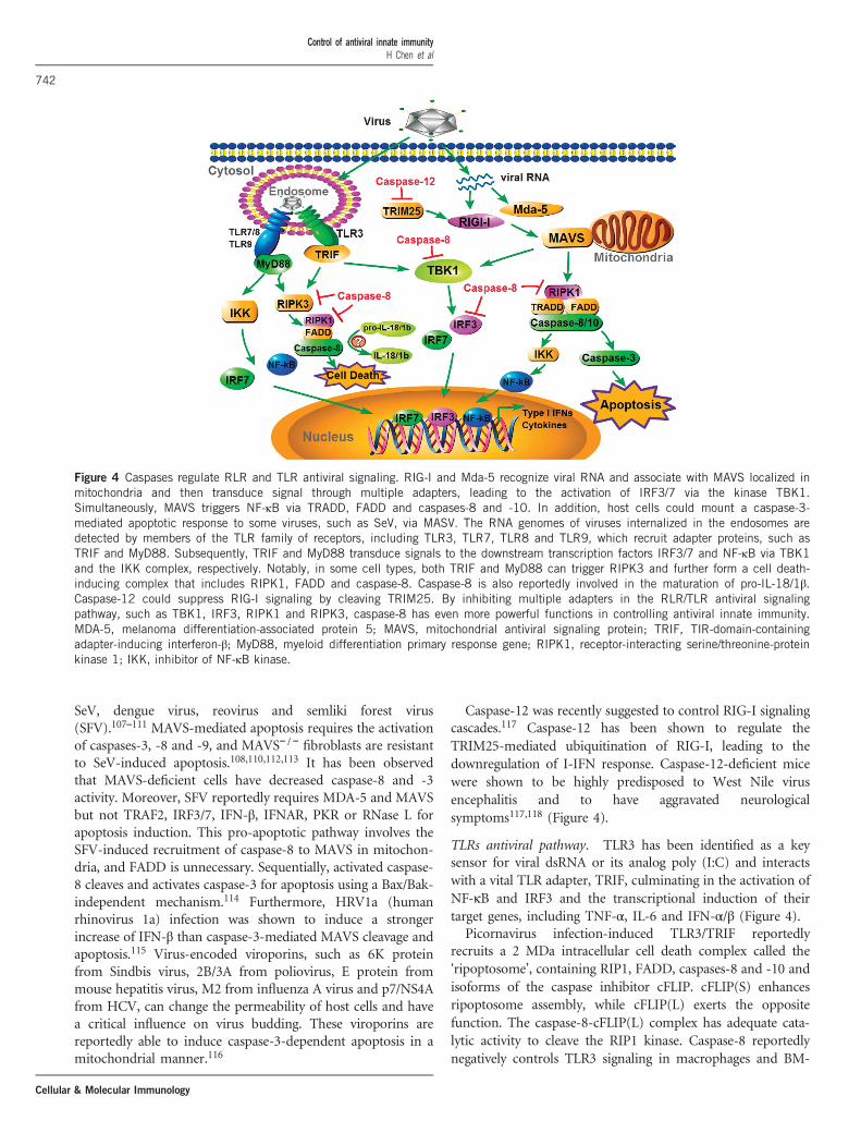
Caspase-12 was recently suggested to control RIG-I signalingcascades.117 Caspase-12 has been shown to regulate theTRIM25-mediated ubiquitination of RIG-I, leading to thedownregulation of I-IFN response. Caspase-12-deficient micewere shown to be highly predisposed to West Nile virusencephalitis and to have aggravated neurologicalsymptoms117,118 (Figure 4).
TLRs antiviral pathway. TLR3 has been identified as a keysensor for viral dsRNA or its analog poly (I:C) and interactswith a vital TLR adapter, TRIF, culminating in the activation ofNF-κB and IRF3 and the transcriptional induction of theirtarget genes, including TNF-α, IL-6 and IFN-α/β (Figure 4).
Picornavirus infection-induced TLR3/TRIF reportedlyrecruits a 2 MDa intracellular cell death complex called the‘ripoptosome’, containing RIP1, FADD, caspases-8 and -10 andisoforms of the caspase inhibitor cFLIP. cFLIP(S) enhancesripoptosome assembly, while cFLIP(L) exerts the oppositefunction. The caspase-8-cFLIP(L) complex has adequate cata-lytic activity to cleave the RIP1 kinase. Caspase-8 reportedlynegatively controls TLR3 signaling in macrophages and BM-
Figure 4 Caspases regulate RLR and TLR antiviral signaling. RIG-I and Mda-5 recognize viral RNA and associate with MAVS localized inmitochondria and then transduce signal through multiple adapters, leading to the activation of IRF3/7 via the kinase TBK1.Simultaneously, MAVS triggers NF-κB via TRADD, FADD and caspases-8 and -10. In addition, host cells could mount a caspase-3-mediated apoptotic response to some viruses, such as SeV, via MASV. The RNA genomes of viruses internalized in the endosomes aredetected by members of the TLR family of receptors, including TLR3, TLR7, TLR8 and TLR9, which recruit adapter proteins, such asTRIF and MyD88. Subsequently, TRIF and MyD88 transduce signals to the downstream transcription factors IRF3/7 and NF-κB via TBK1and the IKK complex, respectively. Notably, in some cell types, both TRIF and MyD88 can trigger RIPK3 and further form a cell death-inducing complex that includes RIPK1, FADD and caspase-8. Caspase-8 is also reportedly involved in the maturation of pro-IL-18/1β.Caspase-12 could suppress RIG-I signaling by cleaving TRIM25. By inhibiting multiple adapters in the RLR/TLR antiviral signalingpathway, such as TBK1, IRF3, RIPK1 and RIPK3, caspase-8 has even more powerful functions in controlling antiviral innate immunity.MDA-5, melanoma differentiation-associated protein 5; MAVS, mitochondrial antiviral signaling protein; TRIF, TIR-domain-containingadapter-inducing interferon-β; MyD88, myeloid differentiation primary response gene; RIPK1, receptor-interacting serine/threonine-proteinkinase 1; IKK, inhibitor of NF-κB kinase.
Control of antiviral innate immunity
H Chen et al
742
Cellular & Molecular Immunology

derived dendritic cells via caspase-8-interacting proteins, suchas FADD, cfLIP, RIP and TRAIL-R119 (Figure 4).
NF-κB plays a vital role in the transcriptional activation ofmany inflammation-related and anti-apoptotic genes inresponse to inflammatory cytokines, PAMPs, growth factorsand cellular stress states. NF-κB is located in the cytosol in aninactive form, which is inhibited by IκB. After multiplesignaling cascades, IKK phosphorylates IκB, and the subsequentdegradation of IκB releases NF-κB after it translocates to thenucleus and, finally, activates its targets. In B cells, caspase-1was shown to activate NF-κB to induce inflammation in adifferent manner.120 This novel signaling is mediated by theCARD of procaspase-1 and results in the stimulation of NF-κBand p38 MAPK in a RIP2-dependent manner. In addition, thispathway is not shared by caspase-11 and -12.120,121
Recent data have indicated that caspase-8 is critical for theactivation of NF-κB signaling, which results in the induction ofproinflammatory cytokines. However, many studies of thefunction of caspase-8 in modulating NF-κB signaling havethus far been restricted in anti-bacterial/fungal innate immu-nity, adaptive immunity and cancer.80,122–125 Moreover, todate, there is no consensus on the molecular mechanismunderpinning how caspase-8 mediates NF-κB signaling orwhether caspase-8 can regulate the NF-κB activation involvedin antiviral innate immunity, and these phenomena need to bestudied in more detail. Caspase-8 has also been demonstratedto exert critical functions in B-cell activation and expansion inresponse to TLR3 but not TLR9 by associating with the IKKcomplex and, in turn, modulating NF-κB translocation.124
These findings are consistent with the increased susceptibilityof human patients deficient for caspase-8 to viral infections.122
In addition, the existence of the involvement of TRIF in thematuration of IL-1β upon TLR3 activation has been demon-strated. Surprisingly, poly (I:C)-stimulated-IL-1β productionwas still observed in caspase-1-deficient cells but was sup-pressed by caspase-8 inhibitors and caspase-8 RNAi silencing.Accordingly, researchers proposed that a comparable TRIF-RIP1-FADD-caspase-8 cascade leads to IL-1β production.126
Moreover, melanoma cells and HaCaT human keratinocytesstimulated by poly (I:C) were shown to be susceptible toapoptosis associated with the TLR3/TRIF/caspase-8 complex127
(Figure 4).By contrast, caspase-8 has also been shown to prevent
inflammasome activity, IFN response and necroptosis, aprogrammed cell death resulting from caspase-8, RIPK1 andRIPK3.128,129 Compared to WT or Ripk3−/− BMDMs, Ripk3−/−
Casp8−/− BMDMs have been shown to display decreased levelsof IL-1β and poly (I:C) /TLR3-induced inflammasomepriming.130
TLR7 and TLR9 recognize ssRNA and unmethylated CpGDNA, respectively, in endosomes and are tightly connected tothe pathogenesis of SLE (systemic lupus erythematosus). Exceptfor TLR3, most TLRs signal through MyD88 to activate NF-κBsignaling. Caspase-8 also reportedly serves as a molecularrheostat to restrict MyD88-dependent dendritic cellactivation.131 When stimulated by CpG (TLR9 agonist) or
imiquimod (TLR7 agonist), CreCD11cCasp8fl/fl BMDCs exhibitincreased expression of IL-12/23p40, TNFα, IL-6 and IL-1βwithout inducing cell death. The loss of caspase-8 in DCscauses hyper-responsiveness to TLR activation, indicating thatcaspase-8 suppresses MyD88 signaling. However, it was pro-posed that this suppressive activity of caspase-8 is related toMyD88 recruitment but not its catalytic activity.131 These dataprovide a relationship between caspase-8 and augmented TLRresponses to pathogenetic stimuli.
Caspase-8-deficient keratinocytes exhibit enhanced geneactivation by DNA transfection. Meanwhile, TBK1 and IRF3are constitutively phosphorylated in the epidermis of theCasp8F/−K5-Cre mice, and the well-known ISGs are alsoinduced. Moreover, in response to DNA transfection,caspase-8-deficient keratinocytes yield a higher level of IRF3-targeted genes than the caspase-8-overexpressed cells. Inaddition, since MyD88, TRIF and most TLR adapters wereruled out, caspase-8 was further proven to regulate IRF3 orTBK1 directly, but the exact molecular mechanism needs to befurther clarified132 (Figure 4).
Caspases regulate viral ER stress-induced apoptosisCaspase-12 is an inflammatory caspase located in the endo-plasmic reticulum (ER). Early studies indicated that caspase-12is an ER stress response caspase.133 Unbalanced ER-Ca2+
homeostasis and accumulated misfolded, unassembled oraggregated proteins in the ER lumen reportedly stimulate ERstress signaling. Furthermore, viruses utilize the ER as anintegrated unit for their life cycles, leading to some levels of ERstress. Mild ER stress affects protein synthesis initiation and candownregulate cell proliferation, whereas acute or persistent ERstress can cause cell death induced by caspase-12.134 Studiesincaspase-12-deficient mice emphasized the principle functionof caspase-12 in ER stress-mediated apoptosis.133 In addition,viral infection might induce caspase-12-dependentapoptosis.18,135 The RNA virus BVDV (bovine viral diarrheavirus) and related flavivirus-infected MDBK (Madin-Darbybovine kidney) cells have increased expression of caspase-12and decreased levels of the anti-apoptotic protein Bcl-2.Caspase-12 cleaves pro-caspase-7 to its active caspase-7 form.The detailed mechanisms of how ER stress signaling activatescaspase-12 have not yet been elucidated. However, currentstudies have proven that other cytosolic caspases can berecruited by activated caspase-12 in the ER membrane, andthus, the apoptotic signal is greatly boosted.136 In A549epithelial cells infected with RSV, casapse-9 is not activated,caspases-3 and -8 are modestly activated, and caspase-12 isstrongly activated. Moreover, RSV-stimulated apoptosis seemsto occur through a caspase-12-dependent ER stress response,which is uncoupled from NF-κB activation.135
CONCLUSIONS
Viral PAMPs are recognized differentially by innate PRRs toinitiate antiviral immune responses in a systematic manner.The complicated antiviral innate immune signaling networkhas co-evolved with viruses to defend against and restrain
Control of antiviral innate immunity
H Chen et al
743
Cellular & Molecular Immunology

autoimmunity via a sophisticated state-of-the-art feedbackscheme. The caspase family members are architects of thebody and sit at the nexus of this dynamic regulatory frame-work. In the past few decades, significant advances in under-standing the critical functions of caspases for organismhomeostasis and especially novel and exciting discoveries haveunveiled the multifaceted nature of individual caspases in theantiviral innate immune system. Studies on caspases haveprovided an interesting point of view that caspases act asmolecular ‘decision-makers’ because of their ability to fine-tuneantiviral innate immunity and because caspases have bothproinflammatory and anti-inflammatory functions. Moreover,recent studies have uncovered the capacity of caspases tomediate nearly all the PRRs involved in innate immunity byacting as a molecular switch on whether to respond to an insultby initiating inflammation or potentiating I-IFN response toeliminate invading viruses, or by preventing inflammation tomaintain immunological silence by dismantling insulted ordamaged cells. Increasingly distinguished and sophisticateddiscoveries have challenged the traditional definition of caspasefamily members. However, contradiction and controversyregarding the regulation of innate immunity by caspases exist,and the understanding of the molecular basis of thesephenomena remains in infancy. Caspases are double-edgedswords. The inappropriate activation of caspases and dysregu-lation of the antiviral innate immune signaling controlled bycaspases has dire consequences for human health and isassociated with an extensive list of human diseases. Ongoingstudies will provide us with a more comprehensive under-standing of caspase mechanisms and may shed new light ontherapeutic options for the various caspase-related humandiseases.
CONFLICT OF INTERESTThe authors declare no conflict of interest.
ACKNOWLEDGEMENTS
We thank members of the Jiang Laboratory for helpful discussions.We apologize for being unable to include the important contributionsfrom all researchers in the field due to space limitations. Recent andongoing studies performed in the Jiang Laboratory are supported bygrants from the Chinese Ministry of Science and Technology(2014CB542600 and 2015CC040097) and the China Natural ScienceFoundation (31230023, 91129000 and 81621001).
1 Takeuchi O, Akira S. Pattern recognition receptors and inflammation.Cell 2010; 140: 805–820.
2 Allen IC, Scull MA, Moore CB, Holl EK, McElvania-TeKippe E,Taxman DJ et al. The NLRP3 inflammasome mediates in vivo innateimmunity to influenza A virus through recognition of viral RNA.Immunity 2009; 30: 556–565.
3 Yoneyama M, Kikuchi M, Matsumoto K, Imaizumi T, Miyagishi M,Taira K et al. Shared and unique functions of the DExD/H-boxhelicases RIG-I, MDA5, and LGP2 in antiviral innate immunity.J Immunol 2005; 175: 2851–2858.
4 Yoneyama M, Kikuchi M, Natsukawa T, Shinobu N, Imaizumi T,Miyagishi M et al. The RNA helicase RIG-I has an essential function
in double-stranded RNA-induced innate antiviral responses.Nat Immunol 2004; 5: 730–737.
5 Yoh SM, Schneider M, Seifried J, Soonthornvacharin S, Akleh RE,Olivieri KC et al. PQBP1 is a proximal sensor of the cGAS-dependentinnate response to HIV-1. Cell 2015; 161: 1293–1305.
6 Ferguson BJ, Mansur DS, Peters NE, Ren H, Smith GL. DNA-PK is aDNA sensor for IRF-3-dependent innate immunity. eLife 2012; 1:e00047.
7 Xia P, Wang S, Ye B, Du Y, Huang G, Zhu P et al. Sox2 functions as asequence-specific DNA sensor in neutrophils to initiate innateimmunity against microbial infection. Nat Immunol 2015; 16:366–375.
8 Kondo T, Kobayashi J, Saitoh T, Maruyama K, Ishii KJ, Barber GNet al. DNA damage sensor MRE11 recognizes cytosolic double-stranded DNA and induces type I interferon by regulating STINGtrafficking. Proc Natl Acad USA 2013; 110: 2969–2974.
9 Ablasser A, Goldeck M, Cavlar T, Deimling T, Witte G, Rohl I et al.cGAS produces a 2'-5'-linked cyclic dinucleotide second messengerthat activates STING. Nature 2013; 498: 380–384.
10 Burckstummer T, Baumann C, Bluml S, Dixit E, Durnberger G,Jahn H et al. An orthogonal proteomic-genomic screen identifiesAIM2 as a cytoplasmic DNA sensor for the inflammasome. NatImmunol 2009; 10: 266–272.
11 Fernandes-Alnemri T, Yu JW, Datta P, Wu J, Alnemri ES. AIM2activates the inflammasome and cell death in response tocytoplasmic DNA. Nature 2009; 458: 509–513.
12 Hornung V, Ablasser A, Charrel-Dennis M, Bauernfeind F, Horvath G,Caffrey DR et al. AIM2 recognizes cytosolic dsDNA and forms acaspase-1-activating inflammasome with ASC. Nature 2009; 458:514–518.
13 Kawai T, Akira S. The role of pattern-recognition receptors in innateimmunity: update on Toll-like receptors. Nat Immunol 2010; 11:373–384.
14 Nie Y, Wang YY. Innate immune responses to DNA viruses. ProteinCell 2013; 4: 1–7.
15 Pichlmair A, Reis e Sousa C. Innate recognition of viruses. Immunity2007; 27: 370–383.
16 Ramos HJ, Gale M Jr.. RIG-I like receptors and their signalingcrosstalk in the regulation of antiviral immunity. Curr Opin Virol2011; 1: 167–176.
17 Roberts TL, Idris A, Dunn JA, Kelly GM, Burnton CM, Hodgson Set al. HIN-200 proteins regulate caspase activation in response toforeign cytoplasmic DNA. Science 2009; 323: 1057–1060.
18 Jordan R, Wang L, Graczyk TM, Block TM, Romano PR. Replicationof a cytopathic strain of bovine viral diarrhea virus activates PERK andinduces endoplasmic reticulum stress-mediated apoptosis ofMDBK cells. J Virol 2002; 76: 9588–9599.
19 McIlwain DR, Berger T, Mak TW. Caspase functions in cell death anddisease. Cold Spring Harb Perspect Biol 2013; 5: a008656.
20 Shalini S, Dorstyn L, Dawar S, Kumar S. Old, new and emergingfunctions of caspases. Cell Death Differ 2015; 22: 526–539.
21 Kumar S. Caspase function in programmed cell death. Cell DeathDiffer 2007; 14: 32–43.
22 Latz E, Xiao TS, Stutz A. Activation and regulation of the inflamma-somes. Nat Rev Immunol 2013; 13: 397–411.
23 Lamkanfi M, Dixit VM. Mechanisms and functions of inflammasomes.Cell 2014; 157: 1013–1022.
24 Man SM, Kanneganti TD. Regulation of inflammasome activation.Immunol Rev 2015; 265: 6–21.
25 Degterev A, Boyce M, Yuan J. A decade of caspases. Oncogene 2003;22: 8543–8567.
26 Fuentes-Prior P, Salvesen GS. The protein structures that shapecaspase activity, specificity, activation and inhibition. Biochem J2004; 384 (Pt 2): 201–232.
27 Li P, Nijhawan D, Budihardjo I, Srinivasula SM, Ahmad M, AlnemriES et al. Cytochrome c and dATP-dependent formation of Apaf-1/caspase-9 complex initiates an apoptotic protease cascade. Cell1997; 91: 479–489.
28 Acehan D, Jiang X, Morgan DG, Heuser JE, Wang X, Akey CW.Three-dimensional structure of the apoptosome: implications forassembly, procaspase-9 binding, and activation. Mol Cell 2002; 9:423–432.
29 Ashkenazi A, Dixit VM. Death receptors: signaling and modulation.Science 1998; 281: 1305–1308.
Control of antiviral innate immunity
H Chen et al
744
Cellular & Molecular Immunology

30 Li H, Zhu H, Xu CJ, Yuan J. Cleavage of BID by caspase 8 mediatesthe mitochondrial damage in the Fas pathway of apoptosis. Cell1998; 94: 491–501.
31 Feoktistova M, Geserick P, Kellert B, Dimitrova DP, Langlais C,Hupe M et al. cIAPs block ripoptosome formation, a RIP1/caspase-8containing intracellular cell death complex differentially regulated bycFLIP isoforms. Mol Cell 2011; 43: 449–463.
32 Blander JM. A long-awaited merger of the pathways mediating hostdefence and programmed cell death. Nat Rev Immunol 2014; 14:601–618.
33 Brenner D, Blaser H, Mak TW. Regulation of tumour necrosis factorsignalling: live or let die. Nat Rev Immunol 2015; 15: 362–374.
34 Kanneganti TD. Central roles of NLRs and inflammasomes in viralinfection. Nat Rev Immunol 2010; 10: 688–698.
35 Kanneganti TD, Body-Malapel M, Amer A, Park JH, Whitfield J,Franchi L et al. Critical role for cryopyrin/Nalp3 in activation ofcaspase-1 in response to viral infection and double-stranded RNA.J Biol Chem 2006; 281: 36560–36568.
36 Kanneganti TD, Ozoren N, Body-Malapel M, Amer A, Park JH,Franchi L et al. Bacterial RNA and small antiviral compounds activatecaspase-1 through cryopyrin/Nalp3. Nature 2006; 440: 233–236.
37 Ichinohe T, Lee HK, Ogura Y, Flavell R, Iwasaki A. Inflammasomerecognition of influenza virus is essential for adaptive immuneresponses. J Exp Med 2009; 206: 79–87.
38 Thomas PG, Dash P, Aldridge JR Jr., Ellebedy AH, Reynolds C, FunkAJ et al. The intracellular sensor NLRP3 mediates key innate andhealing responses to influenza A virus via the regulation of caspase-1.Immunity 2009; 30: 566–575.
39 Poeck H, Bscheider M, Gross O, Finger K, Roth S, Rebsamen M et al.Recognition of RNA virus by RIG-I results in activation of CARD9 andinflammasome signaling for interleukin 1 beta production. NatImmunol 2010; 11: 63–69.
40 Hagar JA, Powell DA, Aachoui Y, Ernst RK, Miao EA. CytoplasmicLPS activates caspase-11: implications in TLR4-independentendotoxic shock. Science 2013; 341: 1250–1253.
41 Kayagaki N, Wong MT, Stowe IB, Ramani SR, Gonzalez LC, Akashi-Takamura S et al. Noncanonical inflammasome activation byintracellular LPS independent of TLR4. Science 2013; 341:1246–1249.
42 Vigano E, Diamond CE, Spreafico R, Balachander A, Sobota RM,Mortellaro A. Human caspase-4 and caspase-5 regulate the one-stepnon-canonical inflammasome activation in monocytes. Nat Commun2015; 6: 8761.
43 Broz P, Dixit VM. Inflammasomes: mechanism of assembly,regulation and signalling. Nat Rev Immunol 2016; 16: 407–420.
44 Luthi AU, Cullen SP, McNeela EA, Duriez PJ, Afonina IS, Sheridan Cet al. Suppression of interleukin-33 bioactivity through proteolysis byapoptotic caspases. Immunity 2009; 31: 84–98.
45 Fink SL, Bergsbaken T, Cookson BT. Anthrax lethal toxin andSalmonella elicit the common cell death pathway of caspase-1-dependent pyroptosis via distinct mechanisms. Proc Natl Acad SciUSA 2008; 105: 4312–4317.
46 Schmitz J, Owyang A, Oldham E, Song Y, Murphy E, McClanahan TKet al. IL-33, an interleukin-1-like cytokine that signals via the IL-1receptor-related protein ST2 and induces T helper type 2-associatedcytokines. Immunity 2005; 23: 479–490.
47 Rintahaka J, Wiik D, Kovanen PE, Alenius H, Matikainen S.Cytosolic antiviral RNA recognition pathway activates caspases1 and 3. J Immunol 2008; 180: 1749–1757.
48 Melchjorsen J, Jensen SB, Malmgaard L, Rasmussen SB, Weber F,Bowie AG et al. Activation of innate defense against a paramyxovirusis mediated by RIG-I and TLR7 and TLR8 in a cell-type-specific manner. J Virol 2005; 79: 12944–12951.
49 Rothenfusser S, Goutagny N, DiPerna G, Gong M, Monks BG,Schoenemeyer A et al. The RNA helicase Lgp2 inhibits TLR-independent sensing of viral replication by retinoic acid-inducible gene-I. J Immunol 2005; 175: 5260–5268.
50 Fredericksen BL, Gale M Jr.. West Nile virus evades activation ofinterferon regulatory factor 3 through RIG-I-dependent and -indepen-dent pathways without antagonizing host defense signaling. J Virol2006; 80: 2913–2923.
51 Hornung V, Ellegast J, Kim S, Brzozka K, Jung A, Kato H et al.5'-Triphosphate RNA is the ligand for RIG-I. Science 2006; 314:994–997.
52 Kato H, Takeuchi O, Sato S, Yoneyama M, Yamamoto M, Matsui Ket al. Differential roles of MDA5 and RIG-I helicases in the recogni-tion of RNA viruses. Nature 2006; 441: 101–105.
53 Pichlmair A, Schulz O, Tan CP, Naslund TI, Liljestrom P, Weber Fet al. RIG-I-mediated antiviral responses to single-stranded RNAbearing 5'-phosphates. Science 2006; 314: 997–1001.
54 Liu P, Jamaluddin M, Li K, Garofalo RP, Casola A, Brasier AR.Retinoic acid-inducible gene I mediates early antiviral response andToll-like receptor 3 expression in respiratory syncytial virus-infectedairway epithelial cells. J Virol 2007; 81: 1401–1411.
55 Mikkelsen SS, Jensen SB, Chiliveru S, Melchjorsen J, Julkunen I,Gaestel M et al. RIG-I-mediated activation of p38 MAPK is essentialfor viral induction of interferon and activation of dendritic cells:dependence on TRAF2 and TAK1. J Biol Chem 2009; 284:10774–10782.
56 Rathinam VA, Jiang Z, Waggoner SN, Sharma S, Cole LE, Waggoner Let al. The AIM2 inflammasome is essential for host defense againstcytosolic bacteria and DNA viruses. Nat Immunol 2010; 11:395–402.
57 Ekchariyawat P, Hamel R, Bernard E, Wichit S, Surasombatpattana P,Talignani L et al. Inflammasome signaling pathways exert antiviraleffect against Chikungunya virus in human dermal fibroblasts. InfectGenet Evol 2015; 32: 401–408.
58 Hamel R, Dejarnac O, Wichit S, Ekchariyawat P, Neyret A,Luplertlop N et al. Biology of Zika virus infection in humanskin cells. J Virol 2015; 89: 8880–8896.
59 Kerur N, Veettil MV, Sharma-Walia N, Bottero V, Sadagopan S,Otageri P et al. IFI16 acts as a nuclear pathogen sensor to induce theinflammasome in response to Kaposi Sarcoma-associated herpesvirusinfection. Cell Host Microbe 2011; 9: 363–375.
60 Jakobsen MR, Bak RO, Andersen A, Berg RK, Jensen SB, TengchuanJ et al. IFI16 senses DNA forms of the lentiviral replication cycle andcontrols HIV-1 replication. Proc Natl Acad Sci USA 2013; 110:E4571–E4580.
61 Doitsh G, Galloway NLK, Geng X, Yang ZY, Monroe KM, Zepeda Oet al. Cell death by pyroptosis drives CD4 T-cell depletion in HIV-1infection. Nature 2014; 505: 509–514.
62 Monroe KM, Yang Z, Johnson JR, Geng X, Doitsh G, Krogan NJ et al.IFI16 DNA sensor is required for death of lymphoidCD4 T cells abortively infected with HIV. Science 2014; 343:428–432.
63 Guillot L, Le Goffic R, Bloch S, Escriou N, Akira S, Chignard M et al.Involvement of toll-like receptor 3 in the immune response of lungepithelial cells to double-stranded RNA and influenza A virus. J BiolChem 2005; 280: 5571–5580.
64 Koyama S, Ishii KJ, Kumar H, Tanimoto T, Coban C, Uematsu S et al.Differential role of TLR- and RLR-signaling in the immune responsesto influenza A virus infection and vaccination. J Immunol 2007; 179:4711–4720.
65 Ichinohe T, Pang IK, Iwasaki A. Influenza virus activates inflamma-somes via its intracellular M2 ion channel. Nat Immunol 2010; 11:404–410.
66 Delaloye J, Roger T, Steiner-Tardivel QG, Le Roy D, Knaup ReymondM, Akira S et al. Innate immune sensing of modified vaccinia virusAnkara (MVA) is mediated by TLR2-TLR6, MDA-5 and the NALP3inflammasome. PLoS Pathog 2009; 5: e1000480.
67 Jones JW, Kayagaki N, Broz P, Henry T, Newton K, O'Rourke K et al.Absent in melanoma 2 is required for innate immune recognition ofFrancisella tularensis. Proc Natl Acad Sci USA 2010; 107:9771–9776.
68 Cookson BT, Brennan MA. Pro-inflammatory programmed cell death.Trends Microbiol 2001; 9: 113–114.
69 Jorgensen I, Miao EA. Pyroptotic cell death defends against intracel-lular pathogens. Immunol Rev 2015; 265: 130–142.
70 Zychlinsky A, Prevost MC, Sansonetti PJ. Shigella flexneriinduces apoptosis in infected macrophages. Nature 1992; 358:167–169.
71 Miao EA, Leaf IA, Treuting PM, Mao DP, Dors M, Sarkar A et al.Caspase-1-induced pyroptosis is an innate immune effector mechan-ism against intracellular bacteria. Nat Immunol 2010; 11:1136–1142.
72 Kayagaki N, Stowe IB, Lee BL, O'Rourke K, Anderson K, Warming Set al. Caspase-11 cleaves gasdermin D for non-canonical inflamma-some signalling. Nature 2015; 526: 666–671.
Control of antiviral innate immunity
H Chen et al
745
Cellular & Molecular Immunology

73 Shi J, Zhao Y, Wang K, Shi X, Wang Y, Huang H et al. Cleavage ofGSDMD by inflammatory caspases determines pyroptotic cell death.Nature 2015; 526: 660–665.
74 Agard NJ, Maltby D, Wells JA. Inflammatory stimuli regulate caspasesubstrate profiles. Mol Cell Proteomics 2010; 9: 880–893.
75 Malireddi RKS, Ippagunta S, Lamkanfi M, Kanneganti TD. Cuttingedge: proteolytic inactivation of poly(ADP-ribose) polymerase 1 by theNlrp3 and Nlrc4 inflammasomes. J Immunol 2010; 185:3127–3130.
76 Knodler LA, Crowley SM, Sham HP, Yang HJ, Wrande M, Ma CX et al.Noncanonical inflammasome activation of caspase-4/caspase-11mediates epithelial defenses against enteric bacterial pathogens. CellHost Microbe 2014; 16: 249–256.
77 Aachoui Y, Leaf IA, Hagar JA, Fontana MF, Campos CG, Zak DE et al.Caspase-11 protects against bacteria that escape the vacuole.Science 2013; 339: 975–978.
78 Miao EA, Leaf IA, Treuting PM, Mao DP, Dors M, Sarkar A et al.Caspase-1-induced pyroptosis is an innate immune effector mechan-ism against intracellular bacteria. Nat Immunol 2010; 11:1136–U94.
79 Sellin ME, Muller AA, Felmy B, Dolowschiak T, Diard M, Tardivel Aet al. Epithelium-intrinsic NAIP/NLRC4 inflammasome drivesinfected enterocyte expulsion to restrict salmonella replication inthe intestinal mucosa. Cell Host Microbe 2014; 16: 237–248.
80 Man SM, Kanneganti TD. Converging roles of caspases in inflamma-some activation, cell death and innate immunity. Nat Rev Immunol2016; 16: 7–21.
81 Shi JJ, Zhao Y, Wang YP, Gao WQ, Ding JJ, Li P et al. Inflammatorycaspases are innate immune receptors for intracellular LPS. Nature2014; 514: 187–192.
82 Kayagaki N, Warming S, Lamkanfi M, Vande Walle L, Louie S, Dong Jet al. Non-canonical inflammasome activation targets caspase-11.Nature 2011; 479: 117–U46.
83 Rongvaux A, Jackson R, Harman CCD, Li T, West AP, de Zoete MRet al. Apoptotic caspases prevent the induction of type I interferons bymitochondrial DNA. Cell 2014; 159: 1563–1577.
84 White MJ, McArthur K, Metcalf D, Lane RM, Cambier JC, Herold MJet al. Apoptotic caspases suppress mtDNA-induced STING-mediatedtype I IFN production. Cell 2014; 159: 1549–1562.
85 Cai X, Chiu YH, Chen ZJ. The cGAS-cGAMP-STING pathwayof cytosolic DNA sensing and signaling. Mol Cell 2014; 54:289–296.
86 Schoggins JW, MacDuff DA, Imanaka N, Gainey MD, Shrestha B,Eitson JL et al. Pan-viral specificity of IFN-induced genes reveals newroles for cGAS in innate immunity. Nature 2014; 505: 691–695.
87 Li XD, Wu J, Gao D, Wang H, Sun L, Chen ZJ. Pivotal roles ofcGAS-cGAMP signaling in antiviral defense and immune adjuvanteffects. Science 2013; 341: 1390–1394.
88 Ishikawa H, Ma Z, Barber GN. STING regulates intracellularDNA-mediated, type I interferon-dependent innate immunity. Nature2009; 461: 788–792.
89 Lam E, Stein S, Falck-Pedersen E. Adenovirus detection by the cGAS/STING/TBK1 DNA sensing cascade. J Virol 2014; 88: 974–981.
90 Sunthamala N, Thierry F, Teissier S, Pientong C, Kongyingyoes B,Tangsiriwatthana T et al. E2 proteins of high risk human papilloma-viruses down-modulate STING and IFN-kappa transcription inkeratinocytes. PloS One 2014; 9: e91473.
91 Paijo J, Doring M, Spanier J, Grabski E, Nooruzzaman M,Schmidt T et al. cGAS senses human cytomegalovirus and inducestype I interferon responses in human monocyte-derived cells. PLoSPathog 2016; 12: e1005546.
92 Hu MM, Shu HB. Multifaceted roles of TRIM38 in innateimmune and inflammatory responses. Cell Mol Immunol 2017; 14:331–338.
93 Wu J, Chen ZJ. Innate immune sensing and signaling of cytosolicnucleic acids. Annu Rev Immunol 2014; 32: 461–488.
94 Chen H, Sun H, You F, Sun W, Zhou X, Chen L et al. Activation ofSTAT6 by STING is critical for antiviral innate immunity. Cell 2011;147: 436–446.
95 Christensen MH, Paludan SR. Viral evasion of DNA-stimulated innateimmune responses. Cell Mol Immunol 2017; 14: 4–13.
96 West AP, Khoury-Hanold W, Staron M, Tal MC, Pineda CM, Lang SMet al. Mitochondrial DNA stress primes the antiviral innate immuneresponse. Nature 2015; 520: 553–557.
97 Crawford ED, Seaman JE, Agard N, Hsu GW, Julien O, Mahrus S et al.The DegraBase: a database of proteolysis in healthy and apoptotichuman cells. Mol Cell Proteomics 2013; 12: 813–824.
98 Wang Y, Ning X, Gao P, Wu S, Sha M, Lv M et al. Inflammasomeactivation triggers caspase-1-mediated cleavage of cGAS toregulate responses to DNA virus infection. Immunity 2017; 46:393–404.
99 Tao J, Zhang XW, Jin J, Du XX, Lian T, Yang J et al. Non-specific DNAbinding of cGAS N-terminus promotes cGAS activation. J Immunol2017; 198: 3627–3636.
100 Takahashi K, Kawai T, Kumar H, Sato S, Yonehara S, Akira S.Roles of caspase-8 and caspase-10 in innate immune responses todouble-stranded RNA. J Immunol 2006; 176: 4520–4524.
101 Rajput A, Kovalenko A, Bogdanov K, Yang SH, Kang TB, Kim JCet al. RIG-I RNA helicase activation of IRF3 transcription factor isnegatively regulated by caspase-8-mediated cleavage of the RIP1protein. Immunity 2011; 34: 340–351.
102 Lin Y, Devin A, Rodriguez Y, Liu ZG. Cleavage of the death domainkinase RIP by caspase-8 prompts TNF-induced apoptosis. Genes Dev1999; 13: 2514–2526.
103 Feng S, Yang Y, Mei Y, Ma L, Zhu DE, Hoti N et al. Cleavage of RIP3inactivates its caspase-independent apoptosis pathway by removal ofkinase domain. Cell Signal 2007; 19: 2056–2067.
104 O'Donnell MA, Perez-Jimenez E, Oberst A, Ng A, Massoumi R,Xavier R et al. Caspase 8 inhibits programmed necrosis byprocessing CYLD. Nat Cell Biol 2011; 13: 1437–1442.
105 Sears N, Sen GC, Stark GR, Chattopadhyay S. Caspase-8-mediatedcleavage inhibits IRF-3 protein by facilitating its proteasome-mediated degradation. J Biol Chem 2011; 286: 33037–33044.
106 Staal J, Bekaert T, Beyaert R. Regulation of NF-kappaB signaling bycaspases and MALT1 paracaspase. Cell Res 2011; 21: 40–54.
107 Holm GH, Zurney J, Tumilasci V, Leveille S, Danthi P, Hiscott J et al.Retinoic acid-inducible gene-I and interferon-beta promoterstimulator-1 augment proapoptotic responses following mammalianreovirus infection via interferon regulatory factor-3. J Biol Chem2007; 282: 21953–21961.
108 Lei Y, Moore CB, Liesman RM, O'Connor BP, Bergstralh DT, Chen ZJet al. MAVS-mediated apoptosis and its inhibition by viral proteins.PLoS One 2009; 4: e5466.
109 Chattopadhyay S, Marques JT, Yamashita M, Peters KL, Smith K,Desai A et al. Viral apoptosis is induced by IRF-3-mediatedactivation of Bax. EMBO J 2010; 29: 1762–1773.
110 Yu CY, Chiang RL, Chang TH, Liao CL, Lin YL. The interferonstimulator mitochondrial antiviral signaling protein facilitates celldeath by disrupting the mitochondrial membrane potential and byactivating caspases. J Virol 2010; 84: 2421–2431.
111 Besch R, Poeck H, Hohenauer T, Senft D, Hacker G, Berking C et al.Proapoptotic signaling induced by RIG-I and MDA-5 results in type Iinterferon-independent apoptosis in human melanoma cells. J ClinInvest 2009; 119: 2399–2411.
112 Li HM, Fujikura D, Harada T, Uehara J, Kawai T, Akira S et al.IPS-1 is crucial for DAP3-mediated anoikis induction by caspase-8activation. Cell Death Differ 2009; 16: 1615–1621.
113 Buskiewicz IA, Koenig A, Huber SA, Budd RC. Caspase-8 and FLIPregulate RIG-I/MDA5-induced innate immune host responses topicornaviruses. Future Virol 2012; 7: 1221–1236.
114 El Maadidi S, Faletti L, Berg B, Wenzl C, Wieland K, Chen ZJ et al. Anovel mitochondrial MAVS/Caspase-8 platform links RNA virus-induced innate antiviral signaling to Bax/Bak-independent apoptosis.J Immunol 2014; 192: 1171–1183.
115 Drahos J, Racaniello VR. Cleavage of IPS-1 in cells infected withhuman rhinovirus. J Virol 2009; 83: 11581–11587.
116 Madan V, Castello A, Carrasco L. Viroporins from RNA virusesinduce caspase-dependent apoptosis. Cell Microbiol 2008; 10:437–451.
117 Wang P, Arjona A, Zhang Y, Sultana H, Dai J, Yang L et al. Caspase-12 controls West Nile virus infection via the viral RNA receptor RIG-I.Nat Immunol 2010; 11: 912–919.
118 Morizot A, Saleh M. Non-apoptotic functions of cell death effectors ininflammation and innate immunity. Microbes Infect 2012; 14:1241–1253.
119 Diehl GE, Yue HH, Hsieh K, Kuang AA, Ho M, Morici LA et al.TRAIL-R as a negative regulator of innate immune cell responses.Immunity 2004; 21: 877–889.
Control of antiviral innate immunity
H Chen et al
746
Cellular & Molecular Immunology

120 Lamkanfi M, Kalai M, Saelens X, Declercq W, Vandenabeele P.Caspase-1 activates nuclear factor of the kappa-enhancer in B cellsindependently of its enzymatic activity. J Biol Chem 2004; 279:24785–24793.
121 Launay S, Hermine O, Fontenay M, Kroemer G, Solary E, Garrido C.Vital functions for lethal caspases. Oncogene 2005; 24:5137–5148.
122 Chun HJ, Zheng L, Ahmad M, Wang J, Speirs CK, Siegel RM et al.Pleiotropic defects in lymphocyte activation caused by caspase-8mutations lead to human immunodeficiency. Nature 2002; 419:395–399.
123 Su H, Bidere N, Zheng L, Cubre A, Sakai K, Dale J et al. Requirementfor caspase-8 in NF-kappaB activation by antigen receptor. Science2005; 307: 1465–1468.
124 Lemmers B, Salmena L, Bidere N, Su H, Matysiak-Zablocki E,Murakami K et al. Essential role for caspase-8 in Toll-like receptorsand NFkappaB signaling. J Biol Chem 2007; 282: 7416–7423.
125 Hu WH, Johnson H, Shu HB. Activation of NF-kappaB by FADD,casper, and caspase-8. J Biol Chem 2000; 275: 10838–10844.
126 Maelfait J, Vercammen E, Janssens S, Schotte P, Haegman M,Magez S et al. Stimulation of Toll-like receptor 3 and 4 inducesinterleukin-1beta maturation by caspase-8. J Exp Med 2008; 205:1967–1973.
127 Weber A, Kirejczyk Z, Besch R, Potthoff S, Leverkus M, Hacker G.Proapoptotic signalling through Toll-like receptor-3 involvesTRIF-dependent activation of caspase-8 and is under the control ofinhibitor of apoptosis proteins in melanoma cells. Cell Death Differ2010; 17: 942–951.
128 Holler N, Zaru R, Micheau O, Thome M, Attinger A, Valitutti S et al.Fas triggers an alternative, caspase-8-independent cell death path-way using the kinase RIP as effector molecule. Nat Immunol 2000;1: 489–495.
129 Kang TB, Yang SH, Toth B, Kovalenko A, Wallach D. Caspase-8blocks kinase RIPK3-mediated activation of the NLRP3 inflamma-some. Immunity 2013; 38: 27–40.
130 Allam R, Lawlor KE, Yu EC, Mildenhall AL, Moujalled DM, Lewis RSet al. Mitochondrial apoptosis is dispensable for NLRP3 inflamma-some activation but non-apoptotic caspase-8 is required for inflam-masome priming. EMBO Rep 2014; 15: 982–990.
131 Cuda CM, Misharin AV, Gierut AK, Saber R, Haines GK, Hutcheson Jet al. Caspase-8 acts as a molecular rheostat to limit RIPK1-andMyD88-mediated dendritic cell activation. J Immunol 2014; 192:5548–5560.
132 Kovalenko A, Kim JC, Kang TB, Rajput A, Bogdanov K,Dittrich-Breiholz O et al. Caspase-8 deficiency in epidermal kerati-nocytes triggers an inflammatory skin disease. J Exp Med 2009; 206:2161–2177.
133 Nakagawa T, Zhu H, Morishima N, Li E, Xu J, Yankner BA et al.Caspase-12 mediates endoplasmic-reticulum-specific apoptosis andcytotoxicity by amyloid-beta. Nature 2000; 403: 98–103.
134 Kaufman RJ. Stress signaling from the lumen of the endoplasmicreticulum: coordination of gene transcriptional and translationalcontrols. Genes Dev 1999; 13: 1211–1233.
135 Bitko V, Barik S. An endoplasmic reticulum-specific stress-activatedcaspase (caspase-12) is implicated in the apoptosis of A549epithelial cells by respiratory syncytial virus. J Cell Biochem 2001;80: 441–454.
136 Rao RV, Hermel E, Castro-Obregon S, del Rio G, Ellerby LM,Ellerby HM et al. Coupling endoplasmic reticulum stress to the celldeath program—mechanism of caspase activation. J Biol Chem2001; 276: 33869–33874.
137 Tenev T, Bianchi K, Darding M, Broemer M, Langlais C, Wallberg Fet al. The ripoptosome, a signaling platform that assembles in responseto genotoxic stress and loss of IAPs. Mol Cell 2011; 43: 432–448.
Control of antiviral innate immunity
H Chen et al
747
Cellular & Molecular Immunology
Copyright © 2022 FDOKUMEN