
Hepatitis C: molecular virology and antiviral targets
18
HEPATITIS C: MOLECULAR VIROLOGY AND ANTIVIRAL TARGETS Darius Moradpour Hepatitis C virus (HCV) is now the major cause of chronic hepatitis, liver cirrhosis and hepatocellular carcinoma (HCC) in the western world. In addition, end-stage liver disease due to chronic hepatitis C is the leading indication to liver transplantation. A protective vaccine is not available yet and therapeutic modalities are still limited. As a con- sequence, the number of patients presenting with long-term sequelae of chronic hepatitis C, including HCC, is expected to further increase for the next two decades even if the incidence of new cases has diminished since the introduction of anti-HCV screening of blood and blood pro- ducts in 1990 [1]. Given this scenario, there is an urgent need to develop more effective and better tolerated therapies for chronic hepatitis C. A detailed understanding of the viral life cycle underpins these efforts. HEPATITIS C Epidemiology It is estimated that 120–180 million people worldwide are infected with HCV [2], which corresponds to roughly 4 times the number of indivi- duals infected with HIV and about half the number of persons infected with the hepatitis B virus (HBV). The seroprevalence rate is 1–2% in western Europe and North America, 3– 4% in some Mediterranean and Asian countries and up to 20% in Egypt and parts of Central Africa. HCV is parenterally transmitted. With the introduction of anti-HCV screening, new cases of posttransfusion hepatitis C have virtually dis- appeared. Indeed, over the last 20 years the risk of posttransfusion hepatitis C could be reduced from about 1 per 100 blood units trans- fused to 1 per 2,000,000–10,000,000 [3]. Unfortunately, the lack of systematic screening of blood donors continues to result in HCV trans- mission in countries with developing or transitional economies. In these 16
-
Upload
independent -
Category
Documents
-
view
0 -
download
0
Transcript of Hepatitis C: molecular virology and antiviral targets

HEPATITIS C: MOLECULAR VIROLOGYAND ANTIVIRAL TARGETS
Darius Moradpour
Hepatitis C virus (HCV) is now the major cause of chronic hepatitis,
liver cirrhosis and hepatocellular carcinoma (HCC) in the western
world. In addition, end-stage liver disease due to chronic hepatitis C is
the leading indication to liver transplantation. A protective vaccine is
not available yet and therapeutic modalities are still limited. As a con-
sequence, the number of patients presenting with long-term sequelae of
chronic hepatitis C, including HCC, is expected to further increase for
the next two decades even if the incidence of new cases has diminished
since the introduction of anti-HCV screening of blood and blood pro-
ducts in 1990 [1]. Given this scenario, there is an urgent need to develop
more effective and better tolerated therapies for chronic hepatitis C. A
detailed understanding of the viral life cycle underpins these efforts.
HEPATITIS C
EpidemiologyIt is estimated that 120–180 million people worldwide are infected with
HCV [2], which corresponds to roughly 4 times the number of indivi-
duals infected with HIV and about half the number of persons infected
with the hepatitis B virus (HBV). The seroprevalence rate is 1–2% in
western Europe and North America, 3–4% in some Mediterranean and
Asian countries and up to 20% in Egypt and parts of Central Africa.
HCV is parenterally transmitted. With the introduction of anti-HCV
screening, new cases of posttransfusion hepatitis C have virtually dis-
appeared. Indeed, over the last 20 years the risk of posttransfusion
hepatitis C could be reduced from about 1 per 100 blood units trans-
fused to 1 per 2,000,000–10,000,000 [3]. Unfortunately, the lack of
systematic screening of blood donors continues to result in HCV trans-
mission in countries with developing or transitional economies. In these
16

countries, large-scale immunization and parenteral therapy programs
(e.g., for the treatment of schistosomiasis in Egypt or leishmaniosis in
India) as well as surgical and dental procedures with inadequately steri-
lized equipment have also been important routes of transmission. In the
western world, intravenous drug use is now the major identifiable mode
of HCV transmission. In addition, HCV transmission has been de-
scribed in the nosocomial setting and as a consequence of occupational
exposure. Sexual transmission is rare. The risk of perinatal transmission
is probably less than 5% unless the mother is co-infected with HIV.
Intriguingly, in clinical practice no epidemiologic risk factor can be
identified in up to one third of patients with hepatitis C («sporadic
hepatitis C»).
Natural historyAfter an incubation period of 3–12 weeks HCV infection is usually
followed by a clinically inapparent hepatitis [4]. Only about 25% of
patients are symptomatic. Fulminant hepatitis is very uncommon. One
of the most important clinical features of hepatitis C is its progression
to chronicity in 50–80% (Fig. 1). Typically, patients with chronic hepa-
titis C have few if any symptoms and these are usually nonspecific,
intermittent, and mild.
Figure 1Natural history and management of hepatitis C. HBV, hepatitis B virus; HCC, hepato-cellular carcinoma; LT, liver transplantation; NAFLD, nonalcoholic fatty liver disease.
17

The natural history of chronic hepatitis C has been analyzed in several
retro- and prospective studies. Overall, 2–20% of patients with chronic
hepatitis C will develop liver cirrhosis within 20 years. Factors asso-
ciated with more frequent and rapid progression to cirrhosis are higher
age at the time of infection, male sex, alcohol consumption, coinfec-
tions with HBV or HIV, nonalcoholic fatty liver disease (NAFLD),
hepatic iron overload, smoking and immunosuppression. Comprehen-
sive management of chronic hepatitis C takes these factors into account
and aims at eliminating or improving the ones that can be modified. In
this context, patients with chronic hepatitis C should avoid alcohol con-
sumption and smoking, should be vaccinated against hepatitis A and B,
and should receive counseling for overweight.
Once cirrhosis is established, the rate of HCC development is 1–6% per
year. Indeed, HCV infection is responsible for a substantial proportion
of the increase in HCC incidence and mortality recently observed in
most western countries.
DiagnosisDiagnosis of hepatitis C is based on serological assays which detect
HCV-specific antibodies (anti-HCV) and on molecular assays which
detect HCV RNA. Current enzyme immunoassays (EIAs) are highly
sensitive as well as specific and represent the primary diagnostic tool.
HCV RNA detection by real-time RT-PCR is now standardized, relia-
ble and reproducible, and offers a broad dynamic quantitation range.
HCV becomes positive by RT-PCR as early as 1–2 weeks after in-
fection and 4–6 weeks before anti-HCV seroconversion. HCV RNA
testing is used to confirm active infection in anti-HCV-positive indivi-
duals and to diagnose acute hepatitis C or chronic hepatitis C in the
rare immunocompromised patients that do not develop anti-HCV anti-
bodies. However, the principal role of HCV RNA testing is in the tailor-
ing and monitoring of antiviral therapy. Determination of HCV genotype
is important for the selection of the optimal antiviral regimen.
Liver biopsy allows to determine the necro-inflammatory activity (grad-
ing) and the degree of fibrosis (staging) as well as to recognize or ex-
clude coexisting liver pathology (such as alcoholic liver disease, iron
18

overload or NAFLD). Non-invasive tests are currently being explored
to predict liver fibrosis. These are based on different combinations of
blood tests, transient elastography or magnetic resonance imaging [5].
While promising, current prediction methods remain limited with
respect to the differentiation of intermediate fibrosis stages and may be
able to replace liver biopsy only in selected patients. Moreover, they
will miss the opportunity of molecular profiling as a novel means to
predict treatment outcome [6, 7].
Current therapyCurrent standard therapy of chronic hepatitis C consists of pegylated
interferon-� (PEG-IFN-�), administered once weekly by subcutaneous
injection, combined with ribavirin, which is taken orally on a daily
basis [8]. Both drugs operate through incompletely understood, likely
direct antiviral and immunomodulatory mechanisms. There are a
number of contraindications, and adverse effects, sometimes serious,
are frequent. Standard treatment duration is 48 weeks for HCV genotype
1 and 24 weeks for genotypes 2 and 3. With this treatment, 40–50% of
genotype 1- and about 80% of genotype 2- and 3-infected patients
achieve a sustained virological response (SVR). Current efforts are
aimed at tailoring doses and treatment duration to the individual patient
based on baseline parameters (e.g., genotype, viremia, fibrosis stage)
and on-treatment viral kinetics (viremia at 4, 12 and 24 weeks). Hence,
therapy may be abbreviated in selected patients with favorable baseline
parameters and a rapid virological response (i.e., negative HCV RNA
after 4 weeks of treatment) while others may benefit from prolonged
treatment.
Comprehensive management of hepatitis C includes, besides antiviral
therapy, efforts to prevent the infection, HCC surveillance in patients
with cirrhosis, liver transplantation for selected patients with end-stage
liver disease, and, as pointed out above, the elimination or improvement
of cofactors of disease progression (Fig.1).
Liver transplantation for end-stage chronic hepatitis C is inevitably fol-
lowed by recurrent infection of the graft. Unfortunately, two thirds of
patients will develop recurrent hepatitis C and one third will rapidly
19

develop graft cirrhosis. The scarcity of cadaveric donor organs repre-
sents an immense problem. Living donor liver transplantation is a pos-
sible alternative. However, this necessitates careful consideration of the
potential risks for the donor and of complex ethical issues and, there-
fore, will remain reserved for a limited proportion of patients.
Overall, about 50% of patients with chronic hepatitis C can be cured
with the current treatment. For these patients, ongoing efforts are aimed
at tailoring treatment to the individual needs in order to improve toler-
ability. For the other patients and for the important proportion of
patients who cannot tolerate current treatment, there is an urgent need
to develop more effective and better tolerated therapies. Advances in
the molecular virology and pathogenesis of hepatitis C form the basis
for these efforts.
THE HEPATITIS C VIRUS AND ITS LIFE CYCLE
HCV was identified in 1989 by immunoscreening of an expression
library with serum from a patient with post-transfusion non-A, non-B
hepatitis [9]. However, the virus was not visualized conclusively, the
low titres in serum and liver tissue precluded biochemical characteriza-
tion of native viral products, and, most importantly, it was not possible
to culture HCV efficiently in vitro, impeding elucidation of the viral life
cycle as well as the development of specific antiviral agents and pre-
ventive vaccines. Despite these obstacles, great progress has been made
in the study of HCV over the past almost 20 years using heterologous
expression systems, functional cDNA clones that are infectious in vivoin chimpanzees, pseudoparticles that enable the study of viral entry
and, most recently, complete cell culture systems (reviewed in [10]).
These and other milestones in HCV research are listed in Table 1. With
these advances, the entire life cycle of HCV can now be studied under
reproducible conditions in cell culture.
20
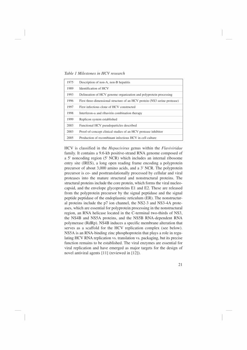
Table 1 Milestones in HCV research
1975 Description of non-A, non-B hepatitis
1989 Identification of HCV
1993 Delineation of HCV genome organization and polyprotein processing
1996 First three-dimensional structure of an HCV protein (NS3 serine protease)
1997 First infectious clone of HCV constructed
1998 Interferon-� and ribavirin combination therapy
1999 Replicon system established
2003 Functional HCV pseudoparticles described
2003 Proof-of-concept clinical studies of an HCV protease inhibitor
2005 Production of recombinant infectious HCV in cell culture
HCV is classified in the Hepacivirus genus within the Flaviviridaefamily. It contains a 9.6-kb positive-strand RNA genome composed of
a 5' noncoding region (5' NCR) which includes an internal ribosome
entry site (IRES), a long open reading frame encoding a polyprotein
precursor of about 3,000 amino acids, and a 3' NCR. The polyprotein
precursor is co- and posttranslationally processed by cellular and viral
proteases into the mature structural and nonstructural proteins. The
structural proteins include the core protein, which forms the viral nucleo-
capsid, and the envelope glycoproteins E1 and E2. These are released
from the polyprotein precursor by the signal peptidase and the signal
peptide peptidase of the endoplasmic reticulum (ER). The nonstructur-
al proteins include the p7 ion channel, the NS2-3 and NS3-4A prote-
ases, which are essential for polyprotein processing in the nonstructural
region, an RNA helicase located in the C-terminal two-thirds of NS3,
the NS4B and NS5A proteins, and the NS5B RNA-dependent RNA
polymerase (RdRp). NS4B induces a specific membrane alteration that
serves as a scaffold for the HCV replication complex (see below).
NS5A is an RNA-binding zinc phosphoprotein that plays a role in regu-
lating HCV RNA replication vs. translation vs. packaging, but its precise
function remains to be established. The viral enzymes are essential for
viral replication and have emerged as major targets for the design of
novel antiviral agents [11] (reviewed in [12]).
21

HCV infection is a highly dynamic process with a viral half-life of only
a few hours and production and clearance of an estimated 1012 virions
per day in a given individual [13]. This high replicative activity,
together with the lack of a proof-reading function of the viral RdRp, is
the basis of the high genetic variability of HCV. These properties are
similar to those of HIV infection and provide a strong rationale for the
development and implementation of antiviral combination therapies.
HCV isolates can be classified into 6 major genotypes and numerous
subtypes [14]. As stated above, patients infected with genotype 1 do not
respond as well to IFN-�-based therapy as those infected with genotype
2 or 3. The term quasispecies refers to the genetic heterogeneity of the
population of HCV genomes coexisting in an infected individual.
Virion structureWhile exciting progress has been made towards determining virion
structures of related viruses, e.g., dengue virus [15], HCV has not
been definitively visualized and its structure remains to be elucidated.
Based on filtration and electron microscopic studies, HCV particles
are 40–70 nm in diameter ([16] and references therein). E1 and E2 are
presumably anchored to a host cell-derived double-layer lipid envelope
that surrounds a nucleocapsid composed of multiple copies of the core
protein and the genomic RNA. HCV circulates in various forms in the
infected host and can be associated with low-density (LDL) and very-
low-density lipoproteins (VLDL), which appears to represent the infec-
tious fraction, virions bound to immunoglobulins, and free virions. This
feature may explain its unusually heterogenous and low buoyant density
(peak infectivity near 1.10g/ml).
Viral entryThe HCV life cycle begins with binding to the cell surface and interna-
lization, as schematically illustrated in Figure 2. Hepatocytes are the
main target cells but infection of B cells, dendritic cells, and other cell
types has also been reported. CD81, a tetraspanin protein found on the
surface of many cell types including hepatocytes, the low density lipo-
protein receptor (LDLR), scavenger receptor class B type I (SR-BI),
and, most recently, claudin-1 [17] have, among others, been proposed
22

as HCV receptors or components of a receptor complex (reviewed in
[10]). CD81, SR-BI and claudin-1 have been found to be required for
HCV entry. However, certain cell types were found to be nonpermissive
despite expression of CD81, SR-BI and claudin-1, indicating that one
or more additional HCV entry factor(s) remain to be discovered.
HCV enters via clathrin-mediated endocytosis, with transit through an
endosomal, low pH compartment and presumed endosomal membrane
fusion. The structural basis for low pH-induced membrane fusion has
been elucidated for related viruses [18, 19]. The envelope proteins of
these viruses have an internal fusion peptide that is exposed during low
pH-mediated domain rearrangement and trimerization of the protein.
The scaffolds of these so called class II fusion proteins are remarkably
similar, suggesting that entry of all viruses in the Flaviviridae family,
23
Figure 2Life cycle of HCV. 1) Virus binding and internalization, 2) cytoplasmic release and un-coating, 3) internal ribosome entry site (IRES)-mediated translation and polyproteinprocessing, 4) RNA replication, 5) packaging and assembly, 6) virion maturation andrelease. The topology of HCV structural and nonstructural proteins at the endoplasmic reti-culum (ER) membrane is shown schematically. HCV RNA replication occurs in a specificmembrane alteration, the membranous web. Note that IRES-mediated translation and poly-protein processing as well as membranous web formation and RNA replication, illustratedhere as separate steps for simplicity, may occur in a tightly coupled fashion (from ref. [10]).

including HCV, may include a class II fusion step. However, the mechan-
isms involved in activating HCV for low pH-induced fusion, the fusion
step, and the identity of the fusion peptide(s) have not yet been cha-
racterized.
Formation of a membrane-associated replication complexOnce liberated into the cytosol, the viral genome is translated by an
IRES-mediated mechanism, yielding a polyprotein precursor that is co-
and posttranslationally processed by cellular and viral proteases. A cha-
racteristic feature of HCV proteins is their association with intracellular
membranes. Indeed, each HCV protein contains a specific determinant
for membrane association. These hydrophobic segments have been dif-
ficult to study and have commonly been deleted in recombinant pro-
teins used for biochemical and structural studies. A primary interest in
our laboratory, therefore, was to identify and characterize the deter-
minants for membrane association of the HCV nonstructural proteins
3-5B, which are required for RNA replication (reviewed in [10]).
Figure 3 illustrates available three-dimensional structures and our cur-
rent understanding of the membrane association of HCV proteins.
Figure 3Structure and membrane association of HCV proteins. Scissors indicate cleavages by theendoplasmic reticulum (ER) signal peptidase, except on the cytosolic side where it indi-cates processing of core by signal peptide peptidase. The cyclic arrow denotes cleavageby the NS2-3 protease. Black arrows indicate processing by the NS3-4A protease com-plex. Known protein structures are shown as ribbon diagrams. The structures and themembrane bilayer are shown at the same scale. Proteins or protein segments of unresolvedstructure are represented as colored spheres or cylinders with their approximate sizes.See ref. [10] for details.
24

25
As an example, we have recently explored the mechanism of membrane
association of the HCV NS3-4A complex [20]. NS3-4A is a multifunc-
tional protein harboring serine protease and RNA helicase activities. It
is an essential component of the HCV replication complex and a prime
target for antiviral intervention. We were able to show through bioche-
mical assays, site directed mutagenesis, HCV RNA replication assays
and circular dichroism as well as nuclear magnetic resonance structural
analyses that membrane association and structural organization of HCV
NS3-4A are ensured in a cooperative manner by two membrane binding
determinants. We demonstrated that the N-terminal 21 amino acids of
NS4A form a transmembrane �-helix that is likely involved in intra-
membrane protein-protein interactions essential for the assembly of a
functional replication complex. In addition, we demonstrated that
amphipathic helix �0, formed by NS3 residues 12–23, serves as a
second essential determinant for membrane association of NS3-4A,
allowing proper positioning of the serine protease active site on the
membrane. These results allowed us to propose a dynamic model for
the membrane association and structural organization of NS3-4A on the
membrane (Figure 4).
An important consequence of our model relates to the proteolytic tar-
geting of host factors whereby the strict positioning of the protease
active site with respect to the membrane confers a high degree of selec-
tivity to potential cellular trans-cleavage substrates. In this context, it
has recently been shown that the NS3-4A protease cleaves and thereby
inactivates two crucial adaptor proteins in innate immune sensing,
namely Trif [21] and Cardif [22] (also known as MAVS, IPS-1 and
VISA), thereby blocking interferon production. Cardif cleavage by
NS3-4A was observed in various experimental systems and in the liver
from patients with hepatitis C ([23] and our unpublished data). Indeed,
the Cardif cleavage site is located close to the membrane surface and
fits well with our structural model of the membrane-associated trans-
cleavage conformation of NS3-4A (Fig. 4, Step 5).
Formation of a membrane-associated replication complex, composed of
viral proteins, replicating RNA, altered cellular membranes, and addi-
tional host components, is a hallmark of all positive-strand RNA viruses

26
Figure 4Dynamic model for the membrane association and structural organization of NS3-4A onthe membrane. In the HCV polyprotein context, translation of NS3 occurs at the mem-brane, as the preceeding NS2 protein is associated with membranes through its N-terminaldomain (Step 1). Induced folding of amphipathic helix �0 due to its interaction with themembrane interface is presumably a cotranslational event, followed by folding of the NS3serine protease and helicase domains (Step 2). A low affinity interaction of the centralsegment of NS4A with NS3 serine protease �-strands A1 and A0 is likely at this point, keep-ing the central segment of NS4A close to the serine protease domain before cleavage.Note that at this stage the in-plane membrane association of helix �0 does not imposeconstraints on the positioning of NS3. Hence, a forward movement of NS3 would bringthe hydrophobic N-terminal segment of NS4A into close contact with the membrane,thereby facilitating its posttranslational insertion into the membrane after processing atthe NS3/NS4A site (Step 3). Final incorporation of the central segment of NS4A into theN-terminal �-barrel stabilizes the interaction of helix �0 with the NS3 serine protease.This complete folding as well as membrane association by amphipathic helix �0 and thetransmembrane segment of NS4A lock the serine protease in a strictly defined positiononto the membrane (Step 4). As shown in the brackets, the hydrophilic helicase domainwould be immersed into the membrane at this stage in the known NS3-4A cis-cleavageconformation. Hence, the helicase domain has to move away from the serine proteasedomain in the final membrane-associated stage through a rotation of the linker segmentconnecting the two domains (Step 5). See [20] for details.

investigated thus far. Depending on the virus, replication may occur on
altered membranes derived from the ER, Golgi apparatus, mitochondria
or even lysosomes. The role of membranes in viral RNA synthesis is
not well understood. It may include (i) the physical support and orga-
nization of the RNA replication complex, (ii) the compartimentaliza-
tion and local concentration of viral products, (iii) tethering of the viral
RNA during unwinding, (iv) provision of lipid constituents important
for replication, and (v) protection of the viral RNA from double-strand
RNA-mediated host defenses or RNA interference.
A specific membrane alteration, designated as membranous web, was
identified as the site of RNA replication in Huh-7 cells containing sub-
genomic HCV replicons [24] (Fig. 5). Formation of the membranous
web was induced by NS4B alone and was very similar to the “sponge-
like inclusions” previously found by electron microscopy in the liver of
HCV-infected chimpanzees [25]. The membranous web is likely derived
27
Figure 5HCV replication complex. (A) Low-power overview of a Huh-7 cell harboring a sub-genomic HCV replicon. A distinct membrane alteration, designated as membranous web(arrows), is found in the juxtanuclear region. Note the circumscript nature of this speci-fic membrane alteration and the otherwise unaltered cellular organelles. Bar, 1 µm. (B)Higher magnification of a membranous web (arrows) composed of small vesicles embed-ded in a membrane matrix. Note the close association of the membranous web with therough endoplasmic reticulum. Bar, 500nm. The membranous web harbors all HCV non-structural proteins and nascent viral RNA in Huh-7 cells harboring subgenomic replicons[24], and therefore represents the subcellular site of HCV RNA replication. N, nucleus;ER, endoplasmic reticulum; M, mitochondria.

from ER membranes. Ongoing studies are aimed at characterizing
the host factors and cellular processes involved in formation of the
HCV replication complex. In this context, we have developed a system
that allows to visualize functional replication complexes in living cells
[26, 27].
Recent studies demonstrate a complex interaction between HCV RNA
replication and the cellular lipid metabolism, presumably via the traf-
ficking and association of viral and host proteins with intracellular
membranes. Such observations suggest that pharmacologic manipula-
tion of lipid metabolism may have therapeutic potential in hepatitis C.
Additional host factors, including cyclophilins, have been found to be
involved in HCV RNA replication, opening new angles for therapeutic
intervention.
Packaging, assembly and releaseLittle is known about the late steps of the viral life cycle, as these have
only recently become amenable to systematic study. Interestingly, the
nonstructural proteins p7, NS2 and NS5A as well yet to be defined
RNA structures are involved in these processes (reviewed in [28]).
Exciting new findings indicate an important role for lipid droplets and
the VLDL secretory pathway in HCV assembly and release [29, 30].
Virions persumably form by budding into the ER or an ER-derived
compartment and leave the cell through the secretory pathway.
IMPLICATIONS FOR THE DEVELOPMENT OF NEW THERAPEUTIC STRATEGIES
In principle, each step of the HCV life cycle illustrated in Figure 2
represents a target for antiviral intervention [12]. Specific inhibitors of
the biochemically and structurally well-characterized NS3-4A serine
protease and NS5B RdRp are currently being developed as antiviral
agents, and the first candidates have already been evaluated in clinical
trials. Serine protease inhibitors seem particularly promising, as they
not only block viral polyprotein processing but may also reverse the
inhibition of innate immune sensing by HCV (see above). In addition,
28

new targets have been uncovered by the recent studies highlighted
above, including, among others, the HCV 5' NCR, viral entry and
fusion, the p7 ion channel, the NS2-3 protease and NS5A. Moreover,
drugs affecting host factors involved in HCV replication are being
explored as antiviral agents. Already at this early stage it is evident that
the genetic variability of HCV, allowing the rapid development of anti-
viral resistance, represents a major challenge to the clinical develop-
ment of specific inhibitors and that, in common with HIV infection,
combination therapy will be necessary for therapeutic success.
CONCLUSIONS AND PERSPECTIVES
The development of powerful model systems enables dissection of the
HCV life cycle. Much work remains to be done with respect to the early
and late steps, the virion assembly and structure, the mechanism and
regulation of RNA replication, and the pathogenesis of HCV-induced
liver disease. Ultimately, these efforts should result in innovative thera-
peutic and preventive strategies for one of the most common causes of
chronic hepatitis, liver cirrhosis, and HCC worldwide.
ACKNOWLEDGMENTS
I wish to express my sincere gratitude to the Cloëtta Foundation for
distinguishing our research efforts with this award. These efforts could
only be accomplished with the guidance by outstanding mentors, the
contributions of highly dedicated collaborators, and the insight and
critique by many loyal colleagues.
I would like to thank Walter Siegenthaler, Hubert E. Blum, Jack R.
Wands and Charles M. Rice for their mentorship, invaluable support
and long-lasting friendship. I am indepted to Pierre Michetti for his
constant support and for creating the environment necessary to pursue
a productive clinical and research activity at the CHUV in Lausanne.
I would like to thank past and present members of my laboratory,
especially Pantxika Bellecave, Volker Brass, Rainer Gosert, Jérôme
29

Gouttenoire, Audrey Kennel, Anja Wahl and BennoWölk as well as my
wife Elke. It has been a priviledge and a source of immense satisfaction
to share their enthusiasm over the years. My sincere gratitude is also
due to outstanding collaboration partners for their numerous contribu-
tions and continuous support over the years, especially François Penin,
Ralf Bartenschlager, Charles M. Rice, Denise Egger and Kurt Bienz,
Markus Heim, Andreas Cerny, Amalio Telenti and Jürg Tschopp.
Nothing would have been possible without my wife Elke and our chil-
dren Henry and Lily. I am grateful for their love and efforts to maintain
the best possible balance between professional commitment and family
life.
I would like to dedicate this award to the memory of my father, Morad
Moradpour, with gratitude and affection. I would give much to have
him with us today.
Finally, I would like to acknowledge the funding agencies that have
supported our research over the years, especially the Swiss National
Science Foundation, the Swiss Cancer League, the Leenaards
Foundation, the Deutsche Forschungsgemeinschaft and the European
Commission.
30

31
REFERENCES
1. Williams R. Global challenges in liver disease. Hepatology 2006; 44:521–526.
2. Shepard C.W., Finelli L., Alter M. J. Global epidemiology of hepatitis C virus infec-
tion. Lancet Infect Dis 2005; 5:558–567.
3. Stramer S.L., Glynn S.A, Kleinman S.H., Strong D.M., Sally C., Wright D. J., et al.
Detection of HIV-1 and HCV infections among antibody-negative blood donors by
nucleic acid-amplification testing. N Engl J Med 2004; 351:760–768.
4. Hoofnagle J. H. Course and outcome of hepatitis C. Hepatology 2002; 36
(Suppl 1):S21–S29.
5. Pinzani M., Vizzutti F., Arena U., Marra F. Noninvasive assessment of liver fibrosis
by biochemical scores and elastography. Nat Clin Pract Gastroenterol Hepatol 2008;
5:95–106.
6. Sarasin-Filipowicz M., Oakeley E. J., Duong F. H., Christen V., Terracciano L.,
Filipowicz W., et al. Interferon signaling and treatment outcome in chronic hepatitis
C. Proc Natl Acad Sci USA 2008; 105:7034–7039.
7. Bellecave P., Moradpour D.A. fresh look at interferon-� signaling and treatment out-
comes in chronic hepatitis C. Hepatology 2008; 48:1330–1333.
8. Hoofnagle J. H., Seeff L. B. Peginterferon and ribavirin for chronic hepatitis C. N
Engl J Med 2006; 355:2444–2451.
9. Choo Q.-L., Kuo G., Weiner A. J., Overby L.R., Bradley D.W., Houghton M.
Isolation of a cDNA clone derived from a blood-borne non-A, non-B viral hepatitis
genome. Science 1989; 244:359–362.
10. Moradpour D., Penin F., Rice C.M. Replication of hepatitis C virus. Nat Rev
Microbiol 2007; 5:453–463.
11. Lamarre D., Anderson P.C., Bailey M., Beaulieu P., Bolger G., Bonneau P., et al. An
NS3 protease inhibitor with antiviral effects in humans infected with hepatitis C
virus. Nature 2003;426:186–189.

32
12. De Francesco R., Migliaccio G. Challenges and successes in developing new the-
rapies for hepatitis C. Nature 2005; 436:953–960.
13. Neumann A.U., Lam N.P., Dahari H., Gretch D.R., Wiley T.E., Layden T. J., et al.
Hepatitis C viral dynamics in vivo and the antiviral efficacy of interferon-� therapy.
Science 1998; 282:103–107.
14. Simmonds P., Bukh J., Combet C., Deleage G., Enomoto N., Feinstone S., et al.
Consensus proposals for a unified system of nomenclature of hepatitis C virus geno-
types. Hepatology 2005; 42:962–973.
15. Kuhn R. J., Zhang W., Rossmann M.G, Pletnev S.V, Corver J., Lenches E., et al.
Structure of dengue virus: implications for flavivirus organization, maturation, and
fusion. Cell 2002; 108:717–725.
16. Wakita T., Pietschmann T., Kato T., Date T., Miyamoto M., Zhao Z., et al. Production
of infectious hepatitis C virus in tissue culture from a cloned viral genome. Nat Med
2005; 11:791–796.
17. Evans M. J., von Hahn T., Tscherne D. M., Syder A. J., Panis M., Wölk B., et al.
Claudin-1 is a hepatitis C virus co-receptor required for a late step in entry. Nature
2007; 446:801–805.
18. Modis Y., Ogata S., Clements D., Harrison S. C. Structure of the dengue virus en-
velope protein after membrane fusion. Nature 2004; 427:313–319.
19. Gibbons D.L., Vaney M.C., Roussel A., Vigouroux A., Reilly B., Lepault J., et al.
Conformational change and protein-protein interactions of the fusion protein of
Semliki Forest virus. Nature 2004; 427:320–325.
20. Brass V., Berke J.M., Montserret R., Blum H.E., Penin F., Moradpour D. Structural
determinants for membrane association and dynamic organization of the hepatitis D
virus NS3-4A complex. Proc Natl Acad Sci USA 2008; 105:14545–14550.
21. Li K., Foy E., Ferreon J. C., Nakamura M., Ferreon A. C, Ikeda M., et al. Immune
evasion by hepatitis C virus NS3/4A protease-mediated cleavage of the Toll-like
receptor 3 adaptor protein TRIF. Proc Natl Acad Sci USA 2005; 102:2992–2997.
22. Meylan E., Curran J., Hofmann K., Moradpour D., Binder M, Bartenschlager R., et
al. Cardif is an adaptor protein in the RIG-I antiviral pathway and is targeted by hepa-
titis C virus. Nature 2005; 437:1167–1172.
23. Loo Y.M., Owen D.M., Li K., Erickson A.K., Johnson C.L., Fish P.M., et al. Viral
and therapeutic control of IFN-� promoter stimulator 1 during hepatitis C virus
infection. Proc Natl Acad Sci USA 2006; 103:6001–6006.

33
24. Gosert R., Egger D., Lohmann V., Bartenschlager R., Blum H.E., Bienz K., et al.
Identification of the hepatitis C virus RNA replication complex in Huh-7 cells har-
boring subgenomic replicons. J Virol 2003; 77:5487–5492.
25. Egger D., Wölk B., Gosert R., Bianchi L., Blum H. E., Moradpour D., et al.
Expression of hepatitis C virus proteins induces distinct membrane alterations in-
cluding a candidate viral replication complex. J Virol 2002; 76:5974–5984.
26. Moradpour D., Evans M. J., Gosert R., Yuan Z. H., Blum H. E., Goff S. P., et al.
Insertion of green fluorescent protein into nonstructural protein 5A allows direct
visualization of functional hepatitis C virus replication complexes. J Virol 2004;
78:7400–7409.
27. Wölk B., Büchele B., Moradpour D., Rice C.M. A dynamic view of hepatitis C virus
replication complexes. J Virol 2008, in press.
28. Murray C.L., Jones C.T., Rice C.M. Architects of assembly: roles of Flaviviridae
non-structural proteins in virion morphogenesis. Nat Rev Microbiol 2008; 6:699–708.
29. Huang H., Sun F., Owen D.M., Li W., Chen Y., Gale M, Jr., et al. Hepatitis C virus
production by human hepatocytes dependent on assembly and secretion of very low-
density lipoproteins. Proc Natl Acad Sci USA 2007; 104:5848–5853.
30. Miyanari Y., Atsuzawa K., Usuda N., Watashi K., Hishiki T., Zayas M., et al. The
lipid droplet is an important organelle for hepatitis C virus production. Nat Cell Biol
2007; 9:1089–1097.